title o - opus.lib.uts.edu.au
TRANSCRIPT

Page 1
TITLE
Evidence for the Key Role of H3O+ in Phospholipid Membrane Morphology
Charles G. Cranfield1*, Thomas Berry1, Stephen A. Holt2, Khondker R.
Hossain1,2, Anton P. Le Brun2, Sonia Carne3, Heba Al Khamici1, Hans Coster4,
Stella M. Valenzuela1 and Bruce Cornell3
AUTHOR AFFILIATIONS 1School of Life Sciences, University of Technology Sydney, Ultimo, NSW 2007, Australia. 2Australian Nuclear Science and Technology Organisation, Locked Bag 2001 Kirrawee DC,
New South Wales 2232, Australia. 3SDx Tethered Membranes Pty Ltd, Unit 6 30-32 Barcoo
St, Roseville NSW 2069, Australia. 4School of Chemical and Biomolecular Engineering,
University of Sydney, NSW 2006, Australia.
CORRESPONDING AUTHOR *Charles Cranfield, School of Life Sciences, University of Technology Sydney, Ultimo, NSW
2007, Australia. Ph +61 9514 4034. E-mail: [email protected]
KEYWORDS
Tethered bilayer lipid membranes (tBLMs), hydrogen bond, pH regulation, impedance spectroscopy,
neutron reflectrometry.

Page 2
ABSTRACT
This study identifies the importance of the phosphate moiety and H3O+ in controlling the ionic
flux through phospholipid membranes. We show that despite increasing the H3O+
concentration when lowering the pH, the ionic conduction through phospholipid bilayers is
reduced. Through modifying the lipid structure we show the dominant determinant of
membrane conduction is hydrogen bonds between the phosphate oxygens on adjacent
phospholipids. The modulation of conduction with pH is proposed to arise from the varying
H3O+ concentrations altering the molecular area per lipid and modifying the geometry of
conductive defects already present in the membrane. Given the geometrical constraints that
control the lipid phase structure of membranes, these area changes predict that organisms
evolving in environments of different pH will select for different phospholipid chain lengths,
such as is found for organisms near highly acidic volcanic vents (short chains) or in highly
alkaline salt lakes (long chains). The stabilizing effect of the hydration shells around phosphate
groups also accounts for the prevalence of phospholipids across biology. Measurement of ion
permeation through lipid bilayers was made tractable using sparsely tethered bilayer lipid
membranes (tBLMs) with swept frequency electrical impedance spectroscopy (EIS) and
ramped DC amperometry. Additional evidence for the effect of pH change on lipid packing
density is obtained from neutron reflectometry data of tethered membranes containing
perdeuterated lipids.

Page 3
INTRODUCTION
In the study of the effects on membrane permeability of both ion channel proteins and peptides
it is important to identify all factors that impact the membrane conductivity. The objective of
the present study is to characterise one of the major variables determining membrane structure,
namely the hydrogen bond strength within and between lipid molecules within the membrane.
A key variable in hydrogen bond strength is the pH of the bathing solution. Despite the fact
that the concentration of the hydronium ion being three to four orders of magnitude lower than
that of others ions (mainly Na+ and Cl-) in the bulk electrolyte solutions, its small size and high
electric field gradients can result in it being a dominant factor in membrane integrity. To date,
to the best of our knowledge, the impact of pH on membrane conduction remains unreported.
In this study we report the dependence of membrane conduction on pH in the range 5 -9 log
units. We relate changes in membrane conduction to structural features underlying membrane
morphology.
The structures formed by aqueous dispersions of lipids are determined by the balance of forces
within the hydrophobic, hydrophilic and interfacial zones of lipid-water aggregates. For a fluid
Lα lipid lamellar structure this balance of forces has been described in terms of the hydrated
molecular area (ao), the lipid leaflet thickness (l) and hydrated lipid molecular volume (v) 1.
The measure v/(aol) is a qualitative guide to the lipid phases formed by surfactants. The
structures formed for v/(aol) ~1 are lamella bilayers, for v/(aol) ~ 1/3 the structures formed are
micelles, for v/(aol) ~ 1/2 cylindrical tubes, and for v/(aol) > 1 a variety of inverted phases 2-3.
In maintaining an appropriate v/(aol) close to unity for lipid bilayers, the area per phospholipid
and the resultant membrane thickness will dictate the conformation of all other membrane
associated proteins and their physiological functions. This balance is dependent on
contributions from both the hydrophobic and hydrophilic components of the membrane and is
significantly dependent on the hydration shells surrounding the polar head groups.
The role of hydrogen bonding in determining the balance of forces is evident in all aqueous
surfactant assemblies. Increasing the hydrogen ion concentration through lowering the pH is
known to cause a decrease in the critical micelle concentration (CMC) of sodium dodecyl
sulphate (SDS) 4. Rupert et al (1998) reported significant changes in vesicle fusion and head
group clustering for didodecyl phosphate (DDP) vesicles around the effective pKa of 5.2 5.
They interpret these effects as arising from protonation of the non-ester phosphate of the DDP.
Siegal et al (1989) have also shown that lowering the pH of a DOPE dispersion caused a phase
transition from the Lα lamellar phase to the Hexagonal II inverted phase (HII) 6. This is
consistent with a reduction in the area of the hydrated polar group driving the Lα phase into the
highly curved inverted HII lipid phase.
The major impediment to the passage of ions across lipid membranes is the hydrophobic, low
dielectric constant membrane core of the lipid chains. This is a consequence of the high Born
energy required to cause an ion to partition into the hydrophobic interior of a lipid bilayer.7

Page 4
That there is a source of the background ionic current despite this high energy cost, has been
suggested to arise from sparse and fluctuating “defects” in the bilayer morphology in which
hydrophilic pores traverse the non-polar core of the membrane.8 The incidence of such defects
in homogenous fluid phase lipid bilayers will depend on the lipid molecular geometry. The
inter-relationship of lipid dimensions, v/(aol), indicates that longer chain lipids with smaller
areas per molecule will tend to possess fewer pores than short chain lipids with large areas per
molecule. This is a consequence of the curvature energy required to form a highly curved
toroidal pore traversing the membrane from the inner and to the outer leaflets of the bilayer.
The lifetime of toroidal pores is discussed by Karatekin et al (2003).9 They address the rupture
and sealing of liposomes by modelling a balance between the surface pressure arising from
intense optical illumination balanced by the line tension of a toroidal pore. The steric
contribution, a consequence of the geometrical constraints embodied in v/(aol), is termed by
these authors as the curvature, co . For planar membranes co = 0. For membranes possessing
positive curvature co > 0 and for negative curvature co < 0. This term is then added or subtracted
from the line tension permitting a calculation of the pore lifetime.
The measurement of ionic permeability through lipid bilayer membranes is experimentally
challenging. Patch clamp electrophysiology measures are typically performed on 1 µm
diameter membrane patches which, at 1 MΩ cm2 membrane leakage, would require
conductance measures to be performed at impedances of greater than 105 GΩ, far in excess of
the typically measured values of 1 - 10 GΩ. This suggests the intrinsic membrane conduction
in patch clamp experiments is dominated by mechanisms other than the intrinsic membrane
resistance 10. Solvent-based black lipid membranes (BLMs) suffer not only from high
background resistances due to their small areas, but also from the residual solvent in free
exchange with the membrane plateau border.11-12 Further approaches to measuring the intrinsic
membrane conduction includes liposomal release assays, which at best, are limited to
qualitative measures of conduction due to the uncertainty in the liposome stability. Monolayer
techniques performed at the interface between two immiscible electrolyte solutions (ITIES) 13
or measurements employing Langmuir films report on the interfacial properties, but by
definition, are unable to describe bilayer permeability or conduction.
The use of electrical impedance spectroscopy (EIS) and ramped direct current amperometry to
determine the conduction and capacitance of lipid bilayers provides a unique approach to
quantifying ionic permeability. In the present study the conductance of tethered bilayer lipid
membranes (tBLMs) is measured in the presence of a range of pH values from 5.0 – 9.0 log
units.
In summary we show that decreased pH (increased H3O+ concentration) reduces the intrinsic
membrane conduction by an order of magnitude (pH 9 to pH 5) and significantly decreases
water penetration into the membrane. These effects are interpreted as arising from an increase
in the hydrogen bond stability caused by neighbouring H3O+ ions. This interpretation suggests
that changes in pH will affect the intrinsic conduction of all phospholid membranes. Further

Page 5
data based on neutron reflectometry measures, report on the associated changes in membrane
geometry.
MATERIALS AND METHODS
Tethered bilayer lipid membranes
Using the solvent exchange technique,14 tBLMs with 10% tethering lipids and 90% spacer
lipids (T10 tBLM) were formed. The procedure involves using 2.1 mm2 pre-prepared tethered
benzyl-disulfide (tetra-ethyleneglycol)n=2 C20-phytanyl tethers (DLP) : benzyl-disulfide-tetra-
ethyleneglycol-OH spacers in the ratio of 1:9 coated onto 2 mm2 patterned, 100 nm thick fresh
5N5 gold surfaces sputter coated onto a polycarbonate slide (SDx Tethered Membranes Pty
Ltd, Australia) 15. The ratio of tethering molecules to spacer molecules (eg 1:9) is termed here
as T10. Similarly, ratios of 1:99 and 0:100 are termed T1 and T100 respectively. Following air
drying of this tethered monolayer, 8 μL of a 3 mM solution of a mobile lipid phase dissolved
in ethanol is added to a 0.1 mm high, 1µL volume flow cell chamber, and, after a 2 minute
incubation, is washed 3 times with 2 x 200 μL of 100 mM NaCl. Mobile lipid phases
investigated were: 1-palmitoyl-2-oleoyl-sn-glycero-3-phosphocholine (POPC) (Avanti Lipids,
USA); 1,2-dioleoyl-sn-glycero-3-ethylphosphocholine (EPC) (Avanti Lipids USA; 100%
diphytanyl-glycero-phosphatidylcholine (DPEPC) (SDx Tethered Membranes Pty Ltd,
Australia) (See Fig 2A). As a non-phosphate fully tethered lipid control, tBLMs were prepared
employing the fully tethered membrane spanning lipid terminated with an OH group
(MSLOH) previously described 16 (SDx Tethered Membranes Pty Ltd, Australia). Diagrams of
the tethering chemistries are provided in Figure S1 of the Supplementary Material.
The pH of the 100mM NaCl solutions was adjusted by the addition of concentrated HCl or
NaOH to create stock solutions of pH ~ 5.0, pH ~ 7.0 and pH ~ 9.0. Each of these solutions
was kept in an air-tight container prior to use, and used in within 5 min to minimise any
subsequent acidification due to atmospheric CO2. The pH of the stock solutions was regularly
monitored.
Gramicidin containing membranes were formed as described previously.15 Briefly, 100 nM of
Gramicidin-A (BioAustralis Pty Ltd, Australia) was included in the 3mM mobile phase lipid
from which the tBLM was formed. Covalently linked bis-gramicidin tBLMs were synthesized
as described previously 17.
AC impedance spectroscopy measures were performed using an SDx tethaPod™ operated with
SDx tethaQuick™ software (SDx Tethered Membranes Pty Ltd). Swept frequency impedance
spectrometry was employed using a 50 mV peak-to-peak AC excitation at 0.1, 0.2, 0.5, 1, 2, 5,
40, 100, 200, 500, and 1000 Hz and zero bias potential. The data were fitted to a Constant
Phase Element in series with an Resistor/Capacitor network 15 using a proprietary adaptation
of a Lev Mar fitting routine. This model provided excellent agreement with the data. The use
of more complex equivalent circuits16, 18 to model the defect distribution upon which the

Page 6
membrane conduction is based was considered to be beyond the scope of the present study.
Examples of Bode plots are given in Figure S2 and the equivalent circuit in Figure S3 of the
Supplementary Material.
DC ramped amperometry measures were performed an eDAQ ER466 Potentiostat.14 Ramps of
100 V sec-1 from 0 to 500 mV over 5 ms were applied to T1, T10 and T100 DPEPC tBLMs
and to T100 MSLOH tBLMs at pH 5, 7 and 9. The initial current step provides an estimate of
the membrane capacitance and the following slope an estimate of the membrane conduction
and the onset of additional voltage dependent changes in the membrane conduction.
Neutron Reflectometry (NR) and Data Fitting
The NR data was collected on the PLATYPUS time-of-flight instrument19 located at the 20
MW OPAL research reactor (Australian Nuclear Science and Technology Organisation
(ANSTO), Sydney, Australia). PLATYPUS is located on a cold neutron guide and as such is
supplied with a neutron bandwidth ranging from 2.5 – 18.0 Å with the cut-off being determined
by the instrument disc choppers. The Q resolution of the experiment was set at ΔQ/Q of 3.3 %
where Q, the scattering vector is defined as
Q = (4π sinθ) / λ
where θ is the scattering angle and λ is the wavelength of the incident radiation. Incident angles
of 0.5, 0.85 and 3.8° were used with the data spliced together after normalisation to the direct
beam and background subtraction to produce a single absolutely scaled reflectivity dataset over
a Q range of 0.006 to 0.25 Å-1 using the SLIM reduction package.20
Tethered bilayers were formed on 50 mm diameter, 7mm thick, conductive silicon disks that
had been coated with chromium then gold at the Melbourne Centre for Nanotechnology. Half
membrane spanning T100 tBLMs comprising an inner monolayer of tethered hydrogenated
DLP and a perdeuterated DPEPC outer monolayer.21 Coated disks were clamped to a bespoke
perfusion chamber for assembly and measurement at the PLATYPUS sample position.
The hydrogenated and deuterated lipid tails were employed to give neutron contrast between
the two leaflets. Further contrast was provided by bathing solutions of 100 mM NaCl in either
D2O, H2O or a HDO mixtures that nominally matched the gold layer. Because of the necessity
for measures over extended time periods (~3 hours) the effects arising from dissolved CO2
causing a drop in pH, 100 mM buffer solutions were employed. For pH 5 acetate buffer was
used which was adjusted to the final pH using glacial acetic acid; for pH 7 phosphate buffer
was used, and for pH 9 [(cyclohexylamino)ethane sulfonic acid] CHES buffer was used, and
both were adjusted to their designated pH value using a NaOH solution. The difference between
pH and pD was taken into account when preparing D2O-containing buffers and adjusted
accordingly so that they were equivalent.
Page 7
The absolutely scaled data sets were modelled using RasCAL (version 1, A. Hughes, ISIS
Spallation Neutron Source, Rutherford Appleton Laboratory) operating within the MATLAB
environment. RasCAL uses the standard optical matrix approach of Abeles22 to calculate the
reflectivity. The data modelling was carried out assuming that the interface was composed of
a series of parallel layers where the model parameters fitted to the data were scattering length
density (SLD), layer thickness and interfacial roughness. Data were simultaneously fitted
whereby the structural parameters of the layers were constrained to be equivalent but the SLD
allowed to vary with buffer contrast. The SLD for silicon was constrained to 2.07 ×10⁻⁶ Å-2.
The SLD of the D2O buffer solution was fitted based upon the position of the critical edge in
the datasets. The final data fits were then assessed via a Markov Chain Monte Carlo resampling
method. In this procedure the fitting stating points are randomised and the data fitting repeated
40,000 times The output from these fits are histogrammed for each parameter with the midpoint
taken as the parameter value and the 95% confidence level determined from the distribution.
In order to maximise the observable effects of pH change on the membrane lipid geometry, a
fully tethered hydrogenated inner monolayer membrane leaflet and a mobile deuterated outer
membrane leaflet was employed. Outer tail thickness and hydration, and headgroup hydration
were permitted to vary for a layer model.
RESULTS
Tether density
Figures 1A-C show the effect of pH values of 5, 7 and 9 on the EIS derived conduction values
obtained from a phytanyl ether phosphatidyl choline (DPEPC). Tether densities ranged from 1
tether to 99 mobile lipids (1% tethered) to 10 per 90 (10% tethered) to a tBLM in which the
inner monolayer was fully tethered and the outer monolayer was mobile (100% tethered).
Figure 1D shows similar EIS derived conduction data obtained from MSLOH tBLMs, a full
membrane spanning non-phosphate containing control. Figures 1E-H show current voltage
curves (I-V) measures on the same samples using DC ramped amperommetry. DC ramped
amperommetry provides a measure of the voltage dependent changes in conduction for each
membrane configuration. It is evident that the overall membrane conduction increases with
increasing pH in all cases. In addition, as the pH is raised the membrane conduction becomes
more sensitive to the voltage ramp. However, with the increase in tethering density both the
pH dependence and the voltage dependencies are reduced. In the extreme case of a fully
tethered membrane spanning MSLOH, tBLM, the membrane conduction is effectively
unaltered by pH or the voltage ramp. In Figures 1E-H, the electroporative responses to a 5 ms
linear ramp 0 - 500 mV, the initial step reports on the capacitance of the tBLM and the
subsequent slope reports on the membrane conduction. A steeper slope reflects a greater
conduction. The non-linearity in slope seen at higher voltages14, 23 reflect an increase in
conduction. Smith et al (1984)24 using free standing lipid bilayers, and later Valincius et al
(2008)25 using sparsely tethered tBLMs, interpret the low activation energy observed for the

Page 8
intrinsic membrane conduction as ruling out electric field induced channel formation as the
mechanism for the induced changes in conduction. Thus, we propose that the non-linear current
increases observed here arise, not from the creation of new pores, but from the modulation of
existing pores. It is significant that with reduced tethering density the effects of electroporative
responses are seen at progressively lower voltages. In particular the voltage dependent
background conduction rises steeply as the tether density is lowered below 10% tethers. This
is consistent with the dimension required to form a membrane spanning pore between the
membrane tethers. These results support the now generally accepted notion that toroidal pores
provide the primary mechanism for ionic transport across lipid bilayers.
Lipid class
The effect of pH on the membrane conduction for bilayers formed from, POPC, DPEPC, EPC
and MSLOH is shown in Figure 2. The number of potential interfacial hydrogen bonding sites
is systematically varied across this family of tBLMs. POPC possesses four types of hydrogen
bonding identified in the figure and suffers the largest change (X245) in conduction between
pH 5 and pH 9 (Figure 2C). DPEPC has the carbonyl sites replaced by the non-hydrogen
bonding ethers which results in an intermediate change in conduction (X8.4) between pH 5 and
pH 9, and EPC has the non-ester phosphate oxygen-hydrogen bonding site blocked by a ethyl
group has a small change in the conduction ratio between pH 5 and pH 9 (X2.9). The MSLOH
is a hydroxyl terminated membrane spanning lipid with the potential for only a single hydrogen
bond at the outer surface of the tBLM interface with the bulk solvent. This resulted in the
smallest change in the conduction ratio between pH 5 and pH 9 (X1.3). Relatively little
variation was observed for the membrane capacitance, however, as also seen in Figure 2C, the
same trend occurs for the change in capacitance as occurred for conduction across the four lipid
types.
pH dependence of the reservoir space
Krishna et al (2003)16 and Cornell et al (1997)26 have identified conditions under which the
apparent membrane conduction is largely determined by the properties of the reservoir space
between the membrane and the supporting electrode. However, as described in Krishna et al
(1997), provided all-ether chemistries are employed in the fabrication of the tethers these
effects are essentially eliminated. In addition, by contrast to these earlier studies, the present
report focuses on the pH dependence of membrane conduction rather than the ionic species
dependence of membrane conduction. Were the pH dependence of conduction primarily arising
from the properties of the reservoir space between the tBLM and the tethering gold electrode,
the observed effects would be independent of the presence of an ion channel, or of variations
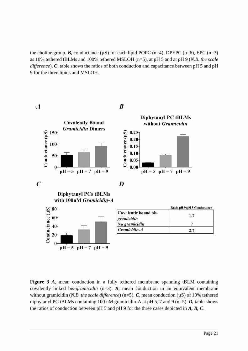
in the lipid class employed to form the tBLM. In Figure 3 we show the pH dependence of
conduction for diphytanyl PC tBLMs with and without a covalently linked dimer analogue of
the bacterial ion channel gramicidin-A. The concentration of the ion channel employed here is
such that the conduction has increased by an amount in excess of two orders of magnitude. As

Page 9
seen in Figure 3A, the pH dependence is less than two-fold when the pH is adjusted from 5 to
9. This is to be compared with the seven-fold change in conduction over the same pH range
when employing a diphytanyl PC tBLM (Figure 3B), and in excess of a 200-fold change when
employing POPC membrane lipids. Furthermore, the reduction of the pH dependence of
conduction to less than a two-fold variation over this pH range is consistent with the reported
pH dependence of the intrinsic cesium conduction of gramicidin-A channels within diphytanyl
PC bilayers.27 These data argue against a pH dependence of the conduction of the reservoir
space as the primary source of conduction variation observed in the current studies. To quantify
in detail any residual reservoir effects would require insertion of an ion channel with even
smaller pH dependent conductance.
The effect of pH on the conduction of monomeric gramicidin-A channels is shown in Figure
3C. Unlike the case of the covalently linked gramicidin dimer, the membrane conduction will
now depend upon membrane thickness as has been reported by Mobashery et al (1997).28 The
conduction caused by monomeric Gramicidin-A within lipid bilayers arises from the
gramicidin monomers aligning within each membrane leaflet to form a conducting dimer.29
Increasing the H3O+concentration from pH 9 to pH 5 caused a 2.7 fold decrease in the
gramicidin-A induced conduction. The greater pH dependence seen here compared to that for
the covalently linked gramicidin dimer (Figure 3A) is interpreted as arising from a modulation
of the dimer lifetime. The proposed reduction in the area per molecule on reducing the pH from
9 to 5 is further suggested to cause a consequent thickening of the membrane, leading to a
reduction in the monomer to dimer on-rate due to a weakening of the hydrogen bonds between
the gramicidin-A monomers.28 In addition, thickening the membrane will increase the dimer to
monomer off-rate of the channel. The relatively high concentration of gramicidin-A employed
here will minimise these effects, however, it is evident that lowering the pH still induces a
significant additional conduction decrease beyond the intrinsic pH dependence of gramicidin
conduction.
Neutron Reflectometry
In order to further explore the effect of pH on the geometry of lipid bilayers, neutron
reflectometry measures were acquired using a tBLM comprising a fully hydrogenated inner
leaflet and a fully deuterated outer leaflet. These measures were obtained for aqueous bathing
solutions comprising various ratios of HDO. The results are presented as the one-
dimensional SLD plot for solutions at pH 5, 7 and 9 for three HDO ratios (Figure 4A).
Figure 4B shows the outer tail thickness. As pH is reduced from 9 to 5, the outer leaflet tail
thickness is increased by ~4%. Associated with this area thickening of the outer layer leaflet
tails is an increase in the lipid volume fraction of 0.95 at pH 5 from 0.89 at pH 9 (Figure
4C). In addition, the volume fraction of the outer layer leaflet head groups show an even
more dramatic dependence on pH ranging from ~0.87 at pH 5 to ~0.59 at pH 9 (Figure 4D
and Table S1 in Supplementary Material). At pH = 9 the outer head group volume fraction
was essentially indeterminate.

Page 10
DISCUSSION
Relative ionic impact of H3O+
At pH 5 the 55 M concentration of water and the 100 mM Na+ concentration greatly exceed
the 10 µM H3O+ concentration. However, the biological significance of pH arises from the
smaller dimensions of the H3O+ resulting in higher field strengths, and thus more potent non-
covalent interactions. Kotyńska and Figaszewski (2005) have reported the electrophoretic
measures of monovalent cations across liposomal surfaces of phosphatidylcholine.30 From
these measures they derive the binding density of Na+ at the membrane surface for a pH
range from 0-11. Over the range of pH 6 – 8 they observe a shift in the sodium ion ‘degree
of coverage’ from essentially zero to approaching 90%, respectively, indicating a dominant
contribution by the H3O+ ions to the Na+ distribution. These observations are consistent with
the results reported here in which the H3O+ concentration also dominates the membrane
conduction. The change in the surface Na+ concentration, as a result of pH changes, may
well contribute to morphological changes in the membrane resulting in variations in
membrane conduction. Others have proposed, based on molecular dynamics modelling, that
the distribution of Na+ and K+ at the membrane surface are very different.31 It is suggested
that the Na+ is located adjacent to the PO4- groups whereas K+ is excluded from the lipid
interface. Our conduction results fail to show any significant difference between Na+ and K+
at 100 mM despite a similar pH dependence (data not shown). This suggests that the H3O+
ions have the dominant effect on conduction.
Area per lipid
In the major biological phospholipid families, the fluid Lα phase area per lipid molecule is in
the range of 65 ~75 Ǻ2 .32 Lewis and Engelman (1983) report that for lipid chains from C10 –
C24:1 the area per molecule (ao) was 68 ± 2 Ǻ2 and the lipid bilayer thickness (l) was proportional
to acyl chain length.33 In the crystalline Lβ phase the area per lipid molecule is typically in the
range of ~50 Ǻ2 and dominated by the inter-chain packing 34. Paresgian et al (1979) have
shown, for the fluid phase, that below ~68 Ǻ2 a significant increase in energy is required to
further reduce the hydrated molecular area.35 Pasenkiewicz-Gierula et al. (1997) report a
molecular dynamics simulation of the hydrogen bonding of water to phosphatidylcholine lipids 36. From their simulation the radial distribution function for the non-ester oxygens bound to the
phosphate are within 0.3 % as that for liquid water.37 Collectively, these reports indicate that
the area per lipid in a fluid phospholipid bilayer is dominated by the hydrated lipid headgroup.
Hydration shells
Pasenkiewicz-Gierula et al. (1997) identify the dominant hydrogen bonding pattern for the
water populations surrounding the PC headgroup as water molecules directly hydrogen-bonded
to the non-ester oxygens of the phosphate group.38 A further population of associated water
molecules, not hydrogen bonded to the lipids, are identified as being consistent with transient

Page 11
clathrate cages surrounding the choline group. Lopez et al (2004) have extended the
Pasenkiewicz-Gierula et al (1997) study and include the extended lifetimes of both the
hydrogen bonding and diffusional jumps associated with water in the lipid hydration shells 39.
These molecular dynamic simulations closely correspond to the experimental observations of
White and King (1985)40 who identify a hydration barrier comprising 11-13 water molecules
per lipid. One of the few approaches to permit measure of the order of the various populations
of water molecules is Sum Frequency Generation Spectroscopy which observes the water order
on a time that is short compared to the exchange rate.41 Using this technique Sovago et al (2009)
identify a water population that is isolated from the bulk water and buried between the
phosphate and acyl groups of the phospholipid. Collectively, these studies demonstrate the
existence of a population of water molecules surrounding the lipid head-groups that is
significantly more ordered that bulk water.
Effect of pH on phospholipid area
The results presented here from EIS, amperometry and neutron reflectometry support a model
in which the area per molecule within a lipid assembly is modulated by pH. The primary
mechanism for these effects is interpreted in terms of the interaction of the lipid phosphate
groups with a hydrating network at the lipid-water interface (Figure 5).
Molecular dynamic studies have identified potential hydrogen bonding patterns of water
bridging between adjacent phospholipid molecules via the non-ester oxygens of the lipid
phosphate groups.36, 38 Further hydrogen bonded bridging water molecules are proposed to exist
between the non-ester phosphate oxygens and the carbonyl oxygens in esterified phospholipids.
In the present study it is shown that substantial changes in conduction occur over the two to
four orders of magnitude change in the H3O+ concentration, with the conduction being reduced
at the highest H3O+ concentrations (low pH) across all phospholipid species studied here. It is
proposed that the H3O+ ions compete with and disrupt the hydrogen bonding pattern of the
intermolecular bridging water molecules causing the observed variation in membrane
conductivity, primarily through variations in the molecular area. A further insight by molecular
dynamics is the contribution made by bridging water molecules for an ether or ester linked
phospholipid.36, 38 Figure 2A shows a mechanism whereby the hydrogen bonds between
individual water molecules and their neighbouring phospholipid phosphate or carbonyl
oxygens can be disrupted by a less anisotropic H3O+ ion causing a disruption in the bridging
hydrogen bonds and a condensation of the phospholipid onto the positively charged H3O+
resulting in a reduction in the area occupied per phospholipid. In addition, the conduction at
pH ~ 5 in sparsely 10% tethered tBLMs comprising 100% EPC or 100% DPEPC or 100%
POPC is shown in Figure 2B. The conduction is found to progressively increase in the order
of POPC < DPEPC < EPC. In the case of POPC it is proposed that the conduction is the smallest
due to the attraction of the H3O+ ions to both carbonyl and non-ester phosphate oxygens. The
~ 2-fold higher conduction seen in DPEPC is proposed to be as a result of the elimination of
the negative carbonyl oxygen creating a smaller reduction in the molecular area. The ~ 5-fold

Page 12
higher conduction seen in EPC is proposed to arise from the blocking effects of the non-ester
phosphate oxygen bound ethyl group both reducing the attractive charge and sterically
impeding the area reduction.
Further consequences of this model are demonstrated in Figure 2B where the conduction at
pH ~ 9 is shown for the same family of lipids. At this high pH the POPC the carbonyl and
oxygen sites would thus be surrounded by un-dissociated water resulting in the largest
molecular area and therefore highest conductivity. The lower conductances seen for EPC and
DPEPC would arise from the hydrophobic and steric blocking caused by the non-ester
phosphate oxygen ethyl group for the EPC, and the absence of the ester carbonyls for the
DPEPC.
Membrane Thickness and pH
The effect of pH on tBLM thickness was further investigated by including the ion channel
gramicidin-A 28. It can be seen in Figure 3C that lowering the pH caused a decrease in
conduction consistent with a thicker membrane causing a reduction in the gramicidin-A dimer
lifetime. It should be noted, the similar reduction seen for the same tBLM in the absence of
gramicidin-A also arises from a reduction in the molecular area, however, we propose these
effects in the absence of gramicidin-A arise from the modulation of the toroidal pore defect
diameter driven by changes in the molecular geometry of the lipid. These observations are
supported by a direct measure of the contraction of the area of a monolayer of phospholipid on
a Langmuir trough as reported by Gong et al (2002). 42 Measurements of variations in
membrane thickness based on capacitance observations are complicated by uncertainties of the
interfacial dielectric constant for the different values of pH. Other contributions to the pH
dependence of the gramicidin-A conduction may arise from a small modulation of the diffusion
coefficient of both the lipid and gramicidin-A impacting the monomer to dimer on-rate.
From the neutron reflectometry results additional evidence is available on the pH dependence
of the outer bilayer lipid leaflet. It was observed that over the pH range from 5 – 9 log units the
outer bilayer leaflet thickness decreased by ~3 – 4 %. This is an indication that the molecular
area has increased over this pH range by ~6 – 8 %. These changes in molecular geometry will
be reflected in changes in the geometry of membrane spanning defects described here as
toroidal pores. Increasing the intrinsic molecular area across the tBLM drives an increase in
the average pore diameter, causing an increase in conduction.
The Modulation of Toroidal Pores by pH
The tBLM conduction is modelled here as arising from the modulation of fluctuating toroidal
pores that are already present within the membrane and traverse the otherwise essentially ion
impermeable hydrocarbon core of the lipid bilayer. That the conduction variation arises from
existing toroidal pores rather than the creation of new pores is demonstrated by the
approximately 35-38 kJ/mol activation energy for the intrinsic bilayer conduction.24-25 This low

Page 13
value of activation energy for conduction eliminates other explanations requiring the creation
of new pores for which activation energies in excess of 100 kJ/mol are required.24, 43
Increasing the pH results in both an increase in the molecular area per hydrated molecule and
in the intrinsic membrane conduction. We assign the conduction increase to the primary effect
of pH increasing the local hydrated molecular area resulting in a re-distribution of the
membrane lipids between the bilayer regions and the toroidal pores favouring greater numbers
within toroidal pore defects already within the membrane. This causes the average diameter
and conduction of these defects to increase. The origin of the increase in diameter of the
toroidal pores is a result of relaxing the critical packing parameter (CPP), or v/(a0l), to nearer
unity as the pH drives an increase in hydrated molecular area. Within the toroidal pore the CPP
is between 1/2 and 1/3. With the increased hydrated molecular area driving the CPP away from
the bilayer geometry a lateral pressure is introduced that is relaxed by the lipids diffusing into
the curved regions of the pore. With the increased area caused by the increased pH, an increase
in the pore diameter occurs, permitting more membrane lipids to be accommodated within the
highly curved low CPP region, with the associated effect of causing an increase in the
membrane conduction (Figure 6). This effect will relax the geometrical constraints on the
overall membrane until the lateral redistribution of membrane lipid causes the pore diameter to
approach the bilayer thickness at which point the process is no longer reversible and the
membrane conduction irreversibly increases. This effect was evident following excursions to
pH values exceeding 9 log units (data not shown).
CONCLUSIONS
A clear conclusion from this study is the significant decrease in membrane conduction at low
pH across all phospholipid types. In the absence of a phosphate group there was no significant
change in conduction with the same changes in pH. We propose the role of the phosphate in
membrane geometry and stability arises from the hydration shells associated with the
phosphate groups being commensurate with the hydrogen bonding geometry of water
molecules. The area decrease according to this model primarily arises from the progressively
greater attractive force between the phosphate and carbonyl oxygens mediated by the different
charged states of water. The introduction of H3O+ ions sequestered to the region of the
negatively charged phosphate oxygens strengthens the hydrogen bonding network, reducing
the hydrated molecular area, increasing membrane thickness, and reducing the ionic
conduction. In this model the observed decrease in membrane conduction is proposed to arise
from a decrease in the average toroidal pore diameter due to a redistribution of lipids between
the highly curved region within the toroidal pores (CPP 1/2 - 1/3) and the planar regions (CPP
~ 1) of the bilayer. This supports the proposition that an important factor in the evolution of
stable biological membranes across plants, bacteria and animals has been the occurrence of a
phosphate group within the primordial surfactant population. It could be significant that
bacteria found in highly alkaline salt lakes possess longer chained lipids 44. This may be in
order to sustain the geometrical constraint of the CPP close to unity to permit the formation of

Page 14
lamellar structures at high pH. Similarly, some extremophile bacteria found adjacent to
volcanic vents may possess short chain lipids 45 to accommodate the same geometrical
constraint for the very low pH conditions resulting from vented sulphur dioxide. In conclusion,
the ubiquity of phospholipid bilayers in biology poses the question of the evolutionary
advantage of a phosphate group and its hydration shell within the composition of lipidic
biomembranes. As the present study suggests, this phosphate hydration shell plays an important
role in determining the membrane structure, stability and conductivity.
ACKNOWLEDGEMENTS
We wish to thank Dr Paul Duckworth for valuable discussions concerning this work.
Experiments were undertaken by TB, CGC, HA, KRH, and SC. Neutron Reflectometry
experiments were undertaken by TB, CGC, SAH and APLB with data fitting by SAH.
Experiments were designed by CGC, BC, SAH. HC and SV assisted with theoretical
discussions. Manuscript was written by CGC and BC and subsequently edited by all authors.
This work was supported by the Australia Research Council Linkage Program (LP120200078)
Discovery Program (DP160101664). APLB is supported by an Australian Research Council
Development Early Career Researcher Award (DE140101788). Access to the PLATYPUS
Neutron Reflectometer was supported by the Australian Nuclear Science and Technology
Organisation (ANSTO) beam proposals P4473 and P4469. BC is a shareholder, and SC an
employer, of SDx Tethered Membranes Pty Ltd.
SUPPORTING INFORMATION AVAILABLE
Supplementary material for this work provides diagrams of the tethering chemistries used for
the creation of tBLMs used in this study (Figure S1); sample Bode plots of tBLMs at pH 5, 7
and 9 (Figure S2) fitted to an equivalent circuit (Figure S3); a Table detailing the 95%
confidence intervals for the neutron scattering fits (Table S1); and an inset with expanded scale
of Figure 4A, which is a plot of scattering length density versus distance at different pH values
with varying contrasts (Figure S4). This information is available free of charge via the Internet
at http://pubs.acs.org/.
REFERENCES
1. Israelachvili, J. N.; Mitchell, D. J.; Ninham, B. W., Theory of self-assembly of hydrocarbon
amphiphiles into micelles and bilayers. J. Chem. Soc., Faraday Trans. 2 1976, 72, 1525-1568.
2. Wennerström, H.; Lindman, B., Micelles. Physical chemistry of surfactant association. Phys.
Rep. 1979, 52 (1), 1-86.
3. Israelachvili, J.; Marčelja, S.; Horn, R. G., Physical principles of membrane organization.
Quarterly reviews of biophysics 1980, 13 (02), 121-200.
4. Rahman, A.; Brown, C., Effect of pH on the critical micelle concentration of sodium dodecyl
sulphate. J. Appl. Polym. Sci 1983, 28 (4), 1331-1334.
5. Rupert, L. A. M.; Van Breemen, J. F. L.; Hoekstra, D.; Engberts, J. B. F. N., pH-dependent
fusion of didodecyl phosphate vesicles: role of hydrogen-bond formation and membrane fluidity. J.
Phys. Chem. 1988, 92 (15), 4416-4420.

Page 15
6. Siegel, D.; Burns, J.; Chestnut, M.; Talmon, Y., Intermediates in membrane fusion and
bilayer/nonbilayer phase transitions imaged by time-resolved cryo-transmission electron microscopy.
Biophys. J. 1989, 56 (1), 161.
7. Parsegian, A., Energy of an ion crossing a low dielectric membrane: solutions to four relevant
electrostatic problems. Nature 1969, 221 (5183), 844-846.
8. Bennett, W. F. D.; Sapay, N.; Tieleman, D. P., Atomistic Simulations of Pore Formation and
Closure in Lipid Bilayers. Biophys. J. 2014, 106 (1), 210-219.
9. Karatekin, E.; Sandre, O.; Guitouni, H.; Borghi, N.; Puech, P.-H.; Brochard-Wyart, F.,
Cascades of Transient Pores in Giant Vesicles: Line Tension and Transport. Biophys. J. 2003, 84 (3),
1734-1749.
10. Hamill, O. P.; Marty, A.; Neher, E.; Sakmann, B.; Sigworth, F., Improved patch-clamp
techniques for high-resolution current recording from cells and cell-free membrane patches. Pflügers
Archiv 1981, 391 (2), 85-100.
11. Ashcroft, R.; Coster, H.; Smith, J., The molecular organisation of bimolecular lipid
membranes. The dielectric structure of the hydrophilic/hydrophobic interface. Biochim. Biophys. Acta,
Biomembr. 1981, 643 (1), 191-204.
12. Naumowicz, M.; Figaszewski, Z. A.; Poltorak, L., Electrochemical impedance spectroscopy
as a useful method for examination of the acid-base equilibria at interface separating electrolyte
solution and phosphatidylcholine bilayer. Electrochim. Acta 2013, 91, 367-372.
13. Arrigan, D. W., Bioanalytical detection based on electrochemistry at interfaces between
immiscible liquids. Anal. Lett. 2008, 41 (18), 3233-3252.
14. Cranfield, Charles G.; Cornell, Bruce A.; Grage, Stephan L.; Duckworth, P.; Carne, S.;
Ulrich, Anne S.; Martinac, B., Transient Potential Gradients and Impedance Measures of Tethered
Bilayer Lipid Membranes: Pore-Forming Peptide Insertion and the Effect of Electroporation. Biophys.
J. 2014, 106, 182-189.
15. Cranfield, C. G.; Bettler, T.; Cornell, B., Nanoscale Ion Sequestration To Determine the
Polarity Selectivity of Ion Conductance in Carriers and Channels. Langmuir 2015, 31, 292-298.
16. Krishna, G.; Schulte, J.; Cornell, B. A.; Pace, R. J.; Osman, P. D., Tethered Bilayer
Membranes Containing Ionic Reservoirs: Selectivity and Conductance. Langmuir 2003, 19 (6), 2294-
2305.
17. Prashar, J.; Sharp, P.; Scarffe, M.; Cornell, B., Making lipid membranes even tougher. J.
Mater. Res. 2007, 22 (08), 2189-2194.
18. Valincius, G.; Meškauskas, T.; Ivanauskas, F., Electrochemical impedance spectroscopy of
tethered bilayer membranes. Langmuir 2012, 28, 977-90.
19. James, M.; Nelson, A.; Holt, S.; Saerbeck, T.; Hamilton, W.; Klose, F., The multipurpose
time-of-flight neutron reflectometer “Platypus” at Australia's OPAL reactor. Nucl. Instrum. Methods
Phys. Res., Sect. A 2011, 632 (1), 112-123.
20. Nelson, A. In Motofit–integrating neutron reflectometry acquisition, reduction and analysis
into one, easy to use, package, J. Phys.: Conf. Ser., IOP Publishing: 2010; p 012094.
21. Yepuri, N. R.; Holt, S. A.; Moraes, G.; Holden, P. J.; Hossain, K. R.; Valenzuela, S. M.;
James, M.; Darwish, T. A., Stereoselective synthesis of perdeuterated phytanic acid, its phospholipid
derivatives and their formation into lipid model membranes for neutron reflectivity studies. Chem.
Phys. Lipids 2014, 183, 22-33.
22. Abelès, F., La théorie générale des couches minces. J. Phys. Radium 1950, 11 (7), 307-309.
23. Hoiles, W.; Krishnamurthy, V.; Cranfield, C. G.; Cornell, B., An engineered membrane to
measure electroporation: effect of tethers and bioelectronic interface. Biophys. J. 2014, 107, 1339-51.
24. Smith, J.; Laver, D.; Coster, H., The conductance of lecithin bilayers: The dependence upon
temperature. Chem. Phys. Lipids 1984, 34 (3), 227-236.
25. Valincius, G.; Heinrich, F.; Budvytyte, R.; Vanderah, D. J.; McGillivray, D. J.; Sokolov, Y.;
Hall, J. E.; Lösche, M., Soluble amyloid β-oligomers affect dielectric membrane properties by bilayer
insertion and domain formation: implications for cell toxicity. Biophys. J. 2008, 95 (10), 4845-4861.

Page 16
26. Cornell, B. a.; Braach-Maksvytis, V. L.; King, L. G.; Osman, P. D.; Raguse, B.; Wieczorek,
L.; Pace, R. J., A biosensor that uses ion-channel switches. Nature 1997, 387, 580-583.
27. Rostovtseva, T. K.; Aguilella, V. M.; Vodyanoy, I.; Bezrukov, S. M.; Parsegian, V. A.,
Membrane surface-charge titration probed by gramicidin A channel conductance. Biophys. J. 1998, 75
(4), 1783-1792.
28. Mobashery, N.; Nielsen, C.; Andersen, O. S., The conformational preference of gramicidin
channels is a function of lipid bilayer thickness. FEBS Lett. 1997, 412 (1), 15-20.
29. Wallace, B. A.; Veatch, W. R.; Blout, E. R., Conformation of gramicidin A in phospholipid
vesicles: circular dichroism studies of effects of ion binding, chemical modification, and lipid
structure. Biochemistry 1981, 20 (20), 5754-5760.
30. Kotyńska, J.; Figaszewski, Z., Adsorption equilibria between liposome membrane formed of
phosphatidylcholine and aqueous sodium chloride solution as a function of pH. Biochim. Biophys.
Acta, Biomembr. 2005, 1720 (1), 22-27.
31. Vácha, R.; Siu, S. W.; Petrov, M.; Bockmann, R. A.; Barucha-Kraszewska, J.; Jurkiewicz, P.;
Hof, M.; Berkowitz, M. L.; Jungwirth, P., Effects of Alkali Cations and Halide Anions on the DOPC
Lipid Membrane†. J. Phys. Chem. A 2009, 113 (26), 7235-7243.
32. Luzzati, V.; Husson, F., The structure of the liquid-crystalline phases of lipid-water systems.
J. Cell Biol. 1962, 12 (2), 207-219.
33. Lewis, B. A.; Engelman, D. M., Lipid bilayer thickness varies linearly with acyl chain length
in fluid phosphatidylcholine vesicles. J. Mol. Biol. 1983, 166 (2), 211-217.
34. Sun, W.-J.; Suter, R.; Knewtson, M.; Worthington, C.; Tristram-Nagle, S.; Zhang, R.; Nagle,
J., Order and disorder in fully hydrated unoriented bilayers of gel-phase
dipalmitoylphosphatidylcholine. Phys. Rev. E 1994, 49 (5), 4665.
35. Parsegian, V. a.; Fuller, N.; Rand, R. P., Measured work of deformation and repulsion of
lecithin bilayers. Proc. Natl. Acad. Sci. 1979, 76, 2750-2754.
36. Pasenkiewicz-gierula, M.; Takaoka, Y.; Miyagawa, H.; Kitamura, K., Hydrogen Bonding of
Water to Phosphatidylcholine in the Membrane As Studied by a Molecular Dynamics Simulation :
Location , Geometry , and Lipid - Lipid Bridging via Hydrogen-Bonded Water. J. Phys. Chem. A
1997, 5639, 3677-3691.
37. Soper, A. K., The radial distribution functions of water and ice from 229 to 673 K and at
pressures up to 400 MPa. Chem. Phys. 2000, 258, 121-137.
38. Pasenkiewicz-Gierula, M.; Takaoka, Y.; Miyagawa, H.; Kitamura, K.; Kusumi, A., Charge
pairing of headgroups in phosphatidylcholine membranes: a molecular dynamics simulation study.
Biophys. J. 1999, 76 (3), 1228-1240.
39. Lopez, C. F.; Nielsen, S. O.; Klein, M. L.; Moore, P. B., Hydrogen bonding structure and
dynamics of water at the dimyristoylphosphatidylcholine lipid bilayer surface from a molecular
dynamics simulation. J. Phys. Chem. B 2004, 108 (21), 6603-6610.
40. White, S. H.; King, G. I., Molecular packing and area compressibility of lipid bilayers. Proc.
Natl. Acad. Sci. 1985, 82 (19), 6532-6536.
41. Sovago, M.; Vartiainen, E.; Bonn, M., Observation of buried water molecules in phospholipid
membranes by surface sum-frequency generation spectroscopy. J. Chem. Phys. 2009, 131 (16),
161107.
42. Gong, K.; Feng, S.-S.; Go, M. L.; Soew, P. H., Effects of pH on the stability and
compressibility of DPPC/cholesterol monolayers at the air–water interface. Colloids Surf., A 2002,
207 (1), 113-125.
43. Coster, H., The physics of cell membranes. J. Biol. Phys. 2003, 29 (4), 363-399.
44. de Rosa, M.; Gambacorta, a.; Nicolaus, B.; Grant, W. D., A C25,C25 Diether Core Lipid
from Archaebacterial Haloalkaliphiles. Microbiology 1983, 129, 2333-2337.
45. Mykytczuk, N.; Trevors, J.; Ferroni, G.; Leduc, L., Cytoplasmic membrane fluidity and fatty
acid composition of Acidithiobacillus ferrooxidans in response to pH stress. Extremophiles 2010, 14
(5), 427-441.

Page 17

Page 18
Figure 1 A-D, conductance (µS) at pH 5, 7, and 9. A, 1% mol:mol tethers:spacers (n=4); B,
10% mol:mol tethers:spacers (n=6); C, 100% tethers anchoring a mobile DPEPC lipid

Page 19
bilayer (n=5). In C, the DPEPC forms a monolayer over the fully tethered inner leaflet. D,
100% MSLOH fully membrane spanning tBLM (n=5). E-H, select examples of ramped
amperometry spectra of the same membrane architectures at pH 5, 7 and 9. All measures
taken at room temperature of 20-22°C.

Page 20
Figure 2. A POPC, DPEPC, EPC and MSLOH molecules in which 4 hydrogen bonding sites
are identified: at 1) the non-ester phosphate oxygen, at 2) the ester phosphate oxygen at 3) the
carbonyl oxygens and at 4) the hydroxyl group of the MSLOH; all of which may hydrogen
bond cross-link through water to similar sites on adjacent phospholipids. At 5) a further
population is identified with a weakly hydrogen-bonded clathrate hydration shell surrounding
Page 21
the choline group. B, conductance (µS) for each lipid POPC (n=4), DPEPC (n=6), EPC (n=3)
as 10% tethered tBLMs and 100% tethered MSLOH (n=5), at pH 5 and at pH 9 (N.B. the scale
difference). C, table shows the ratios of both conduction and capacitance between pH 5 and pH
9 for the three lipids and MSLOH.
Figure 3 A, mean conduction in a fully tethered membrane spanning tBLM containing
covalently linked bis-gramicidin (n=3). B, mean conduction in an equivalent membrane
without gramicidin (N.B. the scale difference) (n=5). C, mean conduction (µS) of 10% tethered
diphytanyl PC tBLMs containing 100 nM gramicidin-A at pH 5, 7 and 9 (n=5). D, table shows
the ratios of conduction between pH 5 and pH 9 for the three cases depicted in A, B, C.

Page 22
Figure 4 A, Distance versus SLD plots at pH values of 5, 7 and 9 for three contrasts of H2O,
D2O and a gold matched mixture of H2O (25%) and D2O (75%). An enlarged version of this
figure from 250 Å – 500 Å is provided as S4 in Supplementary Material. B, histogram of
40,000 modelled fits of the outer deuterated lipid leaflet tail thickness at pH 5, 7 and 9. C,
histogram of 40,000 modelled fits of the outer deuterated lipid leaflet tails volume fraction at
pH 5, 7 and 9. D,. histogram of 40,000 modelled fits of the outer deuterated lipid leaflet head
group volume fraction at pH 5, 7 and 9.

Page 23
Figure 5, Schematic of phosphatidyl cholines hydrogen bonded through the phosphate and
carbonyl oxygens of adjacent lipids through water intermediaries. A, at high pH the charged
intermediary being a hydroxyl molecule (OH-). B, at neutral pH the prevalent intermediary
being a water molecule (H2O). C, at low pH the charged intermediary being an hydronium
ion (H3O+).

Page 24
Figure 6, Schematic of proposed toroidal pore variations due to pH. In order to accommodate
a decrease in the CPP at high pH more lipids diffuse into the curved regions of toroidal pores
increasing their surface area and making the bilayer more conductive to ions.

Page 25
TOC Graphic