tissues introduction epithelial tissue classification glands
TRANSCRIPT

Tissues IntroductionTissues Introduction
Epithelial Tissue Epithelial Tissue ClassificationClassification
GlandsGlands

Cell SpecializationCell Specialization
Multicellularity requires a Multicellularity requires a division of labordivision of labor
Cells look and function Cells look and function differently (specialize) in differently (specialize) in different parts of the body different parts of the body (ex. bone cell vs. nerve cell)(ex. bone cell vs. nerve cell)
Cells specialize into types Cells specialize into types of tissues, then form of tissues, then form organs. organs.

HistologyHistology
Study of tissues (groups of Study of tissues (groups of cells that are similar in cells that are similar in structure and function)structure and function)
4 Tissue Types4 Tissue Types
1. Connective Tissue1. Connective Tissue
Support Support Connect layers of tissue to Connect layers of tissue to each othereach other
Bone, ligaments, fatBone, ligaments, fat
(we will study these in great detail in class (we will study these in great detail in class later…)later…)

2. Nervous Tissue2. Nervous Tissue
ControlControl Brain, nerves, Brain, nerves, spinal cordspinal cord
Highly Highly specialized specialized cells that cells that generate and generate and conduct nerve conduct nerve impulsesimpulses

3. Muscular Tissue3. Muscular Tissue
MovementMovement Highly vascularHighly vascular Contraction involves myosin Contraction involves myosin protein filament and cytoskeleton protein filament and cytoskeleton microfilament, actinmicrofilament, actin
2 categories:2 categories: Striated muscle tissue (voluntary or Striated muscle tissue (voluntary or partly voluntary control)partly voluntary control)
Smooth muscle tissue (involuntary)Smooth muscle tissue (involuntary)

3. Muscular Tissue3. Muscular Tissue Striated MuscleStriated Muscle
Ex. SkeletalEx. Skeletal Attached to skeletal bonesAttached to skeletal bones Long, cylindrical multi-nucleated Long, cylindrical multi-nucleated cellscells
Visible striationsVisible striations Voluntary controlVoluntary control
Ex. CardiacEx. Cardiac Also striated, but uni-nucleatedAlso striated, but uni-nucleated Branching cells fit tightly with Branching cells fit tightly with special junctions called intercalated special junctions called intercalated discsdiscs

3. Muscular Tissue3. Muscular Tissue
Smooth muscleSmooth muscle No striationsNo striations Spindle shapedSpindle shaped One central nucleusOne central nucleus Involuntary muscleInvoluntary muscle
Ex. digestive systemEx. digestive system

4. Epithelial Tissue4. Epithelial Tissue
InterfaceInterface tissue that forms tissue that forms boundaries boundaries between environments between environments and lines surfacesand lines surfaces
““epithe-” means “laid on”epithe-” means “laid on”
Coverings and Protection Coverings and Protection (ex. skin)(ex. skin)
Excretion & Secretion Excretion & Secretion (ex. glands)(ex. glands)
Filtration Filtration ((ex. kidneys)ex. kidneys)
Absorption Absorption (ex. digestive system)(ex. digestive system)

Identifying Identifying Characteristics of Characteristics of Epithelial TisuesEpithelial Tisues
1. Tight 1. Tight fitting fitting sheetssheets
Regardless of cell Regardless of cell shape or number shape or number of layersof layers

Identifying Identifying CharacteristicsCharacteristics
2. Apical-Basal Polarity2. Apical-Basal Polarity Apical Surface = top Apical Surface = top
surface that borders surface that borders an “open” space called an “open” space called LUMENLUMEN
Basal Surface = bottom Basal Surface = bottom surface that borders surface that borders underlying supportive underlying supportive connective tissueconnective tissue
LUMEN
Connective Tissue

Identifying Identifying CharacteristicsCharacteristics
Apical Surface Apical Surface Often w/ Often w/
microvilli (brush microvilli (brush border)border)
Increases SA in Increases SA in areas that need to areas that need to absorb or secreteabsorb or secrete
Some with cilia to Some with cilia to move substances move substances along lumenalong lumen
LUMEN
Connective Tissue

Identifying Identifying CharacteristicsCharacteristics
Basal Surface Basal Surface Has adhesive sheet of Has adhesive sheet of
glyco-proteins secreted glyco-proteins secreted by epithelial cells by epithelial cells called the basal laminacalled the basal lamina
Connective Tissue Connective Tissue beneath secretes beneath secretes collagen, creating the collagen, creating the Reticular Lamina.Reticular Lamina.
Basal Lamina + Basal Lamina + Reticular Lamina = Reticular Lamina = Basement MembraneBasement Membrane (defines the epithelial (defines the epithelial boundary)boundary)
LUMEN
Connective Tissue
LUMEN

Identifying Identifying CharacteristicsCharacteristics
3. Avascular (a = 3. Avascular (a = without)without) Lacks blood vesselsLacks blood vessels Nourished by Nourished by connective tissueconnective tissue
But InnervatedBut Innervated w/ nerve fibersw/ nerve fibers
4. Regeneration and 4. Regeneration and repair quicklyrepair quickly
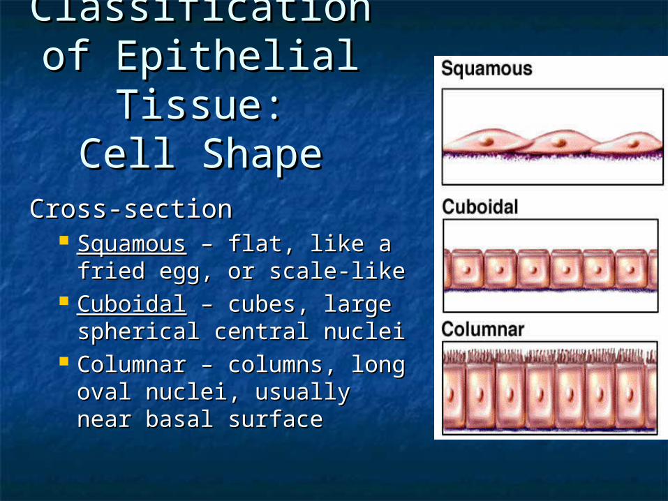
Classification Classification of Epithelial of Epithelial
Tissue:Tissue:Cell ShapeCell Shape
Cross-sectionCross-section SquamousSquamous – flat, like a – flat, like a fried egg, or scale-likefried egg, or scale-like
CuboidalCuboidal – cubes, large – cubes, large spherical central nucleispherical central nuclei
Columnar – columns, long Columnar – columns, long oval nuclei, usually oval nuclei, usually near basal surfacenear basal surface
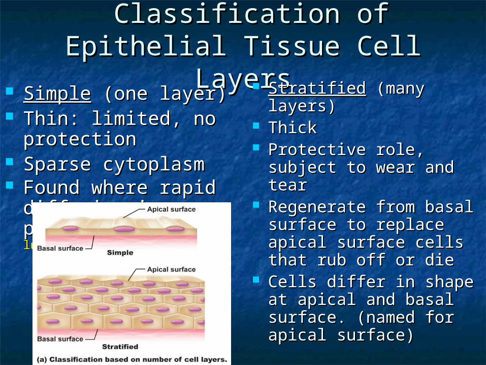
Classification of Classification of Epithelial Tissue Cell Epithelial Tissue Cell
LayersLayers SimpleSimple (one layer) (one layer) Thin: limited, no Thin: limited, no protectionprotection
Sparse cytoplasmSparse cytoplasm Found where rapid Found where rapid diffusion is a diffusion is a priority priority (ex. kidneys, (ex. kidneys, lungs)lungs)
StratifiedStratified (many (many layers)layers)
ThickThick Protective role, Protective role, subject to wear and subject to wear and teartear
Regenerate from basal Regenerate from basal surface to replace surface to replace apical surface cells apical surface cells that rub off or diethat rub off or die
Cells differ in shape Cells differ in shape at apical and basal at apical and basal surface. (named for surface. (named for apical surface)apical surface)

Pseudo-stratifiedPseudo-stratified
Shapes vary in height Shapes vary in height Nuclei at different levels – Nuclei at different levels – appear stratified, but aren’t.appear stratified, but aren’t.
All cells reach basement All cells reach basement membrane; only a few reach the membrane; only a few reach the surfacesurface
false

Simple Squamous Simple Squamous EpitheliumEpithelium
Function and LocationFunction and Location Areas of high diffusion rates:Areas of high diffusion rates:
gasses gasses (ex lungs)(ex lungs)nutrients and waste exchange nutrients and waste exchange (blood vessels and surrounding cells)(blood vessels and surrounding cells)
filtrates filtrates (kidneys)(kidneys) Makes lubricating fluid in Makes lubricating fluid in lining of body cavities lining of body cavities (ex. (ex. serous membranes)serous membranes)
One layer Flat

Simple Squamous EpitheliumSimple Squamous Epithelium(Top View) – cells fit (Top View) – cells fit
like tiled floorlike tiled floor

Figure 4.2
LUMEN
Simple Squamous Simple Squamous EpitheliumEpithelium(side view/cross (side view/cross section) section) – – cells look like cells look like fried eggfried egg
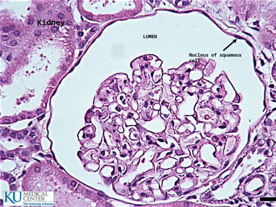
LUMEN
Nucleus of squamous cell
Kidney


Simple Cuboidal Simple Cuboidal EpitheliumEpithelium
Function and LocationFunction and Location Secretion and AbsorptionSecretion and Absorption Covers walls of SMALL ducts, Covers walls of SMALL ducts, glands, kidney tubules, ovariesglands, kidney tubules, ovaries
One layer Cubed
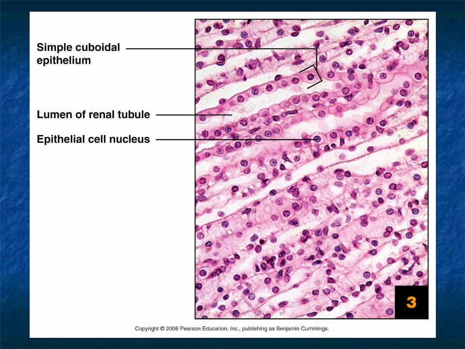

LUMEN
Spherical, large nuclei
Basement membrane
Cuboidal Cell
Apical surface

LUMENSpherical, large nuclei Apical surface
Basal surface


Simple Columnar Simple Columnar EpitheliumEpithelium
Function and LocationFunction and Location Absorption & Secretion Absorption & Secretion (ex. digestive (ex. digestive tract)tract)
When in open to body cavities – When in open to body cavities – called mucous membranescalled mucous membranes
Special FeaturesSpecial Features Often w/ Often w/ microvillmicrovilli on apical surface i on apical surface (brush border)(brush border)
Goblet cellsGoblet cells, single cell glands, , single cell glands, produce protective mucus.produce protective mucus.
One layer columns


LUMEN
Apical surface
Basal surface

Pseudostratified Pseudostratified EpitheliumEpithelium
FunctionFunction AbsorptionAbsorption Secretion of mucus by goblet cellsSecretion of mucus by goblet cells Cilia (larger than microvilli) Cilia (larger than microvilli) sweep mucus sweep mucus
LocationLocation Respiratory Linings & Reproductive Respiratory Linings & Reproductive tracttract

LUMEN

CiliaBasement Membrane
Multilevel nuclei
LUMEN

Stratified Squamous Stratified Squamous EpitheliumEpithelium
StructureStructure Cells often cuboidal or columnar Cells often cuboidal or columnar below apical squamous layerbelow apical squamous layer
Function and LocationFunction and Location ProtectionProtection Keratin (protein) is accumulated in Keratin (protein) is accumulated in older cells near the surface – older cells near the surface – waterproofs and toughens skinwaterproofs and toughens skin
LocationLocation Skin (keratinized), mouth & throatSkin (keratinized), mouth & throat
Multi-layer (thick!)
Flat (only cells on apical surface)
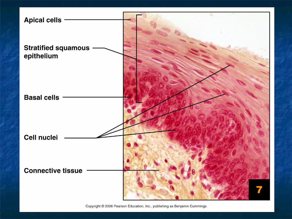


keratin
Dense-Irregular Connective Tissue
Basement Membrane
Squamous
Cuboidal
Columnar

TransitionaTransitional l
EpitheliumEpithelium
StructureStructure Multi-layerMulti-layer Basal surface cells are cuboidal Basal surface cells are cuboidal or columnaror columnar
Apical surface cells vary: changes Apical surface cells vary: changes shape to accommodate for change in shape to accommodate for change in volume due to stretchingvolume due to stretching
FunctionFunction Allows stretchingAllows stretching
LocationLocation Urinary bladder, ureters & urethraUrinary bladder, ureters & urethra
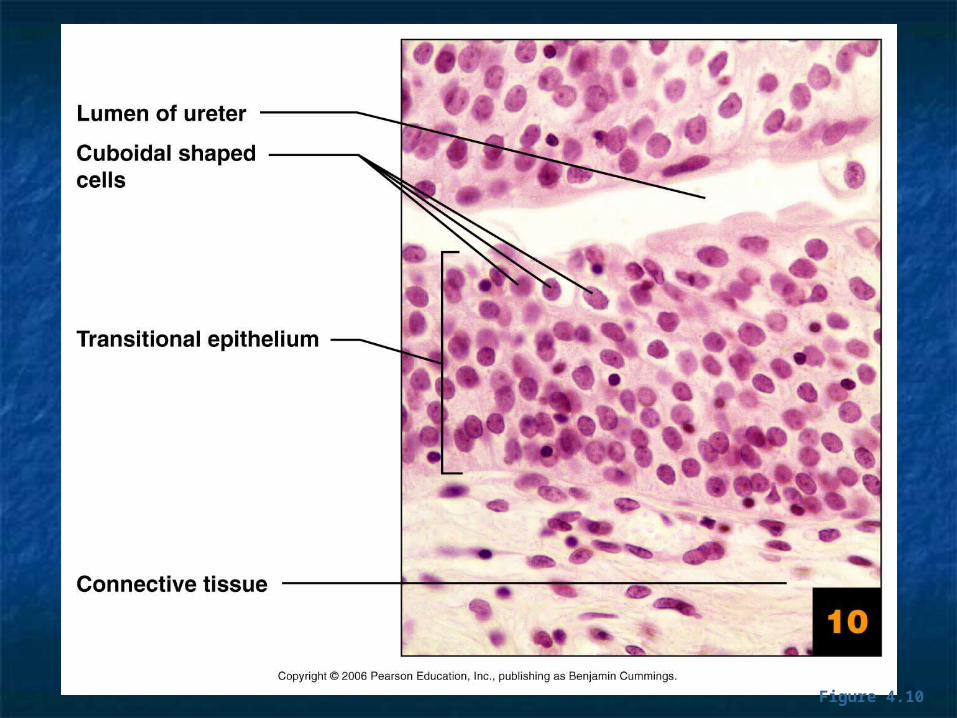
Figure 4.10


Nervous TissueNervous Tissue

Muscular TissueMuscular Tissue

GlandsGlands
Cells that secrete or export a product.Cells that secrete or export a product. Secretion = protein, lipids, hormones, Secretion = protein, lipids, hormones, steroids, acidssteroids, acids
Endocrine glands (internally secreting)Endocrine glands (internally secreting) No duct, release secretion into blood No duct, release secretion into blood vessels vessels
Often hormonesOften hormones Thyroid, adrenal and pituitary glandsThyroid, adrenal and pituitary glands
Exocrine glands (externally secreting)Exocrine glands (externally secreting) Contain ducts, empty onto epithelial surfaceContain ducts, empty onto epithelial surface Sweat, Oil glands, Salivary glands, Mammary Sweat, Oil glands, Salivary glands, Mammary glands.glands.

Shapes of Exocrine Shapes of Exocrine glandsglands
Branching
Simple – single, unbranched duct
Compound – branched.
Shape: tubular or alveolar
Tubular – shaped like a tube
Alveolar – shaped like flasks or sacs
Tubuloalveolar – has both tubes and sacs in gland
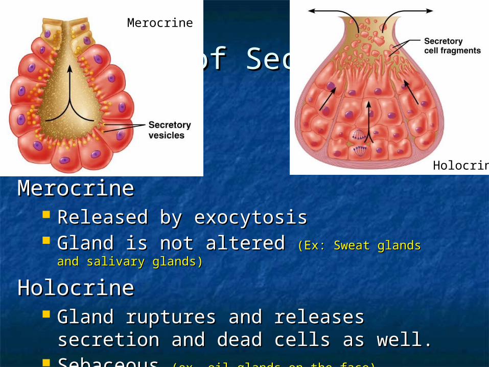
Modes of SecretionModes of Secretion
MerocrineMerocrine Released by exocytosis Released by exocytosis Gland is not altered Gland is not altered (Ex: Sweat glands (Ex: Sweat glands
and salivary glands)and salivary glands)
HolocrineHolocrine Gland ruptures and releases Gland ruptures and releases secretion and dead cells as well.secretion and dead cells as well.
Sebaceous Sebaceous (ex. oil glands on the face)(ex. oil glands on the face)
Merocrine
Holocrine