tissue optics - guided therapeutics. optical properties 2. how to measure optical properties 3....
TRANSCRIPT

Scott Prahl
= graduated
Ricky Wang Steven Jacques Sean Kirkpatrick
Don Duncan
Tissue Optics
Steven L. Jacques [email protected] http://omlc.ogi.edu
Depts. of Biomedical Engineering and Dermatology Oregon Health & Science University, Portland OR, USA

1. Optical properties
2. How to measure optical properties
3. Light transport
4. Complex tissues
Tissue Optics
Steven L. Jacques [email protected] http://omlc.ogi.edu
Depts. of Biomedical Engineering and Dermatology
Oregon Health & Science University, Portland OR, USA

1. where tissue affects photons, used for diagnostic sensing, imaging, and spectroscopy of tissues and biomaterials
We replaced heel stick tests with pain-free opical measurement of bilirubin
(FDA approved, marketed as Bilichek)
Normal light
polarized light Guiding skin
cancer surgery with polarized light camera
True cancer margin
BCC = basal cell carcinoma
Cancer margin seen by Doctor’s eye
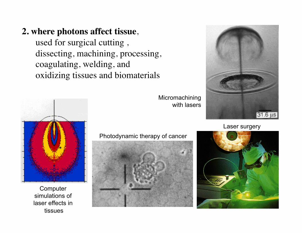
2. where photons affect tissue, used for surgical cutting , dissecting, machining, processing, coagulating, welding, and oxidizing tissues and biomaterials
Micromachining with lasers
Laser surgery Photodynamic therapy of cancer
Computer simulations of laser effects in
tissues
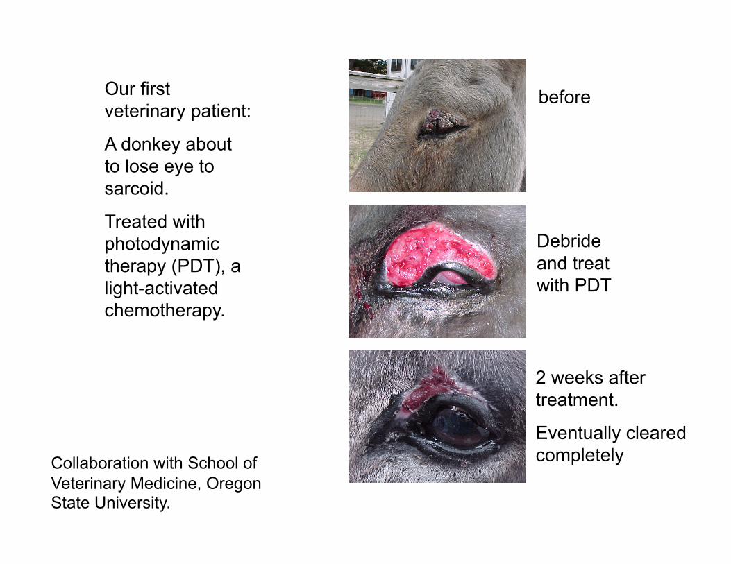
Our first veterinary patient:
A donkey about to lose eye to sarcoid.
Treated with photodynamic therapy (PDT), a light-activated chemotherapy.
before
Debride and treat with PDT
2 weeks after treatment.
Eventually cleared completely Collaboration with School of
Veterinary Medicine, Oregon State University.
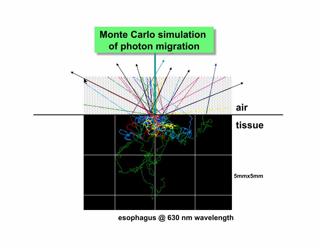
tissue
air
5mmx5mm
esophagus @ 630 nm wavelength
Monte Carlo simulation of photon migration

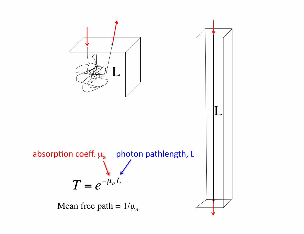
A photon’s path is tortuous due to multiple scattering, like a ball of string.
Nevertheless, there is a total pathlength L, like the length of the string.
L
€
T = e−µa L
Mean free path = 1/µa
L
L
€
T = e−µa L
Mean free path = 1/µa
L
absorp'oncoeff.µa photonpathlength,L

cm2W
per Wincident
power
r [mm]
z [mm]
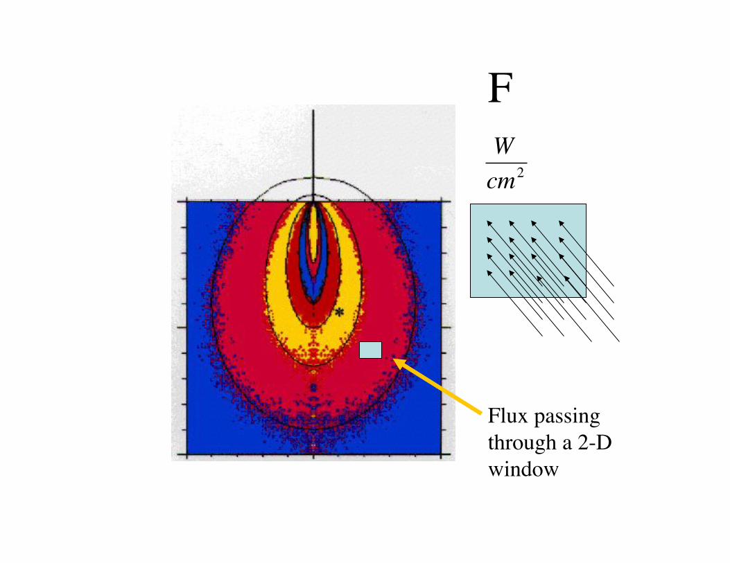
Monte Carlo
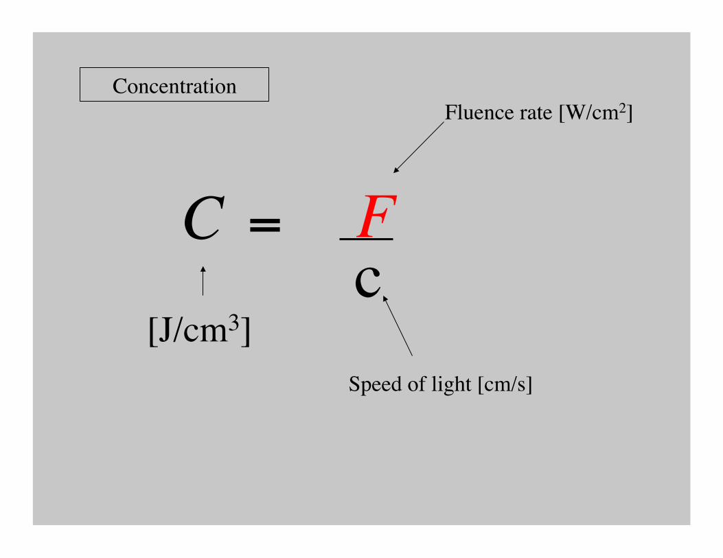
photon diffusion Fluence rate [W/cm2]
€
Wcm2
F
Flux passing through a 2-D window

€
Wcm2 =
Jcm3
cms
F = C c
Energy density within a 3-D volume

1 W cm-2
(c [cm/s])(44 ps)
1 cm
1 cm
1 cm
for λ = 488 nm, n = 1.33
Fluence rate = speed of light x photon concentration F = c C
€
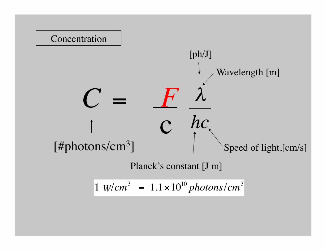
Cph =Enc
λhc
= 1.1×10¹⁰ photons / cm3
=Enc
λhc
1000Nav
= 1.8×10⁻¹³moles / liter
€
1 W / cm2
€
(c [cm / s])(44 ps)
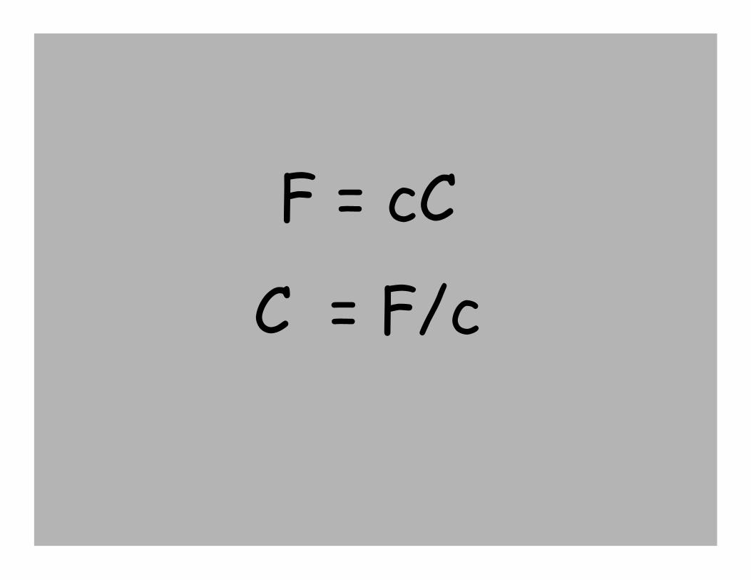
F = cC
C = F/c

Photochemical
Photothermal
Photomechanical
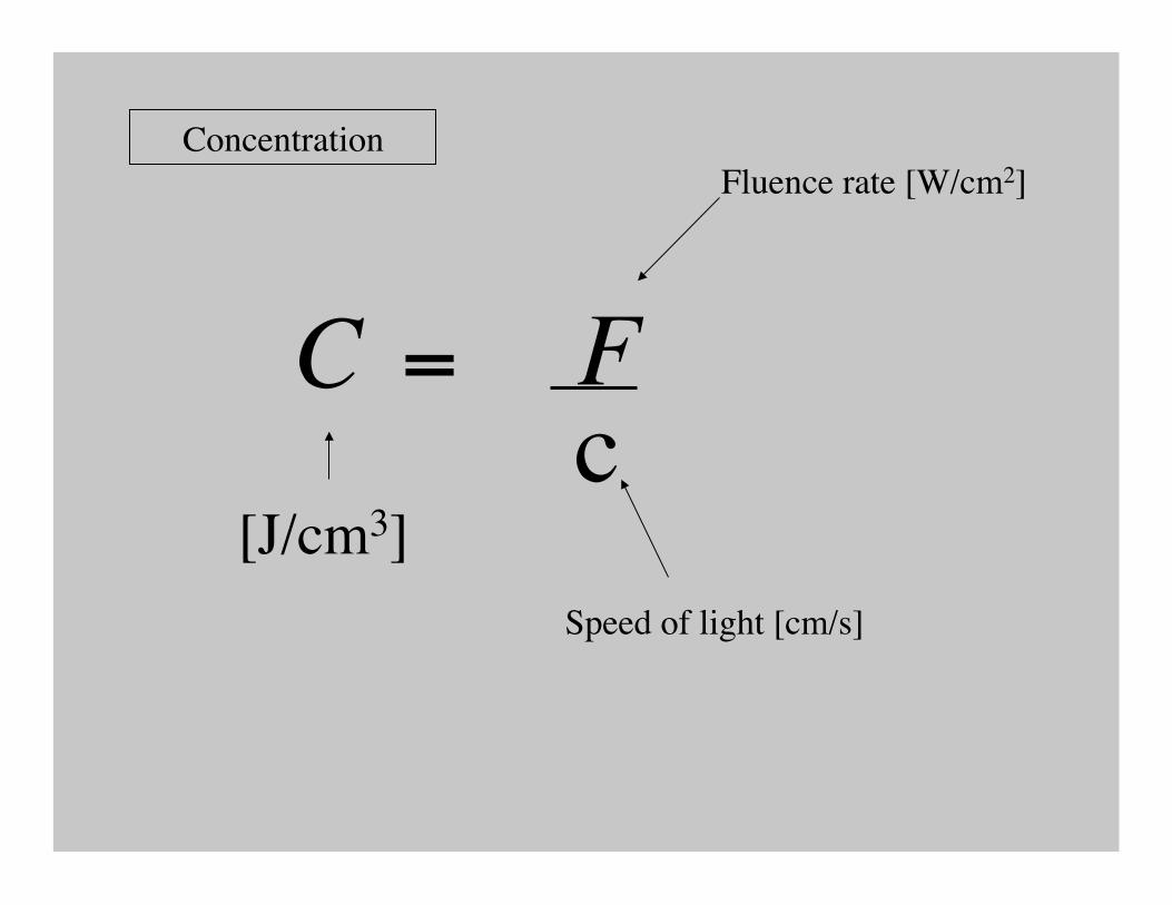
Concentration Fluence rate [W/cm2]
Speed of light [cm/s]
[J/cm3]
= C F
c
Concentration Fluence rate [W/cm2]
Speed of light [cm/s]
[J/cm3]
= C F
c
Concentration
[#photons/cm3]
= C F
c
€
λhc1000Nav
Wavelength [m]
Speed of light,[cm/s]
Planck’s constant [J m]
[ph/J]
€
1 J /cm3 = 1.1×10¹⁰ photons /cm3
= 1.8 ×10⁻¹³moles / literW

Concentration
= C F
c
€
λhc1000Nav
[moles/liter]
# [cm3/liter]
# [1/mole]
€
1 J /cm3 = 1.1×10¹⁰ photons /cm3
= 1.8 ×10⁻¹³moles / literW

Energy deposition
time [s]
Absorption coefficient [1/cm]
[J/cm3]
= Q Ft
Fluence rate [W/cm2]
µ a
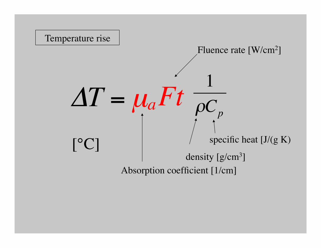
ΔT = µ a Ft
Temperature rise
€
1ρCp
density [g/cm3]
specific heat [J/(g K) [°C]
Fluence rate [W/cm2]
Absorption coefficient [1/cm]
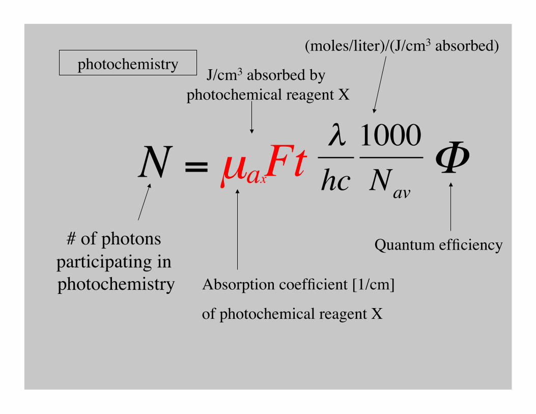
N = µ ax Ft
(moles/liter)/(J/cm3 absorbed)
# of photons participating in photochemistry
photochemistry
€
λhc1000Nav
Φ
Quantum efficiency
J/cm3 absorbed by photochemical reagent X
Absorption coefficient [1/cm]
of photochemical reagent X
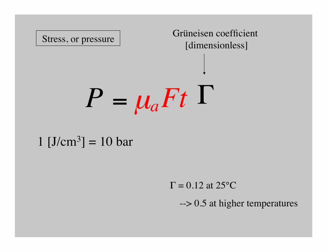
P = µ a Ft
€
Γ
Grüneisen coefficient [dimensionless]
1 [J/cm3] = 10 bar
Stress, or pressure
Γ = 0.12 at 25°C
--> 0.5 at higher temperatures
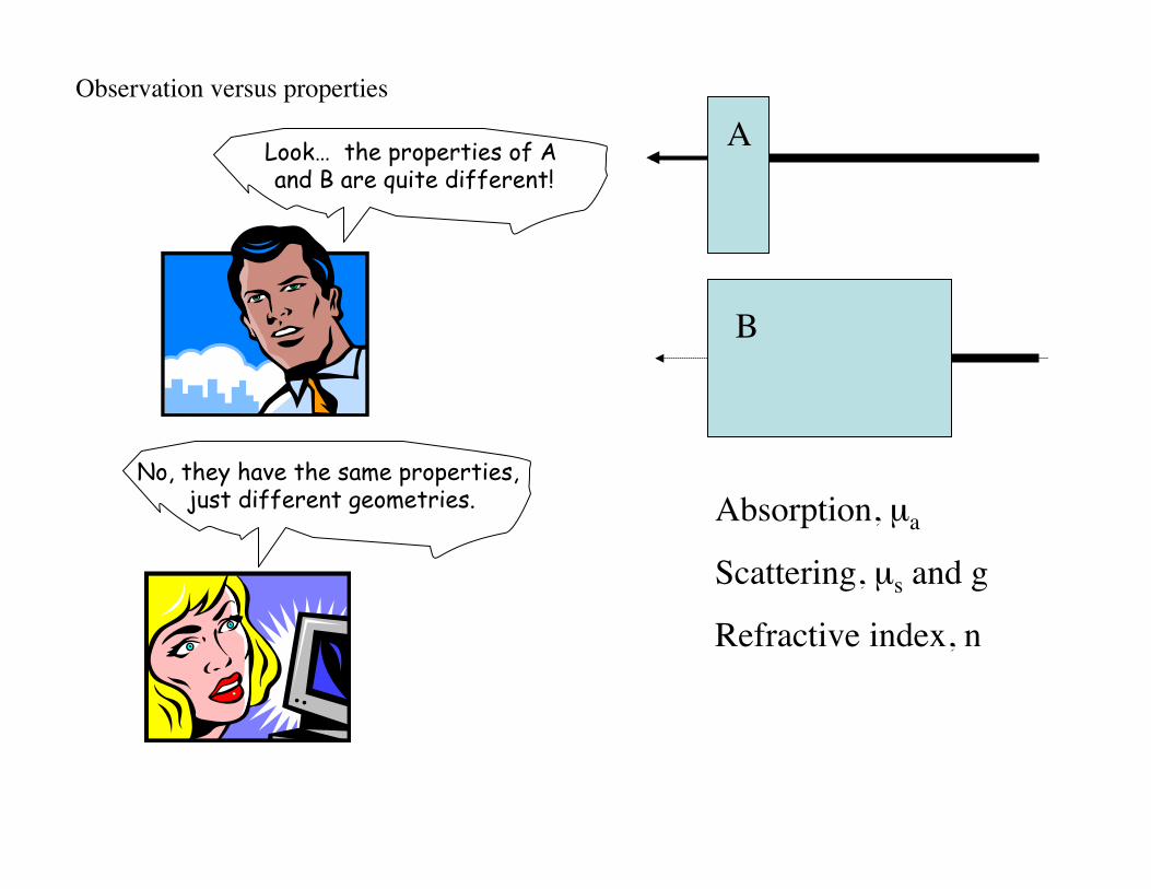
A
B
Look… the properties of A and B are quite different!
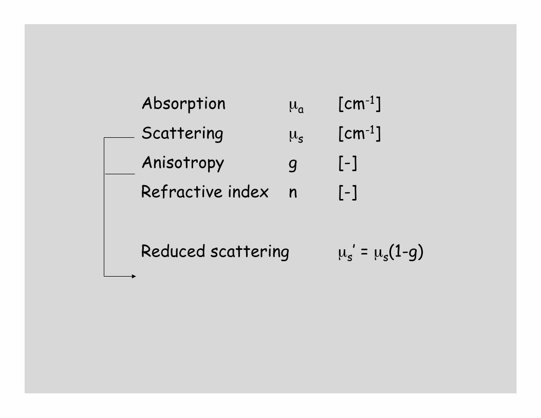
No, they have the same properties, just different geometries. Absorption, µa
Scattering, µs and g
Refractive index, n
Observation versus properties
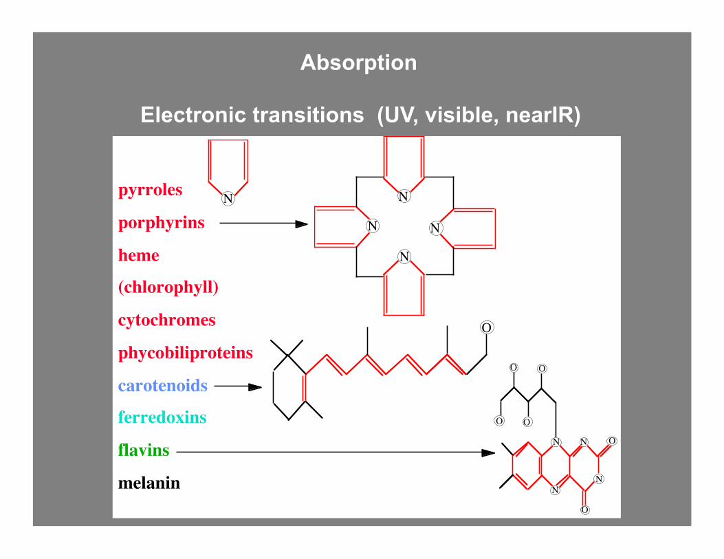
Electronic transitions (UV, visible, nearIR)
N
N
N
N
pyrroles
porphyrins
heme
(chlorophyll)
cytochromes
phycobiliproteins
carotenoids
ferredoxins
flavins
melanin
N
O
N N
N
O
N
O
OO
OO
Absorption

vibrational transitions (nearIR, midIR, farIR)
Absorption

absorption cross-sectional area efficiency
geometrical area
Ageometrical
cross-sectioneffective
cross-section
σ = Q Aa a
σa = Qa A
[cm2] [-] [cm2]

absorption coefficient
density cross-sectional area
µa = ρa σa
[cm-1] [cm-3][cm2] σa = Qa A[cm2] [-][cm2]
efficiency area
T = exp(-µaL)

1
10
100
1,000
10,000
100 1,000 10,000Abs
orpt
ion
coef
ficie
nt [c
m-1
]
Wavelength [nm]
KrFexcimer
XeClexcimer
ArFexcimer
Ho:YAG
Er:YAG
CO2
Vis
aorta
UV IR
Nd:YAG
whole blood
dye laserargon
skin
75% water
melanosomeepidermis
1 µm
10 µm
100 µm
1 mm
10 mm
1 cm
mfp = 1/µa
µa
€
mfp =1
µa
mean free path
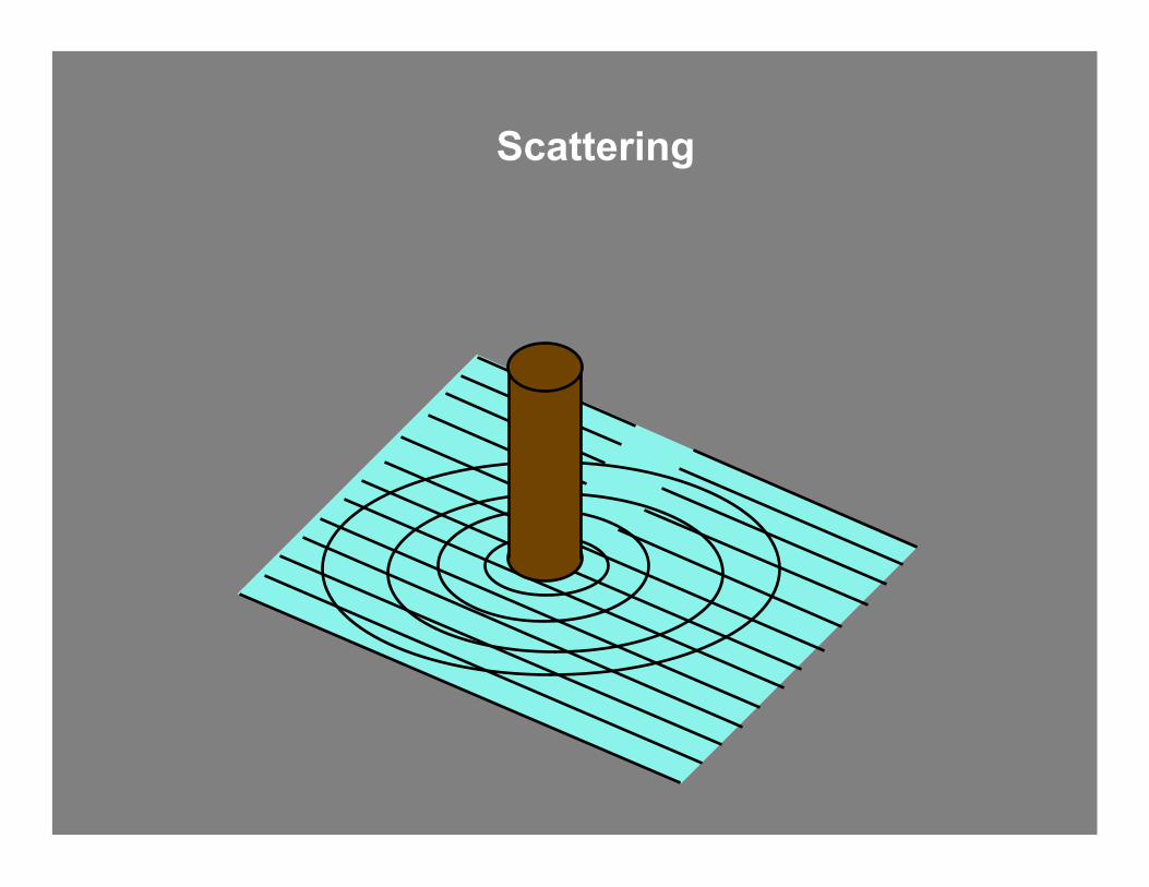
Scattering

dipole

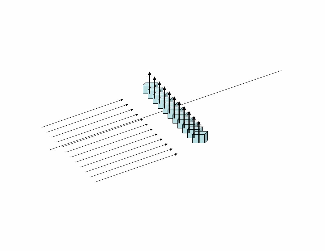

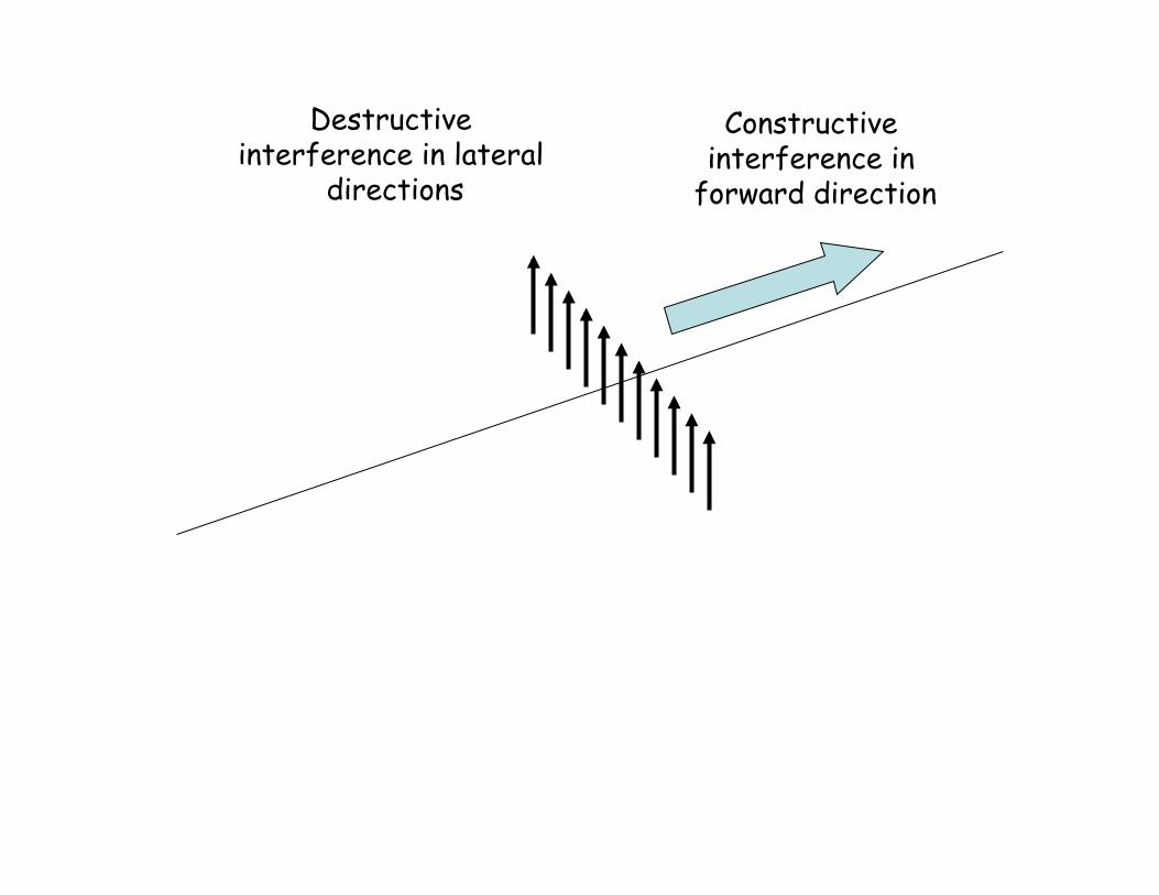
Constructive interference in
forward direction
Destructive interference in lateral
directions

Non-uniform density of dipoles yields scattering
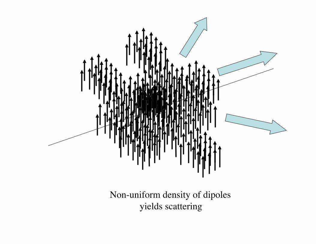
Non-uniform density of dipoles yields scattering
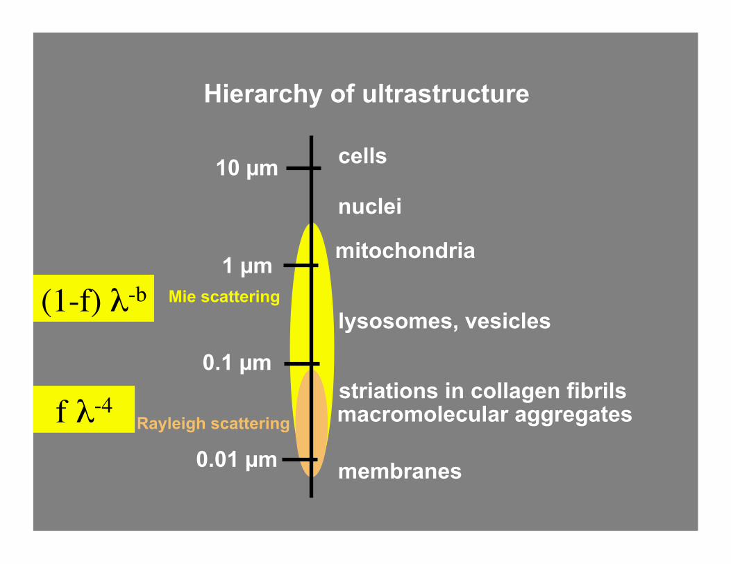
Hierarchy of ultrastructure
striations in collagen fibrils
membranes
mitochondria
nuclei
cells 10 µm
1 µm
0.1 µm
lysosomes, vesicles Mie scattering
Rayleigh scattering macromolecular aggregates
0.01 µm
(1-f) λ-b
f λ-4
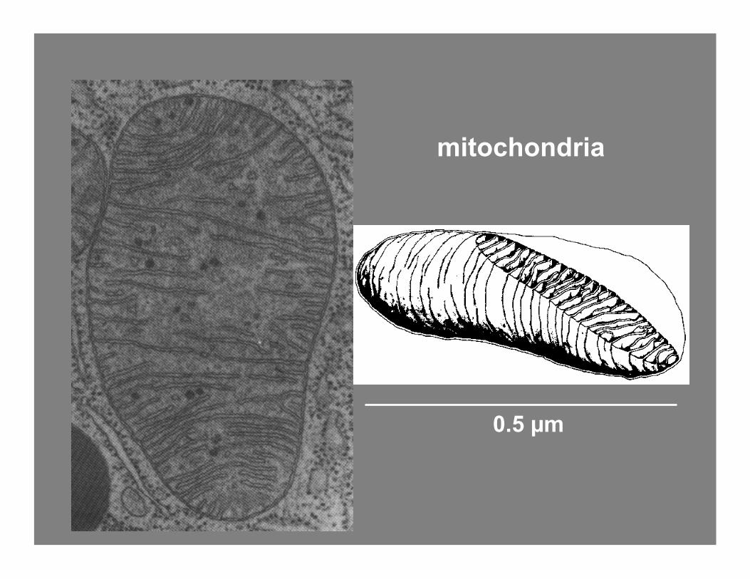
mitochondria
0.5 µm

Collagen fibrils, fibers

Ageometrical
cross-sectioneffective
cross-section
σ = Q As s
σs = Qs A
[cm2] [-] [cm2]
scattering cross-section efficiency
cross-sectional area

scattering coefficient
density cross-sectional area
µ s = ρ s σ s
[ c m - 1 ] [ c m - 3 ] [ c m 2 ] σ s = Q s A [ c m 2 ] [ - ] [ c m 2 ]
efficiency area
T = e x p ( - µ s L )

Anisotropy, g
cos(θ)cos(θ)
azimuthalangle ψψ
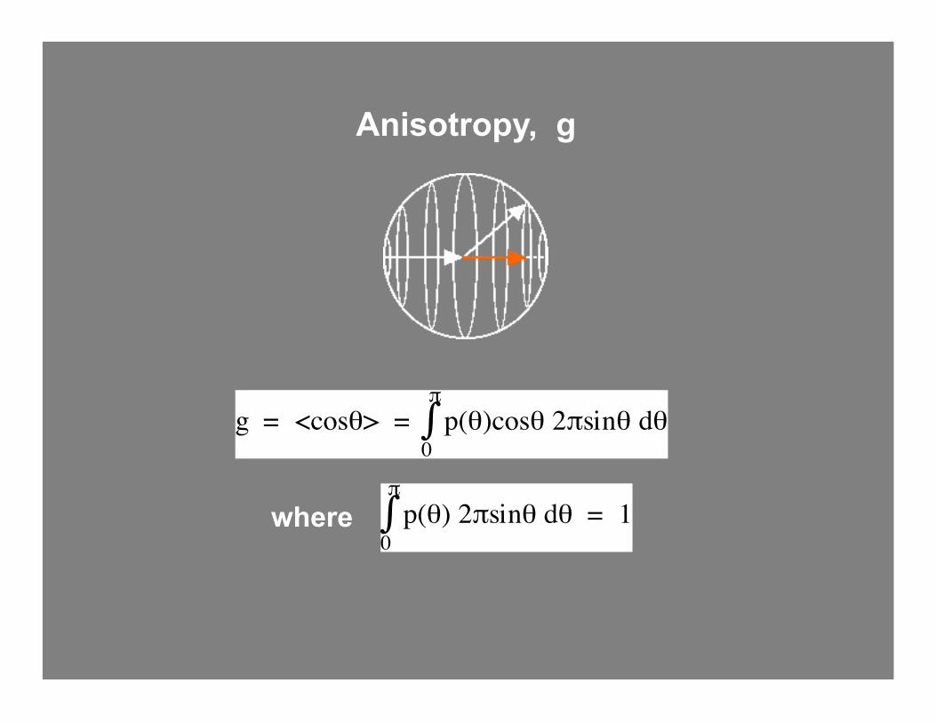
g = <cosθ> = ∫0
π p(θ)cosθ 2πsinθ dθ
∫0
π p(θ) 2πsinθ dθ = 1where
Anisotropy, g
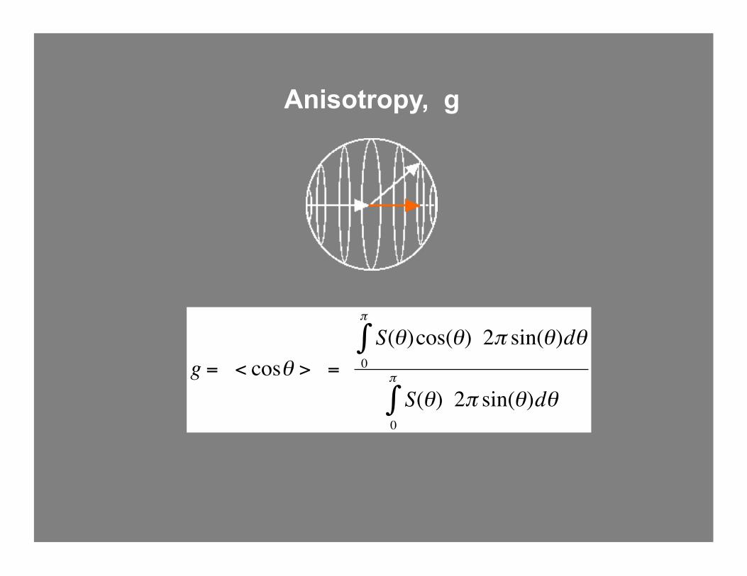
Anisotropy, g
€
g = < cosθ > = S(θ)cos(θ) 2π sin(θ)dθ
0
π
∫
S(θ) 2π sin(θ)dθ0
π
∫
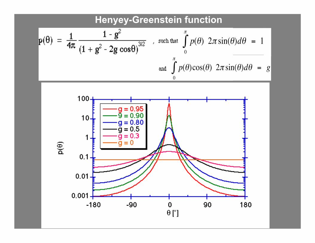
Henyey-Greenstein function
€
p(θ)cos(θ) 2π sin(θ)dθ = g0
π
∫
€
p(θ) 2π sin(θ)dθ = 10
π
∫
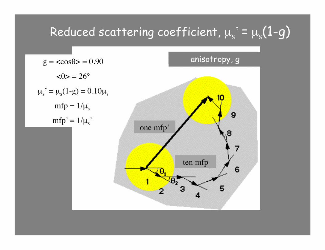
26°g = <cosθ> = 0.90
<θ> = 26°
µs’ = µs(1-g) = 0.10µs
mfp = 1/µs
mfp’ = 1/µs’
anisotropy, g
one mfp’
ten mfp
Reduced scattering coefficient, µs’ = µs(1-g)

equivalent Random Walk

Mie
Rayleigh
total µs’
skin
Reduced scattering coeff. of skin
µs‘ = µs(1-g)
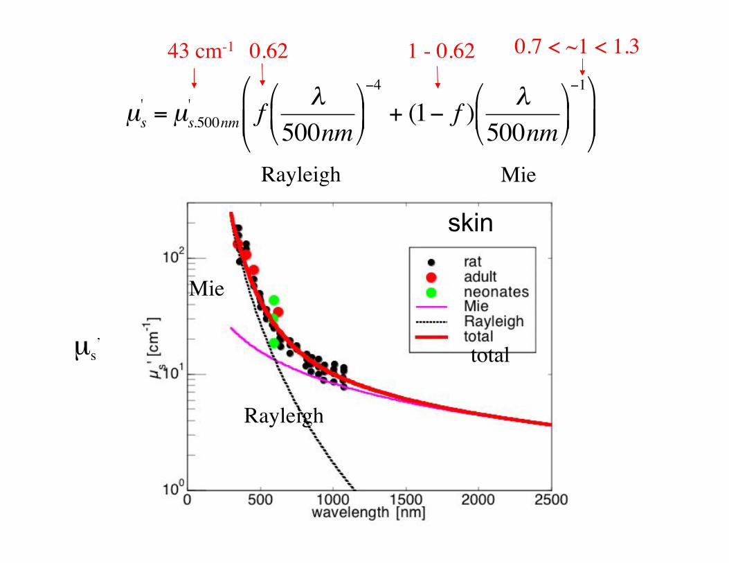
€
µs' = µs.500nm
' f λ500nm
−4
+ (1− f ) λ500nm
−1
Mie
Rayleigh
total µs’
43 cm-1 0.62 1 - 0.62 0.7 < ~1 < 1.3
Rayleigh Mie
skin
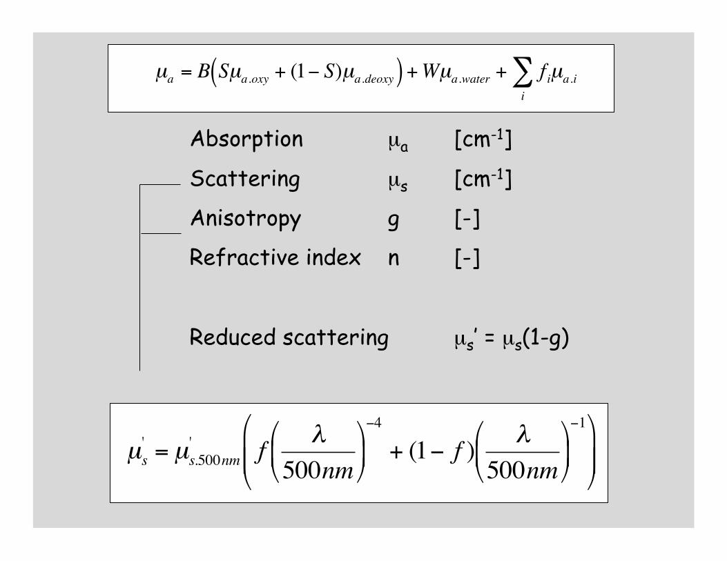
Absorption µa [cm-1]
Scattering µs [cm-1]
Anisotropy g [-]
Refractive index n [-]
Reduced scattering µs’ = µs(1-g)

Absorption µa [cm-1]
Scattering µs [cm-1]
Anisotropy g [-]
Refractive index n [-]
Reduced scattering µs’ = µs(1-g)
€
µa = B Sµa.oxy + (1− S)µa.deoxy( ) +Wµa.water + f iµa.ii∑
Absorption µa [cm-1]
Scattering µs [cm-1]
Anisotropy g [-]
Refractive index n [-]
Reduced scattering µs’ = µs(1-g)
€
µa = B Sµa.oxy + (1− S)µa.deoxy( ) +Wµa.water + f iµa.ii∑
€
µs' = µs.500nm
' f λ500nm
−4
+ (1− f ) λ500nm
−1

1. Optical properties
2. How to measure optical properties
3. Light transport
4. Complex tissues
Tissue Optics
Steven L. Jacques [email protected] http://omlc.ogi.edu
Depts. of Biomedical Engineering and Dermatology
Oregon Health & Science University, Portland OR, USA