time course of endothelium-dependent and -independent ... · time course of endothelium-dependent...
TRANSCRIPT
E
5(wpdaNa
Coc
J A C C : C A R D I O V A S C U L A R I N T E R V E N T I O N S V O L . 5 , N O . 7 , 2 0 1 2
© 2 0 1 2 B Y T H E A M E R I C A N C O L L E G E O F C A R D I O L O G Y F O U N D A T I O N I S S N 1 9 3 6 - 8 7 9 8 / $ 3 6 . 0 0
P U B L I S H E D B Y E L S E V I E R I N C . h t t p : / / d x . d o i . o r g / 1 0 . 1 0 1 6 / j . j c i n . 2 0 1 2 . 0 3 . 0 2 1
Time Course of Endothelium-Dependent and-Independent Coronary Vasomotor Responseto Coronary Balloons and StentsComparison of Plain and Drug-Eluting Balloons and Stents
Christian A. Plass, MD,* Inna Sabdyusheva-Litschauer, MSC,* Andreas Bernhart, MD,*slam Samaha, MD,† Örs Petnehazy, PHD,‡ Eszter Szentirmai, VMD,‡
Zsolt Petrasi, PHD,‡ Victor Lamin, MSC,* Noemi Pavo, MD, MSC,* Noemi Nyolczas, MD,*Andras Jakab, MD,§ Zsolt Murlasits, PHD,� Jutta Bergler-Klein, MD,* Gerald Maurer, MD,*Mariann Gyöngyösi, MD, PHD*
Vienna, Austria; Kaposvar and Debrecen, Hungary; and Memphis, Tennessee
Objectives This study sought to determine the time dependency of the endothelium-dependentand -independent vascular responses after percutaneous coronary intervention (PCI) with drug-eluting (DEB) or plain balloons, bare-metal (BMS), and drug-eluting (DES) stents, or controls.
Background Long-term endothelial dysfunction after DES implantation is associated with delayedhealing and late thrombosis.
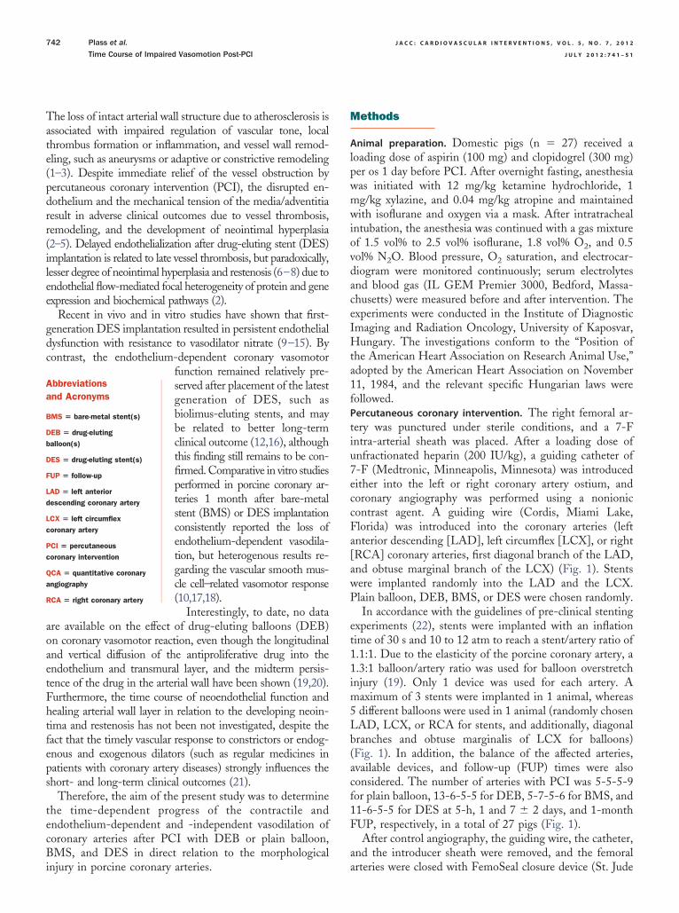
Methods Domestic pigs underwent PCI using DEB or plain balloon, BMS, or DES. The dilated andstented segments, and the proximal reference segments of stents and control arteries were ex-planted at 5-h, 24-h, 1-week, and 1-month follow-up (FUP). Endothelin-induced vasoconstriction andendothelium-dependent and -independent vasodilation of the arterial segments were determined invitro and were related to histological results.
Results DES- and BMS-treated arteries showed proneness to vasoconstriction 5 h post-PCI. The endothelium-dependent vasodilation was profoundly (p � 0.05) impaired early after PCI (9.8 � 3.7%, 13.4 � 9.2%,.7 � 5.3%, and 7.6 � 4.7% using plain balloon, DEB, BMS, and DES, respectively), as compared with controls49.6 � 9.5%), with slow recovery. In contrast to DES, the endothelium-related vasodilation of vessels treatedith plain balloon, DEB, and BMS was increased at 1 month, suggesting enhanced endogenous nitric oxideroduction of the neointima. The endothelium-independent (vascular smooth muscle–related) vasodilationecreased significantly at 1 day, with slow normalization during FUP. All PCI-treated vessels exhibited imbal-nce between vasoconstriction–vasodilation, which was more pronounced in DES- and BMS-treated vessels.o correlation between histological parameters and vasomotor function was found, indicating complex inter-ctions between the healing neoendothelium and smooth muscle post-PCI.
onclusions Coronary arteries treated with plain balloon, DEB, BMS, and DES showed time-dependent lossf endothelial-dependent and -independent vasomotor function, with imbalanced contraction/dilationapacity. (J Am Coll Cardiol Intv 2012;5:741–51) © 2012 by the American College of Cardiology Foundation
From the *Department of Cardiology, Medical University of Vienna, Vienna, Austria; †Department of Pulmonology, MedicalUniversity of Vienna, Vienna, Austria; ‡Institute of Diagnostic Imaging and Radiation Oncology, University of Kaposvar,Kaposvar, Hungary; §Department of Biomedical Laboratory and Imaging Science, Faculty of Medicine, University of Debrecen,Debrecen, Hungary; and the �Exercise Biochemistry Laboratory, Department of Health, Sport Sciences, the University ofMemphis, Memphis, Tennessee. The study was supported by the Ludwig Boltzmann Institute for Cardiovascular Research,Vienna, Austria. The guiding catheters and the FemoSeal were donated by Medtronic Inc., Minneapolis, Minnesota, and St. JudeMedical, St. Paul, Minnesota. The authors have reported that they have no relationships relevant to the contents of this paper todisclose.
Manuscript received October 30, 2011; revised manuscript received March 7, 2012, accepted March 29, 2012.
lpwmwiov
tiu7eccFa[awP
et11im5Lb(acf1F
a
J A C C : C A R D I O V A S C U L A R I N T E R V E N T I O N S , V O L . 5 , N O . 7 , 2 0 1 2
J U L Y 2 0 1 2 : 7 4 1 – 5 1
Plass et al.
Time Course of Impaired Vasomotion Post-PCI
742
The loss of intact arterial wall structure due to atherosclerosis isassociated with impaired regulation of vascular tone, localthrombus formation or inflammation, and vessel wall remod-eling, such as aneurysms or adaptive or constrictive remodeling(1–3). Despite immediate relief of the vessel obstruction bypercutaneous coronary intervention (PCI), the disrupted en-dothelium and the mechanical tension of the media/adventitiaresult in adverse clinical outcomes due to vessel thrombosis,remodeling, and the development of neointimal hyperplasia(2–5). Delayed endothelialization after drug-eluting stent (DES)implantation is related to late vessel thrombosis, but paradoxically,lesser degree of neointimal hyperplasia and restenosis (6–8) due toendothelial flow-mediated focal heterogeneity of protein and geneexpression and biochemical pathways (2).
Recent in vivo and in vitro studies have shown that first-generation DES implantation resulted in persistent endothelialdysfunction with resistance to vasodilator nitrate (9–15). Bycontrast, the endothelium-dependent coronary vasomotor
function remained relatively pre-served after placement of the latestgeneration of DES, such asbiolimus-eluting stents, and maybe related to better long-termclinical outcome (12,16), althoughthis finding still remains to be con-firmed. Comparative in vitro studiesperformed in porcine coronary ar-teries 1 month after bare-metalstent (BMS) or DES implantationconsistently reported the loss ofendothelium-dependent vasodila-tion, but heterogenous results re-garding the vascular smooth mus-cle cell–related vasomotor response(10,17,18).
Interestingly, to date, no dataare available on the effect of drug-eluting balloons (DEB)on coronary vasomotor reaction, even though the longitudinaland vertical diffusion of the antiproliferative drug into theendothelium and transmural layer, and the midterm persis-tence of the drug in the arterial wall have been shown (19,20).Furthermore, the time course of neoendothelial function andhealing arterial wall layer in relation to the developing neoin-tima and restenosis has not been not investigated, despite thefact that the timely vascular response to constrictors or endog-enous and exogenous dilators (such as regular medicines inpatients with coronary artery diseases) strongly influences theshort- and long-term clinical outcomes (21).
Therefore, the aim of the present study was to determinethe time-dependent progress of the contractile andendothelium-dependent and -independent vasodilation ofcoronary arteries after PCI with DEB or plain balloon,BMS, and DES in direct relation to the morphological
Abbreviationsand Acronyms
BMS � bare-metal stent(s)
DEB � drug-elutingballoon(s)
DES � drug-eluting stent(s)
FUP � follow-up
LAD � left anteriordescending coronary artery
LCX � left circumflexcoronary artery
PCI � percutaneouscoronary intervention
QCA � quantitative coronaryangiography
RCA � right coronary artery
injury in porcine coronary arteries. a
Methods
Animal preparation. Domestic pigs (n � 27) received aoading dose of aspirin (100 mg) and clopidogrel (300 mg)er os 1 day before PCI. After overnight fasting, anesthesiaas initiated with 12 mg/kg ketamine hydrochloride, 1g/kg xylazine, and 0.04 mg/kg atropine and maintainedith isoflurane and oxygen via a mask. After intratracheal
ntubation, the anesthesia was continued with a gas mixturef 1.5 vol% to 2.5 vol% isoflurane, 1.8 vol% O2, and 0.5ol% N2O. Blood pressure, O2 saturation, and electrocar-
diogram were monitored continuously; serum electrolytesand blood gas (IL GEM Premier 3000, Bedford, Massa-chusetts) were measured before and after intervention. Theexperiments were conducted in the Institute of DiagnosticImaging and Radiation Oncology, University of Kaposvar,Hungary. The investigations conform to the “Position ofthe American Heart Association on Research Animal Use,”adopted by the American Heart Association on November11, 1984, and the relevant specific Hungarian laws werefollowed.Percutaneous coronary intervention. The right femoral ar-ery was punctured under sterile conditions, and a 7-Fntra-arterial sheath was placed. After a loading dose ofnfractionated heparin (200 IU/kg), a guiding catheter of-F (Medtronic, Minneapolis, Minnesota) was introducedither into the left or right coronary artery ostium, andoronary angiography was performed using a nonionicontrast agent. A guiding wire (Cordis, Miami Lake,lorida) was introduced into the coronary arteries (leftnterior descending [LAD], left circumflex [LCX], or rightRCA] coronary arteries, first diagonal branch of the LAD,nd obtuse marginal branch of the LCX) (Fig. 1). Stentsere implanted randomly into the LAD and the LCX.lain balloon, DEB, BMS, or DES were chosen randomly.In accordance with the guidelines of pre-clinical stenting
xperiments (22), stents were implanted with an inflationime of 30 s and 10 to 12 atm to reach a stent/artery ratio of.1:1. Due to the elasticity of the porcine coronary artery, a.3:1 balloon/artery ratio was used for balloon overstretchnjury (19). Only 1 device was used for each artery. A
aximum of 3 stents were implanted in 1 animal, whereasdifferent balloons were used in 1 animal (randomly chosenAD, LCX, or RCA for stents, and additionally, diagonalranches and obtuse marginalis of LCX for balloons)Fig. 1). In addition, the balance of the affected arteries,vailable devices, and follow-up (FUP) times were alsoonsidered. The number of arteries with PCI was 5-5-5-9or plain balloon, 13-6-5-5 for DEB, 5-7-5-6 for BMS, and1-6-5-5 for DES at 5-h, 1 and 7 � 2 days, and 1-monthUP, respectively, in a total of 27 pigs (Fig. 1).After control angiography, the guiding wire, the catheter,
nd the introducer sheath were removed, and the femoral
rteries were closed with FemoSeal closure device (St. Jude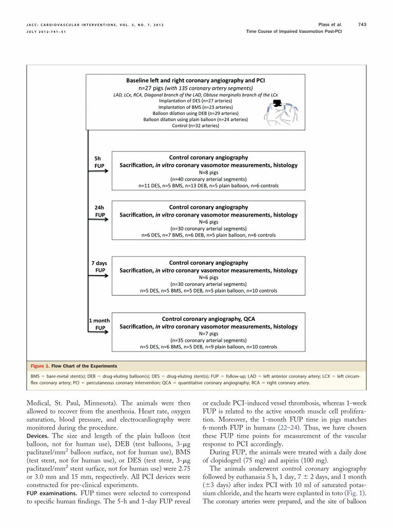
bp
tative
J A C C : C A R D I O V A S C U L A R I N T E R V E N T I O N S , V O L . 5 , N O . 7 , 2 0 1 2 Plass et al.
J U L Y 2 0 1 2 : 7 4 1 – 5 1 Time Course of Impaired Vasomotion Post-PCI
743
Medical, St. Paul, Minnesota). The animals were thenallowed to recover from the anesthesia. Heart rate, oxygensaturation, blood pressure, and electrocardiography weremonitored during the procedure.Devices. The size and length of the plain balloon (testalloon, not for human use), DEB (test balloons, 3-�gaclitaxel/mm2 balloon surface, not for human use), BMS
(test stent, not for human use), or DES (test stent, 3-�gpaclitaxel/mm2 stent surface, not for human use) were 2.75or 3.0 mm and 15 mm, respectively. All PCI devices wereconstructed for pre-clinical experiments.FUP examinations. FUP times were selected to correspond
Figure 1. Flow Chart of the Experiments
BMS � bare-metal stent(s); DEB � drug-eluting balloon(s); DES � drug-elutingflex coronary artery; PCI � percutaneous coronary intervention; QCA � quanti
to specific human findings. The 5-h and 1-day FUP reveal
or exclude PCI-induced vessel thrombosis, whereas 1-weekFUP is related to the active smooth muscle cell prolifera-tion. Moreover, the 1-month FUP time in pigs matches6-month FUP in humans (22–24). Thus, we have chosenthese FUP time points for measurement of the vascularresponse to PCI accordingly.
During FUP, the animals were treated with a daily doseof clopidogrel (75 mg) and aspirin (100 mg).
The animals underwent control coronary angiographyfollowed by euthanasia 5 h, 1 day, 7 � 2 days, and 1 month(�3 days) after index PCI with 10 ml of saturated potas-sium chloride, and the hearts were explanted in toto (Fig. 1).
(s); FUP � follow-up; LAD � left anterior coronary artery; LCX � left circum-coronary angiography; RCA � right coronary artery.
stent
The coronary arteries were prepared, and the site of balloon
a
qm
am
msrs1H(cpim
J A C C : C A R D I O V A S C U L A R I N T E R V E N T I O N S , V O L . 5 , N O . 7 , 2 0 1 2
J U L Y 2 0 1 2 : 7 4 1 – 5 1
Plass et al.
Time Course of Impaired Vasomotion Post-PCI
744
dilation, the stented segments, and the proximal segmentsadjacent to stenting were cut and flushed with physiologicalsaline solution, then the vasomotor response of the cut (stent-free) segments was measured. After completion of the in vitromeasurements, the balloon-dilated segments, the segmentswith stents, and the proximal parts were fixed in 4% formalin.The stented arteries were embedded in Technovit 9100(Heraeus Kulzer GmbH, Wehrheim, Germany), whereas thenative coronary arteries were embedded in paraffin, cut into 4-to 6-�m-thick slices, and then stained with hematoxylin-eosinnd MOVAT pentachrome stains (22,25).Quantitative coronary angiography. Post-stent and FUPuantitative coronary angiography (QCA) parameters wereeasured by means of a computer-assisted quantitative
Figure 2. Schematic Illustration of the In Vitro Measurements of Vessel Va
(A) L-shaped metal pins in a myograph with a vessel (arrow). (B) Bath chamboxide). (D) Longitudinal section of the arteries and changes in response to thethe arteries in response to the applied solution and drugs (schematic illustratio
to the applied drugs. (G) Schematic illustration of the wall tension in response to thrteriographic edge-detection algorithm (ACOM.PC, Sie-ens, Erlangen, Germany) (Online Methods 1).
Measurements of vasomotor responses. The dilated seg-ent of the coronary arteries as well as the proximal
egments adjacent to the stents were isolated, and after theemoval of fat and connective tissue, 4-mm-long ringegments were cut and mounted in a temperature-controlled5-ml tissue bath (37°C) containing a modified Krebs–enseleit buffer solution (Fig. 2), as published previously
8) (Online Methods 2). Briefly, to measure the isometricircular wall tension of the vessels, each segment was sus-ended between 2 L-shaped metal pins (0.4 mm in diameter)n a myograph (8). After approximately 1 h, the vessels were
aximally contracted with endothelin-1 (30 nmol/l).
tor Reaction
h the L-shaped pins. (C) Applied (arrows) solution and drugs (NO � nitriced solution and drugs (schematic illustration). (E) Cross-sectional changes of) Pathophysiological mechanism of the changes of the arteries in response
somo
er witapplin). (F
e applied drugs (blue line).
daimtma
twnew
diriM
R
s0aa
0vaitDr
cp
Daphl((
td(r
J A C C : C A R D I O V A S C U L A R I N T E R V E N T I O N S , V O L . 5 , N O . 7 , 2 0 1 2 Plass et al.
J U L Y 2 0 1 2 : 7 4 1 – 5 1 Time Course of Impaired Vasomotion Post-PCI
745
Maximum endothelium-dependent vasodilation wasachieved by a bolus application of substance P directly intothe organ bath with increasing concentration, up to a finalconcentration of 1 nmol/l. Sensitivity of smooth muscle toexternal nitric oxide (NO) was investigated by subsequentaddition of sodium nitroprusside (4 mmol/l). The vasocon-striction, corresponding to media injury and/or endothelialdysfunction in regulation of vascular tone, was expressed asmilliNewtons (mN). The endothelium-dependent and-independent vasodilation (stiffness of vascular smoothmuscle after overstretch injury of PCI) were expressed inpercent change of steady-state level contraction, and inmN/s/mN units, respectively.Histopathology and histomorphometry. Each arterial seg-ment (stented, balloon-dilated, and proximal to stents) wascut into 3 parts (proximal, middle, and distal) and investigatedhistopathologically and histomorphometrically. Stented seg-ments were evaluated using recommendation of Schwartz et al.(22) and Virmani et al. (6) (Online Methods 3).
Dilated segments or the segments proximal to stents wereanalyzed similarly to the score system after stenting, adaptedfor balloon injury only (19,25) (Online Methods 4). Endo-thelial coverage was expressed as the percentage of thelumen circumference covered by endothelium, measured bycomputerized planimetry (ImageJ version 1.440, NIH, Be-thesda, Maryland).
The following quantitative histomorphometric parame-ters were measured: lumen, internal and external elasticlamina area, and the maximal neointimal thickness. Thecalculated histomorphometric parameters included the neo-intima area (difference between internal elastic lamina andlumen area), media area (difference between external andinternal elastic lamina area), and % area stenosis [(neointi-mal area/internal elastic lamina area) � 100].Statistics. In the absence of normal values with standard
eviation or expected significant intraindividual changend, therefore, no possible sample size calculation regard-ng the in vitro vasomotor function measurements, a
inimum number of 5 samples per group and 1 FUPime were attempted. No adjustments were made forultiple comparisons or for multiple observations within
nimals.Continuous parameters were tested for normal distribu-
ion and expressed as mean � SD. Differences in variancesere tested, and if the Levene test had a p value �0.05, aonparametric test was used for comparison. In case ofqual variances, the analysis of variance was supplementedith 2-sided Student t test.Correlation between vessel constriction and endothelium-
ependent and -independent vasodilation or between histolog-cal and vasomotor parameters was evaluated using linearegression analysis. A p value of �0.05 was considered signif-cant. The statistical analyses were performed with SPSS for
acintosh version 17 (SPSS Inc., Chicago, Illinois). (
esults
FUP angiography. Coronary angiography at 5 h, 1 day and7 � 2 days revealed no stenosis nor suspected thrombosis.At the 1-month FUP, no angiographic abnormalities of thedilated segments were seen. However, QCA documented15 � 10% and 8 � 9% diameter stenosis of the stentedegments with BMS and DES, with a late lumen loss of.78 � 0.32 mm and 0.24 � 0.21 mm, respectively. Therterial segments proximal or distal to the stents showed nongiographic abnormalities.Time dependency of the vasomotor reaction post-PCI.Endothelin induced proneness to vasoconstriction in arter-ies with DES or BMS (22.6 � 4.7 mN or 20.6 � 4.6 mN)at the earliest time point (5 h) post-PCI, compared withcontrol (12.2 � 3.6 mN), or balloon-dilated arteries (p �.05) (Figs. 3 and 4). Interestingly, 1-day post-PCI, theasoconstrictive response seemed to be normalized in DESnd BMS arteries. At 7 � 2 days post-PCI, the endothelin-nduced vasoconstriction increased in drug-eluting device–reated arteries (p � 0.05 between controls and DEB/ES), which was normalized at 1 month, as the vessel
epair was completed.The endothelium-dependent vasodilation was signifi-
antly (p � 0.05) decreased in all treated arteries 5 host-PCI (49.6 � 9.5% vs. 9.8 � 3.7% or 13.4 � 9.2% or
5.7 � 5.3% or 7.6 � 4.7% in controls vs. plain balloon,EB, BMS, or DES, respectively) compared with controls,
s an obvious sign of immediate endothelial layer injuryost-PCI, with slow recovery (Fig. 4). At 1-month FUP, aypersensitivity of the neoendothelium-dependent vasodi-
ation to substance P was observed in PCI-treated arteriesbut not DES-treated vessels) as compared with controlsp � 0.05) (68.6 � 10.0% or 76.0 � 13.1% or 78.7 � 18.3%
in plain balloon, DEB, or BMS). By contrast, theendothelium-dependent vasodilation remained significantlydepressed in DES-treated vessels (33.3 � 7.4%).
The endothelium-independent (muscular layer–related)vasodilation was profoundly impaired 1 day post-PCI inDEB/BMS/DES-treated arteries (0.062 � 0.045 mN/s/mN,0.054 � 0.041 mN/s/mN, and 0.023 � 0.003 mN/s/mN,respectively) compared with controls (0.142 � 0.047 mN/s/mN),but to a lesser extent in plain balloon arteries, indicating lessmedia (muscular) damage with the plain balloon when com-pared with DEB, BMS, or DES (Fig. 4). At 7-day FUP, thedysfunction in endothelium-independent vasodilation still per-sisted in DEB and DES-arteries.Vasoconstriction–vasodilation balance of the epicardialcoronary vessels. A significant correlation was found be-ween the vasoconstriction response and endothelium-ependent vasodilation of the noninstrumented arteriesFig. 5), indicating a physiological balance between contraction–elaxation. However, after pooling the data for 1 device
independent from FUP time), no association could be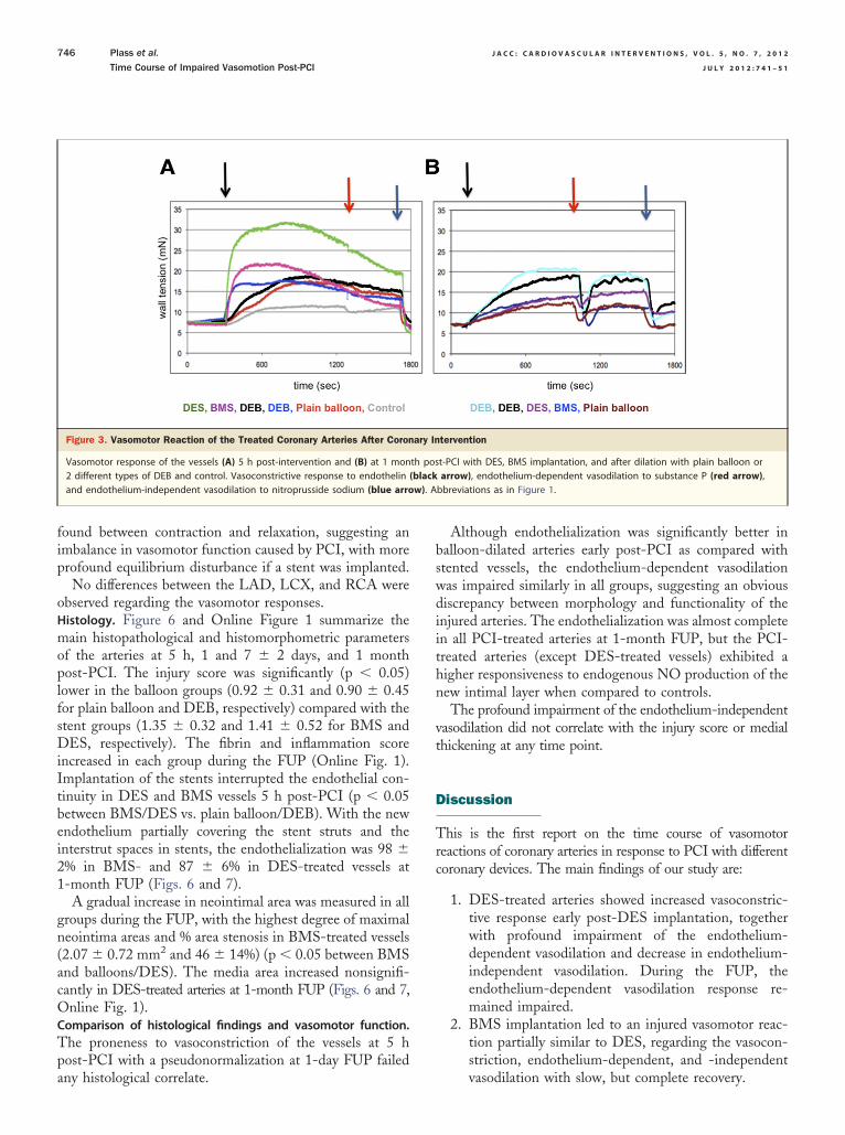
moplfsDiItbei21
gn(acO
w). A
J A C C : C A R D I O V A S C U L A R I N T E R V E N T I O N S , V O L . 5 , N O . 7 , 2 0 1 2
J U L Y 2 0 1 2 : 7 4 1 – 5 1
Plass et al.
Time Course of Impaired Vasomotion Post-PCI
746
found between contraction and relaxation, suggesting animbalance in vasomotor function caused by PCI, with moreprofound equilibrium disturbance if a stent was implanted.
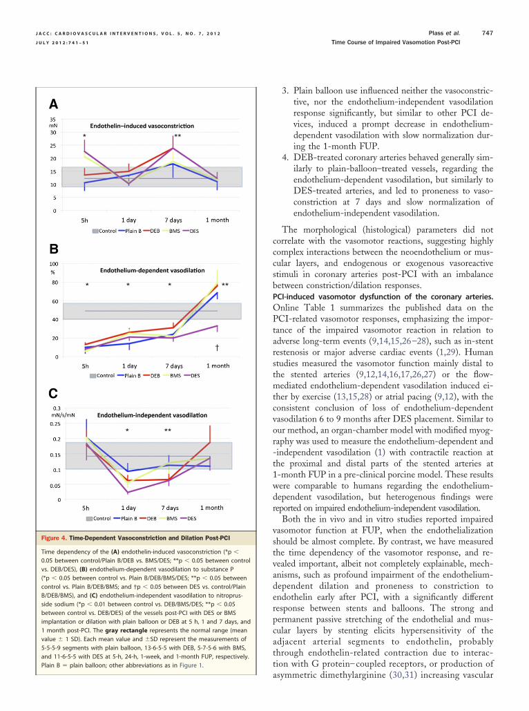
No differences between the LAD, LCX, and RCA wereobserved regarding the vasomotor responses.Histology. Figure 6 and Online Figure 1 summarize the
ain histopathological and histomorphometric parametersf the arteries at 5 h, 1 and 7 � 2 days, and 1 monthost-PCI. The injury score was significantly (p � 0.05)
ower in the balloon groups (0.92 � 0.31 and 0.90 � 0.45or plain balloon and DEB, respectively) compared with thetent groups (1.35 � 0.32 and 1.41 � 0.52 for BMS andES, respectively). The fibrin and inflammation score
ncreased in each group during the FUP (Online Fig. 1).mplantation of the stents interrupted the endothelial con-inuity in DES and BMS vessels 5 h post-PCI (p � 0.05etween BMS/DES vs. plain balloon/DEB). With the newndothelium partially covering the stent struts and thenterstrut spaces in stents, the endothelialization was 98 �% in BMS- and 87 � 6% in DES-treated vessels at-month FUP (Figs. 6 and 7).A gradual increase in neointimal area was measured in all
roups during the FUP, with the highest degree of maximaleointima areas and % area stenosis in BMS-treated vessels2.07 � 0.72 mm2 and 46 � 14%) (p � 0.05 between BMSnd balloons/DES). The media area increased nonsignifi-antly in DES-treated arteries at 1-month FUP (Figs. 6 and 7,nline Fig. 1).
Comparison of histological findings and vasomotor function.The proneness to vasoconstriction of the vessels at 5 hpost-PCI with a pseudonormalization at 1-day FUP failed
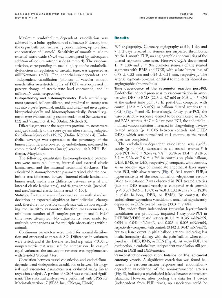
Figure 3. Vasomotor Reaction of the Treated Coronary Arteries After Coron
Vasomotor response of the vessels (A) 5 h post-intervention and (B) at 1 mon2 different types of DEB and control. Vasoconstrictive response to endothelinand endothelium-independent vasodilation to nitroprusside sodium (blue arro
any histological correlate.
Although endothelialization was significantly better inballoon-dilated arteries early post-PCI as compared withstented vessels, the endothelium-dependent vasodilationwas impaired similarly in all groups, suggesting an obviousdiscrepancy between morphology and functionality of theinjured arteries. The endothelialization was almost completein all PCI-treated arteries at 1-month FUP, but the PCI-treated arteries (except DES-treated vessels) exhibited ahigher responsiveness to endogenous NO production of thenew intimal layer when compared to controls.
The profound impairment of the endothelium-independentvasodilation did not correlate with the injury score or medialthickening at any time point.
Discussion
This is the first report on the time course of vasomotorreactions of coronary arteries in response to PCI with differentcoronary devices. The main findings of our study are:
1. DES-treated arteries showed increased vasoconstric-tive response early post-DES implantation, togetherwith profound impairment of the endothelium-dependent vasodilation and decrease in endothelium-independent vasodilation. During the FUP, theendothelium-dependent vasodilation response re-mained impaired.
2. BMS implantation led to an injured vasomotor reac-tion partially similar to DES, regarding the vasocon-striction, endothelium-dependent, and -independent
tervention
t-PCI with DES, BMS implantation, and after dilation with plain balloon orarrow), endothelium-dependent vasodilation to substance P (red arrow),bbreviations as in Figure 1.
ary In
th pos(black
vasodilation with slow, but complete recovery.
Plain B � plain balloon; other abbreviations as in Figure 1.
J A C C : C A R D I O V A S C U L A R I N T E R V E N T I O N S , V O L . 5 , N O . 7 , 2 0 1 2 Plass et al.
J U L Y 2 0 1 2 : 7 4 1 – 5 1 Time Course of Impaired Vasomotion Post-PCI
747
3. Plain balloon use influenced neither the vasoconstric-tive, nor the endothelium-independent vasodilationresponse significantly, but similar to other PCI de-vices, induced a prompt decrease in endothelium-dependent vasodilation with slow normalization dur-ing the 1-month FUP.
4. DEB-treated coronary arteries behaved generally sim-ilarly to plain-balloon–treated vessels, regarding theendothelium-dependent vasodilation, but similarly toDES-treated arteries, and led to proneness to vaso-constriction at 7 days and slow normalization ofendothelium-independent vasodilation.
The morphological (histological) parameters did notcorrelate with the vasomotor reactions, suggesting highlycomplex interactions between the neoendothelium or mus-cular layers, and endogenous or exogenous vasoreactivestimuli in coronary arteries post-PCI with an imbalancebetween constriction/dilation responses.PCI-induced vasomotor dysfunction of the coronary arteries.Online Table 1 summarizes the published data on thePCI-related vasomotor responses, emphasizing the impor-tance of the impaired vasomotor reaction in relation toadverse long-term events (9,14,15,26–28), such as in-stentrestenosis or major adverse cardiac events (1,29). Humanstudies measured the vasomotor function mainly distal tothe stented arteries (9,12,14,16,17,26,27) or the flow-mediated endothelium-dependent vasodilation induced ei-ther by exercise (13,15,28) or atrial pacing (9,12), with theconsistent conclusion of loss of endothelium-dependentvasodilation 6 to 9 months after DES placement. Similar toour method, an organ-chamber model with modified myog-raphy was used to measure the endothelium-dependent and-independent vasodilation (1) with contractile reaction atthe proximal and distal parts of the stented arteries at1-month FUP in a pre-clinical porcine model. These resultswere comparable to humans regarding the endothelium-dependent vasodilation, but heterogenous findings werereported on impaired endothelium-independent vasodilation.
Both the in vivo and in vitro studies reported impairedvasomotor function at FUP, when the endothelializationshould be almost complete. By contrast, we have measuredthe time dependency of the vasomotor response, and re-vealed important, albeit not completely explainable, mech-anisms, such as profound impairment of the endothelium-dependent dilation and proneness to constriction toendothelin early after PCI, with a significantly differentresponse between stents and balloons. The strong andpermanent passive stretching of the endothelial and mus-cular layers by stenting elicits hypersensitivity of theadjacent arterial segments to endothelin, probablythrough endothelin-related contraction due to interac-tion with G protein– coupled receptors, or production of
Figure 4. Time-Dependent Vasoconstriction and Dilation Post-PCI
Time dependency of the (A) endothelin-induced vasoconstriction (*p �
0.05 between control/Plain B/DEB vs. BMS/DES; **p � 0.05 between controlvs. DEB/DES), (B) endothelium-dependent vasodilation to substance P(*p � 0.05 between control vs. Plain B/DEB/BMS/DES; **p � 0.05 betweencontrol vs. Plain B/DEB/BMS; and †p � 0.05 between DES vs. control/PlainB/DEB/BMS), and (C) endothelium-independent vasodilation to nitroprus-side sodium (*p � 0.01 between control vs. DEB/BMS/DES; **p � 0.05between control vs. DEB/DES) of the vessels post-PCI with DES or BMSimplantation or dilation with plain balloon or DEB at 5 h, 1 and 7 days, and1 month post-PCI. The gray rectangle represents the normal range (meanvalue � 1 SD). Each mean value and �SD represent the measurements of5-5-5-9 segments with plain balloon, 13-6-5-5 with DEB, 5-7-5-6 with BMS,and 11-6-5-5 with DES at 5-h, 24-h, 1-week, and 1-month FUP, respectively.
asymmetric dimethylarginine (30,31) increasing vascular

Figur
J A C C : C A R D I O V A S C U L A R I N T E R V E N T I O N S , V O L . 5 , N O . 7 , 2 0 1 2
J U L Y 2 0 1 2 : 7 4 1 – 5 1
Plass et al.
Time Course of Impaired Vasomotion Post-PCI
748
resistance. The application of DEB also sensitized thearteries to endothelin-induced vasoconstriction 7 dayspost-PCI, likely due to the accumulation of the drugwithin the vessel wall.
Interesting is the time dependency of the endothelium-dependent vasodilation without correlation with the re-endothelialization progress. Similar to our findings, Li et al.(10) and Pendyala et al. (5) showed an increasedendothelium-dependent dilation of the coronary arteries atFUP, but without presenting comparative baseline data. Bycontrast, other authors described a decrease of endothelium-dependent vasodilation long after endothelial regrowth (17).In our experiment, the increased endothelium-dependentvasodilation at FUP can be assigned to the combination ofthe original and newly developed endothelium, exceedingthe values of the native coronary arteries, indicating analtered phenotype of the newly developed endothelium withhigher sensitivity to endogenous NO, with the exception ofDES-treated vessels.
The DEB exposes the arterial wall to the drug for alimited time; usually, the drug is eliminated within days. By
Figure 5. Association Between Endothelin-Induced Vasoconstriction and Eto Maintain the Normal Vascular Tone
Pooled data of measurements of 1 device-type–related artery, independently fdilation capacities of the normal (control) vessel (n � 32). No correlation betw(C) DEB (n � 29), (D) BMS (n � 23), and (E) DES (n � 27). Abbreviations as in
contrast, the mechanical characteristics of the plain balloon
and DEB are similar. This might be the reason for thesimilarity of some functional and histological findings be-tween the 2 balloon types.
The obvious mismatch between vasoconstriction andvasodilation found in all PCI-treated vessels suggests thatPCI of any type disturbs the physiological harmony betweenthe contractile and vasodilatory capacities to a much higherextent if stents are implanted. Moreover, the localizedhypersensitivity or hyposensitivity of the newly developedneointima to endogenous NO might result in turbulence inthe dilated and stented segment, further aggravating thelocal shear stress.Endothelium-independent vasodilation of PCI-treated coronaryvessels. The endothelium-independent vasodilation showed pro-found impairment at 1 day, with slow recovery during theFUP, suggesting a PCI-triggered time-dependent reor-ganization of the muscular (medial and adventitial) layerafter arterial overstretch injury. Implantation of DESimpaired the smooth muscle cell–related vasodilation to alarger extent than the other PCI device–treated arteries,indicating a stronger dysfunction of the thickened media
lium-Dependent Vasodilation of the Vessels
e follow-up time. (A) Significant linear correlation between the contraction/ntraction and relaxation after implantation of (B) plain balloon (n � 24),e 1.
ndothe
rom theen co
and a pathological signaling pathway from the endothe-
smdstieTerm(wmaps
sstw
(hw
sads
steTret
iocoof
aa
J A C C : C A R D I O V A S C U L A R I N T E R V E N T I O N S , V O L . 5 , N O . 7 , 2 0 1 2 Plass et al.
J U L Y 2 0 1 2 : 7 4 1 – 5 1 Time Course of Impaired Vasomotion Post-PCI
749
lium to the medial smooth muscle cells, which may alsobe influenced by the storage of the drug within the vesselwall (1).Association between vasomotor response and histologicalchanges. Stent implantation resulted in a higher injurycore than balloons, likely leading to a difference in vaso-otor function between the stents and balloons. The
ifference is device-specific, as DEB use is associated withhort-term overstretch with a short-term (days) high pacli-axel level in the tissue (9,10), in contrast to the DES withts permanent arterial stretch and long-term slow- or mod-rate release and penetration of drugs into the vessel wall.he differences in vasomotor response of the various drug-
luting PCI devices can be attributed to the altered drugelease kinetics, activation of P2 receptors, and other flow-ediated mechanotransduction or cell-signaling processes
2,31). These local biological mechanisms in connectionith the systemic inflammatory changes, proliferation, andigration of the smooth muscle cells or local NO bioavail-
bility are thoroughly investigated in cell culture and inre-clinical experiments (2,5,9,11,32), revealing device-pecific profiles of the arteries.
Additionally, in our experiment, DES-treated arterieshowed susceptibility to vasoconstriction at 7-day (time ofmooth muscle cell activation) and incomplete recovery ofhe endothelium-dependent vasodilation at 1-month FUP,
Figure 6. Time-Course of the Histopathological and Histomorphometric Cha
(A) Endothelialization and (B) neointimal area of the coronary arteries treatedmeasurements of 5-5-5-9 segments with plain balloon, 13-6-5-5 with DEB, 5-7-respectively. Abbreviations as in Figure 1.
hich might be due to the incomplete endothelialization m
33). However, the time course of histopathological andistomorphometric changes was not completely in accordith the time course of the vasomotor reactions.
Study limitations. We have investigated paclitaxel-coatedtents and balloons; a “limus” group–coated device was notvailable for this experiment. However, the endothelium-ependent vasodilation disturbances were reported to beimilar in both drug-eluting groups.
We have only tested the proximal segment adjacent to thetent, not the distal part, although it has been shown thathe distal part demonstrates inferior dilation capacity. How-ver, such a comparison is already published (12) (Onlineable 1) pointing out that the vasomotion of the nonstented
eference segments reflects both the local and the systemicndothelial functions, influenced by the local antiprolifera-ive drug storage in case of DES.
All PCI devices were constructed for pre-clinical exper-ments, not for human use. Using the test coronary devicesf different companies, we must take confidentiality intoonsideration. However, we have given the size and lengthf the balloons and stents, and the paclitaxel concentrationf the drug-eluting balloons and stents, which design andeatures are similar to the devices used in human PCI.
The coronary stents were implanted into the healthyrteries of pigs, which is not representative of thetherosclerotic human coronaries. Being similar to hu-
Post-PCI
ither plain balloon, DEB, BMS, or DES. Each mean � SD value represents theith BMS, and 11-6-5-5 with DES at 5-h, 24-h, 1-week, and 1-month FUP,
nges
with e5-6 w
ans, the anatomy and pathophysiology of the coronary
J A C C : C A R D I O V A S C U L A R I N T E R V E N T I O N S , V O L . 5 , N O . 7 , 2 0 1 2
J U L Y 2 0 1 2 : 7 4 1 – 5 1
Plass et al.
Time Course of Impaired Vasomotion Post-PCI
750
arteries of pigs make this model attractive for basicclinical translational research, and is well accepted as apre-clinical model of PCI.
Conclusions
Coronary arteries treated with plain balloon, DEB, BMS,and DES showed time-dependent loss of endothelium-dependent vasodilation with imbalanced increases inendothelin-induced vasoconstriction and impaired endothelium-independent vasodilation post-PCI with slow recovery,which might influence the long-term outcome of PCI,regarding re-stenosis, vessel remodeling, and thrombosis.The novelty of our experiment is the serial observation, inparallel with the 4 most-used PCI devices, and therelationship between the functional and histological find-
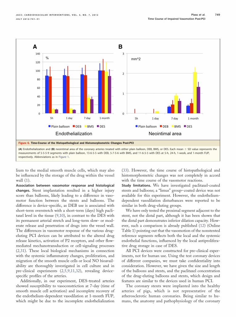
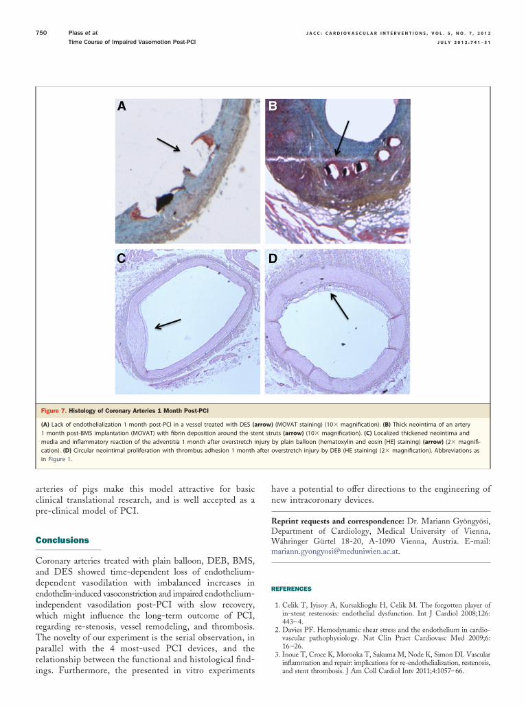
Figure 7. Histology of Coronary Arteries 1 Month Post-PCI
(A) Lack of endothelialization 1 month post-PCI in a vessel treated with DES (a1 month post-BMS implantation (MOVAT) with fibrin deposition around the stmedia and inflammatory reaction of the adventitia 1 month after overstretch ication). (D) Circular neointimal proliferation with thrombus adhesion 1 monthin Figure 1.
ings. Furthermore, the presented in vitro experiments
have a potential to offer directions to the engineering ofnew intracoronary devices.
Reprint requests and correspondence: Dr. Mariann Gyöngyösi,Department of Cardiology, Medical University of Vienna,Währinger Gürtel 18-20, A-1090 Vienna, Austria. E-mail:[email protected].
REFERENCES
1. Celik T, Iyisoy A, Kursaklioglu H, Celik M. The forgotten player ofin-stent restenosis: endothelial dysfunction. Int J Cardiol 2008;126:443–4.
2. Davies PF. Hemodynamic shear stress and the endothelium in cardio-vascular pathophysiology. Nat Clin Pract Cardiovasc Med 2009;6:16–26.
3. Inoue T, Croce K, Morooka T, Sakuma M, Node K, Simon DI. Vascularinflammation and repair: implications for re-endothelialization, restenosis,
(MOVAT staining) (10� magnification). (B) Thick neointima of an arteryuts (arrow) (10� magnification). (C) Localized thickened neointima andby plain balloon (hematoxylin and eosin [HE] staining) (arrow) (2� magnifi-overstretch injury by DEB (HE staining) (2� magnification). Abbreviations as
rrow)ent strnjuryafter
and stent thrombosis. J Am Coll Cardiol Intv 2011;4:1057–66.
J A C C : C A R D I O V A S C U L A R I N T E R V E N T I O N S , V O L . 5 , N O . 7 , 2 0 1 2 Plass et al.
J U L Y 2 0 1 2 : 7 4 1 – 5 1 Time Course of Impaired Vasomotion Post-PCI
751
4. Dohi T, Miyauchi K, Iesaki T, et al. Candesartan with pioglitazoneprotects against endothelial dysfunction and inflammatory responses inporcine coronary arteries implanted with sirolimus-eluting stents. CircJ 2011;75:1098–106.
5. Pendyala LK, Yin X, Li J, Chen JP, Chronos N, Hou D. Thefirst-generation drug-eluting stents and coronary endothelial dysfunc-tion. J Am Coll Cardiol Intv 2009;2:1169–77.
6. Virmani R, Kolodgie FD, Farb A, Lafont A. Drug eluting stents: arehuman and animal studies comparable? Heart 2003;89:133–8.
7. Farhan S, Hemetsberger R, Matiasek J, et al. Implantation ofpaclitaxel-eluting stent impairs the vascular compliance of arteries inporcine coronary stenting model. Atherosclerosis 2009;202:144–51.
8. Plass CA, Schmid W, Holy EW, Kreatschitsch U, Laggner H, Volf I.Redox-sensitive impairment of porcine coronary artery vasodilation byhypochlorite-modified LDL. Atherosclerosis 2007;190:330–7.
9. Hamilos M, Sarma J, Ostojic M, et al. Interference of drug-elutingstents with endothelium-dependent coronary vasomotion: evidence fordevice-specific responses. Circ Cardiovasc Interv 2008;1:193–200.
10. Li J, Jabara R, Pendyala L, et al. Abnormal vasomotor function ofporcine coronary arteries distal to sirolimus-eluting stents. J Am CollCardiol Intv 2008;1:279–85.
11. Jabs A, Göbel S, Wenzel P, et al. Sirolimus-induced vascular dysfunc-tion. Increased mitochondrial and nicotinamide adenosine dinucleotidephosphate oxidase-dependent superoxide production and decreasedvascular nitric oxide formation. J Am Coll Cardiol 2008;51:2130–8.
12. Hamilos MI, Ostojic M, Beleslin B, et al. Differential effects ofdrug-eluting stents on local endothelium-dependent coronary vasomo-tion. J Am Coll Cardiol 2008;51:2123–9.
13. Kitta Y, Nakamura T, Kodama Y, et al. Endothelial vasomotordysfunction in the brachial artery is associated with late in-stentcoronary restenosis. J Am Coll Cardiol 2005;46:648–55.
14. Obata JE, Kitta Y, Takano H, et al. Sirolimus-eluting stent implan-tation aggravates endothelial vasomotor dysfunction in the infarct-related coronary artery in patients with acute myocardial infarction.J Am Coll Cardiol 2007;50:1305–9.
15. Akcakoyun M, Kargin R, Tanalp AC, et al. Predictive value ofnoninvasively determined endothelial dysfunction for long-term car-diovascular events and restenosis in patients undergoing coronary stentimplantation: a prospective study. Coron Artery Dis 2008;19:337–43.
16. Fuji K, Kawasaki D, Oka K, et al. Endothelium-dependent coronaryvasomotor response and neointimal coverage of zotarolimus-elutingstents 3 months after implantation. Heart 2011;97:977–82.
17. Hofma SH, van der Giessen WJ, van Dalen BM, et al. Indication oflong-term endothelial dysfunction after sirolimus-eluting stent implan-tation. Eur Heart J 2006;27:166–70.
18. Pendyala LK, Li J, Shinke T, et al. Endothelium-dependent vasomotordysfunction in pig coronary arteries with paclitaxel-eluting stents isassociated with inflammation and oxidative stress. J Am Coll CardiolIntv 2009;2:253–62.
19. Posa A, Nyolczas N, Hemetsberger R, et al. Optimization of drug-eluting balloon use for safety and efficacy: evaluation of the 2nd
generation paclitaxel-eluting DIOR-Balloon in porcine coronary arter-ies. Catheter Cardiovasc Interv 2010;76:395–403.20. Creel CJ, Lovich MA, Edelman ER. Arterial paclitaxel distributionand deposition. Circ Res 2000;86:879–84.
21. Finn AV, Nakazawa G, Joner M, et al. Vascular responses to drugeluting stents: importance of delayed healing. Arterioscler ThrombVasc Biol 2007;27:1500–10.
22. Schwartz RS, Edelman E, Virmani R, et al. Drug-eluting stents inpreclinical studies: updated consensus recommendations for preclinicalevaluation. Circ Cardiovasc Interv 2008;1:143–53.
23. Johnson LL, Schofield LM, Weber DK, Kolodgie F, Virmani R, KhawBA. Uptake of 111In-Z2D3 on SPECT imaging in a swine model ofcoronary stent restenosis correlated with cell proliferation. J Nucl Med2004;45:294–9.
24. Nakazawa G, Otsuka F, Nakano M, et al. The pathology of neoath-erosclerosis in human coronary implants: bare-metal and drug-elutingstents. J Am Coll Cardiol 2011;57:1314–22.
25. Rosenthal EA, Bohlmeyer TJ, Monnet E, et al. An iron-bindingexochelin prevents restenosis due to coronary artery balloon injury in aporcine model. Circulation 2001;104:2222–7.
26. Fuke S, Maekawa K, Kawamoto K, et al. Impaired endothelialvasomotor function after sirolimus-eluting stent implantation. Circ J2007;71:220–5.
27. Caramori PR, Lima VC, Seidelin PH, Newton GE, Parker JD,Adelman AG. Long-term endothelial dysfunction after coronary arterystenting. J Am Coll Cardiol 1999;34:1675–9.
28. Togni M, Windecker S, Cocchia R, et al. Sirolimus-eluting stentsassociated with paradoxic coronary vasoconstriction. J Am Coll Cardiol2005;46:231–6.
29. Thanyasiri P, Kathir K, Celermajer DS, Adams MR. Endothelialdysfunction and restenosis following percutaneous coronary interven-tion. Int J Cardiol 2007;119:362–7.
30. Yanagisawa M, Kurihara H, Kimura S, et al. A novel potent vasocon-strictor peptide produced by vascular endothelial cells. Nature 1988;332:411–5.
31. Hedegaard ER, Stankevicius E, Simonsen U, Fröbert O. Non-endothelial endothelin counteracts hypoxic vasodilation in porcine largecoronary arteries. BMC Physiol 2011;11:8.
32. Seye CI, Kong Q, Yu N, Gonzalez FA, Erb L, Weisman GA. P2receptors in atherosclerosis and postangioplasty restenosis. PurinergicSignal 2007;3:153–62.
33. Van den Heuvel M, Sorop O, Batenburg WW, et al. Specific coronarydrug-eluting stents interfere with distal microvascular function after singlestent implantation in pigs. J Am Coll Cardiol Intv 2010;3:723–30.
Key Words: endothelium-dependent vasodilation � per-cutaneous coronary intervention � pre-clinical experiment �vasoconstriction.
APPENDIX
For supplementary methods, figures, and tables, please see the online
version of this article.