tibiotalocalcaneal arthrodesis - sport foot &...
TRANSCRIPT

4-28-2011 0713 FrolaquoiNHC HS LIBRARY 202 B77 6757 To919705697613 P26
FOOT $ ANKIr iKTTpnNKTICWt
Copyright 4) 2000 by Utti American Orthopaedic hoot H AnMo Suuivly Iiw
Tibiotalocalcaneal Arthrodesis
Loretta B Chou M 0 Roger A Mann MD Burt Yaszay BS1 Stanley C Grams M D William T McPcakc III Sharon M Dreebert MDb Greg A Horton MD OevtdA Katehorlan MD Thorn O Ciwnlon MD Richard A
MD John W Van Marion
ABSTRACT The purpose of this multicenter retrospective study of 55 patients (56 ankles) who underwent simultaneous tibiotalocalcaneal arthrodeeie with eevere disease involving the anKie and subtalar joints was to determine improvement of pain and function The surgical Indicashytions included osteoarthritis posttraumatic injury failed previous surgery talar avascular necrosis osteoarthrishytis and rheumatoid arthritis involving the ankle and subtalar joints The average age at the time of the opershyation was 53 years The average time o( follow-up was 26 months after the operation Fusion was achieved in 49 ankles with an average time of fusion of 18 weeks Forty-eight of the 55 patients were satisfied with the proshycedure The average leg length discrepancy was 14 cm The average amount of dorsiflexion was 2 degrees and planter flexion woa 5 degrees Following surgery 42 patient complained of pain 40 patients required Shoe modification or an orthotic device and 34 patients had a limp Fourteen patients described their activity as unlimshyited Based on the AOFAS evaluation the patients scored an average of 66 on the ankle-hind foot scale folshylowing surgery The mo6t common complications were nonunion (8 ankles) and wound Infection (6 ankles) This study demonstratecopy that tibiotalocalcaneal arthrodesis is an effective salvage procedure for patients with disshyease both Involving the ankle and subtalar joints
INTRODUCTION
Patients wilh disease involvement of both tho ankle and subtalar joints can have symptoms of pain deforshymity and limited ambulatory capactity on the affected
Corrasponainrj miihoi LOrtffla R fihou MD (It 300 Pasteur Drive R W Stanford California S4305 Telephone mraquoiirlaquogtr (650) 490-7526 FAX nnmhoi (650) 725-9592 ft-mafl inJJtess mtrrjccopy1oiSylrififitantltiiit laquodu
i OeMiiiid CA PhrxmU AZ 4 KnoxvBle TN G Sin Owyu CA 6 Kjmswj City K37 Wflrti Biuomlioia Ml 0 Houston I X 9 Albuquerque MM 10 Richmond VA
limb Treatment options are limited rtonope measures may help decrease some of the symp but much of pain and deformity remain Surgical ment is aimed at obtaning a painless brace plantigrade foot and (ibioialocalcaneal arthro offers an effective surgical treatment This metholt discussed by Russotti and Johnson1 in 1988 a not commonly performed It has been reported quently with the largest series being 30 cases end most series involve Charcot foot problRms purpose of this study was to determine the freqi Of this procedure and report on the cl nical results use of the AOFAS evaluation for Ilie ankle-hindfc
MATERIALS AND METHODS
Between 1991 and 1998 tibiotalocaicc arthrodesis was performed by 9 surgeons at respective 9 tnstitut ons on 55 pat ents (56 ankle which 30 patients were women (31 ankles) an were men (25 ankles) The average age ot the pat at the time of surgery was 53 years (range 19 I years)
The indications for surgery were severe arrh and associated deformity and pain involving boti ankle and subtalar joints from one of the folio posttraumatic injury (14 ankles) failed previous gery (12 ankles) osteoarthritis (11 ankles) nvas necrosis of the taus (7 ankles) rheumatoid arthri ankles) failed total ankle replacement (2 ant Charcot-Marie-Tooth disease (2 ankles) and Ch foot (2 ankes) All patients had failed nonsur treatment
All patients were interviewed and underwent p cal and radiographic examination The avarage io up time was 26 months after the time of oper (range 12 to 168 months) The physical examin involved evaluation ot the limb foi tenderness j lion and range of motion The position of trie foot assessed in the standing position with a hand
804
To919705697613
KM amp Ankle InternationalVol 21 No wociobw 2000
Igoniomeier Weighl-bearing radiographs were taken of the operated ankle in the anteroposterior lateral arid Mortise planes and of the foot in the anteroposterior Jsnrl oblique planes Fusion was determined by radishyographic consolidation at the arthrodesis site
The American Orthopaedic Foot and Ankle Society Clinical Mating System Ankle-Hind Foot Scale was used for evaluation This includes subjective and objective factors into numerical scales to describe function alignment and pain-
SURGICAL TECHNIQUE
An intramedullary rod was used in 37 procedures screws in 17 procedures an external fixator in one procedure and a plate and screws in one procedure
Autogenous bone grafting was utilized in 42 cases and allograft in one case
In most cases the intramedullary fixation device was used Alransfibular approach is utilised between the sural nerve and the lateral branch of the superficial peroneal nerve similar to the method described lor ankle fusion The articular cartilage was removed
lfrom the tibiotalar joint with a saggitai saw Frequently a small medial incision was used to remove the medishyal malleolus Next the articular surface was removed from the subtalar joint Any deformity involving the ankle or hindfoot was corrected with the saw cuts The hind foot was placed in approximately 5 degrees of valgus with neutral dorsi-plantar flexion The amount of external rotation was equal to the contralateral foot approximately 5 to 10 degrees If there was extensive bone loss iliac crest bone graft was utilized
When an intramedullary rod was used a small transshyverse incision was made on the plantar aspect of the heel at the level of the distal and middle third of the heel pad A guide wire was placed through the calcashyneus talus and into ihe tibia and the position was confirmed with image intensification Reaming was accomplished over the guide pin and then the rod was placed in a retrograde fashion The interlocking screws were drilled and filled The wound was closed in layers over a drain and a bulky compression dressshying with splints was placed
The drain was removed after 24 hours The dressing bullbull and sutures were removed 10 to 14 days following surshy
gery A short leg cast was placed and nonweight-bearshying was continued for a total of 3 months following which progressive weight-bearing was begun in a short leg walking cast The extremity was protected with a cast until clinical and radiographic healing was satisfactory
TiBIOTALOCALCANEAL ARTHRODtSlS
RESULTS 805
Fusion was achieved clinically and rarJiorjraptiicaUy in 47 patients (48 ankles) with an average lime to lusion of 19 weeks (range 12 to 65 weeks) These patients had no motion at the arthrodesis sile and conshysolidation was seen radiographically Tine average time of immobilization was 20 weeks
Forty-eight of the 55 patients were satisfied with the procedure Seven patients were not satisfied with ihe results because of nonunion of the ankle arthrodesis in 5 patients residual sevore- pain in one and limited activity level in one Two patients complained of sevens pain 7 of moderate 33 of mild and 13 had no pain
Nine patients use an AFO of which 2 had anterior tibial pain from a stress reaction to the rod Thirty-one patients requ re some form of shoe modification Must ol the modifications consisted of a soft insert or orthotshyic device Forty-one patients had limited activity ot which 2 used a wheelchair and one was house bound The remaining 14 patients described their activity as unlimited
The average ieg length discrepancy was 14 cm as measured with blocks The average position of fusion was 3 degrees of valgus The average range of motion ol dorsillexion was 2 degrees and plantar flexion was 5 degrees which took plate through the distal tarsal joints Wearing shoes 35 patients had a flatfoot gait and limped
The AOFAS anklehind foot score is based on pain (40 of the total score of 100 points) function (28) motion (22) end alignment (10) The overage postoperative ankle-hind foot scale was 66
COMPLICATIONS
Thirty-eight ankles healed without complications There was nonunion of the ankle arthrodess in 8 patients superficial wound infection in 5 and there was one case of each of the following complications deep wound infection (in one case involv ng a nonunion) skin necrosis sural neuroma wuturc gran-uoma delayed union of the ankle arthrodesis and stress tracture Of the 8 cases with nonunion 3 had fixation with screws and 5 had fixation wild an intramndullary rod Additional Surgery was required in 16 patients which included hardware removal in 11 cases revision lor nonunion in 2 and one case of each of the following removal of a bone growth stimshyulator placement ot autograft for a nonunion and resection of a postoperative aural neuroma The patients with the superficial Infections healed with antibiotics and local wound care The patient with the
t
pound8-2011 0714 FromWHC HS LIBRARY B0P B77 6757 To919705697613 P 4 6
806 CHOUETAL
deep infection was treated with antibiotics and wet to dry dressings The patient with the stress fracture healed after 1 month
DISCUSSION
Arthrosis involving both the ankle and subtalar joint is one of the most difficult problems facing the orthopaedic foot and ankle surgeon Patients complain of moderate to severe pain deformity and disability The goal of tibiotalocalcaneai arthrodesis is to allevishyate pain and provide a stable plantigrade foot for ambulation Often bracing this type of deformity is not possible because of the severe deformity In cases of failed ankle arthrodesis failed total ankle replacement and avascular necrosis of the talus pain and instabilishyty progress It would be preferablo to avoid performing a fusion of both joints because of the significant loss ot motion6 Following a pantalar arthrodesis dorsiflexshyion is decreased 63 and plantar flexion 82 which results in a significant increase in the amount of stress placed on the surrounding joints However arthrodesis ol only the ankle joint when the subtalar joint is arthritshyic will likely result in residual symptoms in the subtalar joint
Tibiotalocalcaneai arthrodesis has been reported infrequemiyultlM1 until recently and generally show good results The aim of this study was to obtain data 1rom multiple institutions to evaluate the frequency of the use of this procedure (56 cases from 9 orthopaedic loot and ankle practices) and determine the clinical outcome of this procedure The AOFAS anklehindfoot-score was used to provide a value from a system that is widely utilized To our knowledge this is the largest reported series of tibiotalocalcaneai arthrodesis
Tibiotalocalcaneai arthrodesis has been described by Johnson11 to treat severe pain and deformity involvshying the hind part of the foot and ankle Johnson initialshyly reported on this procedure using multiple internal screws or an external fixator The indications were failed arthrodesis of the ankle failed total ankle arthroshyplasty osteonecrosis of the talus infra-articular fracshyture at the ankle that was un-unlted or malaligned and neuropathy involving both joints Satisfactory results were obtained in 75 per cent of 21 patients and fusion was achieved in 18 patients
Johnson later devised an intramedullary fixation device (Revision Nail Smith amp Nephew Richards Inc Memphis TN) for this procedure to improve on the stashybility of the fixation and would avoid compications associated with external fixation devices Kile4 reportshying on using this intramedullary fixation device obtained 87 satisfactory result in 30 patients and fusion was complete in 28 patients Twenty-six
Foot amp Ankle InternationalAol 21 No 10October 200
patients felt that the operation had been worthwhilt There was 1 superficial skin 8lough 2 deep infection 1 prominent plantarward rod and one death fro pneumonia There were 2 patients with stress rea tions at the proximal end ot the nail that healed w immobilization
In a similar report by Moore et al retrogra intramedullary redding was evaluated retrospecttrade in 19 ankle arthrodesis in 16 patients The procedi was done as a salvage procedure In each patient significant posttraumatic arthrosis and bone loss ct comitant subtalar arthrosis and severe osteoper Union occured in 14 ankles The comp ications w nonunion in 5 ankles one deep infection and one b ken rod Thirteen of the 16 patients were ambuiau and 9 used an AFO or shoe modification
This study only had one patient with a Charcot jt of the 51 ankles In contrast Plnzur and Ketikii reported on 20 patients (21 ankles) with severe nei pathio (Charcot) ankle deformities who were trea with a retrograde locked intramedullary nail Nineu ot the 21 ankles went on to fusion at an average of months In ten of the patients the talus was retain There were 6 patients who developed late poslopt tfve wound infections of which 3 required remova the nail One patient with an infection elected to unr go an ankle disarticulation The authors stated t use of the retrograde locked intramedullary nail is excellent method of obtaining ankle fusion in Charcot patient
Papa et al reported on a cases of tibiotaio caneal arthrodesis for intractable diabetic neuropa arthropathy ot the foot and ankle One patient ha partial wound slough but fused in 4 to 5 months T preferred internal fixation for their patient popuia who are prone to infection In another study by P-et al 13 patients underwent tibiotalocalcaneai tus for posttraumatic osteoarthrosis ol the ankle and r fool They included patients who underwent pant arthrodesis and the results were not separated from each type of fusion Eighty-one percent much improved but 95 had residual pain Ove the union rate was 86 and the mean time to fus was 14 weeks The mean amount of shortening 15 cm There were 3 nonunions (pantalar and li talocaicaneal) The authors found that their nonur rate was low considering that extended arthrodi was performed They concluded that ft is a com[ and technically demanding procedure but a reas able alternative to amputation
Felix and Kitaoka report the resut of 26 ai arthrodeses performed for rheumatoid arthrit s or patients In their series tibiotalocalcaneai arthrori was performed in 12 ankles They did not distmgi
Foot amp Ankle InternatlonalAd 21 No 10Octobot 2000
results of the ankle arthrodesis from the tibiotalocalshycaneai arthrodesis patients Fixation was achieved wth external fixation or internal fixation with multiple screws Nearly all patients were satisfied and union
[was achieved in 96 Their union and complication [rale were found to be comparable with rates for [ arthrodesis (or posttraumatic and degenerative arthrishytis The authors concluded that for the treatment of rheumatoid arthritis arthrodesis provides more relishyable long term function and remains the standard of treatment They also emphasize that patients be edu-
i caled that they will not have a normal joint function and will continue to have limitations
Rigid internal fixation with good bony apposition is important for successful fusion Biomechanical analy-
sis of hindfoot fixation using an intramedullary rod was compared to three cross-cannulated screws The t intramedullary rod with one distal screw inserted pro-i vided more stiffness to the hindfoot We prefer the use j of the intramedullary rod when possible External fixa-[ton may be used if the bone stock Is insuffcient [ Autogenous corticocancellous bone graft may be nec- essary with severe bone deficits | The time to lusion in ihis study 19 weeks is pro--
bull longed as compared to ankle arthrodesis (138 1 weeks) This is not surprising because the patients in this study had more severe disease In a retrospective
study on arthrodesis of 81 ankles by Mann and Rongsiad there were 10 nonunions (12) The avershyage postoperative score for ankle-hinclloot on the AOFAS evaluation was 74 points and the rate of the patient satisfaction was 65 (89) ot the 73 patients in our study the AOFAS clinical rating system score was consistent with expectations of improved pain and function but with some residual limitations Many of these patients have co-morbidities that further limit (unction such as rheumatoid arthritis Also with extensive disease there is usually soft-tissue problems trial affect healing and symptoms
The scores obtained in this study will be helpful to compare to longer follow-up studies for this procedure In addition these values may be used to compare to other procedures involving the ankle and subtalar joints for example combined total ankle replacement and subtalar arthrodesis
The position of the tibiotalocalcaneai arthrodesis is extremely important as with other fusions of the foot and ankle The optimum position is neutral flexion and 5 degrees of valgus and 5 to 10 degrees of external rotation1 The final result of fusion is dependent on the bony cuts and quality of the bone stock It is not uncommon for bone affected by chronic disease such as posttraumatic arthritis or rheumatoid arthritis to have some shifting or settling during the healing
TBIOTALOCAlCANLzAL ARTHRODESIS 807
process Rigid Internal fixation may help to avoid this situation The average amount of shortening of the operated limb was 14 cm and was tolerable for most ot the patients Most did not require a heel lift
The most common complications with arthrodesis of the foot and ankle are infection skin slough neuroma nonunion or malunion The postoperative complicashytion is higher for tibiotalocalcaneai arthrodesis because of previous operations loss of adequate bone stock and co-morbidities The incidence of nonunion in this study is similar to other reports of th s procedure The incidence of infection was slightly higher than seen with ankle fusions This probably is caused by already compromised soft tissue of the foot and ankle that limits healing capacity Thus it is imporshytant to evaluate the patientis vascular status and note the risk of complications
CONCLUSION
Tibiotalocalcaneai arthrodesis is a salvage operation ID treat a difficult problem normal function is not expected with arthrodesis of these two major joints However It can be concluded that it is a good treatshyment option to improve pain and function As stated by previous studies we emphasize that patients must be informed lhat they will not have a normal joint and will continue to have limitations
REFERENCES
1 Buck f Money BF Chalt ETS The optimum position of arthrodesis of the ankle A gait study ot Ihe Knee ann ankle J Bone Joint Surg 69A10010621987
2 Felix NA Kttaoka He- Ankle arthrodesis in patlante with rheumatoid arthritis Clin Orthop 34958-84 t99fl
3 Fleming SS Moore TJ Hutton WC Biomechanics I analysis ot hindfoot llxailon using an intramedullary rod J Souihern Orthop Assoc 719-26 1998
4 Kile TA Donnelly RE Gehrkc JC Wuner ME Johnson KA Tibintalocalcaneat arthrodoeie with an inimmncMlary device Fool Ankle Int 15660-6731994
5 KHaoka Ha Alexander IJ Adeiaar R3 Nunley JA Mycrsoti MS Sanders M Clinical rating 3y3leins for ths awe-hindfoot mimrwt hallux and lesser loss Foot Ankle Int 15349-363 1994
6 Mann HA Arthrodesis of the toot and ankle in Mann HA Coughlin MJ (eds) Surgery ot the Foot and Ankle Ed ltS Si Lojis CV Mostly 1993 pp 673-713
7 Mann RA Rongsiad KM Arthrodesis of the ankle A critical analysis Foot Ankle Int 1930 1998
a Mann RA Van Marten JW Wepnei K Martin J Ankle fusion Clin Orthop 26849-55 1991
9 Moore TJ Prince R Fochatko 0 Smith JW Flaming 8 Retrograde intramedullary nailing for ankle arthrodesis Foot Ankle int ISM33-4361995
10Papa JA Myarson MS Pantalar and libiotalocalcsnsal arthrodesis lor poet-traumatic osteoarthrosis ol the ankla awl hind toot 1 Bone Joint Surg 74A1042-KW9 1992
11 Papa JA Myarson MS GHrard p SalvHlaquojlaquo with arthrodesis In
-28-2011 0715 FromWHC HS LIBRARY 202 877 6757 To919705697613 P 6-6
808 CHQUETAL
Intractable diabetic neuropathic arthropathy ol ihe font raquonrj anklA J Rone Joint Surg 76A1056-10661993
12Plnzur US Kelikian A Charcot ankla fusion with a retrograde locked intramedullary nail Foot Ankle Ini iB6Hraquo-704 1997
Fool amp Anklo IntcmatlonaWol 21 No 10Octobet m
13Ruaeottl QM Johnson KA Case JR IibioralocaJcarc arthrodesis for arthritis and dolormily raquoi the hinrt part of the fo J Bone Joint Surg 70Ai J04-1307 IBRA
u n 2 8 2011 101CAM No 7193 P 1 9 i age u u i i j
31306566 Request 31306566 Email (PDF) To mitchellvvmccom
VAIL VALLEY MEDICAL CENTER Library-Shaw Regional Cancer Center 322 Beard Creek Road PO Box 2559 Edwards CO 81632
JUN 272011
DOCLINE Journal Copy Techniques In foot amp ankle surgery 20054()214-21 Tendon fixation in flexor hallucis long us transfer clanton 101141485 Verify LocatorPlus 1536-0644 (Print) 1538-1943 (Electronic) Any format Upplncott Williams amp Wiikins Hagerstown MD Copyright Compliance Law Kim Lyons-Mitchell NA $2000 CRIntern [crlnternsprivailorg] CCMLFreeShare 1970569-7607 1970569-7613 mitchellvvmccom Email(TIFF)FaxMallWeb(PDF)Web(TIFF) Prefer to work with Free Share program Prefer fax or
PDF Routed to MOUSJK In Serial Routing - cell 1
Jun27 2011 ( 0525 PMET) St Joseph Medical Center KANSAS CITY MO USA (MOUSJK)
This material may be protected by copyright law (TITLE 17US CODE) Bill to COUVYA
VAIL VALLEY MEDICAL CENTER Library-Shaw Pavilion P0 Box 2559 Edwards CO 81632
Title Citation Article Author NLM Unique ID ISSN Fill from Publisher Copyright Authorization Need By Maximum Cost Patron Name Library Groups Phone Fax Email Alt Delivery
Comments
Routing Reason
Received Lender
httpSdoc]inegovdodinerequestsreceiptreceiptcfmProgram=Docamptype=nampt=08641 6272011
un 28 2011 101CAM Mo 7193 P 2 9
Ttdmtpus di Van and MMt Snrampjy 4(4i2li-UI 2005 OiOOi UppImait Wtllaiu 6 Wkbu HtlMeWit
5 F E C I A L F O C U sect
Tendon Fixation in Flexor Hallucis Longus Transfer A Biomechanical Study Comparing a Traditional Technique Versus Biobasorbable Interference Screw Fixation J M Cohn M D E P Sabonghy MD C A Godlewsld M D T 0 Clanton M D and W C McGarvey MD Ocfvawitm of Onhofitieilic Surgery Division of rant and Ankle Surgery1
The VnixtnUy of Texas w Houston Health Science Center Houston TX
m A B S T R A C T
Augmentation of the Achilles mechanism utilizing Ihe flexor hallucis longus (FHL) tendon transfer to the calcashyneus is a welKdescribcd procedure Traditional methods Tor this procedure require suturing the tendon onto iiseif after passing ii through tin osseous tunnel Tendon fixashytion techniques thai reduce dissection and thus operative lime while allowing adequate fixation would be advantashygeous in reducing patient morbidity from die aforemenshytioned extended operative limes The authors suggest a new technique for transfer of the FHL lo the calcaneus as a treatment of chronic Achilles tendon insufficiency The objective of this fresh cadaver study is to compare ihe tendon fixation pulloui strength of A traditional tendon transfer technique versus bioabsorbable interference screw fixation and subsequently propose a less invasive bvt stronger and more efficient technique for FHL transshyfer and fixation Clinical implications suggest more relishyable fixation that may allow faster rehabilitation after the procedure Ten cadaver fool and ankle matched pairs were used after undergoing bone densitometry A specishymen from each cadaver pair had the flexor hallucis longus tendon sutured to itself with 1 Ticron suture (Ethicon) after being pulled through an osseous tunnel These 10 specimens were assigned to group A In the contralateral ankle specimen the flexor hallucis longus tendon was placed into a 65-mm osseous drill hole and fixed with a x 25-mrn bioabsorbable interference screw These
AddrcKcom^ponilcnce rtnd reprint requests loCtiritiophtrA CHKIIBWSIU MD University or TcMS-Housion Mwictil School Derwrinwni or Orthopaedics 64Jl Pannin Room 6144 Houston Texas 7703 E-mail ChrilaquolaquotphWAOiiltlleltkiltBittmcetliJ
comprised group B Mechanical testing of pulloui strength was then performed pulloui strength and mode of failure were recorded during this testing Tendon fixshyation in group A averaged 1276 N and group B 17028 N By paired 2-tailed Student i test the differences between each mulched pair were statistically significant ( = 004529) In group A failure occurred most often at the bone tunnel (6 of 10) and tendon midsubsiancc (4 out of 10) Failures at the tendon midsubsisince wens not inshycluded in the data analysis All Group B failures occurred at the tendonscrew interface According to Ihe results of our study the bioabsorbable interference screw fixation technique was found to resist significantly higher pulloui forces than the traditional approach to flexor hallucis lonshygus transfer The authors feci that the interference screw technique is technically easier while having the capacity to resist higher loads subsequent lo clinical testing it could prove a superior method of flexor hallucis longus transfer for chronic Achilles tendon rupture or tendinopathy Keywords flexor hallucis longus transfer bioabsorbable screw interference screw
bull B A C K G R O U N D I I I S T O R K A I P E R S P E C T I V E
The treatment of chronic Achilles tendon rupture and tendinopathy is challenging for the general orthopedic surgeon as well as (he foot and ankle subsnecialist Nushymerous surgical procedures have been proposed to adshydress die reconstruction of chronic Achilles tendon rupture and lendJiiopailiywl5lls1 Of these transfer of Ihe flexor hallucis longus (FHL) tendon has become
214 Tfdiiilqut In Foot awl Ankle Stirgtiy
vun 28 201 i 1010AM ^o7193 P 39
Special Focus Tendon Fixation In PHL Transfer
a viable option and has numerous benefits6 The close proximity of the FHL-to the Achilles tendon complex facilitates its transfer with minima] risk for neurovascular injury The FHL is one of lite strongest plantar flexors second only to the gastrocnemius-soleus complex The axis of contractile force of the FHL fires in phase wiih the gaslrocnemios-soleus complex Finally transfer of ihe FHL tendon has the least impact on ihe biomechanics of the foot and ankle us well as gait
Hansen3 and Wapner el al7 have eloquently described the technique of flexor hallucis longus tendon transfer Wapner recommends tendon harvest by way of a 2-inci-sion technique passing the tendon through an osseous tunnel in the calcaneus and teuodesing it to itself Potenshytial complications with this method include technical difshyficulty with graft harvest inadequate length of tendon for icnodesis and wound breakdown andor posteromedial incisions
To avoid these potential complications we propose a technique in which fixation of the FHL tendon is achieved in an osseous tunnel of the calcaneus wiih an interfershyence-fit screw We feel that this method would allow less dissection avoid violation of the plantar surface of the foot and decrease ihe chance of fracture through the osshyseous tunnel
The success of bioabsorbable interference screw fixshyation of lendon transfers has been well documented in the knee1 In addition we have reported interference screw fixation in tendon transfers in the foot-1 We present a biomechanics study comparing interference screw fixshyation with traditional tenodesis of the flexor hallucis longus tendon transfer for the treatment of chronic Achilshyles tendon rupture and tendinopathy
bull MATERIALS AND M E T H O D S
Ten pairs of feet and ankles from fresh cadaveric specishymens were obtained for evaluation Although no case hisshytories were available for review examination of each specimen revealed no obvious abnormalities or evidence of trauma Criteria for inclusion were lower leg specishymens with intact flexor hallucis longus insertions no flexor hallucis longus tendinopathy and minimal osteoshyporosis of the calcaneus AH specimens underwent bone densitomeiric evaluation utilizing a Hologic QDR 4000 PEXA Scanner before manipulation to document variashytion in specimen bone quality
Ten feet were randomly assigned lo group A and (heir counleriMrts to group B Group A represented feet in which a traditional flexor hallucis longus tendon transfer involving tenodesis using 1 Ticron after passage through an osseous uinnel in tlie calcaneus was performed Croup B was the experimenml group in which interference
fixation was used to secure the flexor hallucis longus tenshydon in the osseous tunnel of (lie calcaneus
Specimens were thawed to room temperature for 24 hours In all specimens assigned to groups A and B a lonshygitudinal incision was made just medial to (he Achilles tendon from the musculotendinous junction to approxishymately 2 cm distal to (he insertion of the Achilles tendon on the calcaneus After retraction of Ihe posterior tibial neurovascular bundle medially the deep posterior comshypartment fascia was incised longitudinally lo expose the flexor hallucis longus muscle belly In specimens assigned to group A a second longitudinal incision was made along the medial border of the foot With the abductor hallucis flexor hallucis brevis and medial plantar neurovascular bundle retracted plantar die tenshydons of ihe flexor hallucis longus and flexor digitomm longus were isolated The flexor hallucis longus tendon was harvested distal to ihe knot of Henry and pulled through to the proximal wound In group B specimens FHL harvest was performed through the same posterior incision planned for fixation of the tendon transfer The FHL was transected in the fibroosseous tunnel just beshyneath the sustentaculum tali The fibrous raphe if preshyserved prevents errant procurement of the tibial peivc
In group A a transverse 65-mm drill hole was creshyated in the calcaneus approximately I cm distal and J cm anterior lo the Achilles tendon insertion The flexor halshylucis longus tendon was then passed from medial to latshyeral and tenodesed to itself using a 1 Ticron suture (Figs 1-5)
In group B specimens a vertical 65-mm drill hole was created in the calcaneus approximately 1 cm medial and 5 mm anterior to die Achilles tendon in Ihe superior aspect of the calcaneus This drill hole was foil thickness fhrough the calcaneus to allow for lendon transfer through ihe calcaneus and out through the heel pad The flexor hallucis longus tendon was then passed through this osshyseous runnel and out the plantar heel pad With tension applied to die tendon via a puliout suture i 7 x 25-mm bioabsorbable interference screw was secured next to the tendon within the osseous tunnel (Figs 6-9)
Each specimen was fixated via a smooth transverse metatarsal shaft Steinmann pin (364) through ihe midshyshaft ponion of the firsl second and third metatarsals and a smooth transcalcaneal Steinmann pin (364) through the body of the calcaneus to allow for attachment to the MTS device (MTS Machine Testing System 810) The free portion of the flexor hallucis longus tendon was attached to the load cell using a freeze clamp Following secure fixation each specimen was sequentially loaded to determine ultimate yield strength and made of failure The specimens were loaded physiologically thai is ihe tension was applied vertically on the tendon and perpenshydicular to ihe screw rather than in-line tension parallel lto
Volume ltt (ftvt 4 215
bull un28 2011 1010AM Ho 7193 P V 9
Special Focus Cohn el al
FIGURE 1 Planned incision for FHL transfer
the screw Tensile testing of each specimen using the computer-controlled servo-hydraulic materials tester was performed at a uniform rate of I mrns Testing was concluded when failure of fixation or tendon rupture occurred
The bioabsorbable interference screws used in the study are composed of 82 L-lnclie acid and 18 glyshyceric acid (Aithrotek Warsaw IN) The size of the screw used for inicrferencc fit in the calcaneus was 7 x 25 mm This screw is amorphous wiih uniform degradation ihroughotil its substance The copolymer ratio allows retention of original strength for an average of 6 to 8 weeks
bull INDICATIONS AND CONTRAINDICATIONS
Plexor hallucis tendon transfer is indicated when residual Achilles tendon tissue is deemed inadequate necessitatshying augmentation
Preoperative Planning Flexor hallucis tendon function should be assessed to enshysure that its function is not compromised Tlie extent of Achilles disease should be assessed clinically and with imaging studies Substantial Achilles disease should
prompt (he surgeon lo be prepared for an FHL lendon transferaugmentation
Surgical Procedure B ased on (he results of this study and a clinical desire for the senior authors to minimize trauma from surgical disshysection as well as to maximize efficiency of (his proceshydure some modifications have been implemented lo arrive at Ihe final proposed surgical method
The procedure is currently perfonucd through I incishysion This is typically placed just medial to the midline for nomnsenional Achilles problems but may be shifted to directly midline for insertions pathology in an effon to better access the diseased portion of ihe tendon14 Reshygardless of whether the pathology lies al the insertion or Ihe more classic watershed region of the tendon exshytensive debridement is necessary lo excise all diseased tissue for rapid symptom palliation Debridement should take place through the substance of the Achilles tendon itself and no residual or suspect tissue should be preserved
The FHL may now be harvested through this split in ihe tendon The muscle belly is most easily identified afshyter the fascial covering is incised The body of the muscle is used to trace its course into the region of ihe tarsal
FIGURE 2 Midline Achilles lendort-splilting Incision is made full thickness through ihe tendon
216 TV-liijtijittr Jti Feat unit Mhlt Sittgsty
un 28- 2011 1011AM Mo 7193 P 5 9
Special Pocus Tendon Fixation m FHL Transfer
FIGURE 3 Tne retrocalcaneal bursa is visualized through Ihe tendon
tunnel where it becomes tendinous Care should be taken to avoid careless identification of presumed tendon alone without meticulous inspection as the shiny whitshyish yellow tubular appearance of the tibial nerve can be deceptive IO the unassuming surgeon We often spend a moment dissecting the neurovascular structures to acshycurately identify and isolate them from harms way beshyfore proceeding with the FHL harvest Once revealed the FHL tendon should be tracked into its fibroosseous tunnel directly beneath the sustentaculum tali and freed of any adhesions Now with ihe ankle in maximal equimis and the hallux plontarflexed the tendon may be transected with cither tenotomy scissors or more prefshyerably a sharp scalpel with the blnde beveled toward the calcaneus to ensure safely of the neurovascular bundle One caveat here is that the icmpiation lo harvest a longer tendon with a blind pass of a tendon stripper should be avoided because this may result in an injury to one Or more of the plantar branches of die tibial nerve
Once the tendon is harvested a whip stitch is pershyformed in its free end The surgeon should he aware that Ihe amount of tendinous remnant on the harvested end of tne muscle is much shoier than (hat described for the 2-incision technique however this is uniformly adequate
io provide enough tendon for passage and fixation into the calcaneus below
A tunnel is created just anterior (5-10 mm) lo the Achilles insertion in the central body of the calcaneus In cases of insenional tendinopathy a generous resection of the posterolateral prominence or Haglund deformity is necessary but this has not presented a problem with positioning or fixation The tunnel is drilled using ihe same diameter reamer as the planned implant (most often 7 mm)
The tendon is passed in one of several ways Several systems are available to attempt to prevent plantar cortishycal penetration and these require exact measurement of desired lendon lengib before the passage of the tendon Mora traditionally the plantar cortex is penetrated and a Beith pin is passed through ihe plantar foot The sutures are pulled through the plantar skin until a sufficient amount of lendon has passed into the tunnel The approshypriate umouni of tendon passage is based on ihe ultimate tensioning of the tendon transfer For this we routinely include bodi feet in the sterile field to use the normal contralateral side as a reference of normal resting tenshysion Alternatively one may choose lo arbitrarily position
F I G U R E 4 The FHL tendon and tibial nerve (medial or lo the right) tie parallel and in close proximity lo one another separated by a fibroosseous sheath
Volume t taut 4 217
u n 2 8 2011 1011AM Mo 7 93 P 69
Special Focus Cohn et ltxl
FIGURE s Due lo anatomic proximity and concern over Injury the tibial nerve is routinely Identified
the foot in 15-20 degrees of cquinus and tension at ihis level
Once tensioning is optimized the interference screw is placed Should solid purchase not be obtained the imshyplant may be removed safely and witliout fear of tendon rupture and replaced with a larger-diameter screw Tenshysion should be tested and the foot should come lo at least neutral (Fig 1011)
Closure is performed by Drsi providing a side to-side tenodesis of the remaining Achilles and its gasiroc-soleus muscle belly to the new Achilles anchor the FHL transshyfer (Pig 12) This serves 2 presumed purposes First push-off power is maximized by incorporaiing the strong gashroc-soleus complex into the new heel cord Second Ibe ample blood supply from the rich vascularnetwork in the FHL muscle belly provides a source of nutrition and oxygenation to the previously injured Achilles remnant Careful closure is performed with separate layers of para-icnon if still present (not often) subcutaneous fat and skin
bull POSTOPERATIVE MANAGEMENT
The patient is immobilized in an equina splint at 15-20 degrees for 2 weeks He or she is then placed into a boot
or casl wiih a 2-inch heel lift and allowed to gently touch down for another 2 weeks After I month patients are instructed on gentle activeassisted range of motion and advancement of weightbenring io full as tolerated Over the next 2-3 months the amount of the heel lift is dec-remenially removed until a painless plantigrade foot is achieved Physical therapy for strengthening is begun at the 2-month point
bull RESULTS
The results of bone mineral density studies of the paired cadaveric specimens were as follows average bone minshyeral density for the liaditional tendon fixation technique specimens was 032095 gcm2 the average bone mineral density for the bioabsorbable interference screw techshynique was 033536 gcm3 A repeatability study was pershyformed on each cadaver specimen utilizing the DEXA scanning technique Results of this study revealed a coefshyficient of variation of 24 A Student test revealed no statistical difference between cadaveric matched specishymen pair results (P = 02870)
Itie average pulloui strength of the traditional group was 1276 N (median 1214 N standard deviation 93 N)
FIGURE 6 The FHL Is harvested through Ihe posterior n clslon with great care taken lo avoid injuring ihe nerve which lies just oulskte or medial to the fibroosseous lunnei
218 Tednlrjua in Font jurf Atlkk Suiampry
bull raquolaquo28 2011 1011AM Mo 7193 P 79
Special Focus Tendon Fixation in FHL Transfer
FIGURE 7 A bony tunnel Is reamed lo Ihe size ol ihe screw lo be used
the average pulloui strength of the bioabsorbable interfershyence screw specimens was 17028 N (median 1647 N standard deviation 86 N) (Fig 10) With the data obshytained a paired 2-taiied Student test was performed showing a statistically significant difference between bioabsorbable interference screw fixation and traditional tenodesis techniques (004529) All specimens were inshycluded in the study
Of specimens in group A 6 of JO failures occurred at the bone-tendon interface and 4 out of 10 occurred in Ihe mid substance area of the tendon Data included for analysis were all traditional failures at the site of transfer die failures at the midsubstance area of the tendon were not included in data analysis because they did not make it to failure at the surgical site All specimens in group B were loaded to failure with all failures occurring at the bone-tendon interface
bull POSSIBLE CONCERNS AND FUTURE Ot TECHNIQUE
Tendon transfer for ihe treatment of chronic Achilles rupshyture and teiidinopaiby is an evolving concept The outshycome of several different studies7 has shown the efficacy of flexor hallucis longus transfer for Achilles
lendon pathology The biomechanical and anatomic adshyvantages of flexor hallucis longus have been documented by numerous studies791217 The flexor hallucis longus is a stronger plantar flexor compared will) the flexor halshylucis longus and peroneus brevis its axis of contractile force more closely reproduces that of the Achilles tenshydon il fires in phase wiih the gastrocnemius-soleus comshyplex its anatomic proximity avoids the neurovascular bundle and its original function is- ihe same as that of the Achilles lendon (planiarAexion)
The use of interference screw fixation for lendon transfer has been well documented in ligament reconshystruction in the knee11 Its use in tendon transfer fixation in the foot and ankle is a new concept The potential benefits for bioabsorbable screw fixation are also well known The long-term benefit of bioabsorbable screw fixshyation is complete resorption over a period or six months This optimized the environment for ingrowth of bone at the bone-tendon interface Further slow resorption of the bioabsorbable screw serves to slowly increase physioshylogic tensile forces at the bone-tendon interface io stimshyulate tendon-bone healing Finally use of bioabsorbable materials reduces the potential complications seen with metallic fixation These include host reaction to foreign body and potential problems with magnetic resonance
F I G U R E 8 A Krakow locking sulure Is used lo secure the FHL to be brought Into Ihe lunnei
Vutume 4 Issue - 219
vun 28 2011 1012AM Mo 7193 P 8 9
Special Focus Cohn et ltd
FIGURE 9 Tendon pulled through bony tunnel wiih tenshysion equal lo that of the contralateral leg
FIGURE 11 Compteled FHL Iranslar with interference screw fixation
imaging if future imaging of the fool and ankle complex is required
In the present study the strength of ihe interference screw fixation (group B) was superior to thai of tradishytional suture tenodesis (group A) in all specimens It may be argued thai the innate tendon strength of each individual cadaveric specimen may have affected ihe
FIGURE 10 interference screw fixation
overall outcome of the present study By using matched cadaveric specimen pairs it is assumed that liiis will reshyduce the effect of inteicadraquover variability Further hone mineral density did not significantly affect pulloui strength between malched-pair results In addition it may be argued that the angle of distraction on tlie tendon by the material testing system may not be similar to physishyologic forces and thus may have affected die outcome However this was a constant variable throughout sped men evaluation Finally io minimize screw variability all screws were used from the same production lot from a single manufacturer
bull CONCLUSION
The present study focuses on improving a previously described surgical technique io address ihe treatment of chronic Achilles tendon rupture and tendinopathy The reduction in surgical time avoidance of a secondary inshycision decrease in surgical dissection and increase in initial repair strength show this technique to be a viable surgical oplion The increase in physiologic tensile loadshying at (he bone-tendon interface during screw resorption may increase the strength of transfer over time
220 Tnhiilimii In Fun awl Ankle Surgery
un 28 2011 1012AM Ho 7193 P 9 9
Special Feats Tendon Fixation In FHL Tnnsftr
pound tot--r i
bull P jaSjHff
tBamppamp - bull- ^H
b2+slaquor
ft bull pound
SI bullbull
|y| raquo bullbullbullbullbull
bull 3 bull -V
i - bull-
1 bull
E
life $S bull ( ^ -lv
afe ray -VA
FIGURE 12 A slde-lo-side FHL muscle belly to Achilles remnant tenodesis Is performed In theory Irrs provides vascularity Irom muscle belly to Achillea remnant and in turn power Irom ihe Achil es lo Ihe new lendon Insertion
Clinical studies utilizing this technique are ongoing and will serve to support or refute this addition to a preshyviously successful surgical reconstruction
bull R E F E R E N C E S
1 Dalton Q Chronic Achilles tendon rwpipre Foot Ankle Clin 19961225-236
2 Hansen ST Trtumn to the heel cord In Jahss MH MI Dlsurrters nf the Foot and Ankle 2nd ed Philadelphia WB Saunders 19912355-2360
3 Mcguirc DA Barber FA Elrod BF el al Bioabsorbable inshyterference screw for graft fixation in anterior cruciate ligashyment rcconstrociion Antm-stopy 1999 15463-473
4 Turco VI Spinefla AJ Achilles lendon rupturesmdashperoneus brevis Irfliwter Foot Ankle Int 19877253-259
5 Teuffer A Traumatic rupture of ihe Achilles tendon reconstruction by transplant and gmft using ihe lateral peroneus brevis Orthop Clin North Am l974589-93
6 Wapnor KL Hcchl ft SheaJR el al Anatomy of second muscular layer of ihe foot consideration Tor tendon selection in ttansfer for Achilles and posterior tibial tendon reconstruction Foot Ankle Int 1994 15420-423
7 Wapncr KL Pavlocl GS Hectit PJ et al Repair of chronic Achilles lendon rupture with flexor hallucis longus tendon transfer Foot Ankle 199314443-449
8 Sabonglty poundP Wood RM Ambrose CG el al Tendon transshyfer fixation comparing a lendon to tendon technique versus bioabsorbable interference-fit screw fixation torn Ankle Int 20024260-262
9 Silver RL de In Guru I Rang M The myth of muscle balshyance A study of relative strengths and excursions of normal muscles about the foot and ankle J Bone Joint Snrg Sir l98S67-432-437
ID Wtipner KL Hechi PJ Repair of chronic Achilles lendon rupture with flexor hallucis longus tendon transfer Oper Tech Orthop 19944132-137
11 Wapncr KL ffecht PJ Mills RH Jr Reconstruction of neglected Achilles tendon injury Orthop Gin North Am 199526249-263 Review
12 Wilcox DK Bohay DR Anderson JC Treatment of chronic Achilles tendon disorders will) flexor hallucis longus tenshydon transferwgmeniation Foot Ankle Int 2000211004-1010
13 Maim RA Holmes GB Jr Sealc KS el id Chronic rupmre of the Achilles tendon a new technique of repair J Bone Joint Surg Am 199173214-219
14 Myerson MS Achilles lendon ruptures In AAOS lostmc-lioiwl Come Uclunu 1999219-230
15 Myerson MS McGarvey W Disorders of Ihe Achilles ten-don insertion and Achilles tendinitis In AAOS limine-litmul Course Lectures Rosemoro TL 1999211-218
16 Rupp S Seil R Schneider A et til Ligament gnu fixation strength using biodeeraduble interference screws JBiomed Motet Res 19994870-74
17 Us AK Dilfiin SS Aydin T et al Repair ol neglected Achilles tendon ruptures procedures and functional results Arch Orthop Trauma Surg 1997 116408-413
i
19
Louden KW Ambrose CG Bcoiy SG et al Tendoiunuisfer fixation in the foot and ankle a biomcchuiiicttl study evalshyuating two sizes of pilot holes for bioabsorbable screws Foot Ankle Int 20032467-72
McGxrvcy WC Palumbo RC Baxter DE et al Insenioml Achilles lendinosis surgical treatment through n central tendon splitting approach Foot Ankle int 200223 19-25
Vdwve 4 Itsm 4 221
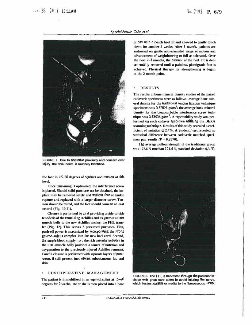
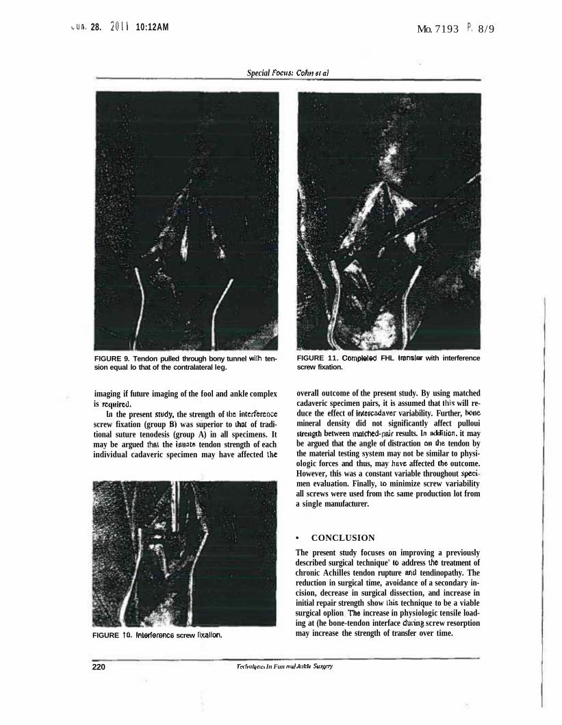