thrombin- and histamine-induced signal transduction in human
TRANSCRIPT

THE JOURNAL OF BIOLOGICAL CHEMISTRY 0 1991 by The American Society for Biochemistry and Molecular Biology, Inc.
Vol. 266, No. 1, Issue of January 5 , pp. 174-181.1991 Printed in U.S.A.
Thrombin- and Histamine-induced Signal Transduction in Human Endothelial Cells STIMULATION AND AGONIST-DEPENDENT DESENSITIZATION OF PROTEIN PHOSPHORYLATION*
(Received for publication, June 11, 1990)
Eugene G . Levin$ and Lydia Santell From the Dewartmnt of Molecular and Exmrimental Medicine, Scripps Clinic and Research Foundation, La Jolla, California 92037
Treatment of human endothelial cells with thrombin, histamine, or dioctanoylglycerol (DiCS), a synthetic diacylglycerol, resulted in the rapid and transient phosphorylation of a M, = 29,000 protein (P29) in a dose-dependent manner. Various tumor promoters also promoted P29 phosphorylation while the adenylate cyclase activator, forskolin, did not. The level of phos- phorylation with all three agonists was similar (2.5-4 fold), and analysis of P29 by two-dimensional gel elec- trophoresis revealed identical patterns in each case. Receptor specificity was demonstrated for the hista- mine-stimulated changes; pyrilamine (lo“? M; HI) but not cimetidine Hz) blocked the response. The thrombin effect was active site-dependent. Phos- phorylation induced by thrombin and histamine oc- curred within 1 min, peaked between 5 and 10 min, and returned to control levels by 1 h. DiC8-induced phosphorylation occurred more slowly but was also reduced by 1 h while phorbol ester treatment pro- longed phosphorylation for at least 4 h. Treatment of these cells with thrombin or histamine for 1 h desen- sitized P29 to further phosphorylation by the homolo- gous agonist although secondary phosphorylation could occur with heterologous compounds. However, if the primary agonist was removed following the onset of a desensitized state, secondary phosphorylation of P29 could be stimulated by the same compound. These same results were observed with two other phospho- proteins M, = 18,000 (P18) and 80,000 (P80) which became more highly phosphorylated in response to thrombin treatment and with histaminelthrombin- stimulated prostaglandin I2 production. In contrast, homologous down-regulation of P29 phosphorylation was not observed with DiC8-treated cells, and the de- cline in phosphorylated P29 was associated with the loss of functional DiC8. The protein kinase inhibitors staurosporine and H-7 blocked P18 and P80 phos- phorylation by thrombin but had no effect on P29 phosphorylation by histamine, thrombin, or DiC8 sug- gesting distinct pathways leading to the phosphoryla- tion of these different proteins. These data suggest that multiple and independent thrombinbistamine-induced
HL30244 and HL40435 and The Council for Tobacco Research. This * This work was supported by National Institutes of Health Grants
is Publication No. 6258-MEM from the Research Institute of Scripps Clinic, La Jolla, CA. The costs of publication of this article were defrayed in part by the payment of page charges. This article must therefore be hereby marked “aduertkement” in accordance with 18 U.S.C. Section 1734 solely to indicate this fact.
$ Recipient of an American Heart Association Established Inves- tigator Award. To whom correspondence and reprint requests should be addressed.
events are susceptible to receptor occupancy-depend- ent homologous down-regulation.
Histamine and thrombin are potent activators of human endothelial cells and have been found to induce numerous changes in endothelial cell function. Some of the changes are common to both of these compounds, e.g. permeability-en- hancing effects (1, 2) and stimulation of the production and/ or release of prostacyclin (3-6), platelet-activating factor (3- 6), and tissue plasminogen activator (7, 8). Similarities also exist in the pathways which transfer the signals generated during histamine/thrombin-receptor interaction. Both are known to activate the phosphoinositide pathway, presumably through the stimulation of phospholipase C, to generate ino- sitol 1,4,5-triphosphate and 1,2-diacylglycerol. These two products of phosphatidylinositol 4,5-bisphosphate hydrolysis cause rapid Ca2+ release from the endoplasmic reticulum and activate protein kinase C, respectively. Both protein kinase C activation and the elevation of [Ca2+]i (through the activation of Ca2+/calmodulin-dependent protein kinase) can lead to protein phosphorylation. Both of these protein kinases phos- phorylate a variety of proteins which function to control various biological responses (9). Nerve signal transduction, muscle contraction, cell shape change, exocytosis, and down- regulation of receptors have all been associated with phos- phorylation of specific proteins by protein kinases. In addi- tion, in some hormone-receptor interactions leading to the onset of transcription, the protein-protein interactions that are necessary may be regulated by phosphorylation.
The potential activation of multiple protein kinases by histamine and thrombin indicates the important role protein phosphorylation must play in mediating signal-response cou- pling after thrombin/histamine treatment of endothelial cells. While the effects of histamine and thrombin on endothelial cell function have been widely studied, little is known of the characteristics of the protein kinase-dependent events which occur in response to treatment with both of these agonists. In addition, the steps along the signal transduction pathway which both of these agonists have in common and which lead to identical responses are largely unexplored. In order to further investigate the properties of histamine/thrombin sig- nal transduction in human endothelial cells, we have studied the events surrounding the phosphorylation of a M, = 29,000 protein (P29) induced by both of these agonists and compared them to other thrombin- and histamine-generated events.
EXPERIMENTAL PROCEDURES
Endothelial Cell Culture-Endothelial cells were isolated from hu- man umbilical cord veins as previously described (7) and were cultured
174

ThrombinlHistarnine-induced Phosphorylation 175
into 75-cm2 tissue culture flasks coated with 20 mg/ml calf skin gelatin. Cells were grown to confluence in RPMI 1640 containing 15% newborn calf serum, 200 units/ml penicillin, 200 pg/ml strepto- mycin, 10 pg/ml endothelial cell growth factor, and 90 pg/ml heparin. Passaged cells were subcultured into 12-well dishes and allowed to grow to confluence under the same conditions as primary cultures except that 50 pg/ml endothelial cell growth factor was used.lAll experiments employed once-passaged cultures. Average cell densities at confluence were 6 X lo4 cells/cm2.
Studies were performed by washing confluent cultures twice with RPMI 1640 and incubating cultures at 37 "C in 0.5 ml of medium containing 5% NuSerum (final serum concentration = 1.25% newborn calf serum), 50 pg/ml endothelial cell growth factor, 90 pg/ml heparin, and the indicated agonist or vehicle. Stock solutions of phorbol esters and forskolin were made in dimethyl sulfoxide. In no case was the final concentration of dimethyl sulfoxide added to cells greater than 0.4%. The presence of endothelial cell growth factor, heparin, or dimethyl sulfoxide had no effect on the phosphorylation or dephos- phorylation of the proteins studied in the presence or absence of any of the compounds employed.
Phosphate Labeling of Endothelial Cells-Confluent monolayers in 12-well tissue dishes were washed twice with phosphate-free RPMI 1640 and incubated in phosphate-free medium containing 50 (for one- dimensional gels) or 200 (for two-dimensional gels) pCi/ml of ["PI orthophosphate for 1 h, and the test compounds were added for the specified times. Experiments to be analyzed by one-dimensional electrophoresis were stopped by removing the medium and adding 500 pl of 10% trichloroacetic acid, the wells were washed once with 1 ml of water, and the protein was solubilized with 200 pl/well of 62.5 mM Tris-HC1, pH 6.8, and 1% SDS.' For two-dimensional electro- phoresis, the cells were lysed (no trichloroacetic acid treatment) in 100 pl of 9.5 M urea, 2% Nonidet P-40, 2% Ampholine, pH 9-11. All lysis buffers were supplemented with protease inhibitors: 1 mM phenylmethylsulfonyl fluoride, 100 pg/ml leupeptin, 100 units/ml aprotinin, 10 mM N-ethylmaleimide. The insoluble material was removed by centrifugation at 100,000 X g for 60 min prior to electro- phoresis.
Electrophoresis-One-dimensional SDS-PAGE was performed ac- cording to the procedure of Laemmli (10) using a 12.5% separating gel and a 4% stacking gel unless otherwise specified. A final protein content of 20 pglsample was reduced with 5% @-mercaptoethanol and boiled for 3 min prior to application to the gel. All gels were stained with Coomassie Brilliant Blue and dried before autoradiography. In some cases, the gels were exposed to film for various lengths of time to attain optimum visualization of the various bands of interest. When the relative intensities of P29 were determined by laser den- sitometry, autoradiographs were scanned perpendicular to the plane of electrophoresis so that no other bands were detected and the signal could return to baseline between each lane. The intensity of the various P29 bands were normalized to a phosphoprotein whose level of phosphorylation did not change in response to any of the agonists employed (M, = 37,000).
Two-dimensional gel electrophoresis was performed with 17.5 pg of cell extract reduced with 5% P-mercaptoethanol employing Am- pholines, pH range 5-8 and 3-10 according to O'Farrell(11). Proteins were separated by isoelectric focusing on cylindrical gels in the first dimension (pH range 5-8) in the presence of 2% Nonidet P-40, and the gels were transferred directly to SDS-PAGE gels consisting of 12% polyacrylamide and 0.1% SDS. Molecular weight markers and a sample of cell extract were applied to the second dimension gel and run simultaneously with the samples in the first dimension gel. The pH gradient after isoelectric focusing was determined by slicing the gel into 0.5-cm sections, soaking each in degassed water for 15 min, and reading the pH of the H,O on a pH meter.
Measurement of Caz+i-Confluent cultures of endothelial cells were detached from their culture dishes using 0.2 M urea, 0.1% EDTA in phosphate-buffered saline with no calcium or magnesium and washed twice by centrifugation at 600 X g in phosphate-buffered saline containing 1.5 mM calcium chloride and 0.5 mM magnesium chloride.
' The abbreviations used are: SDS, sodium dodecyl sulfate; PAGE, polyacrylamide gel electrophoresis; DiC8, dioctanoylglycerob H-7, l-(-isoquinolinylsulfony1)-2-methylpiperazine; MAPTAM, bis(2- amino-5-methylphenoxy)ethane-N,N,N',N"tetraacetic acid tetra- acetoxymethyl ester; PDD, phorbo112,13-didecanoate; PMA, phorbol 12-myristate 13-acetate; pP29, phosphorylated P29; 6-keto-PGF1,, 6- keto-prostaglandin F1,; EGTA, [ethylenebis(oxyethylenenitrilo)] tetraacetic acid.
Cells were resuspended at 4 X lo6 cells/ml in loading buffer as described (12) and incubated with Indo 1/AM at a final concentration of 1.4 mM for 40 min at room temperature. When appropriate, MAPTAM was added 10 min after Indo 1/AM. The cells were washed by centrifugation and resuspended to 4 X lo6 cells/ml of the same buffer and stored at room temperature until used. Assays were per- formed with 0.3 ml of cells at 37 "C. The experiment was initiated and allowed to proceed for 30 s at which time histamine in 3-pl aliquots was added to a final concentration of 10 pg/ml, and the experiments were allowed to proceed for a total of 5 min. Fluorescence was measured at 37 "C in a SLM 8000 three-channel photon counting spectrofluorimeter as described previously (12) using an excitation set at 340 nm and emissions set at 400 nm. Samples incubated in the absence of Indo 1/AM but in the presence of dimethyl sulfoxide were included with each series of experiments in order to control for intrinsic fluorescence. These fluorescence levels were low and were not affected by the addition of thrombin or ionomycin. Fluorescence levels were calibrated after each experiment by adding 4 p~ iono- mycin and measuring fluorescence in the presence and absence of calcium. Calcium concentration was calculated using the previously described formulas (3).
Measurement of 6-Keto-prostaglandin Fl,-Confluent cultures were washed twice, placed in medium containing either 1 unit/ml thrombin or 10 pg/ml histamine for 2 h, and then either the same or opposite agonist was added for 10 min. Medium was removed and assayed for 6-keto-PGF1, by radioimmunoassay. The efficiency of thrombin- or histamine-induced 6-keto-PGF,, release after agonist pretreatment was compared to that released without pretreatment as follows: [agonist (2 h) + agonist (10 min)] - [agonist (2 h) + control (10 min)]/agonist (10 min) = % maximum response.
Materials-All tissue culture reagents were purchased from sources previously described (13). MAPTAM, Indo 1/AM, phorbol esters, bradykinin, and DiC8 were from Calbiochem. H-7 and HA1004 were from Seikagaku America, and staurosporine was from Kamiya Biomedical Co. The radioimmunoassay kit for 6-ketoprostaglandin F,, and [32P]orthophosphate (specific activity of 8500-9120 Ci/mmol) was purchased from Du Pont-New England Nuclear, and Ampholines were obtained from Pharmacia LKB Biotechnology Inc. Histamine and endotoxin were purchased from Sigma. Thrombin was a gift from Dr. J. W. Fenton I1 (New York Department of Health) and had a specific activity of 4014 units/mg of protein.
RESULTS
Phosphorylation of P29"Histamine (10 pg/ml), thrombin (1 unit/ml), various phorbol esters (100 nM-1 pM), and DiC8 (150 pM) were added to cultures for 10 min following incor- poration of 32P04, and the phosphoproteins were fractionated and detected by one-dimensional 12.5% SDS-PAGE and au- toradiography (Fig. 1). Both agonists, all of the active tumor promoters, and DiC8 stimulated an increase in the intensity of a band migrating to a position corresponding to M, = 29,000 (P29). The non-tumor promoter, 4-a-PDD, had no effect, while a small increase was observed in the presence of forskolin, an activator of adenylate cyclase. P29 phosphoryl- ation in the presence of PMA and forskolin was slightly higher than PMA alone. Two other compounds tested, 1 p~ brady- kinin and 10 ng/ml endotoxin, did not have an effect, even when the incubation time was prolonged to 1 h (P29 intensity was equal to control samples). Laser densitometric analysis of the P29 band in multiple gels (n = 5) demonstrated that the increase in the intensity of P29 was similar in thrombin-, histamine-, and DiC8-treated cultures (2.5 to 4.5 times that of control cultures). Dose titration analysis of thrombin, histamine, and DiC8 indicated that the maximum effect oc- curred at the concentrations of 1 unit/ml, 10 pg/ml, and 150 p ~ , respectively (data not shown). Simultaneous addition of thrombin and histamine or histamine and DiC8 showed P29 band intensities identical with samples treated with each agonist alone.
Two-dimensional gel electrophoresis of the histamine-, thrombin-, and DiC8-treated cultures are shown in Fig. 2. In control cultures, three spots migrating to a position corre-


ThrombinlHistamine-induced Phosphorylation 177
r
C 1 5 ’ 10’ C20’ 20 C4OD40 C60 060
”” ”” ”” -P29
-Pl8
lh7t&7lhbair
”” &&a rrolia
1’ 2’ 5’ 1 0 C 20’ C20 40’ C40’ 60’ CEO
0 2 4 6 8 1 0 20 40 - 60
Tim Iminl
FIG. 4. Time course of P29 phosphorylation by histamine/ thrombin and DiCS. A, cultures were labeled for 1 h with 50 pCi/ ml “’PO4 and 1 unit/ml thrombin, 10 pg/ml histamine, and 150 pM DiC8 added. At the indicated times, the reaction was stopped by removing the medium and adding trichloroacetic acid, and the sam- ples were prepared and analyzed on one-dimensional gels as described under “Experimental Procedures.” Due to increased background phosphorylation with longer incubation times, control samples were run simultaneously with the experimental samples for comparative purposes. Presentation of the thrombin and histamine time courses is made with a representative histamine experiment; C, control cul- tures extracted at 0 time; C20’, C40’, etc., cultures incubated in the absence of agonist for the time indicated. Arrows indicate the position of P29. B, relative levels of P29 phosphorylation displayed in A were determined by laser densitometry and normalized to a band not affected by any of the agonists used (M, = 37,000). Each point on the graph represents the fold increase of P29 band intensity over the respective control sample; W, thrombin/histamine; W, DiC8.
h in phosphate-free medium, adding 32P04 directly to the medium for 1 h, and then adding nothing or histamine, thrombin, or DiC8 for 10 min. After 2 h of histamine or thrombin treatment, P29 was not phosphorylated (TH/HIS > C) which is consistent with the disappearance of pP29 during kinetic experiments (Fig. 4; 1 h). Addition of the heterologous agonist resulted in a second phosphorylation of the P29 band (HIS > TH, TH > HIS, or HIS/TH > DiC8) to a level similar to that found in cultures not treated with a primary agonist. In contrast, secondary treatment with the same agonist had no effect on the intensity of P29, indicating that secondary phosphorylation had not taken place (TH > TH or HIS > HIS). Thus. desensitization of P29 Dhosnhorvl- ation was restricted to the homologous agonist and did not affect any of the others tested. Analysis of the thrombin- induced phosphorylation of two other phosphoproteins, P18 and P80, showed the same results we observed with P29 (Fig. 5): a decline in the level of phosphorylation back to steady
I C I I TH I -2 hr c TH HIS 01c8 c TH olC8 -10 min
FIG. 5. Homologous desensitization of protein phosphoryl- ation. Cultures were treated with the indicated agonist (aboue the hatched line) in phosphate-free medium for 1 h, 50 pCi/ml 32P04 added directly for 1 h in the continued presence of the primary agonist, and then a secondary agonist added for 10 min (below the hatched line). Concentrations used for both primary and secondary treatment were 10 pg/ml histamine, 1 unit/ml thrombin, 150 pM DiC8. Samples were analyzed on one-dimensional gels as described under “Experimental Procedures.” The gel was exposed for different lengths of time to allow optimum detection of the three bands indicated on the right side of the figure (P80, P29, P18). C, no agonist added; HIS, histamine; TH, thrombin.
1 2 3 4 5 6 3 4 5 “
DICE HIS
FIG. 6. Loss of DiCS “P29 phosphorylating” activity upon incubation with endothelial cells. Agonist identified under the hatched line refers to that used in the secondary treatment. 1, control; 2, cells incubated with 150 PM DiC8 for 2 h; 3, cells incubated with DiC8 for 2 h and then treated with 150 p~ DiC8 or 10 pg/ml histamine; 4, medium containing 150 PM DiC8 or 10 pg/ml histamine was incubated with cells for 1 h, removed (spent medium), and transferred to fresh cells prelabeled with “‘PO4. After 10 min, the cells were extracted; 5, spent medium prepared as in lune 4 with fresh DiC8 or histamine added; 6, fresh DiC8 added to cells for 10 min. The level of P29 phosphorylation was determined as described under “Experimental Procedures.” HIS, histamine.
state levels by 2 h and a failure to respond to secondary thrombin treatment but not to DiC8 (P80 only). Thus, down- regulation of thrombin-induced phosphorylation appears not to be an event restricted to one protein (P29).
When cultures were pretreated with 150 pM DiC8 for 2 h, readdition of DiC8 or other agonists stimulated P29 phos- phorylation, indicating that desensitization was not induced by DiC8 (Fig. 6, lane 3). The DiC8-generated P29 phosphoryl- ation-dephosphorylation cycle could be repeated at least three times. When cultures were treated with DiC8 at hourly inter- vals, pP29 also declined by the end of the subsequent treat- ment hours but could be regenerated again by the addition of fresh DiC8 (data not shown). Further treatment elevated levels of DMSO beyond acceptable limits.
To determine whether the decline in pP29 occurred in the presence of “functional” agonist (i.e. agonist still capable of inducing P29 phosphorylation), medium containing 150 pM DiC8 or 10 pg/ml histamine was incubated with cells for 1 h, removed from these cultures, and placed directly onto fresh cells prelabeled with “’PO,. After 10 min, the cells were extracted, and the ability of the spent medium (containing the original agonist) to generate pP29 in the new cultures was determined. FnhwinE the 60-min innlhnt.inn, t.hP modillrn containing the DiC8 did not induce an increase in the inten- sity of the P29 band in the fresh cultures while the medium with histamine did (Fig. 6, lune 4 ) . If the medium was removed and fresh 150 PM DiC8 was added to the same spent medium prior to addition, P29 was phosphorylated to the same level

178 ThrombinlHistamine-induced Phosphorylation
as in control cultures (Fig. 6, lane 5 uersus lane 3 ) . Fresh histamine had no further effect on the level of pP29 (Fig. 6, lane 5 ) . DiC8 a t 300 p~ gave identical results. Higher concen- trations were not tested due to adverse effects of the higher dimethyl sulfoxide concentrations.
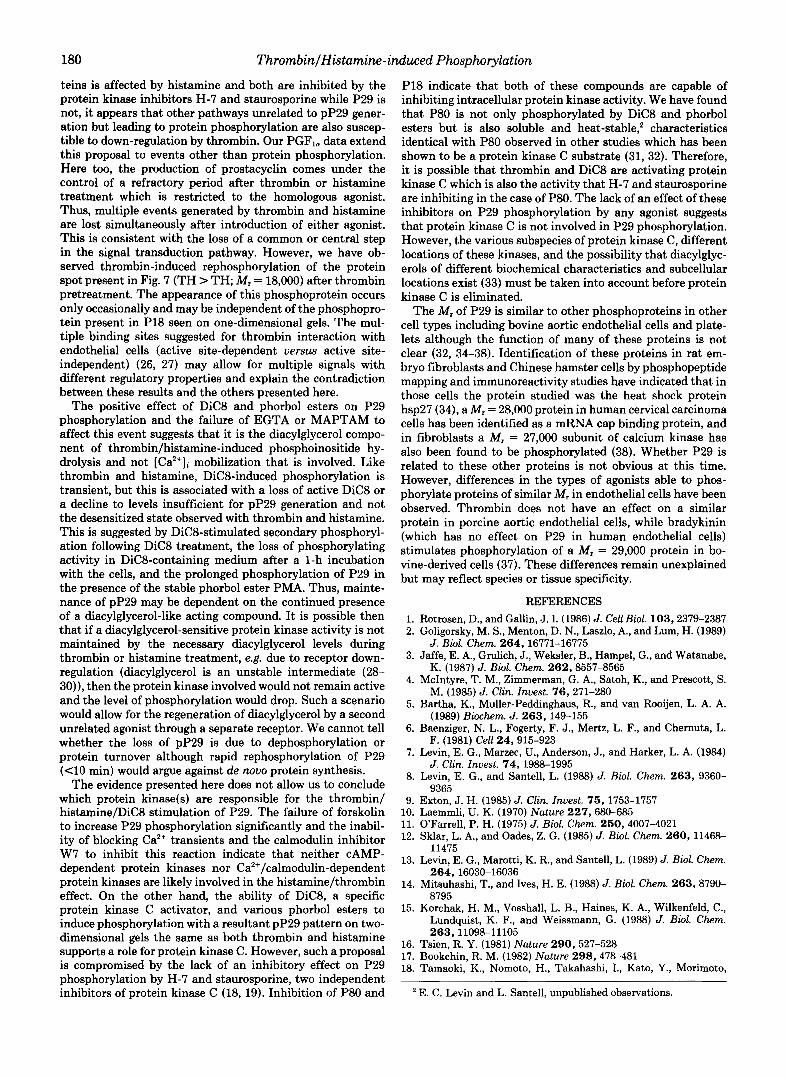
Two-dimensional analysis of the effect of thrombin pre- treatment on the P29 was also performed (Fig. 7). After 2 h of thrombin treatment, addition of the same agonist had no effect on any of the P29 proteins (TH > T H uersus T H > C). When histamine was added to thrombin-treated cultures, enhanced phosphorylation of P29a, -b, and -d occurred (TH > HIS), and the pattern was similar to histamine-only treated samples (Fig. 2). Thus, the rephosphorylation of P29 by heterologous agonists affected the identical species of P29 as did primary treatment.
As an independent measure of the extent of thrombin- and histamine-induced down-regulation, the effect of pretreat- ment on the stimulation of prostacyclin production (measured as 6-keto-PGF,,) was examined (Table I). Experimental con-
I L T H X TH.TH
TH.HIS
FIG. 7. Two-dimensional electrophoresis of P29 phos- phorylation following homologous down-regulation. Endothe- lial cells were exposed to 1 unit/ml thrombin for 1 h, labeled for 1 h with 200 pCi/ml "'PO, in the presence of the primary agonist, and then treated with either thrombin or 10 pg/ml histamine for 10 min. The samples were prepared and analyzed by two-dimensional electro- phoresis as described under "Experimental Procedures." Spots are identified by letters identical with those used in Fig. 2. C, no agonist added; TH, thrombin; HIS, histamine.
TABLE I Homologous down-regulation of 6-keto-PGF,, production by
thrombin/histamine Endothelial cell cultures containing RPMI 1640 were incubated
with 1 unit/ml thrombin, 10 pg/ml histamine, or nothing for 2 h and then vehicle, thrombin, histamine, or 1 p~ ionomycin was added directly to each for 10 min. Medium was removed and assayed for 6- keto-PGF,,. Data are the average of three separate experiments, each performed in duplicate. The net production of 6-keto-PGF1, (agonist - buffer control (value shown in parentheses)) is shown.
Treatment
2 h 10 min 6-Keto-PGF1,
Control Control Control Control Thrombin Thrombin
Thrombin Histamine Histamine Histamine Histamine
Thrombin
Control Thrombin Histamine Ionomycin Control Thrombin
Ionomycin Control Thrombin Histamine Ionomvcin
I-Iiotomino
d m 1 (0.43) 1.07 1.08 3.12 0.88 0.85 1.C7 3.77 0.67 1.72 0.70 4.10
ditions were identical with those used in the phosphorylation experiments. Thrombin and histamine treatment for 10 min resulted in a 3-fold increase in the concentration of 6-keto- PGF],. A 1-h exposure to histamine abrogated any further increase in 6-keto-PGF1, release by the same agonist (com- pare histamine > control with histamine > histamine) while thrombin stimulated a secondary response. The additional 6- keto-PGF,, appearing after thrombin treatment was 92% of that released with no prior treatment (control > thrombin). The same results were observed with thrombin pretreatment although the secondary response to histamine was slightly lower than that released following primary histamine treat- ment (73%). Ionomycin addition following histamine and thrombin pretreatment released 109% and 92% of control levels, respectively.
In the experiments described above, cultures were treated continuously for 2 h with the primary agonist, and the second agonist was added directly to the existing medium. To deter- mine whether removing the primary agonist and washing the cells before secondary treatment would reverse the desensiti- zation of P29 phosphorylation, cultures were treated with histamine or thrombin for 1 h, washed, and incubated for 2 h in phosphate- and agonist-free medium (label was added during the second hour), and then treated a second time for 10 min (Fig. 8). Both histamine and thrombin treatment resulted in a second phosphorylation of P29, P18, and P80 despite the 1-h pretreatment with the same compound.
P29 Phosphorylation Is Not Affected by Calcium Chelators- Both thrombin and histamine elicit a rapid rise in [Ca2+]i which attains peak values 15-30 s after agonist addition (1, 2). A sustained phase of [Ca2+Ii elevation follows the initial rise and is dependent on an influx of extracellular Ca". To determine whether thrombin- and histamine-stimulated P29 phosphorylation might be dependent on changes in the [Ca2+]i, endothelial cells were preloaded with the cell-perme- able chelator of Ca2+-MAPTAM to buffer the increase in [Ca2+]i occurring after thrombin and histamine treatment (14-17). In the absence of MAPTAM, Ca2+ levels in resting cells were 83 nM, increased to 316 nM within 5 s of histamine addition, and then declined to 143 nM by 90 s where it remained throughout the rest of the experiment (average of four experiments). When the cells were loaded with 100 p~ MAPTAM, resting cytosolic Ca2+ levels were reduced by 35%, and no increase in cytosolic Caz+ was observed after histamine addition. With regard to P29 phosphorylation, MAPTAM
FIG. 8. Effect of agonist removal on secondary phosphoryl- ation of P29. Cultures were treated with the agonist indicated above the hush mark a t 10 pg/ml histamine or 1 unit/ml thrombin for 1 h, washed three times with phosphate-free medium, and incubated in the same containing 50 lCi/ml for 2 h. and then exDosed to the second agonist shown under the hash mark for 10 min at the same concentration used above. The gel was exposed for different lengths of time to allow detection of the three bands indicated (P80, P29, P18). Samples were prepared accordingly and analyzed by one-di- mensional electrophoresis. C, no agonist added; TH, thrombin; H I S , histamine.

ThrombinlHistamine-induced Phosphorylation 179
itself had a slight stimulatory effect on pP29 (Fig. 9). This was MAPTAM-specific since control medium was identical with experimental medium except for the presence of the chelator. Despite the reduction in Ca2+ transients, induction of P29 phosphorylation by either thrombin or histamine was not compromised; the level of pP29 generated by histamine or thrombin was equal in the presence and absence of the rise in [Ca2+Ii.
A requirement for extracellular Ca2+ for the maintenance of P29 phosphorylation was examined by depleting culture medium Ca2+ with EGTA 2 min prior to agonist addition. Depletion of extracellular Ca2+ has been shown not to effect the initial increase in [Ca'+]i (0-90 s), which is dependent on intracellular pools, but to eliminate the prolonged [Ca2+Ii elevation (1). Once again, P29 phosphorylation was not com- promised and remained elevated when examined 1 min, 10 min, or 20 min after agonist addition (Fig. 9). Addition of EGTA following P29 phosphorylation did not accelerate the decline in pP29. To determine if calmodulin was involved in thrombin or histamine P29 phosphorylation, cultures were treated with 50 p~ W7, an inhibitor of calmodulin, or W5, an inactive analog of W7, and then stimulated with the agonists. In neither case did the presence of W7 or W5 suppress the
MAPTAM
c
--+" C HIS TH
FIG. 9. Effect of calcium on P29 phosphorylation. Cultures were labeled with "PO4 for 1 h. 1 or 5 mM EGTA was added 2 min prior to agonist addition or 50 PM MAPTAM was added 30 min before agonist and then 10 pg/ml histamine or 1 unit/ml thrombin added. Cultures pretreated with EGTA were extracted 1, 10, or 20 min after stimulation with thrombin or histamine (the 20-min ex- traction is shown), and MAPTAM-treated cultures were extracted a t 10 min. Samples were prepared accordingly and analyzed by one- dimensional electrophoresis. E(l ) , 1 mM EGTA; E(5), 5 mM EGTA; M, MAPTAM; HIS, histamine; TH, thrombin.
DICE DICE TH TH
ST ST + +
P29, P18, and P80. Cultures were labeled with 32P04 for 1 h, and PIG. 10. Effecl uf slauruspurlnt! un the yhuvphurylallun uI
100 nM staurosporine was added 15 min before the end of the labeling period. Thrombin (1 unit/ml) or DiC8 (150 PM) was added for 10 min, and then the cells were extracted and analyzed as described under "Experimental Procedures." TH, thrombin; ST, staurosporine. The three proteins referred to in the text are indicated on the right.
generation of pP29 (data not shown). Staurosporine and H-7 Inhibit Phosphorylation of P18 and
P80 but Not P29"To determine whether phosphorylation of P29 would be affected by the presence of protein kinase inhibitors, staurosporine (18), H-7 (19), and HA1004 (19) were added to the cells 15 min prior to agonist addition, and the cells were treated with either vehicle, histamine, thrombin, or DiC8 for 10 min (Fig. 10). Staurosporine (100 nM) or 100 p~ H-7 (data are shown for staurosporine only) had no effect whatsoever on P29 phosphorylation by thrombin, histamine, or DiC8. In contrast, the increase in P18 phosphorylation in thrombin-treated cultures was depressed. This occurred in a dose-dependent manner with 75 nM staurosporine giving max- imum inhibition. Stimulation of pP80 was affected similarly in both thrombin- and DiC8-treated cultures. In both cases, the enhancement in band intensity which occurred after treat- ment was absent. HA1004 had no effect on any of the proteins examined (data not shown).
DISCUSSION
Generation of diacylglycerol and Ca2+, both activators of protein kinases, following exposure of endothelial cells to thrombin and histamine indicates that protein phosphoryla- tion plays an important role in signal-response coupling. In this present study, we demonstrate that a set of phosphopro- teins which migrate to a position corresponding to M, = 29,000 become more highly phosphorylated following treatment of endothelial cells with histamine, thrombin, DiC8, and various phorbol esters. The band in one-dimensional gels that we refer to as P29 is made up of three detectable spots on two- dimensional gel electrophoresis. These proteins are unevenly phosphorylated under resting conditions with P29a dominat- ing. After stimulation, two of the three spots increase in intensity (P29a and -b), and two more spots become visible.
Comparison of the characteristics of P29 phosphorylation demonstrates a recurring identity between the histamine- and thrombin-induced effects. The level of phosphorylation, the time course of phosphorylation and dephosphorylation, the occurrence of a desensitized state which only affects the homologous agonist, the reversal of desensitization with wash- out, and the failure of the same protein kinase inhibitors to suppress phosphorylation, are all similar when thrombin and histamine are used. This would suggest that these two agonists share the same pathway responsible for P29 phosphorylation. However, the occurrence of homologous desensitization in each case would also indicate that there is a component of the reaction that is specific for each. While the complexity of the pathway that evolves after agonist treatment precludes an obvious choice of this specific component, one possibility is the individual receptors. In various reports describing the onset of homologous desensitization, the loss of responsive- ness to a specific agonist has been linked to impairment of receptor function due to the phosphorylation of that receptor (20-22). Continuous interaction between agonist and receptor is sometimes required for continued down-regulation and is referred to as agonist-dependent desensitization (23-25). This type of down-regulation would be consistent with the char- acteristics we have observed for P29 phosphorylation in this study, i.e. receptor dependency in the case of histamine (HI, Fig. 3), a loss of responsiveness during receptor occupancy, and a reversal of me UeSenSlt~zeU state after washout suggest- ing that the presence of agonist is required for the continued depression of P29 phosphorylation. In addition, the desensi- tized state is not restricted to P29. Thrombin-induced down- regulation also affects other proteins (P18 and P80) in the same manner as P29. Because neither of these phosphopro-
180 ThrombinlHistamine-induced Phosphorylation
teins is affected by histamine and both are inhibited by the protein kinase inhibitors H-7 and staurosporine while P29 is not, it appears that other pathways unrelated to pP29 gener- ation but leading to protein phosphorylation are also suscep- tible to down-regulation by thrombin. Our PGFI, data extend this proposal to events other than protein phosphorylation. Here too, the production of prostacyclin comes under the control of a refractory period after thrombin or histamine treatment which is restricted to the homologous agonist. Thus, multiple events generated by thrombin and histamine are lost simultaneously after introduction of either agonist. This is consistent with the loss of a common or central step in the signal transduction pathway. However, we have ob- served thrombin-induced rephosphorylation of the protein spot present in Fig. 7 (TH > TH; M, = 18,000) after thrombin pretreatment. The appearance of this phosphoprotein occurs only occasionally and may be independent of the phosphopro- tein present in P18 seen on one-dimensional gels. The mul- tiple binding sites suggested for thrombin interaction with endothelial cells (active site-dependent uersus active site- independent) (26, 27) may allow for multiple signals with different regulatory properties and explain the contradiction between these results and the others presented here.
The positive effect of DiC8 and phorbol esters on P29 phosphorylation and the failure of EGTA or MAPTAM to affect this event suggests that it is the diacylglycerol compo- nent of thrombin/histamine-induced phosphoinositide hy- drolysis and not [Ca2+Ii mobilization that is involved. Like thrombin and histamine, DiC8-induced phosphorylation is transient, but this is associated with a loss of active DiC8 or a decline to levels insufficient for pP29 generation and not the desensitized state observed with thrombin and histamine. This is suggested by DiC8-stimulated secondary phosphoryl- ation following DiC8 treatment, the loss of phosphorylating activity in DiC8-containing medium after a 1-h incubation with the cells, and the prolonged phosphorylation of P29 in the presence of the stable phorbol ester PMA. Thus, mainte- nance of pP29 may be dependent on the continued presence of a diacylglycerol-like acting compound. It is possible then that if a diacylglycerol-sensitive protein kinase activity is not maintained by the necessary diacylglycerol levels during thrombin or histamine treatment, e.g. due to receptor down- regulation (diacylglycerol is an unstable intermediate (28- 30)), then the protein kinase involved would not remain active and the level of phosphorylation would drop. Such a scenario would allow for the regeneration of diacylglycerol by a second unrelated agonist through a separate receptor. We cannot tell whether the loss of pP29 is due to dephosphorylation or protein turnover although rapid rephosphorylation of P29 ( 4 0 min) would argue against de m u 0 protein synthesis.
The evidence presented here does not allow us to conclude which protein kinase(s) are responsible for the thrombin/ histamine/DiCS stimulation of P29. The failure of forskolin to increase P29 phosphorylation significantly and the inabil- ity of blocking Ca2+ transients and the calmodulin inhibitor W7 to inhibit this reaction indicate that neither CAMP- dependent protein kinases nor Ca2+/calmodulin-dependent protein kinases are likely involved in the histamine/thrombin effect. On the other hand, the ability of DiC8, a specific protein kinase C activator, and various phorbol esters to induce phosphorylation with a resultant pP29 pattern on two- dimensional gels the same as both thrombin and histamine supports a role for protein kinase C. However, such a proposal is compromised by the lack of an inhibitory effect on P29 phosphorylation by H-7 and staurosporine, two independent inhibitors of protein kinase C (18, 19). Inhibition of P80 and
P18 indicate that both of these compounds are capable of inhibiting intracellular protein kinase activity. We have found that P80 is not only phosphorylated by DiC8 and phorbol esters but is also soluble and heat-stable,' characteristics identical with P80 observed in other studies which has been shown to be a protein kinase C substrate (31, 32). Therefore, it is possible that thrombin and DiC8 are activating protein kinase C which is also the activity that H-7 and staurosporine are inhibiting in the case of P80. The lack of an effect of these inhibitors on P29 phosphorylation by any agonist suggests that protein kinase C is not involved in P29 phosphorylation. However, the various subspecies of protein kinase C, different locations of these kinases, and the possibility that diacylglyc- erols of different biochemical characteristics and subcellular locations exist (33) must be taken into account before protein kinase C is eliminated.
The M, of P29 is similar to other phosphoproteins in other cell types including bovine aortic endothelial cells and plate- lets although the function of many of these proteins is not clear (32, 34-38). Identification of these proteins in rat em- bryo fibroblasts and Chinese hamster cells by phosphopeptide mapping and immunoreactivity studies have indicated that in those cells the protein studied was the heat shock protein hsp27 (34), a M, = 28,000 protein in human cervical carcinoma cells has been identified as a mRNA cap binding protein, and in fibroblasts a M, = 27,000 subunit of calcium kinase has also been found to be phosphorylated (38). Whether P29 is related to these other proteins is not obvious at this time. However, differences in the types of agonists able to phos- phorylate proteins of similar M, in endothelial cells have been observed. Thrombin does not have an effect on a similar protein in porcine aortic endothelial cells, while bradykinin (which has no effect on P29 in human endothelial cells) stimulates phosphorylation of a M, = 29,000 protein in bo- vine-derived cells (37). These differences remain unexplained but may reflect species or tissue specificity.
REFERENCES 1. Rotrosen, D., and Gallin, J. I. (1986) J. Cell Biol. 103,2379-2387 2. Goligorsky, M. S., Menton, D. N., Laszlo, A., and Lum, H. (1989)
3. Jaffe, E. A., Grulich, J., Weksler, B., Hampel, G., and Watanabe,
4. McIntyre, T. M., Zimmerman, G. A., Satoh, K., and Prescott, S.
5. Bartha, K., Muller-Peddinghaus, R., and van Rooijen, L. A. A.
6. Baenziger, N. L., Fogerty, F. J., Mertz, L. F., and Chernuta, L.
7. Levin, E. G., Marzec, U., Anderson, J., and Harker, L. A. (1984)
8. Levin, E. G., and Santell, L. (1988) J. Biol. Chem. 263, 9360-
9. Exton, J. H. (1985) J. Clin. Znuest. 7 5 , 1753-1757
J. Biol. Chem. 264,16771-16775
K. (1987) J. Biol. Chem. 262,8557-8565
M. (1985) J. Clin. Znuest. 7 6 , 271-280
(1989) Biochem. J. 263,149-155
F. (1981) Cell 2 4 , 915-923
J. Clin. Znuest. 7 4 , 1988-1995
9365
10. Laemmli, U. K. (1970) Nature 227,680-685 11. O'Farrell, P. H. (1975) J. Biol. Chem. 250,4007-4021 12. Sklar, L. A., and Oades, Z. G. (1985) J. Biol. Chem. 260 , 11468-
13. Levin, E. G., Marotti, K. R., and Santell, L. (1989) J. Biol. Chem.
14. Mitsuhashi, T., and Ives, H. E. (1988) J. Biol. Chem. 263,8790- 8795
15. Korchak, H. M., Vosshall, L. B., Haines, K. A., Wilkenfeld, C., Lundquist, K. F., and Weissmann, G. (1988) J. Biol. Chem.
11475
264,16030-16036
263,11098-11105 16. Tsien, R. Y. (1981) Nature 290, 527-528 17. Bookchin, R. M. (1982) Nature 298,478-481 18. Tamaoki, K., Nomoto, H., Takahashi, I., Kato, Y., Morimoto,
E. C. Levin and L. Santell, unpublished observations.

Thrombin/Histamine-induced Phosphorylation 181
M., and Tomita, F. (1988) Biochem. Bwphys. Res. Commun.
19. Matsui, T., Nakao, Y., Koizumi, T., Katakami, Y., and Fujita, T. (1986) Cancer Res. 46,583-581
20. Benovic, J . L., Pike, L. J., Cerione, R. A., Staniszewski, C., Yoshimasa, T., Codina, J., Caron, M. G., and Lefkowitz, R. J.
21. Bouvier, M., Leeb-Lundberg, L. M. F., Benovic, J. L., Caron, M. G., and Lefkowitz, R. J. (1987) J. Biol. Chem. 262,3106-3113
22. Lefkowitz, R. J., and Caron, M. G. (1988) J. Biol. Chem. 263,
23. Benovic, J. L., Mayor, F., Jr., Staniszewski, C., Lefkowitz, R. J., and Caron, M. G. (1987) J. Bwl. Chem. 262,9026-9032
24. Mayor, F., Jr., Benovic, J. L., Caron, M. G., and Lefkowitz, R. J. (1987) J. Bwl. Chem. 262,6468-6471
25. Benovic, J. L., Kuhn, H., Weyand, I., Codina, J., Caron, M. G., and Lefkowitz, R. J. (1987) Proc. Natl. Acad. Sci. U. S. A. 84,
26. Awbrey, B. J., Hoak, J. C., and Owen, W. G. (1979) J. Biol. Chem.
27. Lollar, P., Hoak, J. C., and Owen, W. G. (1980) J. Bwl. Chem.
136,397-402
(1985) J. Eiol. Chem. 260,7094-7101
4993-4996
8879-8882
264,4092-4095
256,10279-10283
28.
29. 30.
31.
32.
33.
34.
35. 36.
37.
38.
Davis, R. J., Ganong, B. R., Bell, R. M., and Czech, M. P. (1985)
Rittenhouse-Simmons, S. (1979) J. Clin. Znuest. 63, 580-587 Conn, P. M., Ganong, B. R., Ebeling, J., Staley, D., Neidel, J. E.,
and Bell, R. M. (1985) Biochem. Biophys. Res. Commun. 126,
Issandou, M., and Rozengurt, E. (1989) Biochem. Biophys. Res.
Blackshear, P. J., Witters, L. A., Girard, P. R., Kuo, J. F., and
Rosoff, P. M., Savage, N., and Dinarello, C. A. (1988) Cell 64,
Chretien, P., and Landry, J. (1988) J. Cell. Physwl. 137, 157-
Kaur, P., and Saklatvala, J. (1988) FEES Lett. 241, 6-10 Darbon, J.-M., Tournier, J.-F., Tauber, J.-P., and Bayard, F.
Demolle, D., Lecomte, M., and Boeynaems, J.-M. (1988) J. Bwl.
Marino, M. W., Pfeffer, L. M., Guidon, P. T., Jr., and Donner,
J. Bwl. Chem. 260, 1562-1566
532-539
Commun. 163,201-208
Quamo, S. N. (1985) J. Biol. Chem. 260,13304-13315
73-81
166
(1986) J. Biol. Chem. 261,8002-8008
Chem. 263,18459-18465
D. B. (1989) Proc. Nutl. Acad. Sci. U. S. A. 86,8417-8421