thomas h. chia and michael j. levene - school of...
TRANSCRIPT

102:1310-1314, 2009. First published Jun 3, 2009; doi:10.1152/jn.91208.2008 J NeurophysiolThomas H. Chia and Michael J. Levene
You might find this additional information useful...
10 articles, 5 of which you can access free at: This article cites http://jn.physiology.org/cgi/content/full/102/2/1310#BIBL
including high-resolution figures, can be found at: Updated information and services http://jn.physiology.org/cgi/content/full/102/2/1310
can be found at: Journal of Neurophysiologyabout Additional material and information http://www.the-aps.org/publications/jn
This information is current as of March 25, 2010 .
http://www.the-aps.org/.American Physiological Society. ISSN: 0022-3077, ESSN: 1522-1598. Visit our website at (monthly) by the American Physiological Society, 9650 Rockville Pike, Bethesda MD 20814-3991. Copyright © 2005 by the
publishes original articles on the function of the nervous system. It is published 12 times a yearJournal of Neurophysiology
on March 25, 2010
jn.physiology.orgD
ownloaded from
Innovative Methodology
Microprisms for In Vivo Multilayer Cortical Imaging
Thomas H. Chia and Michael J. LeveneDepartment of Biomedical Engineering, Yale University, New Haven, Connecticut
Submitted 12 November 2008; accepted in final form 29 May 2009
Chia TH, Levene MJ. Microprisms for in vivo multilayer corticalimaging. J Neurophysiol 102: 1310–1314, 2009. First published June3, 2009; doi:10.1152/jn.91208.2008. Cortical slices allow for simul-taneous imaging of multiple cortical layers. However, slices lacknative physiological inputs and outputs. Although in vivo, two-photonimaging preserves the native context, it is typically limited to a depthof �500 �m. In addition, simultaneous imaging of multiple corticallayers is difficult due to the stratified organization of the cortex. Wedemonstrate the use of 1-mm microprisms for in vivo, two-photonneocortical imaging. These prisms enable simultaneous imaging ofmultiple cortical layers, including layer V, at an angle typical of slicepreparations. Images were collected from the mouse motor andsomatosensory cortex and show a nearly 900-�m-wide field of view.At high-magnification imaging using an objective with 1-mm ofcoverglass correction, resolution is sufficient to resolve dendriticspines on layer V neurons. Images collected using the microprism arecomparable to images collected from a traditional slice preparation.Functional imaging of blood flow at various neocortical depths is alsopresented, allowing for quantification of red blood cell flux andvelocity. H&E staining shows the surrounding tissue remains in itsnative, stratified organization. Estimation of neuronal damage usingpropidium iodide and a fluorescent Nissl stain reveals cell damage islimited to �100 �m from the tissue–glass interface. Microprisms area straightforward tool offering numerous advantages for research intoneocortical tissue.
I N T R O D U C T I O N
Cortical slices provide a powerful platform for neurophysi-ology, with easy access to multiple cortical layers for chemical,electrical, and optical measurement and manipulation. How-ever, slices lack important aspects of physiological context,including inputs from intra- and extracortical regions and anactive circulatory system. Although in vivo experiments pro-vide this context, optical access to deeper layers is exceedinglydifficult and almost always provides image planes confined toa single cortical layer (Helmchen and Denk 2005). Here, wedemonstrate that microprisms inserted into mouse cortex en-able simultaneous imaging of multiple cortical layers with a1-mm field of view (FOV), submicron spatial resolution, andan imaging perspective typical of slice preparations.
Attempts to image deep layers of neocortex are made diffi-cult by the highly light scattering nature of brain tissue.Although multiphoton microscopy is well suited for fluores-cence imaging in the brain, imaging depths typically reach�500 �m. Generally, this depth is superficial of cortical layersIV–VI, where many pyramidal cell bodies are located. Giventhe importance of deep cortical layers, there is strong demandto clearly see the unique structures and functions of theselayers in their native context with high resolution and signal-to-noise ratio (SNR).
Two-photon imaging depths of 1,000 �m in cortical tissuehave been obtained using a regenerative amplifier to increaseexcitation power (Theer et al. 2003). However, images atdepths �600 �m suffer from low SNR and poor contrast.Gradient-index (GRIN) lenses, inserted perpendicular to theneocortex, have also imaged deep layers of the brain (Junget al. 2004; Levene et al. 2004). However, the FOV was limitedto �130 �m and apical dendrites belonging to the neuronsimaged directly below the GRIN lens were destroyed. Prismsof 0.5 mm have been used with one-photon excitation tomeasure the net fluorescence emission from layer V apicaldendrites in rats (Murayama et al. 2007, 2009). However,fluorescence was collected from large regions of interests insuperficial cortical layers and did not produce high-resolutionfluorescence images.
M E T H O D S
Microprism optics and imaging parameters
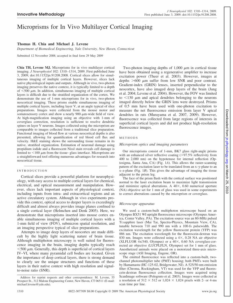
Our microprisms consist of 1-mm, BK7 glass right-angle prismswith an enhanced silver reflective coating (�97.5% reflectivity from400 to 2,000 nm) on the hypotenuse for internal reflection (Op-tosigma, Santa Ana, CA) (Fig. 1A). This allows the raster-scanningpattern of the excitation laser to be translated from an x–y-plane to anx–z-plane (Fig. 1B). This gives the advantage of imaging the tissueadjacent to the prism leg.
The face of the prism flush with the cortical surface was positionednormal to the laser excitation beam to maximize light transmissionand minimize optical aberrations. A 40�, 0.60 numerical aperture(NA) objective set for 1 mm of glass was used in some experimentsto correct aberrations caused by the microprism or coverglass.
Microscope apparatus
We used a custom-built multiphoton microscope based on anOlympus BX51 WI upright fluorescence microscope (Olympus Amer-ica, Center Valley, PA). The excitation source was an 80-MHz pulsedTi:sapphire laser (Mai Tai, Spectra-Physics, Mountain View, CA),tunable between 710 and 990 nm with a 100-fs pulsewidth. Theexcitation wavelength for the yellow fluorescent protein (YFP) was886 nm. The excitation wavelength for the fluorescein-dextran was830 nm. Images were collected using a 4�, 0.28 NA air objective(XLFLUOR 4x/340, Olympus) or a 40�, 0.60 NA coverglass-cor-rected air objective (LUCPLFLN, Olympus) set for 1 mm of glass.Anesthetized animals were placed on a motorized three-axis micro-scope stage (ASI Imaging, Eugene, OR).
The emitted fluorescence was reflected into a custom-built, two-channel photomultiplier tube (PMT) housing; both PMTs were builtby Hamamastu (HC-125-02, Bridgewater, NJ). A 525/50-nm emissionfilter (Chroma, Rockingham, VT) was used for the YFP and fluores-cein-dextran fluorescence collection. Images were acquired usingScanImage software (Pologruto et al. 2003). Images were collected ata resolution of 512 � 512 or 1,024 � 1,024 pixels with 2- or 4-msscan time per line.
Address for reprint requests and other correspondence: M. Levene, 55Prospect St., 312 Malone Engineering Center, New Haven, CT 06511 (E-mail:[email protected]).
J Neurophysiol 102: 1310–1314, 2009.First published June 3, 2009; doi:10.1152/jn.91208.2008.
1310 0022-3077/09 $8.00 Copyright © 2009 The American Physiological Society www.jn.org
on March 25, 2010
jn.physiology.orgD
ownloaded from

Animal surgeries
Mice were anesthetized with an intraperitoneal injection of ket-amine (100 mg/kg) and xylazine (10 mg/kg) to a level suitable forsurgical procedures. Mice were immobilized using a stereotacticdevice (#51625; Stoelting, Wood Dale, IL) to minimize motionartifact during imaging. The skull was properly cleaned and cleared ofskin and hair. The ear bars on the stereotactic device were adjusted totilt the animal’s head until the exposed skull was level. This provideda suitable surface for surgical procedures. A dental bur was used tocreate a craniotomy hole about 2–3 mm in diameter without damagingthe brain tissue. Forceps were used to remove the bone flap. A VonGraefe knife carefully pulled across the side of the exposed brainhelped remove the dura without damaging the cortical tissue. Bleedingwas minimal and primarily occurred only during the initial surgicalprocedure to excise the dura following bone flap removal. Anybleeding that occurred was allowed to continue for about 2 min toallow for coagulation. Once the blood had coagulated, the excessblood could be removed with a dry, triangular surgical sponge withoutpromoting additional bleeding. Subsequent bleeding following prisminsertion was minimal, provided no large blood vessels at the neocor-tical surface were severed. The animal’s body temperature was main-tained at about 36°C using a water-filled heating pad. The sides of themicroprism were held by Dumont forceps away from the verticalimaging face to minimize damage on insertion. In addition, the longside of the forceps handle was parallel with the vertical imaging faceto help guide the prism perpendicular to the cortical surface. Theprism was completely inserted into the cortical tissue with a single,steady motion until the top surface of the microprism was placed flushwith the surface of the cortex. No tissue was excised from the region
where the prism was placed. Any resulting blood was absorbed fromthe region surrounding the prism insertion site using a surgicalsponge. Care was taken not to disturb the prism. The prism remainedin its original position without any additional intervention. In rare in-stances (3 of 25 experiments) the microprism partially displaced itselffrom the surrounding tissue shortly after insertion, resulting in imagingof only superficial layers. The animal was ready for imaging once theregion was clear of any active bleeding. For all experiments, thelocation of the microprism was about 1.5 mm caudal and 1 mm lateralto the bregma. At this location the region imaged is the primary andsecondary motor cortex along with parts of the primary somatosen-sory cortex (hindlimb region) and retrosplenial agranular cortex. Miceused for microprism imaging were between the ages of P30 and P60.The genetic background of the mice used for YFP layer V imagingexperiments were YFP-H (Feng et al. 2000). For blood vessel visu-alization and flow imaging, the microprism was first properly insertedinto the cortex prior to tail vein injection. Then the blood serum wasfluorescently labeled with 5% (wt/vol) fluorescein-dextran (70 kDa,Sigma, FD70) in physiological saline solution. Approximately 70 �lof dye was administered through a tail vein injection. Acquiredimages are �100 �m away from the imaging face of the prism toavoid a region of damaged tissue surrounding the microprism. Allanimal procedures were approved by Yale University InstitutionalAnimal Care and Use Committee.
Histology
For hematoxylin-eosin (H&E) staining, a mouse underwent thetypical surgery to insert a microprism into the cortex. The mouse was
FIG. 1. Microprism size and placement in neo-cortex for 2-photon imaging. A: 1-mm microprismnext to Lincoln’s nose on a U.S. penny. B: illustra-tion depicting the microprism’s placement into theneocortex and the resulting translation of the exci-tation laser for side-on imaging. Brain image in Bused with permission from http://brainmuseum.org.
FIG. 2. Neocortical images of layer V yellow fluo-rescent protein (YFP) neurons taken from a traditionalslice preparation, a slice with 1-mm of coverglass, andin vivo preparations using the microprism. Low-magni-fication (A, C, E, F, G) and high-magnification (B, D, H)images of layer V YFP pyramidal neurons with apicaldendrites extending into layer I. Arrows point to den-dritic spines. A and B: slice images without coverglass.C and D: slice images with 1-mm coverglass to simulateoptical aberrations. E–H: in vivo images collected with1-mm microprism (G from a different animal). F: de-tailed image of a single layer V neuron. H: high-magnification image from box in F. Scale bars: 200 �m(A, C, E, G), 100 �m (F), and 15 �m (B, D, H). Sliceimages taken about 200 �m from surface. Microprismimages taken about 150 �m from the vertical imagingface in E, F, and H and about 200 �m away in G.
Innovative Methodology
1311MICROPRISMS FOR IN VIVO MULTILAYER CORTICAL IMAGING
J Neurophysiol • VOL 102 • AUGUST 2009 • www.jn.org
on March 25, 2010
jn.physiology.orgD
ownloaded from

properly anesthetized for 3 h with the prism inserted. At the end of theexperiment, mice were perfused with PBS followed by 4% parafor-maldehyde. The microprism was taken out of the brain followingfixation. The brain was then carefully removed from the cranium andembedded in paraffin for slicing and staining. Slices were cut 5 �mthick.
Quantification of neurons damaged by the microprism was accom-plished using a combination of a fluorescent Nissl stain (NeuroTrace435/455, Molecular Probes) and a DNA intercalating dye, propidiumiodide (PI; 10% wt/vol in dH2O, Molecular Probes). The Nissl stainlabels neurons, whereas the PI will enter any cells with compromisedcell membranes. This procedure was adapted from Blanche et al.(2005), in which neurons with damaged membranes are colabeledwith both dyes. PI-coated prisms were inserted into the neocortex ofwild-type mice using the previously described procedure. After themicroprism had been in place for 1 h, the microprism was carefullyremoved from the neocortex and the animals were immediatelydecapitated. Their brains were rapidly removed and placed in anice-cold cutting solution containing (in mM): sucrose, 219; NaHCO3,28; KCl, 2.5; CaCl2, 0.5; MgSO4, 7.0; NaHPO4, 1.25; and glucose, 7for 1–2 min. Brains were then blocked into a section including theneocortex where the microprism was inserted and mounted on thestage of a Vibratome (St. Louis, MO). Sagittal neocortical slices werecut 300 �m in thickness. Slices were transferred to PBS and stainedwith NeuroTrace according to the manufacturer’s instructions. Oncompletion of staining, slices were imaged with a two-photon micro-scope using an excitation wavelength of 770 nm for the PI and 830 nmfor the NeuroTrace. In all, 167 neurons were analyzed from threedifferent tissue slices from the same animal. The percentage ofdamaged neurons was calculated within each 25-�m region locatedaway from the imaging face of the microprism.
Neocortical slice imaging
Mice were anesthetized with a pentobarbital sodium solution (50mg/kg) and then decapitated. Their brains were rapidly removed andplaced in an ice-cold cutting solution for 1–2 min and then blockedinto a section including the neocortex and cut 400 �m in thicknessusing a Vibratome. Slices were allowed to recover in oxygenated(95% O2-5% CO2) artificial cerebral spinal fluid (ACSF) for �2 hprior to imaging. Following recovery, slices were placed into animaging chamber (Warner Instruments, Hamden, CT) and perfusedwith oxygenated ACSF at a rate of 0.8 ml/min using a two-channelperistaltic pump (Cole-Parmer, Vernon Hills, IL). The ACSF con-tained (in mM) NaCl, 130; KCl, 3.0; CaCl2, 2.0; MgSO4, 1.25;NaHCO3, 28; NaH2PO4, 1.25; and glucose, 10. For slice imaging withcoverglass, a standard 1-mm-thick microscope slide (12-422-1; FisherScientific) was placed over the sample inside the imaging chamber.
R E S U L T S
Using mice expressing YFP in layer V cortical neurons, wepresent images from several experiments showing a clear bandof layer V pyramidal neurons about 900 �m below the corticalsurface (Fig. 2, E and G). The apical dendrites extend from thecell soma and eventually undergo bifurcations in the upper layersbefore branching to tufts in layer I (Fig. 2, E and F). We couldresolve dendritic spines on layer V pyramidal cells (Fig. 2H).
Imaging through 1-mm of glass is expected to induce sig-nificant amounts of spherical aberration. Therefore we show acomparison between traditional cortical slice images, sliceimages with 1-mm coverglass to simulate the optical aberra-tions of the microprism, and in vivo “slice” images collectedthrough the microprism. Under a low NA, the microprism and1-mm coverglass have minimal impact on image quality when
compared with the direct slice image (Fig. 2, A, C, E, and G).At high NA values, the 1-mm microprism and coverglass stillpermit imaging of dendritic spines. However, the ability toresolve smaller spines appears limited when compared withdirect slice imaging (Fig. 2, B, D, and H).
A 70-�l tail vein injection of fluorescein-dextran (5% wt/volin physiological saline) in conjunction with the microprismallowed for the visualization of neocortical blood vessels froma new perspective. Images produced using this technique show10- to 50-�m-diameter vessels extending from deeper layerstoward the pial surface (Fig. 3, D and I). This method clearly
FIG. 3. Imaging cortical blood vessels and blood flow. A: fluorescentlylabeled blood vessels at the pia mater prior to microprism insertion. Boxindicates location of capillary for line-scanning in B. B: close-up image of acapillary. Black streaks correspond to red blood cells (RBCs) flowing throughthe capillary. C: line scan of blood vessel along dotted line shown in B.Measurements show a velocity of 0.59 � 0.06 mm/s and an average flux of44.7 � 2.5 RBCs/s. D: fluorescently labeled blood vessels visualized throughthe microprism. Horizontal line indicates a depth of 500 �m from the brainsurface. E: image of a capillary from the superficial part of the neocortex(�500 �m deep). Box indicates line-scan location. F: line scan of blood vesselin E measures a velocity of 0.65 � 0.04 mm/s and an average flux of 54.7 �10 RBCs/s. G: image of a capillary from the deep neocortex (�500 �m deep).Box indicates line-scan location. H: line scan of blood vessel in G measures avelocity of 0.56 � 0.03 mm/s and an average flux of 49 � 4.5 RBCs/s. I: imageof fluorescently labeled blood vessels visualized through the microprism froma different animal preparation. J: cortical blood vessels imaged about 50 �maway from the vertical imaging face. Boxes indicate severed vessels leakingfluorescent dye into the extracellular matrix. Scale bars: 100 �m (A), 10 �m(B), 10 �m (C, x-axis) and 200 ms (C, y-axis), 100 �m (D), 10 �m (E), 5 �m(F, x-axis) and 100 ms (F, y-axis), 10 �m (G), 5 �m (H, x-axis) and 100 ms(H, y-axis), 200 �m (I), and 200 �m (J). Microprism images taken about 100�m from the vertical imaging face in D, about 150 �m in I, and about 50 �min J.
Innovative Methodology
1312 T. H. CHIA AND M. J. LEVENE
J Neurophysiol • VOL 102 • AUGUST 2009 • www.jn.org
on March 25, 2010
jn.physiology.orgD
ownloaded from

reveals the delicate network of small-caliber vessels spanningthe entire FOV. Line scans of a capillary in the direction ofblood flow created an image containing information on redblood cell (RBC) flux and velocity. Measurements through themicroprism taken in three different animals were divided intotwo groups to compare values at the superficial layers (�500�m deep) and the deep layers (�500 �m deep) (Fig. 3, E–H).Flux and velocity measurements were also obtained at the piamater, before a microprism was inserted (Fig. 3, A–C). RBCvelocity at the pia mater was 0.59 � 0.06 mm/s with a flux of44.7 � 2.5 RBCs/s. For comparison, blood flow parameters inthe superficial layers following microprism insertions showeda velocity of 0.65 � 0.04 mm/s with a flux of 54.7 � 10RBCs/s. Blood flow in deep cortical blood vessels measured avelocity of 0.56 � 0.03 mm/s with a flux of 49 � 4.5 RBCs/s.Measurements taken pre- and post-insertion compare well witheach other and do not indicate major damage to the vasculatureby the microprism. All measured values are consistent withmeasurements obtained by other groups (Kleinfeld et al. 1998).However, imaging �100 �m from the microprism’s verticalface reveals dye leakage from severed vessels (Fig. 3J).
H&E histology shows the imaged tissue in a native state andretaining its stratified organization (Fig. 4, A and B). Deeper
structures such as the hippocampus also appear unaffected, asevident by the smooth curvature of the cell bodies in the CAfields (Fig. 4C). Some accumulation of blood is seen at thedeepest point where the microprism was placed. However, thevast majority of this blood is found on the nonimaging side(backside) of the microprism (Fig. 4, A and C). Using the com-bination of a fluorescent Nissl stain and PI, results show themajority of damaged neurons occur within the first 50 �m fromthe imaging face of the microprism (Fig. 5, A and B). Only about7% of neurons are damaged at a distance of 75–100 �m away. Noevidence of neuronal damage is present beyond 100 �m from theimaging face (Fig. 5C). Labeled cells within the microprismregion are due to cells pulled into the volume during the prismremoval process. It is important to note these estimations ofneuronal damage are only approximate. Blanche et al. (2005)claim this method will overestimate damage since some PI-labeled neurons will not actually be dead, but rather only havesevered processes that allow the uptake of PI into the cell. Inaddition, the tissue–glass boundary point cannot be exactly pin-pointed and the surrounding brain tissue decompresses followingthe removal of the microprism, leading to its distortion relative tothe cortical tissue.
FIG. 4. H&E staining of tissue surrounding microprism. A: histology reveals imaged tissue remains in native context and the stratified organization of differentcell types are preserved. B: large layer V pyramidal neurons are easily seen at a higher magnification (region inside the box from A). C: histological images ofthe hippocampus immediately below the site of microprism insertion reveal the smooth curvature of the cell bodies in the CA fields, indicating little to nocompression damage. Some blood accumulation is located on the nonimaging side of the microprism. Scale bars: 200 �m (A, C), 100 �m (B).
FIG. 5. Estimating neuronal damage in tissue imaged through the micro-prism. A: neurons are labeled in green with a fluorescent Nissl stain. Damagedcells (neurons and glia) are labeled in red with propidium iodide. Yellow cellsare colabeled with both dyes and indicate damaged neurons. The white linesdenote the former location of the microprism in the cortex. The thick, solid lineis the back side (nonimaging side) of the microprism; the thin, solid line is thefront side (imaging side) of the microprism. The distance between each dottedline is 25 �m. B: a close-up image from A. Arrows point to damaged neuronsin yellow. Healthy neurons (green) and damaged glia (red) are also visualizedusing this method. C: neuronal damage rolls off as a function of distance fromthe imaging face of the microprism. Approximately 7% of neurons aredamaged at a distance of 75–100 �m. No indication of neuronal damage ispresent at distances �100 �m from the imaging face. Scale bars: 25 �m (A),10 �m (B). Error bars in C are SE, n � 3.
FIG. 6. Field-dependent spatial resolution. Imaging away from the centralplane leads to a gradual roll-off of spatial resolution. Differences in resolutionare attributed to the numerical aperture (NA) decreasing due to the partialclipping of the excitation light toward the edges of the prism. Therefore theeffective NA is determined by calculating the fraction of the excitation lightcone able to reach the sample. The effective NA (left y-axis) and spatialresolution (right y-axis) also depend on the distance of the focal plane from theface of the microprism. Calculations assume an 800-nm excitation wavelengthand an upper NA limit of 0.60 due to the microscope objective. NA and spatialresolution are calculated using standard formulas.
Innovative Methodology
1313MICROPRISMS FOR IN VIVO MULTILAYER CORTICAL IMAGING
J Neurophysiol • VOL 102 • AUGUST 2009 • www.jn.org
on March 25, 2010
jn.physiology.orgD
ownloaded from

D I S C U S S I O N
Slice imaging provides an ideal imaging angle for neocor-tical tissue, but lacks the functional context of in vivo exper-iments. Traditional in vivo cortical imaging required largez-stacks to create an x-, z-projection. However, generatingz-stacks can be highly sensitive to motion artifact, result inuneven image intensities, overlook important features if thesampling interval is too large, and is unable to rapidly measurefluorescent transients in both superficial and deep parts of acortex during a single-image acquisition. Microprism imagingprovides a viewing angle similar to that of slice, but in anin vivo preparation. Furthermore, this technique allows fordeep cortical imaging with uniform image intensity, a wideFOV, and spatial resolutions sufficient to resolve dendriticspines.
The optimal spatial resolution and maximum numericalaperture are located in the center of the microprism’s imagingplane. The resolution gradually decreases toward the edges ofthe FOV due to clipping of the excitation beam (Fig. 6).However, overall image quality is comparable to that of imagesobtained in slice preparations using a 0.6 NA objective lens.Using a microprism made from a higher refractive index wouldallow improved light collection and increased imaging resolu-tion. Resolution differences between direct slice imaging ver-sus imaging though 1-mm coverglass or a 1-mm microprismmay be due to imperfect spherical aberration correction by themicroscope objective’s correction collar. There is no need tochange the objective’s correction collar while collecting animage stack. The additional thickness of the cortical tissue hasa relatively minimal effect on the spatial resolution comparedwith the aberrations induced by 1-mm of glass. With thecorrection collar optimized for imaging tissue 100–200 �maway from the imaging face of the prism, all the images in athree-dimensional image stack have comparable spatial reso-lutions. Further imaging quality differences seen in the micro-prism may be attributed to nonnormal incidence of excitationlight onto the top prism surface. Although care is taken toproperly align the microprism during in vivo experiments, it isdifficult to achieve the same precision of alignment as that inslice imaging.
Our experiments have allowed imaging �300–350 �maway from the face of the prism. However, it is expected thatthis imaging “depth limit” depends less on the microprismitself and more on traditional two-photon imaging parametersthat factor into imaging depth such as: age of the animal,numerical aperture of the objective, and optimization of lightcollection in the microscope, for example. The cortical tissuebeing imaged remains steady over the course of the experiment(�1 h). Although some tissue expansion following the prisminsertion might be expected, our experiments have not revealedany significant changes in the FOV while imaging. It is likelythe majority of the tissue rebound occurs immediately after theprism is inserted and has stabilized by the time the imagingexperiment has commenced (�15 min postinsertion).
A reasonable concern with any invasive probe is the extentof damage to structures of interest. The insertion of the micro-
prism into the cortical surface creates a volume of tissue that isdamaged and should be avoided in imaging experiments. Thisis similar to traditional sliced brain tissue that exhibits heavilydamaged cells and structures at the incision surfaces. Thisregion was found to be within 100 �m of the glass–braininterface (Fig. 6C). All images collected in these experimentsare from �100 �m away from the imaging face of the prism toavoid this region of damage. In addition, blood flow datacollected using the microprism indicated that the vasculatureremains functional throughout the 1-mm depth of imaging.Murayama et al. (2007) using 0.5-mm microprisms, foundnormal resting potentials of layer V neurons surrounding themicroprism. These data are not surprising given the imagingface of the microprism could cleanly shear the tissue. Evidenceof a clean cut by the microprisms is seen in the H&E stainingwhere the layered organization of cortical cells is preserved.The thickness of cortex varies depending on the brain region,specimen’s age, and animal model used. Therefore with con-sideration of these factors, it should be possible to simulta-neously image all six cortical layers with the microprism.
Microprisms are an elegant approach to studying manyfacets of neocortical tissue. Our data have shown capabilitiesof imaging large FOVs, dendritic spines, and RBCs flowingthrough capillaries. This simple, easy-to-implement techniquewill open new doors to imaging studies of the mammalianneocortex.
A C K N O W L E D G M E N T S
We thank J. P. Zinter for technical assistance on the multiphoton microscopeinstrumentation, A. J. Koleske for providing the YFP mice, and A. Williamsonfor advice.
R E F E R E N C E S
Blanche TJ, Spacek MA, Hetke JF, Swindale NV. Polytrodes: high-densitysilicon electrode arrays for large-scale multiunit recording. J Neurophysiol93: 2987–3000, 2005.
Feng G, Mellor RH, Bernstein M, Keller-Peck C, Nguyen QT, Wallace M,Nerbonne JM, Lichtman JW, Sanes JR. Imaging neuronal subsets intransgenic mice expressing multiple spectral variants of GFP. Neuron 28:41–51, 2000.
Helmchen F, Denk W. Deep tissue two-photon microscopy. Nat Methods 2:932–940, 2005.
Jung JC, Mehta AD, Aksay E, Stepnoski R, Schnitzer MJ. In vivomammalian brain imaging using one- and two-photon fluorescence microen-doscopy. J Neurophysiol 92: 3121–3133, 2004.
Kleinfeld D, Mitra PP, Helmchen F, Denk W. Fluctuations and stimulus-induced changes in blood flow observed in individual capillaries in layers 2through 4 of rat neocortex. Proc Natl Acad Sci USA 95: 15741–15746, 1998.
Levene MJ, Dombeck DA, Kasischke KA, Molloy RP, Webb WW. In vivomultiphoton microscopy of deep brain tissue. J Neurophysiol 91: 1908–1912, 2004.
Murayama M, Perez-Garci E, Luscher HR, Larkum ME. Fiberopticsystem for recording dendritic calcium signals in layer 5 neocortical pyra-midal cells in freely moving rats. J Neurophysiol 98: 1791–1805, 2007.
Murayama M, Perez-Garci E, Nevian T, Bock T, Senn W, Larkum ME.Dendritic encoding of sensory stimuli controlled by deep cortical interneu-rons. Nature 457: 1137–1141, 2009.
Pologruto TA, Sabatini BL, Svoboda K. ScanImage: flexible software foroperating laser scanning microscopes. Biomed Eng Online 2: 13, 2003.
Theer P, Hasan MT, Denk W. Two-photon imaging to a depth of 1000micron in living brains by use of a Ti:Al2O3 regenerative amplifier. Opt Lett28: 1022–1024, 2003.
Innovative Methodology
1314 T. H. CHIA AND M. J. LEVENE
J Neurophysiol • VOL 102 • AUGUST 2009 • www.jn.org
on March 25, 2010
jn.physiology.orgD
ownloaded from