thirty-one flavors of drosophila rab proteins · caused by a mutation in rab geranylgeranyl...
TRANSCRIPT

Copyright � 2007 by the Genetics Society of AmericaDOI: 10.1534/genetics.106.066761
Thirty-One Flavors of Drosophila Rab Proteins
Jun Zhang,* Karen L. Schulze,† P. Robin Hiesinger,†,‡ Kaye Suyama,* Stream Wang,*Matthew Fish,* Melih Acar,§ Roger A. Hoskins,** Hugo J. Bellen†,§ and Matthew P. Scott*,1
*Departments of Developmental Biology, Genetics, and Bioengineering, Howard Hughes Medical Institute, Stanford UniversitySchool of Medicine, Stanford, California 94305, †Howard Hughes Medical Institute, Department of Molecular
and Human Genetics, Stanford University School of Medicine, Stanford, California 94305, §Program inDevelopmental Biology, Baylor College of Medicine, Houston, Texas 77030, **Department of GenomeBiology, Lawrence Berkeley National Laboratory, Berkeley, California 94720-3200 and ‡Department
of Physiology Green Center Division for Systems Biology, University of Texas SouthwesternMedical Center, Dallas, Texas 75390-9040
Manuscript received October 12, 2006Accepted for publication March 21, 2007
ABSTRACT
Rab proteins are small GTPases that play important roles in transport of vesicle cargo and recruitment,association of motor and other proteins with vesicles, and docking and fusion of vesicles at definedlocations. In vertebrates, .75 Rab genes have been identified, some of which have been intensively studiedfor their roles in endosome and synaptic vesicle trafficking. Recent studies of the functions of certain Rabproteins have revealed specific roles in mediating developmental signal transduction. We have begun asystematic genetic study of the 33 Rab genes in Drosophila. Most of the fly proteins are clearly related tospecific vertebrate proteins. We report here the creation of a set of transgenic fly lines that allow spatiallyand temporally regulated expression of Drosophila Rab proteins. We generated fluorescent protein-tagged wild-type, dominant-negative, and constitutively active forms of 31 Drosophila Rab proteins. Wedescribe Drosophila Rab expression patterns during embryogenesis, the subcellular localization of someRab proteins, and comparisons of the localization of wild-type, dominant-negative, and constitutively ac-tive forms of selected Rab proteins. The high evolutionary conservation and low redundancy of DrosophilaRab proteins make these transgenic lines a useful tool kit for investigating Rab functions in vivo.
THE process of intracellular transport is importantfor almost every aspect of cellular function and for
proper organism development. In highly compartmen-talized eukaryotic cells, a large group of monomericsmall GTPases, termed Rab proteins, orchestrate vesicletrafficking among distinct cellular membrane compart-ments, including cargo selection, vesicle budding, mov-ing, tethering, docking, and targeting (Pfeffer 2001;Pfeffer and Aivazian 2004; Ali and Seabra 2005;Jordens et al. 2005; Pfeffer 2005). Rab proteins aremembers of the larger family of Ras-like GTPases, whichregulate vesicle trafficking, transmembrane signal trans-duction, and cytoskeletal rearrangements, among otherfunctions (Satoh et al. 1992a,b; Hernandez-Alcoceba
et al. 2000).Like most other small GTPases, Rab proteins undergo
two alternate conformational transitions upon bindingto either GDP or GTP. In response to signal stimuli,guanine nucleotide exchange factors interact with RabGTPases, trigger their binding to GTP, and enable theirinteractions with various targets and effector proteins.
GTPase-activating proteins work in the opposite di-rection, accelerating GTP hydrolysis and leaving GDP-bound Rab proteins inactive. In the GTP-bound activeform, each Rab can interact with a different complex ofproteins (effectors) to facilitate the delivery of transportvesicles to different acceptor membranes (Molendijk
et al. 2004; Pfeffer and Aivazian 2004).Mutations in Rab genes can affect cell growth, mo-
tility, and other biological processes. The first memberof the Rab subfamily GTPases to be studied, Sec4p, wasidentified in yeast as an essential protein required forsecretory vesicle exocytosis (Salminen and Novick
1987). Mammalian relatives of this yeast protein wereidentified and formally designated Rab (ras-like genesin rat brain) proteins. Different Rab proteins are foundto be specifically associated with distinct subcellularmembrane compartments and some have become stan-dard markers for these compartments. Rab1 is presentin the endoplasmic reticulum, Rab6 in the Golgi,Rab3 in synaptic vesicles, Rab5 in early endosomes,Rab7 and Rab9 in late endosomes, and Rab11 in therecycling endosome (Pfeffer 2001; Pfeffer andAivazian 2004; Ali and Seabra 2005; Jordens et al.2005; Pfeffer 2005).
Mutations affecting Rab GTPases and their regulatoryproteins and effectors have been identified in multiple
1Corresponding author: Departments of Developmental Biology, Ge-netics, and Bioengineering, Howard Hughes Medical Institute, ClarkCenter, West Wing W252, 318 Campus Dr., Stanford University Schoolof Medicine, Stanford, CA 94305-5439. E-mail: [email protected]
Genetics 176: 1307–1322 ( June 2007)

developmental disorders and malignancies. These in-clude Griscelli syndrome, an autosomal recessive disor-der caused by a mutation in Rab27a and characterizedby pigment dilution in the hair and uncontrolled T-cellactivation; choroideremia, an X-linked form of retinaldegeneration with slow onset and progression caused bya mutation in Rab escort protein-1; and Hermansky–Pudlak syndrome, an autosomal recessive disordercaused by a mutation in Rab geranylgeranyl transferaseand characterized by partial albinism and a tendency tobleed (Pereira-Leal et al. 2001a; Seabra et al. 2002).Mounting evidence also shows that Rab proteins mayinfluence the progression of some cancers (Lanzetti
et al. 2000; Cheng et al. 2004; Amillet et al. 2006). Forexample, Rab32, which is an A-kinase-anchoring pro-tein, has recently been shown to be hypermethylatedand inactivated by epigenetic silencing in colorectal andother cancers (Mori et al. 2004).
Most prior studies of Rab proteins have been carriedout in extracts, yeast cells, or cultured mammalian cells.Although the different Rab proteins have similar se-quences and share GTP/GDP recycling mechanisms,their upstream triggers, binding proteins, and down-stream effects vary greatly. Much remains to be learnedabout how Rab proteins coordinate the control ofvesicle movement/targeting with other key players andhow proper cellular signaling is transduced by Rab-regulated vesicle trafficking.
Recent studies led to the appreciation that Rab pro-teins modulate signal transduction in development.Early embryonic cell fates are regulated by secretedsignaling proteins such as Hedgehog (Hh), Wnt (int-1in the mouse and wingless in Drosophila), and TGF-b/Dpp (Decapentaplegic). The spatial and temporal con-trol of signal concentration is critical for normal de-velopment, and Rab-regulated intracellular traffickingregulates signal gradients and transduction. The signalingrange of Dpp, a secreted protein that controls anterior–posterior patterning during Drosophila wing develop-ment, depends on the activity of Rab5, which controlsearly endocytic trafficking. Rab5 modulates Wnt signal-ing by targeting the Wnt protein to early endosomes(Seto and Bellen 2006). Constitutively active Rab7causes increased destruction of Dpp signal and shortensits range of action (Entchev et al. 2000; Entchev andGonzalez-Gaitan 2002). In the Hh pathway, Smooth-ened (Smo), a transmembrane protein that transducesHh signals, translocates to the plasma membrane uponHh stimulation. This relocalization affects its activitylevel, which can be blocked in vivo by inhibiting en-docytosis with constitutively active Rab7. In contrast,dominant-negative Rab5 stabilizes Smo in the plasmamembrane (Zhu et al. 2003). These results suggest thatRab proteins modulate Smo localization by regulatingendocytosis and perhaps also exocytosis. Mouse Rab23 isa negative regulator of Hh signaling in the developingneural tube (Eggenschwiler et al. 2001, 2006; Evans
et al. 2003, 2005; Guo et al. 2006; Wang et al. 2006).These studies clearly indicate that Rab proteins are im-portant for controlling developmental signals to ensureproper morphogenesis and organismal growth.
We have chosen to create a tool kit for Drosophila Rabproteins to take advantage of three key opportunities.First, there are fewer Rab proteins in Drosophila than invertebrates. Hence, there is less likelihood of redundantgene functions that may confound genetic analyses.Second, Drosophila genetics will be useful in identifyinginteracting genes and proteins. Third, most develop-mental signaling pathways are evolutionarily conservedfrom Drosophila to humans and are easily studied in thefly, offering opportunities to understand the roles ofRab proteins in developmental signal transduction.Characterization of Rab functions in flies is thereforelikely to improve our understanding of the normalcellular functions of Rab proteins and the molecularnature of Rab-related diseases.
Most Drosophila Rab protein sequences can beclearly related to one or a few of the .75 vertebrateRab genes. We identified 33 fly Rab genes and isolatedcDNA clones representing 31 of them. We generatedtransgenic flies that can be stimulated to produce yellowfluorescent protein (YFP)-tagged wild-type, dominant-negative (DN; a T/S / N change that is GTP bindingdefective) and constitutively active (CA; a Q / Lchange that is GTPase defective) forms of each of the31 Drosophila Rab proteins. Here we describe the gen-eration of fluorescently tagged Drosophila Rab proteinsand the transgenic animals, as well as the verificationand initial characterization of the subcellular local-izations of some of the tagged Drosophila Rab proteinsboth in vitro and in vivo.
MATERIALS AND METHODS
Bioinformatics studies: To construct the tree of Rab proteinsequence relationships, first the whole set of Drosophila Rabprotein sequences were aligned using ClustalW 1.83. Second,pairwise distances of all Rab protein sequences were calculatedusing Blosum62. A neighbor-joining tree of Rab proteins wasconstructed on the basis of the distance matrix using MEGA3.1.The confidence in branch structure was ascertained using 1000bootstrap samples from the original alignment, each of whichwas used to construct a Neighbor-Joining tree. The numbershown at each branchpoint indicates the percentage of timethat a particular branch appeared in these 1000 trees. Thelength of each branch indicates the distance calculated basedon Blosum62 between any pair of proteins.
Cloning, construction, germline transformation, andcrosses: cDNAs of 31 Drosophila Rab genes were amplifiedfrom Drosophila embryo total RNA and inserted intopDONR201 (Invitrogen, San Diego) to generate pENTR-Rabconstructs according to the manufacturer’s instructions. NoPCR products were obtained using primers for the remainingtwo Rab genes predicted from the genome sequence. Site-directed mutagenesis was performed to generate the DN andCA versions of each Rab. A T/S / N change was designed toobtain the DN form while a Q / L change was designed toobtain the CA form of each Rab protein (with some exceptions
1308 J. Zhang et al.

as indicated on supplemental Table 2 at http://www.genetics.org/supplemental/). An N-terminal YFP- or dsRed2-taggedpUASp and pUAST construct was fused with the Gateway(Hartley et al. 2000; Walhout et al. 2000) cassette fragmentand cloned into the destination UAS construct. LR recombina-tion assays were performed with each version of pENTR-Rab(now served at the entry vector) and the destination vector(pUAS-YFP-ccd/pUAS-dsRed2-ccd) to generate the final N-terminal YFP- or dsRed2-tagged Rab transgene. YFP and dsRed2tags were purchased from CLONTECH (Palo Alto, CA).
Purified DNA containing each construct was injected toestablish transgenic Drosophila lines. Multiple lines of fliesthat carried each P element were recovered and analyzed.
All fly crosses described here were performed on standardmedia at 22�–25�.
DNA isolation/inverse PCR: Genomic DNA isolation anddetermination of the flanking sequence of the insertions byinverse PCR were performed as described previously (Bellen
et al. 2004). The procedures and primers used were as for theEY collection (P{EPgy2} insertions) described in Bellen et al.(2004).
In situ hybridization: Whole-mount wild-type Drosophilaembryos collected 0–18 hr after egg laying were fixed accord-ing to the standard protocol (Zhu et al. 2003). In situ hybrid-ization was carried out using DIG-labeled riboprobes. Senseand antisense riboprobes were generated by in vitro transcrip-tion using a linearized plasmid of pCR4 containing the full-length coding sequence of each Rab gene.
Cell culture, transfection, and antibody staining Drosoph-ila S2R1 cells (a line derived from embryos) were culturedessentially as described previously (Yanagawa et al. 1998). Atotal of 2 3 105 cells were seeded in a 24-well plate 1 day priorto transfection. Cells were cotransfected with the pUASconstruct and pActin5c-GAL4 using Effectene (QIAGEN,Valencia, CA). A total of 200 ng of DNA was used in total foreach well. Forty-eight hours after transfection, cells were fixedin 4% paraformaldehyde for 20 min at room temperature andexamined with confocal microscopy. Mammalian HeLa cellswere cultured in DMEM (Life Technologies) medium supple-mented with 10% fetal bovine serum. Cells were fixed andimages were taken between 12 and 24 hr on a Leica TCS-SP5confocal microscope. Primary antibody against Myc (SantaCruz Biotechnology) was used at a dilution of 1:500. Anti-mousesecondary antibody conjugated to Alexa Fluor 488 (MolecularProbes, Eugene, OR) was used at a dilution of 1:1000. DAPIwas used to stain the nuclei in all the cell cultures.
Tissue dissection and antibody staining: Tissue collection,fixation, and staining were performed using standard proce-dures (Zhuet al. 2003). Antibody dilutions were used for pri-mary antibodies raised against Chaoptin (mAb 24B10), 1:50(Van Vactor et al. 1988); Drosophila Rab5, 1:50 (Wucherp-
fennig et al. 2003); and mouse Rab11, 1:250 (BD Biosciences).Secondary antibodies conjugated to Cy3 or Cy5 ( JacksonImmunoResearch, West Grove, PA) were used at 1:250. Allantibody incubations were performed at 4� overnight in thepresence of 5% normal goat serum. All fluorescent imageswere taken on a Zeiss LSM510 confocal microscope.
RESULTS
Identification of all members of the Drosophila Rabgene family: By searching the Drosophila melanogaster ge-nome sequence (Release 4.3), we found that the fly Rabgene family consists of 33 members. To identify the Rabgenes, we took advantage of the high evolutionary con-servation of Rab protein sequences. These sequences
have features common to the GTPases of the Ras super-family, as well as Rab-specific motifs that cluster in andaround the ‘‘switch’’ regions. Switch sequences are in-volved in the transition between the GDP- and GTP-bound conformations (Pereira-Leal and Seabra 2001).
The Drosophila Rab proteins were aligned using CLUS-TALW 1.8 Multiple Sequence Alignment ( Jeanmougin
et al. 1998). The alignment is shown in supplementalFigure 1 at http://www.genetics.org/supplemental/. Aneighbor-joining tree was constructed using BLOSUMmatrix and other default parameters (Figure 1). Theneighbor-joining algorithm is an effective method forreconstructing phylogenies. It is capable of clusteringsequences that have substantially variable rates of
Figure 1.—Phylogenetic tree of 33 predicted DrosophilaRab proteins. The number shown between each pair ofbranches is the bootstrap value that measures how consistentthe data are. The value is calculated from a new data set (apseudosample) by randomly copying one character fromthe original data matrix. It represents the percentage of1000 bootstrap pseudosamples with replacement supportingthat branch. Only bootstrap values .40% are shown. Thelength of the unit represents the divergence of proteins.
Functions of Drosophila Rab Proteins 1309

change during evolution (Saitou and Nei 1987). Rabproteins were classified into four major ‘‘branches’’ (A–D in Figure 1). The proteins within branches A–C aremore closely related to each other than to proteins inthe D group.
Compared to a previously published study (Pereira-Leal and Seabra 2001), we have identified four ‘‘new’’Drosophila Rab genes in release 4.3: CG9807, CG32671,CG32673, and CG32678. The sequences of the fourpredicted Drosophila Rab proteins are 98% similar. Thegenes are located in a cluster on the X chromosome atcytological location 9D-F. Two previously identifiedDrosophila Rab genes, RabX2 and RabX3, are locatednearby at 9C1 and 9F13. The six proteins are in branchA of the phylogenetic tree (Figure 1A). Their sequencesimilarity and proximity on the X chromosome suggestthat they evolved relatively recently. The same cluster isalso observed in genomes of other Drosophila species(http://rana.lbl.gov/drosophila/) but not in mouseand human genomes (http://genome.ucsc.edu). An in-teresting feature of these six genes is that they have onlyone protein-coding exon while other Drosophila Rabgenes have multiple coding exons. The six genes may bederived from duplication and rearrangement events(Presgraves 2005).
The expression patterns of Rab genes in the embryo:Many vertebrate Rab genes are widely or ubiquitouslyexpressed, but some are transcribed in tissue- or organ-specific patterns (Ayala et al. 1989; Nagata et al. 1990;Bao et al. 1998). Tissue-specific expression may provideclues about the biological functions of Rab proteins. If atissue has a special secretory role, then a Rab expressedonly in that tissue may control a specific type of secre-tion. For example, mammalian Rab27A protein is pro-duced specifically in melanocytes and cytotoxic Tlymphocytes. In keeping with its expression pattern,this Rab controls melanosome transport in melanocytes(Chen et al. 1997) and lytic granule exocytosis in cyto-toxic T lymphocytes (Stinchcombe et al. 2001). Muta-tion of the Rab27A gene causes the human diseasesGriscelli syndrome, Hermansky–Pudlak syndrome, andchoroideremia. These diseases are characterized bypigment dilution in the hair and uncontrolled T-cellactivation, reflecting the gene’s specific functions in twocell types (Stinchcombe et al. 2001).
To explore when and where Drosophila Rab genes aretranscribed during embryonic development, whole-mountin situ hybridizations were performed. Twenty-one ofthe Drosophila Rab genes are ubiquitously expressed,although in some instances with higher levels in certaintissues (supplemental Table 1 at http://www.genetics.org/supplemental/). Examples of in situ hybridization pat-terns for Rab genes are shown in Figure 2. In embryos(Figure 2A), Drosophila Rab5 mRNA is ubiquitous butmuch more abundant in the garland cells, a group ofcells that may function as nephrocytes and that have arapid rate of fluid-phase endocytosis (Koenig and Ikeda
1990). A similar staining pattern is observed in thirdinstar larvae: Rab5 RNA is enriched in the garland cellsthat surround the esophagus (Figure 2A9). DrosophilaRab3, Rab2, Rab26, and RabX4 (Figure 2, B, C, D, andE, respectively) are expressed mostly in the nervoussystem, whereas Drosophila Rab32 (Figure 2F) is ex-pressed in the Malpighian tubules, which have kidney-like functions. Finally, the expression pattern of Rab30represents the majority of Rab genes; it is expressedin multiple tissues throughout embryogenesis (Figure2G; data not shown). These data suggest that certaintissues and organs may use a distinctive set of traffick-ing or signaling proteins for their development orfunctions.
Generating a set of YFP-tagged Drosophila Rabproteins for determining Rab functions: Of the 33Drosophila Rab genes found in the genome, we suc-ceeded in isolating 31 using RT–PCR to amplify mRNAsequences from total embryo RNA. CG32671 andCG32678 were not recovered. They may be expressedat low levels or not at all in embryos. They are among thefour newly identified Drosophila Rab genes that arehighly similar to each other and were not investigatedfurther. For the other 31 genes, the cDNAs were clonedinto vectors to create fusion proteins with a YFP tag atthe N terminus of the Drosophila Rab protein in anarrangement suitable for P-element-mediated transfor-mation of flies. The transformation vectors are eitherpUAST (Brand and Perrimon 1993; Brand et al. 1994)or pUASp (Rorth 1998) so that the inserted fusiongenes can be expressed under the control of the yeastGAL4 transcription factor, allowing spatio-temporalcontrol of expression with a large number of availableDrosophila GAL4-driver strains (Brand et al. 1994).Multiple transgenic flies were generated and the inser-tion sites were mapped by inverse PCR (information onthe lines contributed to the Bloomington Stock Centeris in Table 1 and supplemental Table 3 at http://www.genetics.org/supplemental/).
To test whether YFP-tagged proteins function as wild-type proteins, we overexpressed UAS-YFP-Rab11WT in aubiquitous manner in flies homozygous for a previouslyisolated Rab11 null mutation (Dollar et al. 2002). Asingle copy of the transgene rescued the zygotic lethalityto adulthood (data not shown). Therefore at least thisYFP-tagged Rab protein is capable of replacing theendogenous gene. Nonetheless, anyone employing thelines that we have made is advised to check the level ofwild-type activity carefully in their particular assay.
To create transgenes encoding DN forms of differentDrosophila Rab proteins, GTP-binding-defective pro-teins were generated by mutating the T/S amino acidsin the GTP-binding domain to N. To obtain CA formsof Drosophila Rab proteins, GTPase-defective Q / Lchanges were created. Although other amino acidchanges have been used to produce DN and CA Rabproteins, these two types of mutation have been used
1310 J. Zhang et al.

extensively in other laboratories and demonstrated tobe effective in many tested Rab proteins (Feng et al.1995; Press et al. 1998; Dinneen and Ceresa 2004a,b;Pasqualato et al. 2004). Some of the Rab proteins,including Rab18, Rab40, RabX2, RabX3, RabX6,CG9807, and CG32673, do not have the conserved T/S or Q amino acid in the GTP- or GDP-binding domain.In these cases we mutated the amino acid in the posi-tion corresponding to N or L (supplemental Figure 1and supplemental Table 2 at http://www.genetics.org/supplemental/). These mutated Rab proteins may ormay not act like typical DN or CA Rab proteins.
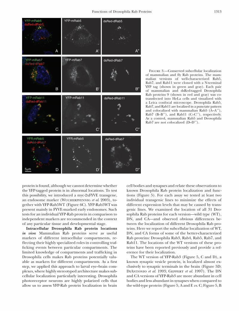
Colocalization of corresponding Mus and Drosoph-ila Rab proteins: The sequence similarity between flyand mammalian Rab proteins raises the question ofwhether corresponding proteins are also functionallyconserved and located in the same intracellular com-partments. We first compared the localization of puta-tive fly and mouse orthologs by cotransfecting mouseand fly Rab genes into mammalian cells and observingthe proteins with double labeling. It is not entirely clearwhich intracellular compartments in fly and mammaliancells are analogous, so differences in localization couldreflect nonconserved protein functions or changes in thenature of cellular compartments. Rab protein localiza-tion may help in resolving the remaining questions aboutwhich compartments are functionally comparable be-tween species.
We used three well-characterized mouse Rab genes(5, 7, and 11), each one encoding an N-terminal YFPtag, for transfection studies. Transgenes encoding pre-sumed mammalian and dsRed2-tagged Drosophila Raborthologs were cotransfected into mammalian HeLacells. Each corresponding pair of mammalian and Dro-sophila versions of Rab5, Rab7, and Rab11 colocalized,suggesting that the targeting systems in mammaliancells are able to recognize and properly transport the flyRab proteins (Figure 3, A–C). The localization patternof each pair of Rab proteins seems different from theothers. To test this, mammalian Rab5 and DrosophilaRab7 were coexpressed and the proteins were observedto be in distinct membrane compartments (Figure 3D).This rules out the possibility that overexpression, andconsequent clogging of cell transport systems, leadsto accumulation of proteins in the same intracellularcompartment.
Comparing the localization of YFP-tagged Rabproteins to endogenous proteins: Functional fluores-cent Rab chimeras using GFP, YFP, CFP, and RFP at-tached to the Rab NH2 terminus have been widely usedin cell biology to observe Rab intracellular movements.In mammalian cultured cells, the presence of fluores-cent proteins at the N terminus of Rab9 causes lessefficient membrane association, probably due to differ-ences in prenylation efficiency and/or differences inrecruiting downstream effectors (Barbero et al. 2002).
Figure 2.—In situ hybridization of DrosophilaRab gene probes to localize transcripts in Dro-sophila whole-mount embryos. For each Rabgene, one stained preparation of a particular em-bryonic stage is shown. The stage that has themost representative staining pattern is shown.(A and A9) Rab5 mRNA signals in embryos (A)and third instar larvae (A9). (B–G) In situ patternsof Rab3, Rab2, Rab26, RabX4, Rab32, and Rab30.
Functions of Drosophila Rab Proteins 1311

To explore this issue in flies, we compared YFP-Rab5and YFP-Rab11 protein localization with endogenousDrosophila Rab5 and Rab11 in wild-type photoreceptorcells. To characterize the YFP signal precisely with re-spect to photoreceptor cell boundaries, we costainedthe photoreceptors (PR) with an antibody that labelsPRs, mAb 24B10 (Zipursky 1982).
Endogenous Drosophila Rab5 has a localizationpattern that closely mimics the YFP-Rab5WT (Figure 4,A and B), including the punctae present in the apicaldomain (arrowheads in Figure 4, A9and B9). We alsocompared the localization of endogenous Rab11 pro-tein and overproduced YFP-Rab11WT in photorecep-tors. Rab11, which is required for the trafficking ofrhodopsin to photoreceptor rhabdomeres, accumulatesin a distinct ring-like pattern at the base of photorecep-tor rhabdomeres (Satoh et al. 2005). YFP-Rab11WT
produced in photoreceptors exhibited a very similardistinct ring-like localization pattern (compare arrowsin Figure 4, C and D).
To further compare the localization of endogenousRab5 and tagged Drosophila Rab5, we produced YFP-tagged Rab5WT in S2R1 cells and stained the cell withanti-Rab5 antibody. This experiment has an inherentlimitation, since the antibody will detect both endoge-nous protein and the YFP-tagged protein. The Rab5antibody signal colocalized with the YFP-Rab5WTsignal,detected by fluorescence (Figure 4E). The interpreta-tion is that the endogenous protein is not in any loca-tion that the YFP-tagged protein fails to reach. Similarly,Rab11 detected by antibody, and YFP-Rab11 detectedby fluorescence, colocalized in S2R1 cells (Figure 4F).In summary, these data show that there is no detect-able cellular location where only the endogenous Rab
TABLE 1
Characterization of selected transgenic UAS-YFP-Rab Drosophila strains
RabAnnotation
symbol WT construct/line information DN construct/line information CA construct/line information
Rab1 3320 UAST/01(80C1) UAST/01(87B8); 04(58B9) UASp/12a(68C13); 12c(1E4)Rab2 3269 UAST/02(86E18) UAST/01(42C7); 05(85E1) UASp/02(61C8); 25(46E6)Rab3 7576 UASp/02(55B9); 05b(95E1) UASp/04L(25C6) UASp/11(76D1); 29a(59E3)Rab4 4921 UASp/9(28B1); 32(100A7) UASp/37(32D2); 46b(72D7) UASp/13(33C1); 28(76A1)Rab5 3664 UASp/02(94A14); 08b(59F5) UASp/01(44B8); 02(75B2) UASp/01a(65A9); 24(24C5)Rab6 6601 UAST/01(57F6) UAST/01(23F3); 03(92B3) UASp/05(61C9); 23(3B6)Rab7 5915 UASp/18(96C1); 21(21B2) UASp/06(79A2); 19(10E6) UASp/14(100D2); 19(22A1)Rab8 8287 UASp/09(68C2); 45(42E7) UASp/09(42E1); 12(83B4) UASp/05(27F4); 10(63F5)Rab9 9994 UASp/13(21B7); 22(79E4) UASp/04(70D7); 11(33A2) UASp/10(31F4); 20(61C9)Rab10 17060 UASp/13(34C4); 21(65A9) UASp/15(23C5); 25a(97D2) UASp/01(35D1); 27a(86C5)Rab11 5771 UASp/32(89B7) UASp/06(88F1); 35(59A3) UASp/24(43C5); 31(61C8)Rab14 4212 UASp/5L(22E1); 12(100D1) UASp/01(36B2); 02(99F6) UASp/02(34D1); 07(67C1)Rab18/RP4 3129 UAST/01(82C5) UAST/02(30B5) 03(87B8) UASp/02(99A11); 12(37C1)Rab19/RP3 7062 UAST/02(39B4) UAST/04(70E2); 05(53D14) UASp/06(62A6); 15(51E2)Rab21 17515/40304 UAST/04(25B1) UAST/01(99A5); 03(58A3) UASp/02(53F8); 09(4F2)Rab23 2108 UASp/01(93D4); 02(21C2) UASp/01(86E11); 02(58E1) UASp/04(66D12)Rab26 7605 UAST/01(73D5); 05(35D1) UAST/02(48A3); 03(74D2) UAST/01(83C4); 04(54E2)Rab27 14791 UASp/01(35D1); 14(86E11) UASp/02(89A5) UASp/05(23A3); 07(3C1)Rab30 9100 UASp/10(53C9) UASp/07(98A7) UASp/11(39E3)Rab32/RP1 8024 UASp/03(27F1); 11(76D5) UASp/04(42C8) UASp/03(85A5); 08(47F8)Rab35 9575 UASp/15(51A2) UAST/01(23C4); 06(68B1) UASp/01(95F1); 11(37B13)Rab39 12156 UASp/13(58D4) UAST/04(37B1); 05(76F1) UASp/02(5C2); 14(92B3)Rab40 1900 UAST/03(61C1); 05(60F5) UAST/01(91F4); 04(26D1) UASp/07(22D1); 13(61B3)RabX1 3870 UASp/10(84F6); 12(27D3) UAST/01(44B3); 14(62B1) UASp/02(92B3); 10(25C6)RabX2 2885 UASp/08(92C1) UASp/02(24A2); 05(67B11) UASp/10(22B1); 21(84F6)RabX3 32670/2532 UASp/24(94A1); 38(50B6) UAST/01(47A7); 03(70C10) UASp/19(98A7)RabX4 31118/13638 UASp/19(94A1) UAST/02(91F11); 03(23F3) UASp/09(44B8); 13a(66D10)RabX5 7980 UASp/13b(27D3); 22b(77B4) UAST/01(47C1) UASp/10(6C8)RabX6 12015 UASp/03(79A2); 04(39B4) UAST/01(31F4); 02(82C3) UASp/13(60A3); 27(73B5)CG9807 9807 UASp/21(chr.2); 25(75E2) UAST/01(49F11); 03(82D5) UASp/04(56F11); 14b(96E6)CG32673 32673 UASp/35(97D7); 42(28C4) UAST/01(57E1); 02(87A2) UASp/05(80A4); 17(26B2)CG32671 32671Rab9D 32678
Only one line from each chromosome (X, II, or III) and a maximal two lines for each gene were selected to send to the stock center.The detailed information is in this table and in supplemental Table 3 at http://www.genetics.org/supplemental/. Columns indicateDrosophila Rab names, CG numbers, the construct that was used to generate the transgenic lines, and the insertion sites identifiedby inverse PCR. CG32671, CG32673, CG9807, and CG32678 are very similar and our PCR identified CG9807 and CG32673 products.
1312 J. Zhang et al.
protein is found, although we cannot determine whetherthe YFP-tagged protein is in abnormal locations. To testthis possibility, we introduced a myc-2xFYVE transgene,an endosome marker (Wucherpfennig et al. 2003), to-gether with YFP-Rab5WT (Figure 6C). YFP-Rab5WT waspresent mainly in FYVE-marked early endosomes. Suchtests for an individual YFP-Rab protein in comparison toindependent markers are recommended in the contextof any particular tissue and developmental stage.
Intracellular Drosophila Rab protein locationsin vivo: Mammalian Rab proteins serve as usefulmarkers of different intracellular compartments, re-flecting their highly specialized roles in controlling traf-ficking events between particular compartments. Thelimited knowledge of compartments and trafficking inDrosophila cells makes Rab proteins potentially valu-able as markers for different compartments. As a firststep, we applied this approach to larval eye–brain com-plexes, where highly stereotyped architecture makes sub-cellular localization particularly interesting. Drosophilaphotoreceptor neurons are highly polarized cells thatallow us to assess YFP-Rab protein localization in brain
cell bodies and synapses and relate these observations toknown Drosophila Rab protein localization and func-tions (Figure 5). For each assay we tested at least twoindividual transgenic lines to minimize the effects ofdifferent expression levels that may be caused by trans-genic lines. We examined the location of all 31 Dro-sophila Rab proteins for each version—wild type (WT),DN, and CA—and observed obvious differences be-tween the localization of different Drosophila Rab pro-teins. Here we report the subcellular localization of WT,DN, and CA forms of some of the better-characterizedRab proteins: Drosophila Rab3, Rab4, Rab5, Rab7, andRab11. The locations of the WT versions of these pro-teins have been reported previously and provide a ref-erence for their localization.
The WT version of YFP-Rab3 (Figure 5, C and D), aknown synaptic vesicle protein, is localized almost ex-clusively to synaptic terminals in the brain (Figure 5D;DiAntonio et al. 1993; Geppert et al. 1997). The DNand CA versions of YFP-Rab3 are more abundant in cellbodies and less abundant in synapses when compared tothe wild-type protein (Figure 5, A and E vs. C; Figure 5, B
Figure 3.—Conserved subcellular localizationof mammalian and fly Rab proteins. The mam-malian versions of well-characterized Rab5,Rab7, and Rab11 were cloned with a N-terminalYFP tag (shown in green and gray). Each pairof mammalian and dsRed-tagged DrosophilaRab proteins 9 (shown in red and gray) was co-transfected into HeLa cells and visualized witha Leica confocal microscope. Drosophila Rab5,Rab7, and Rab11 are localized in a punctate patternand colocalized with mammalian Rab5 (A–A99),Rab7 (B–B99), and Rab11 (C–C99), respectively.As a control, mammalian Rab5 and DrosophilaRab7 are not colocalized (D–D99).
Functions of Drosophila Rab Proteins 1313

and F vs. D), suggesting a defect in protein trafficking ororganelle distribution (or both) in loss- and gain-of-function mutants.
Mammalian Rab4 has been reported to accumulate atthe cytosolic surface of endosomes in Chinese hamsterovary cultured cells (van der Sluijs et al. 1991, 1992).We find that YFP-Rab4WT exhibits a punctate localiza-tion pattern in the cytoplasm of cell bodies (Figure 5I),with little protein at synaptic terminals (Figure 5J). DNand CA versions of YFP-Rab4 are present at higher levelsin axons than is YFP-Rab4WT (Figure 5, H and L vs. J).Furthermore, both the CA and the DN version lack thecharacteristic punctate cell body distribution (Figure 5,G and K vs. I). In mammalian cells, the production ofWT or CA versions of Rab4 changes the morphology ofthe transferrin compartment and causes the formation
of membrane tubules, while production of Rab4DNsignificantly reduces vesicle recycling and degradation(McCaffrey et al. 2001). Our data also suggest thatthere is aberrant formation and/or distribution of thecellular compartment with which Rab4 is normally as-sociated. Alternatively, the mutant proteins are unableto reach their normal location.
Rab5 is an endosome protein that is critical for endo-some fusion along the endocytic pathway. The nativeRab5 protein is present in endosomes at synaptic ter-minals as well as in cell bodies (Wucherpfennig et al.2003). We observed the YFP-Rab5WT fusion protein ina punctate pattern in cell bodies and in nerve termi-nals (Figure 5, O and P) in agreement with previ-ously published data (Wucherpfennig et al. 2003). Themutations in YFP-Rab5DN and YFP-Rab5CA affect Rab5
Figure 4.—Colocalization of YFP-Rab5WT andYFP-Rab11WT with endogenous Rab5 and Rab11proteins. (A and B) Longitudinal sections ofthird instar larval eye discs. Photoreceptor stain-ings with mAb 24B10 are in magenta. A and A9show Rab5 antibody staining in green and gray,respectively, revealing a punctate localization thatis enriched distally (between arrows). B and B9show YFP-Rab5WT driven in photoreceptors withGMR-GAL4. The overexpressed fusion proteinexhibits the same localization pattern as theendogenous protein. (C and D) Anti-Rab 11 stain-ing (C) and photoreceptor-driven YFP-Rab11WT(D) in third instar eye imaginal discs. The crosssections reveal very similar localization patterns.Note the ring-like structure of Rab11-positive ves-icles around the rhabdomeres (arrows). (E andE99) Colocalization of endogenous Rab5 and exog-enous tagged Rab5 proteins in S2R1 cells. Cellswere transfected with YFP-Rab5WT and then fixedand stained with anti-Rab5 antibody (red andgray). (F and F99) Colocalization of endogenousRab11 and exogenous tagged Rab11 proteins inS2R1 cells. Cells were transfected with YFP-Rab11WT and then fixed and stained with anti-Rab11 antibody (red and gray).
1314 J. Zhang et al.
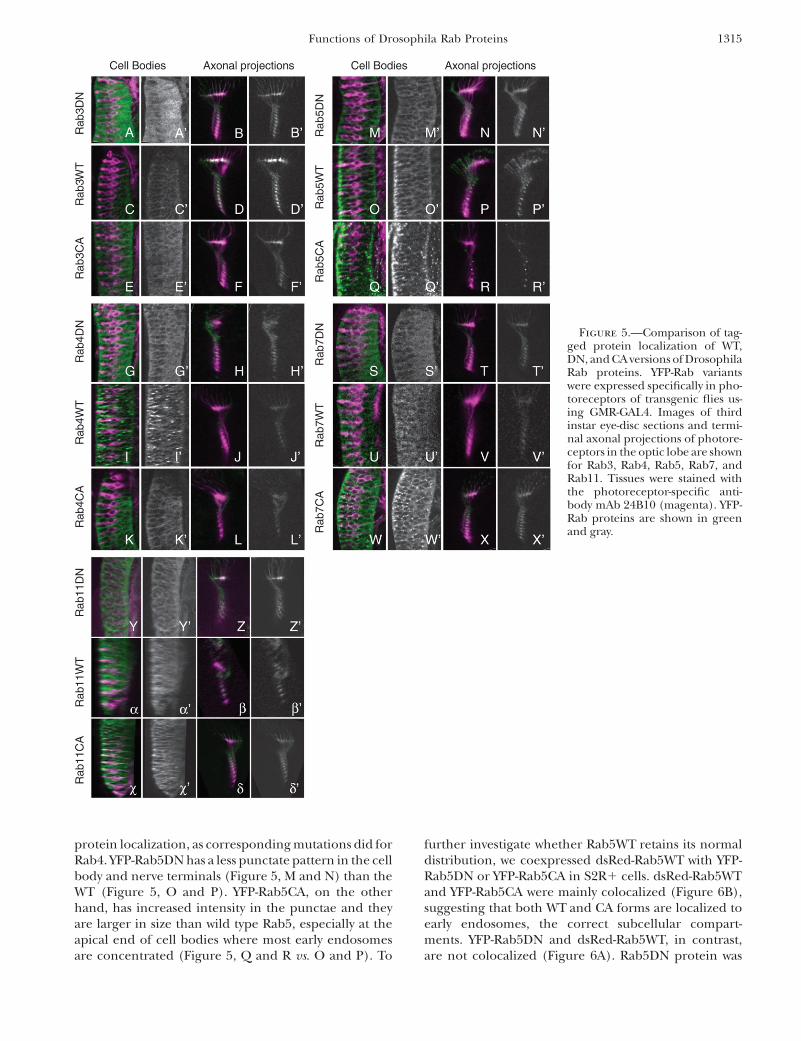
protein localization, as corresponding mutations did forRab4. YFP-Rab5DN has a less punctate pattern in the cellbody and nerve terminals (Figure 5, M and N) than theWT (Figure 5, O and P). YFP-Rab5CA, on the otherhand, has increased intensity in the punctae and theyare larger in size than wild type Rab5, especially at theapical end of cell bodies where most early endosomesare concentrated (Figure 5, Q and R vs. O and P). To
further investigate whether Rab5WT retains its normaldistribution, we coexpressed dsRed-Rab5WT with YFP-Rab5DN or YFP-Rab5CA in S2R1 cells. dsRed-Rab5WTand YFP-Rab5CA were mainly colocalized (Figure 6B),suggesting that both WT and CA forms are localized toearly endosomes, the correct subcellular compart-ments. YFP-Rab5DN and dsRed-Rab5WT, in contrast,are not colocalized (Figure 6A). Rab5DN protein was
Figure 5.—Comparison of tag-ged protein localization of WT,DN, and CA versions of DrosophilaRab proteins. YFP-Rab variantswere expressed specifically in pho-toreceptors of transgenic flies us-ing GMR-GAL4. Images of thirdinstar eye-disc sections and termi-nal axonal projections of photore-ceptors in the optic lobe are shownfor Rab3, Rab4, Rab5, Rab7, andRab11. Tissues were stained withthe photoreceptor-specific anti-body mAb 24B10 (magenta). YFP-Rab proteins are shown in greenand gray.
Functions of Drosophila Rab Proteins 1315

more dispersed (Figure 6A99), suggesting that the GDP-bound Rab5DN shifts to a different location or that thecells making it have altered compartmentalization.
The late endosome marker YFP-Rab7WT exhibits apunctate localization pattern (Figure 5U) similar to thatof Rab5. Again, the CA form of Rab7 displays a much morepronounced punctate localization pattern (Figure 5W),with stronger staining intensity, than the WT form. As inthe case of Rab5, YFP-Rab7DN protein exhibits a moredispersed localization pattern and YFP-Rab7CA proteinaggregates in more and larger punctae (Figure 5S).
We investigated in detail the localization of the WT(Figure 5, a and b), DN (Figure 5, Y and Z), and
CA (Figure 5, x and d) fusion proteins of the well-characterized recycling endosome protein Rab11. Whilethe WT (Figure 5, a and b) and CA (Figure 5, x and d)forms of YFP-Rab11 were located in cell bodies andsynapses, DN YFP-Rab11 caused a loss of photoreceptorstructure in the eye disc (Figure 5Y; data not shown).Adults making YFP-Rab11DN in photoreceptor cellsexhibited developmental defects. Low levels of Rab11DNgave mild developmental defects (supplemental Figure2B at http://www.genetics.org/supplemental/) comparedto control flies (supplemental Figure 2A at http://www.genetics.org/supplemental/) and high levels caused mas-sively reduced eyes or even lethality (data not shown),
Figure 6.—Reduction of YFP-Rab5DN endoso-mal localization. (A and B) dsRed-Rab5WT (redand gray) and YFP-Rab5DN (A; green and gray)or YFP-Rab5CA (B; green and gray) were coex-pressed in cultured Drosophila S2R1 cells.dsRed-Rab5WT and YFP-Rab5CA are mainly colo-calized while dsRed-Rab5WT and YFP-Rab5DNare not. (C–E) A pUAST-myc-2xFYVE (red andgray) construct that labels early endosomes wascotransfected with pUAST-YFP-Rab5WT (C),pUAST-YFP-Rab5DN (D), or pUAST-YFP-Rab5CA(E). YFP-Rab5WT and YFP-Rab5CA are colocal-ized mainly with Myc-2xFYVE whereas YFP-Rab5DN is not.
1316 J. Zhang et al.

confirming a previously reported cell lethality of dom-inant-negative Rab11 (Emery et al. 2005; Jafar-Nejad
et al. 2005).Our data show that DN and CA versions of Drosophila
Rab proteins often have distributions that differ fromWT. Either the mutant proteins affect the structure oforganelles with which they typically associate or theproteins become mis-localized. To determine whetherthe mutant forms alter targeting or the morphology ofthe compartments themselves, we used Rab5 fusionproteins (Figure 6, C–E). We coexpressed the myc-2xFYVE transgene with YFP-Rab5WT, YFP-Rab5DN, orYFP-Rab5CA. Compared to Rab5WT, Rab5DN displayeda more dispersed pattern and was not found where myc-2xFYVE labeled early endosomes. Rab5WTand Rab5CAwere mainly present in FYVE-marked early endosomes(Figure 6, C and E, respectively). The data indicate that,at least for Rab5, the DN proteins are mislocalized. It ispossible that GDP-bound Rab5 is trapped in the cytosolby RabGDIs (Feng et al. 1995; Press et al. 1998; Dinneen
and Ceresa 2004a,b; Pasqualato et al. 2004).In summary, our in vivo localization data indicate that
YFP-Rab5 fusion proteins faithfully recapitulate endog-enous protein localization and that CA and/or DNversions of Rab proteins may accumulate in aberrantcompartments. Together, the data show a high level ofspecificity in protein localization, but this should beexamined in detail with independent markers for anyparticular tissue type.
Identifying intracellular locations of the novel RabXproteins: Twenty-three of the Drosophila Rab proteinsare at least 80% similar to their mammalian counter-parts, while 6 Drosophila Rab proteins have ,40% se-quence similarity to any Rab protein in other species.We refer to the latter as Drosophila RabX proteins. Thedistant relationship of the 6 RabX proteins to other Rabproteins makes it difficult to predict the functions orlocations of the proteins. We have carried out a pre-liminary characterization of YFP-RabX protein localiza-tion to initiate the study of their biology.
Overproducing YFP-tagged wild-type RabX proteinsin either Drosophila or mammalian cells produced apunctate pattern for RabX1, RabX4, and RabX5 (datanot shown). These patterns are likely to report thelocations of the untagged proteins, since other YFP-tagged WT Rab proteins localize to their proper cellularcompartments (Figures 4 and 6 and data not shown). Tolocalize RabX4, we cotransfected Drosophila S2R1 cellswith several dsRed-tagged Rab genes and YFP-taggedRabX4. RabX4 colocalized mainly with DrosophilaRab5 (Figure 7A), which is involved in early endosometrafficking, suggesting that Drosophila RabX4 actsin the same compartment as Rab5. RabX4 is highlyexpressed in the nervous system and less expressedelsewhere (Figure 2E). The RabX4 protein did not co-localize with YFP-tagged Rab7, Rab9, or Rab11 (Figure7, B–D). RabX1 transcripts were barely detectable by
in situ hybridization to embryos, and RabX5 showedsignals in a very limited region (supplemental Table 1at http://www.genetics.org/supplemental/). Their YFP-tagged proteins have a distinctive localization in S2R1
cells that does not overlap with YFP-tagged Rab5, Rab7,Rab9, or Rab11 (data not shown). We conclude that thenovel RabX4 is likely to function in the endosomes ofneural cells, while the other RabX proteins are in dif-ferent, not easily identified, compartments.
DISCUSSION
Rab proteins are crucial in the control of targeted ves-icle trafficking, movements of organelles, and assemblyof subcellular compartments and cytoskeletal elements.Understanding how vesicle trafficking is regulated isimportant in answering many basic questions aboutintracellular events. Our understanding of Rab functionsto date is based on studies of only a few Rab proteins,mostly in yeast and cultured mammalian cells. In thiswork, we have endeavored to create a powerful new toolset that will help researchers explore the fascinatingbiology of Rab proteins in a systematic way.
The new transgenic Rab fly strains will allow research-ers to screen for loss- and gain-of-function phenotypesassociated with altered Drosophila Rab functions inchosen tissues and at defined stages. These tools will beuseful in investigating how vesicle trafficking affectsdevelopmental signaling and other cellular functions,as we have demonstrated with the initial characteriza-tions of Rab3, Rab4, Rab5, Rab7, and Rab11 transgenicinsertions in this study. Furthermore, as we have shownin Figures 3–7, most Rab proteins are located in apunctate pattern and define a compartment of the cellin flies. Therefore, our YFP-tagged Rab proteins canserve as a set of intracellular markers to label differentsteps of the endocytic and exocytic pathways for diversestudies. Our data show that most of the YFP-tagged Rab5is accurately localized to endosomes, where it residestogether with the endogenous protein (Figures 4 and6). Similarly, YFP-Rab11 exhibits the same localization asendogenous Rab11 (Figure 4). Therefore, these linescan serve as cellular markers for different compart-ments. We will deposit these strains in the BloomingtonStock Center for public use.
‘‘New’’ Drosophila Rab genes: We report the identi-fication of four previously unknown Drosophila Rabgenes: CG9807, CG32671, CG32673, and CG32678.These genes are located in a cluster on the X chromo-some at cytological location 9D-F with two other Rabgenes, RabX2 and RabX3. On the basis of a previousstudy that investigated X-linked small GTPases, thecluster of Drosophila Rab genes in the middle of theD. melanogaster X chromosome, along with nearby genesencoding oxidative phosphorylation proteins, are can-didates for the genetic basis of hybrid inviability. Hybrid
Functions of Drosophila Rab Proteins 1317

inviability is a form of reproductive isolation in whichoccasional mating events between two species give riseto a hybrid that is fertile but nevertheless does not leaveany offspring. The RabX genes may be responsible forhybrid inviability among Drosophila species due to theirpotential functions in mitosis (Hutter 2002).
Implications of expression patterns of DrosophilaRab genes for ascertaining function: A Rab gene that isdetectably transcribed only in certain tissues probablyfunctions mainly in those tissues. Our in situ hybrid-izations with probes for different Drosophila Rab genesshow that transcripts from 21 of the genes are ubiqui-tous throughout the embryonic stages. Seven Rab genesare preferentially transcribed in specific tissues ororgans. Ubiquitously expressed Rab genes presumablyfunction in cellular vesicle trafficking, a process that ishighly conserved from yeast to mammals and requiredin all eukaryotic cell types.
Some mammalian Rab genes are expressed in specifictissues to perform highly specialized functions. Rab27ais required in melanocytes to control melanosome traf-
ficking (Izumi et al. 2003), and Rab3 is required in neu-rons to regulate synaptic vesicle trafficking (Schluter
et al. 2002). Thus even seemingly fundamental traffick-ing events required in all cells may be altered by adedicated set of Rab proteins to refine the events for aspecific task. The seven Drosophila Rab genes that aredifferentially expressed are members of all four evolu-tionary branches (Figure 1). Rab2, Rab3, Rab26, andRabX4 transcripts are enriched mainly in neural cells(Figure 2). The expression patterns of Rab3 and Rab26are consistent with a previous study that shows highexpression of mammalian Rab3A and Rab26 in braintissues (Gurkan et al. 2005).
During the analysis of Rab gene expression patternswe identified a previously uncharacterized gene, RabX4,which is specifically transcribed in the nervous system.RabX4 is a member of the A branch of proteins (Figure1). No clear mammalian ortholog of RabX4 exists.Wild-type YFP-RabX4 colocalizes with Rab5 within cells(Figure 7A), so RabX4 may function in endocytosisspecifically in neurons.
Figure 7.—The subcellular localization of anewly studied type of Drosophila Rab, RabX4.(A) dsRed-tagged Rab5 (red and gray) and YFP-tagged RabX4 (green and grays) were cotrans-fected into Drosophila S2R1 cells. The majorityof the RabX4 protein colocalized with Rab5, aprotein reported to be located in the early endo-some. (B–D) dsRed-tagged Rab7 (B), Rab9 (C),or Rab11(D) and YFP-tagged RabX4 were co-transfected into Drosophila S2R1 cells, respec-tively. No colocalization was observed betweeneach pair of proteins.
1318 J. Zhang et al.

Some fly Rab gene expression patterns do not cor-relate well with their mammalian counterparts. For ex-ample, Drosophila Rab32 is specifically transcribed inthe Malpighian tubules, which serve a function similarto that of a kidney, while mammalian Rab32 is highlyexpressed in lymph, trachea, uterus, ovary, and liver, butnot in the kidney (Gurkan et al. 2005). The discrepancybetween fly and mammalian expression patterns ofRab32 suggests that the two genes have functionally di-verged. Since the molecular functions of Rab proteinsare likely to be conserved, dependent as they are on pre-cise protein structures, the evolutionary difference maylie in which tissues employ that particular Rab function.
Functional tests of dominant-negative constructs:Several types of tests can be applied to ascertain whetherthe dominant-negative Drosophila transgenes are infact succeeding in specifically reducing the function ofthe corresponding endogenous proteins.
First, the phenotype caused by the dominant-negativeconstruct can be compared to the phenotype of aconventional mutation in the same gene. Mutationshave been made only in Drosophila Rab5, Rab6, andRab11. Many other uncharacterized P-element insertionstrains that potentially affect some other Rab genes arealso available (Bellen et al. 2004). Our experiments re-veal that the properties of at least one dominant-negative Rab protein, Rab11DN, are in agreement withknown properties of its loss-of-function phenotype, de-spite the possibly incomplete inactivation that is ob-tained with a dominant-negative Rab11. Both Rab11DNand the Rab11 mutant have external sensory organ de-velopment problems that cause a loss-of-bristle pheno-type (supplemental Figure 2D at http://www.genetics.org/supplemental/; Emery et al. 2005; Jafar-Nejad
et al. 2005) and both cause cell lethality in the eyes(supplemental Figure 2B at http://www.genetics.org/supplemental/; Satoh et al. 2005). The similar pheno-types indicate that Rab11DN specifically interferes withthe function of the corresponding endogenous wild-type protein.
The Rab5DN and Rab7DN flies were generated bymutating the same amino acids that were mutated forother studies (Feng et al. 1995; Press et al. 1998;Dinneen and Ceresa 2004a,b; Pasqualato et al. 2004).An experiment with Rab5 that has been reported pre-viously (Zhu et al. 2003) was repeated and confirmedwith our lines (data not shown) and indicates that the DNflies are functioning equivalently to other lines availablein the fly community. Hence, we used these same aminoacid changes in all other DN/CA Rab lines that wedeveloped. The Rab proteins that do not have the par-ticular conserved T/S or Q in the GTP/GDP-bindingdomain may not have the expected DN or CA effects.
The phenotype caused by a dominant-negative pro-tein can be compared to the phenotype of the corre-sponding loss-of-function mammalian mutants to seewhether similar defects are observed. Not many mam-
malian Rab proteins have been studied in vivo due to thedifficulty in making mutants when the genes have po-tential or known redundant functions. Mice carryingRab mutations are available only for Rab3 (Schluter
et al. 2004), Rab23 (Eggenschwiler et al. 2001), andRab27 (Wilson al. 2000). The phenotypes obtained withfly dominant-negative proteins can be compared to thephenotype of the corresponding loss-of-function mam-malian mutants, particularly at the molecular andcellular level. Our initial studies of DN versions of flyRab23 and -27 have not revealed many defects (data notshown), so the endogenous proteins may be too abun-dant to be inhibited or may be redundant with otherproteins. Work on Rab3DN is still in progress.
We must emphasize two issues. First, the DN con-structs may not specifically affect one protein, especiallyin the cases of RabX2, RabX3, and the four ‘‘new’’ Rabproteins where the Rab proteins are very similar. Sec-ond, some dominant-negative Rab proteins do not alwaysfunction properly due to protein instability. For example,it has been reported that the dominant-negative Rab27protein is rapidly degraded in vivo, which precludes theuse of transgenic mouse models to study Rab27 function(Ramalho et al. 2002). Using dominant-negative formsof Rab proteins to reveal the functions of the normal pro-tein requires caution with respect to specificity, level ofinactivation, redundancy among proteins, and proteinstability.
Relationship between Drosophila Rab protein local-ization and function: Protein localization data provideclues about two connected issues. One is whether aparticular cellular compartment is formed under theinfluence of the Drosophila Rab protein, in which casethe compartment may be absent if the protein is notfunctioning. A second possibility is that a mutant formof the Rab protein may not prevent the formation of acertain intracellular compartment, e.g., endosomes, butthe amino acid change may prevent the protein fromaccumulating in the proper compartment. These twopossibilities can be distinguished by using other markersthat define the presence or absence of a compartmentin the presence of various forms of Rab proteins and bylooking at the ultrastructure of each candidate com-partment with electron microscopy.
We compared the subcellular localization of WT, DN,and CA versions of the whole set of Drosophila Rabproteins in vivo. In most cases, the DN versions of theproteins were distributed differently within cells com-pared to the wild-type protein. Data from photoreceptorneurons in the developing third instar larval eye discsshow these differences quite dramatically. YFP-Rab3WT,for example, is almost exclusively located at synapticterminals in the brain. This is consistent with thefunction of Rab3 as a synaptic vesicle regulator. Incontrast, the YFP-Rab3DN and YFP-Rab3CA versions ofthe protein are localized mostly to the cell bodies. Thismislocalization of Rab3 has been observed with the
Functions of Drosophila Rab Proteins 1319

dominant-negative version of mammalian Rab3D. Inpancreatic acini, dominant-negative Rab3D does notaccumulate in the secretory granules where most wild-type Rab3D is located (Chen et al. 2003). There are noreports about the localization of Rab3CA. Our datashow that both loss- and gain-of-function mutations cancause defects in protein localization.
The three versions of Rab5 have distinct patterns ofintracellular staining. The YFP-Rab5WT fusion proteinis present in a punctate pattern both in the cell bodiesand in the nerve terminals of the photoreceptor neu-rons, which is consistent with previously published datathat Drosophila Rab5 is associated with early endosomesin the presynaptic terminal of the neuromuscular junc-tion (Wucherpfennig et al. 2003). Strikingly, the CAand DN mutations of Drosophila Rab5 cause oppositeeffects on the protein localization pattern. The YFP-Rab5DN version of the protein loses the punctate patternin the cell body and nerve terminals while the YFP-Rab5CA version of the protein displays increased intensityin larger punctae, especially at the apex of cell bodies.The same dispersed pattern of Rab5DN is observedin the neuromuscular junction (Wucherpfennig et al.2003). A previous study using mammalian cells shows thatloss of Rab5 causes loss of the endosome compartment,and gain of function leads to an enlarged early endo-some compartment (Bucci et al. 1992). Therefore, ourobservations of loss of punctate staining with Rab5DNand enhanced punctate staining with Rab5CA are con-sistent with previous observations.
Some DN and CA Drosophila Rab proteins, such asRab4DN and Rab4CA, do not show the stereotypicalpunctate pattern. Rab4 is an endosome protein (van
der Sluijs et al. 1991, 1992) and this redistribution ofDN and CA forms may reflect the malfunction of endo-some assembly or trafficking. Although no obvious loss-or gain-of-function phenotypes have been observed withoverexpression of Rab4DN in our assays (data not shown),further detailed studies need to be performed to lookfor subtle changes in vesicle trafficking. On the otherhand, the effects of Rab11CA, Rab11WT, and Rab11DNare in full agreement with previous reports about Rab11function, especially the cell lethality associated with lossof Rab11 function (Emery et al. 2005; Jafar-Nejad et al.2005) . The dynamic localization of endosome Rab pro-teins over time was observed in a study that comparedRab4, Rab5, and Rab11 localization (Sonnichsen et al.2000). Three major endosome populations were iden-tified. The first group contains only Rab5, a secondcontains Rab4 and Rab5, and a third contains Rab4 andRab11. The possibly overlapping functions of Rab4 withRab5 and with Rab11 may explain the lack of obviousdefects caused by Rab4DN, while Rab5 and Rab11, inmost cases, play major roles in endosome trafficking.
The late endosome protein YFP-Rab7WT exhibits apunctate localization pattern similar to that seen withRab5. The CA form of Rab7 displays a pronounced
punctate localization pattern with stronger stainingintensity than its WT variant, while the DN form hasmore dispersed and lesser staining intensity comparedto WT. This pattern is similar to what we observed withsome Rab dominant-negative proteins, such as Rab5DN,suggesting that the DN forms of Rab proteins mayabolish binding to modulators and prevent recruitmentof downstream effectors. We infer that the GDP-boundRab proteins are mislocalized in the cytosol, while theGTP-bound CA forms may have increased activity sincethe proteins are enriched in specifically targeted vesi-cles or other intracellular compartments.
Differences in pUAST and pUASp expressionvectors: We used the well-established UAS/GAL4 regu-latory system (Brand and Perrimon 1993) to generateRab transgenic lines. The GAL4 system has proven tobe an extremely useful tool for spatial and temporalcontrol of Drosophila gene expression. We began usingthe pUASp vector, since pUASp drives expression ingermline cells and is suitable for detecting defects atrelatively early embryonic stages. Unfortunately, thelevel of protein production that we observed usingpUASp is often lower than that with another vector,pUAST (Brand and Perrimon 1993). The differencebetween pUASp and pUAST is that the basal promoterand the 39-UTR in the GAL4-responsive expressionvector are changed for the purpose of driving germlineexpression (Rorth 1998). pUASp contains a P trans-posase promoter and a fs(1)K10 39 terminator, whilepUAST has a hsp70 promoter and a SV40 39 terminator.Although it is difficult to compare transgenic lines sinceexpression levels are determined by multiple factors,we did observe generally weaker phenotypes, even inzygotic tissue, with pUASp in cases in which both pUASpand pUAST lines are available (data not shown). Inaddition, we found that we were unable to enhance theexpression level significantly in lines using pUASp bykeeping the flies at 29� where GAL4 is more active.
We are grateful to Yuchun He for DNA injections, Gabriela David forassistance in generating some of the transgenic lines, Martha Evans-Holm and Joseph Carlson for mapping transgene insertion sites, andBingshan Li for help with bioinformatics analysis. Thanks are alsogiven to Xun Huang, Joel Hyman, Erika Bustamante, and LeticiaBritos for help with initial experiments. We are grateful to M. GonzalezGaitan for kindly providing reagents. P.R.H. acknowledges assistanceby Victor Galanis and Endowed Scholar funds of the University ofTexas Southwestern Medical Center at Dallas during revision experi-ments. Confocal microscopy performed at Baylor College of Medicinewas supported by the Baylor College of Medicine Mental Retardationand Developmental Disabilities Research Center. J.Z. was supportedby a Jane Coffin Childs Memorial Fund for Medical Research Fellow-ship. The research reported here was supported by the HowardHughes Medical Institute (HHMI). M.P.S. and H.J.B. are Investigatorsof the HHMI.
LITERATURE CITED
Ali, B. R., and M. C. Seabra, 2005 Targeting of Rab GTPases to cel-lular membranes. Biochem. Soc. Trans. 33(Pt 4): 652–656.
1320 J. Zhang et al.

Amillet, J. M., D. Ferbus, F. X. Real, C. Antony, M. Mularis et al.,2006 Characterization of human Rab20 overexpressed in exo-crine pancreatic carcinoma. Hum. Pathol. 37(3): 256–263.
Ayala, J., B. Olofsson, A. Tavitian and A. Prochiantz, 1989 De-velopmental and regional regulation of rab3: a new brain specific‘‘ras-like’’ gene. J. Neurosci. Res. 22(3): 241–246.
Bao, S., J. Zhu and W. T. Garvey, 1998 Cloning of Rab GTPases ex-pressed in human skeletal muscle: studies in insulin-resistant sub-jects. Horm. Metab. Res. 30(11): 656–662.
Barbero, P., L. Bittova and S. R. Pfeffer, 2002 Visualization ofRab9-mediated vesicle transport from endosomes to the trans-Golgi in living cells. J. Cell Biol. 156(3): 511–518.
Bellen, H. J., R. W. Levis, G. Liao, Y. He, J. W. Carlson et al.,2004 The BDGP gene disruption project: single transposon in-sertions associated with 40% of Drosophila genes. Genetics 167:761–781.
Brand, A. H., and N. Perrimon, 1993 Targeted gene expression as ameans of altering cell fates and generating dominant pheno-types. Development 118(2): 401–415.
Brand, A. H., A. S. Manoukian and N. Perrimon, 1994 Ectopic ex-pression in Drosophila. Methods Cell Biol. 44: 635–654.
Bucci, C., R. G. Parton, I. H. Mather, H. Stunnenberg, K. Simons
et al., 1992 The small GTPase rab5 functions as a regulatoryfactor in the early endocytic pathway. Cell 70(5): 715–728.
Chen, D., J. Guo, T. Miki, M. Tachibana and W. A. Gahl,1997 Molecular cloning and characterization of rab27a andrab27b, novel human rab proteins shared by melanocytes andplatelets. Biochem. Mol. Med. 60(1): 27–37.
Chen, X., S. A. Ernst and J. A. Williams, 2003 Dominant negativeRab3D mutants reduce GTP-bound endogenous Rab3D in pan-creatic acini. J. Biol. Chem. 278(50): 50053–50060.
Cheng, K. W., J. P. Lahad, W. L. Kuo, A. Lapuk, K. Yamada et al.,2004 The RAB25 small GTPase determines aggressiveness ofovarian and breast cancers. Nat. Med. 10(11): 1251–1256.
DiAntonio, A., R. W. Burgess, A. C. Chin, D. L. Deitcher, R. H.Scheller et al., 1993 Identification and characterization ofDrosophila genes for synaptic vesicle proteins. J. Neurosci.13(11): 4924–4935.
Dinneen, J. L., and B. P. Ceresa, 2004a Continual expression ofRab5(Q79L) causes a ligand-independent EGFR internalizationand diminishes EGFR activity. Traffic 5(8): 606–615.
Dinneen, J. L., and B. P. Ceresa, 2004b Expression of dominantnegative rab5 in HeLa cells regulates endocytic trafficking dis-tal from the plasma membrane. Exp. Cell Res. 294(2): 509–522.
Dollar, G., and E. Struckhoff et al., 2002 Rab11 polarization ofthe Drosophila oocyte: a novel link between membrane traffick-ing, microtubule organization, and oskar mRNA localization andtranslation. Development 129(2): 517–526.
Eggenschwiler, J. T., E. Espinoza and K. V. Anderson, 2001 Rab23is an essential negative regulator of the mouse Sonic hedgehogsignalling pathway. Nature 412(6843): 194–198.
Eggenschwiler, J. T., O. V. Bulgakov, J. Qin, T. Li and K. V. Anderson,2006 Mouse Rab23 regulates hedgehog signaling from smooth-ened to Gli proteins. Dev. Biol. 290(1): 1–12.
Emery, G., A. Hutterer, D. Berdnik, B. Mayer, F. Wirtz-Peitz et al.,2005 AsymmetricRab11endosomesregulatedeltarecyclingandspecifycell fate in theDrosophilanervous system.Cell122(5):763–773.
Entchev, E. V., and M. A. Gonzalez-Gaitan, 2002 Morphogengradient formation and vesicular trafficking. Traffic 3(2): 98–109.
Entchev, E. V., A. Schwabedissen and M. Gonzalez-Gaitan,2000 Gradient formation of the TGF-beta homolog Dpp. Cell103(6): 981–991.
Evans, T. M., C. Ferguson, B. J. Wainwright, R. G. Parton, andC. Wicking, 2003 Rab23, a negative regulator of hedgehog sig-naling, localizes to the plasma membrane and the endocyticpathway. Traffic 4(12): 869–884.
Evans, T. M., F. Simpson, R. G. Parton and C. Wicking,2005 Characterization of Rab23, a negative regulator of sonichedgehog signaling. Methods Enzymol. 403: 759–777.
Feng, Y., B. Press and A. Wandinger-Ness, 1995 Rab 7: an impor-tant regulator of late endocytic membrane traffic. J. Cell Biol.131(6 Pt. 1): 1435–1452.
Geppert, M., Y. Goda, C. F. Stevens and T. C. Sudhof, 1997 Thesmall GTP-binding protein Rab3A regulates a late step in synapticvesicle fusion. Nature 387(6635): 810–814.
Guo, A., T. Wang, E. L. Ng, S. Aulia, K. H. Chong et al., 2006 Openbrain gene product Rab23: expression pattern in the adultmouse brain and functional characterization. J. Neurosci. Res.83(6): 1118–1127.
Gurkan, C., H. Lapp, C. Alory, A. I. Su, J. B. Hogenesch et al.,2005 Large-scale profiling of Rab GTPase trafficking networks:the membrome. Mol. Biol. Cell 16(8): 3847–3864.
Hartley, J. L., G. F. Temple and M. A. Brasch, 2000 DNA cloningusing in vitro site-specific recombination. Genome Res. 10(11):1788–1795.
Hernandez-Alcoceba, R., L. del Peso and J. C. Lacal, 2000 TheRas family of GTPases in cancer cell invasion. Cell. Mol. Life Sci.57(1): 65–76.
Hutter, P., 2002 X-linked small GTPase and OXPHOS genes arecandidates for the genetic basis of hybrid inviability in Drosoph-ila. Dev. Genes Evol. 212(10): 504–512.
Izumi, T., H. Gomi, K. Kasai, S. Mizutani and S. Torii, 2003 Theroles of Rab27 and its effectors in the regulated secretory path-ways. Cell Struct. Funct. 28(5): 465–474.
Jafar-Nejad, H., H. K. Andrews, M. Acar, V. Bayat, F. Wirtz-Peitz
et al., 2005 Sec15, a component of the exocyst, promotes notchsignaling during the asymmetric division of Drosophila sensoryorgan precursors. Dev. Cell 9(3): 351–363.
Jeanmougin, F., J. D. Thompson, M. Gouy, D. G. Higgins and T. J.Gibson, 1998 Multiple sequence alignment with Clustal X.Trends Biochem. Sci. 23(10): 403–405.
Jordens, I., M. Marsman, C. Kuijl and J. Neefjes, 2005 Rab pro-teins, connecting transport and vesicle fusion. Traffic 6(12):1070–1077.
Koenig, J. H., and K. Ikeda, 1990 Transformational process of the en-dosomal compartment in nephrocytes of Drosophila melanogaster.Cell Tissue Res. 262(2): 233–244.
Lanzetti, L., V. Rybin, M. G. Malabarba, S. Christoforidis, G. Scita
et al., 2000 The Eps8 protein coordinates EGF receptor signal-ling through Rac and trafficking through Rab5. Nature 408(6810): 374–377.
McCaffrey, M. W., A. Bielli, G. Cantalupo, S. Mora, V. Roberti
et al., 2001 Rab4 affects both recycling and degradative endoso-mal trafficking. FEBS Lett. 495(1–2): 21–30.
Molendijk, A. J., B. Ruperti and K. Palme, 2004 Small GTPases invesicle trafficking. Curr. Opin. Plant Biol. 7(6): 694–700.
Mori, Y., J. Yin, F. Sato, A. Sterian, L. A. Simms et al., 2004 Iden-tification of genes uniquely involved in frequent microsatellite in-stability colon carcinogenesis by expression profiling combinedwith epigenetic scanning. Cancer Res. 64(7): 2434–2438.
Nagata, K., T. Satoh, H. Itoh, T. Kozasa, Y. Okano et al., 1990 Theram: a novel low molecular weight GTP-binding protein cDNAfrom a rat megakaryocyte library. FEBS Lett. 275(1–2): 29–32.
Pasqualato, S., F. Senic-Matuglia, L. Renault, B. Goud, J. Salamaro
et al., 2004 The structural GDP/GTP cycle of Rab11 reveals anovel interface involved in the dynamics of recycling endosomes.J. Biol. Chem. 279(12): 11480–11488.
Pereira-Leal, J. B., and M. C. Seabra, 2001 Evolution of the Rabfamily of small GTP-binding proteins. J. Mol. Biol. 313(4):889–901.
Pereira-Leal, J. B., A. N. Hume and M. C. Seabra, 2001 Prenylationof Rab GTPases: molecular mechanisms and involvement in ge-netic disease. FEBS Lett. 498(2–3): 197–200.
Pfeffer, S. R., 2001 Rab GTPases: specifying and deciphering organ-elle identity and function. Trends Cell Biol. 11(12): 487–491.
Pfeffer, S., 2005 A model for Rab GTPase localization. Biochem.Soc. Trans. 33(Pt. 4): 627–630.
Pfeffer, S., and D. Aivazian, 2004 Targeting Rab GTPases to dis-tinct membrane compartments. Nat. Rev. Mol. Cell Biol. 5(11):886–896.
Presgraves, D. C., 2005 Evolutionary genomics: new genes for newjobs. Curr. Biol. 15(2): R52–R53.
Press, B., Y. Feng, B. Hoflack and A. Wandinger-Ness, 1998 MutantRab7 causes the accumulation of cathepsin D and cation-independent mannose 6-phosphate receptor in an early endo-cytic compartment. J. Cell Biol. 140(5): 1075–1089.
Functions of Drosophila Rab Proteins 1321

Ramalho, J. S., R. Anders, G. B. Jaissle, M. W. Seeliger, C. Huxley
et al., 2002 Rapid degradation of dominant-negative Rab27 pro-teins in vivo precludes their use in transgenic mouse models.BMC Cell Biol. 3: 26.
Rorth, P., 1998 Gal4 in the Drosophila female germline. Mech.Dev. 78(1–2): 113–118.
Saitou, N., and M. Nei, 1987 The neighbor-joining method: a newmethod for reconstructing phylogenetic trees. Mol. Biol. Evol.4(4): 406–425.
Salminen, A., and P. J. Novick, 1987 A ras-like protein is requiredfor a post-Golgi event in yeast secretion. Cell 49(4): 527–538.
Satoh, A. K., J. E. O’Tousa, K. Ozaki and D. E. Ready, 2005 Rab11mediates post-Golgi trafficking of rhodopsin to the photosensi-tive apical membrane of Drosophila photoreceptors. Develop-ment 132(7): 1487–1497.
Satoh, T., M. Nakafuku and Y. Kaziro, 1992a Function of Ras as amolecular switch in signal transduction. J. Biol. Chem. 267(34):24149–24152.
Satoh, T., Y. Uehara and Y. Kaziro, 1992b Inhibition of inter-leukin 3 and granulocyte-macrophage colony-stimulating factorstimulated increase of active ras.GTP by herbimycin A, a spe-cific inhibitor of tyrosine kinases. J. Biol. Chem. 267(4): 2537–2541.
Schluter, O. M., M. Khvotchev, R. Jahn and T. C. Sudhof,2002 Localization versus function of Rab3 proteins. Evidencefor a common regulatory role in controlling fusion. J. Biol.Chem. 277(43): 40919–40929.
Schluter, O. M., F. Schmitz, R. Jahn, C. Rosenmund and T. C. Sudhof,2004 A complete genetic analysis of neuronal Rab3 function.J. Neurosci. 24(29): 6629–6637.
Seabra, M. C., E. H. Mules and A. N. Hume, 2002 Rab GTPases,intracellular traffic and disease. Trends Mol. Med. 8(1): 23–30.
Seto, E. S., and H. J. Bellen, 2006 Internalization is required forproper Wingless signaling in Drosophila melanogaster. J. Cell Biol.173(1): 95–106.
Sonnichsen, B., S. De Renzis, E. Nielsen, J. Rietdorf and M. Zerial,2000 Distinct membrane domains on endosomes in the recy-cling pathway visualized by multicolor imaging of Rab4, Rab5,and Rab11. J. Cell Biol. 149(4): 901–914.
Stinchcombe, J. C., D. C. Barral, E. H. Mules, S. Booth, A. N.Hume et al., 2001 Rab27a is required for regulated secretionin cytotoxic T lymphocytes. J. Cell Biol. 152(4): 825–834.
van der Sluijs, P., M. Hull, A. Zahraoui, A. Tavitian, B. Goud
et al., 1991 The small GTP-binding protein rab4 is associatedwith early endosomes. Proc. Natl. Acad. Sci. USA 88(14):6313–6317.
van der Sluijs, P., M. Hull, P. Webster, P. Male, B. Goud
et al., 1992 The small GTP-binding protein rab4 controls an earlysorting event on the endocytic pathway. Cell 70(5): 729–740.
Van Vactor, D., Jr., D. E. Krantz, R. Reinke and S. K. Zipursky,1988 Analysis of mutants in chaoptin, a photoreceptor cell-specific glycoprotein in Drosophila, reveals its role in cellularmorphogenesis. Cell 52(2): 281–290.
Walhout, A. J., G. F. Temple, M. A. Brasch, J. L. Hartley, M. A.Lorson et al., 2000 GATEWAY recombinational cloning: appli-cation to the cloning of large numbers of open reading frames orORFeomes. Methods Enzymol. 328: 575–592.
Wang, Y., E. L. Ng and B. L. Tang, 2006 Rab23: What exactly does ittraffic? Traffic 7(6): 746–750.
Wilson, S. M., R. Yip, D. A. Swing, T. N. O’Sullivan, Y. Zhang et al.,2000 A mutation in Rab27a causes the vesicle transport de-fects observed in ashen mice. Proc. Natl. Acad. Sci. USA 97(14):7933–7938.
Wucherpfennig, T., M. Wilsch-Brauninger and M. Gonzalez-Gaitan, 2003 Role of Drosophila Rab5 during endosomal traf-ficking at the synapse and evoked neurotransmitter release.J. Cell Biol. 161(3): 609–624.
Yanagawa, S., J. S. Lee and A. Ishimoto, 1998 Identification andcharacterization of a novel line of Drosophila Schneider S2 cellsthat respond to wingless signaling. J. Biol. Chem. 273(48):32353–32359.
Zhu, A. J., L. Zheng, K. Suyama and M. P. Scott, 2003 Alteredlocalization of Drosophila Smoothened protein activates Hedge-hog signal transduction. Genes Dev. 17(10): 1240–1252.
Zipursky, A., 1982 Mechanisms of hemolysis. Mead Johnson Symp.Perinat. Dev. Med. 19: 17–24.
Communicating editor: J. A. Lopez
1322 J. Zhang et al.