the xcelligence system discover what you’ve been · pdf filecell proliferation 9 ... the...
TRANSCRIPT

www.-applied-science.com
The xCELLigence System Discover what you’ve been missing
For life science research only. Not for use in diagnostic procedures.

The xCELLigence System from ACEA Biosciences is a microelectronic
biosensor system for cell-based assays, providing dynamic, real-time, label-free
cellular analysis for a variety of research applications in drug development,
toxicology, cancer, medical microbiology, and virology. This pioneering technology
allows researchers to increase productivity and exceed the limits of endpoint
analysis by capturing data throughout the entire time course of an experiment
and obtaining more physiologically relevant data. Choose from multiple
instrument formats and benefit from cost-effective, user-friendly cell-based
assays designed to analyze compound effects in a context as close as possible
to the natural environment.
Introduction A new way to look at cellular analysis
The xCELLigence System 3
Instrument Description 4
System Components 5
Technology 7
Applications 8 Cell Proliferation 9 Cell Adhesion and Morphology 10 Correlation of xCELLigence Data and WST-1 Assays 11 Monitoring of Cytotoxic Compounds with Impedance Technology 12 Calculation of IC50 Values 13
xCELLigence System Q and A 15
Additional Information 17 The xCELLigence Special Interest Site 17 Product Specifications 18 Ordering Information 19
2

3
xCELLigence System
Instrument Description
System Components
Applications
Q and A
Additional Information
Technology
The xCELLigence System Discover what you’ve been missing
The xCELLigence System monitors cellular events in real time without the
incorporation of labels by measuring electrical impedance across interdigitated
microelectrodes integrated on the bottom of its special tissue culture plates.
The impedance measurement improves on conventional endpoint assays and
provides quantitative information about the biological status of the cells,
including cell number, adhesion, viability, and morphology.
Obtain dynamic real-time data that focuses assay development and improves attrition rates.
Continuously monitor cell status over the entire time period of the experiment.
Easily measure both short-term (approximately 30 minutes) and long-term (2–3 days) cellular effects.
Non-invasively analyze compound effects in a context as close as possible to the natural environment.
Benefits
The xCELLigence System serves the increasing needs of the life science research and drug discovery markets. The benefits offered by this exciting technology include:
Broad applications: The technology is suitable for many different applications requiring cellular analysis.
High data quality: Rely on reproducible results with <0.3 % well failure and <15 % inter- and intra-plate variation.
Complete data record: Obtain real-time measurements throughout the experiment, from start to finish.
Convenience: Enjoy fully automated recording of experimental data for online viewing.
Physiological relevance: Use the instrument in a fully controlled environment (e.g., standard cell culture incubator), increasing the duration of applicable experiments.
Versatile software: Benefit from a user-friendly interface, online monitoring, and various analysis options like IC50/EC50 calculation, data normalization, and various statistical data analysis tools.
xCELLigence System
3
4
Instrument Description
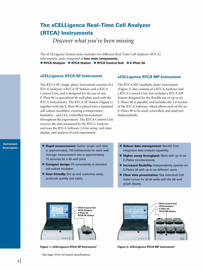
RTCA Analyzer RTCA Station RTCA Control Unit E-Plate 96
The xCELLigence Real-Time Cell Analyzer (RTCA) Instruments Discover what you’ve been missing
xCELLigence RTCA MP Instrument
The RTCA MP (multiple-plate) Instrument (Figure 2) also consists of a RTCA Analyzer and a RTCA Control Unit, but includes a RTCA MP Station designed for the flexible use of up to six E-Plates 96 in parallel, and includes the 2.0 version of the RTCA Software, which allows each of the six E-Plates 96 to be used, controlled, and analyzed independently.
Rapid measurement: Gather single-well data in approximately 150 milliseconds for each well. Average measurement rate is approximately 15 seconds for a 96-well plate.
Compact design: Fit conveniently in standard cell culture incubator.
User-friendly: Set up and customize assay protocols quickly and easily.
Robust data management: Benefit from integrated data analysis capability.
Higher assay throughput: Work with up to six E-Plates simultaneously.
Increased flexibility: Independently operate six E-Plates 96 with up to six different users.
Clear data presentation: See individual Cell Index curves for all 96 wells with the 96-well graph display.
The xCELLigence System series includes two different Real-Time Cell Analyzer (RTCA) instruments, each composed of four main components:
xCELLigence RTCA SP Instrument
The RTCA SP (single-plate) Instrument consists of a RTCA Analyzer, a RTCA SP Station, and a RTCA Control Unit, and is designed for the use of one E-Plate 96 (a specialized 96-well plate used with the RTCA Instrument). The RTCA SP Station (Figure 1) together with the E-Plate 96 is placed into a standard cell culture incubator, creating a temperature-, humidity-, and CO2-controlled environment throughout the experiment. The RTCA Control Unit receives the data measured by the RTCA Analyzer and uses the RTCA Software 2.0 for setup, real-time display, and analysis of each experiment.
Figure 1: xCELLigence RTCA SP Instrument*
* See page 18 for full system specifications.
Figure 2: xCELLigence RTCA MP Instrument*
RTCA Control UnitRTCA AnalyzerE-Plate 96RTCA MP Station
RTCA Control UnitRTCA AnalyzerE-Plate 96RTCA SP Station

5
System Components
The xCELLigence System Components Precision components, versatile options
rapid measurement. The average measurement rate is approximately 15 seconds for a 96-well plate, or approximately 150 milliseconds for each well.
RTCA Control Unit
The RTCA Control Unit consists of a laptop computer with a mobile port replicator. The operating system and all software tools (including the RTCA Software Package) necessary to run the RTCA SP or MP Instrument are already preinstalled.
RTCA Software Package 2.0
The RTCA SP and MP Instruments are driven by powerful, dedicated software. The RTCA Software Package 2.0 provides a user-friendly interface for instrument control and operation, flexible experiment setups, and data acquisition, display, output, and analysis.
User-friendly GUI (Graphical User Interface) – Easy-to-use drop-down and selection menus – Intuitive layout and design
Flexible set-up of experimental protocols – Rapidly configurable experimental design – Supports multistage experiments
Real-time data acquisition – Hands-free data acquisition throughout the entire time course of the experiment
Real-time numerical and graphical data display – Informed experimental decisions based on real-time data
Multiple output formats – Real-time IC50 or EC50 via Cell Index – Easy transfer of raw data for external analysis – Simple copy and paste function for presenta-
tions and communications
The individual components that make up each RTCA Instrument function in precise harmony to provide you with more physiologically relevant cellular analysis data than traditional cell analysis techniques.
RTCA Station
RTCA SP StationThe RTCA SP Station is located inside a standard cell culture incubator and serves to transmit signals from an E-Plate 96 to the RTCA Analyzer. Using the software of the RTCA Control Unit, the RTCAAnalyzer can automatically select wells for measure-ment and continuously transfer measured impedance data to the computer. Cell Index values, derived from the measured impedances, are continuously displayed on the Software user interface.
RTCA MP StationThe RTCA MP Station is located inside a regular cell culture incubator and is capable of switching any one of the wells on any of six E-Plates to the RTCA Analyzer for impedance measurement. Each of the six E-Plate 96 holders can be used independently under the RTCA Software. The RTCA Analyzer can automatically select wells for measurement and continuously transfer measured impedance data to the computer. Cell Index values, derived from the measured impedances, are continuously displayed on the Software user interface.
RTCA Analyzer
The RTCA Analyzer is an electronic analyzer that can measure, under the control of RTCA Software, elec-tronic impedance of sensor electrodes at various sig-nal frequencies. The RTCA Analyzer is capable of computer-controlled signal generation, processing and analysis, automatic frequency scanning and

6
System Components
E-Plate 96
The E-Plate 96 is a single use, disposable device used for performing cell-based assays on the RTCA SP and MP Instruments. The E-Plate 96 is similar to commonly used 96-well plates. Each of the 96 wells on the E-Plate 96 contains integral sensor electrode arrays (Figure 3) so that cells inside each well can be monitored and assayed. The electrodes cover approximately 80% of the area of each well bottom. The plate lid is designed to ensure low evaporation. The plate is designed to be used at temperatures between +15°C and +40°C, and at relative humidity up to 98% maximum without condensation.
E-Plate VIEW 96
Combine real-time impedance-based monitoring of cell behavior with visual inspection in a single 96-well plate. The new E-Plate VIEW 96 for xCELLigence RTCA SP and MP Instruments features both microelectrodes that measure cell response and a clear inspection window on the well bottoms – enabling you to see cellular changes (e.g., morphology, proliferative state) indicated by dynamic monitoring with the xCELLigence System.
Figure 3: Enlarged section of an E-Plate 96
E-Plate 96 E-Plate VIEW 96
500 µm
Figure 5: Easily visualize cells while measuring cell response with xCELLigence System E-Plate VIEW technology. A modified version of the standard E-Plate 96, the new E-Plate VIEW 96 enables image acquisition using microscopes or automated cell-imaging systems. For the modification, 4 rows of microelectrode sensors were removed in each well to create a window for visualizing cells. Approximately 70% of each well bottom is covered by the microelectrodes, providing cell impedance measurements nearly identical to those obtained with the standard E-Plate 96. Both plate types can be used in parallel.
Figure 4: Observe cell status with the E-Plate VIEW 96. Pictures were obtained using the Cellavista System for the brightfield views and an upright microscope for the Crystal Violet staining. PC3-GFP cells were observed under bright-field microscopy at 10x magnification.
Brightfield Crystal Violet
12,500 cells

7
Technology
How it Works
The presence of cells on top of the E-Plate 96 electrodes affects the local ionic environment at the electrode/solution interface, leading to an increase in electrode impedance (Figure 5). The more cells that are attached on the electrodes, the larger the increases in electrode impedance. In addition, the impedance can vary based on the quality of the cell interaction with the electrodes; for example, increased cell adhesion or spreading will lead to a larger change in electrode impedance. Thus, electrode impedance, which is displayed as Cell Index (CI) values, can be used to monitor cell viability, number, morphology, and adhesion degree in a number of cell-based assays.
A dimensionless parameter called Cell Index (CI) is derived as a relative change in measured electrical impedance to represent cell status.
When cells are not present or are not well-adhered on the electrodes, the CI is zero.
Under the same physiological conditions, when more cells are attached on the electrodes, the CI values are larger. Thus, CI is a quantitative measure of cell number present in a well.
Additionally, change in a cell status, such as cell morphology, cell adhesion, or cell viability will lead to a change in CI.
Based on this innovative technology, a wide range of cell-based applications for both high-throughput screening and research laboratory environments can be performed on the xCELLigence System.
Technology of the xCELLigence System Don’t miss the effect you want to analyze
Figure 6: Schematic drawing of the interdigitated microelectrodes on the well bottom of an E-Plate 96

8
xCELLigence System Applications Discover new horizons
The xCELLigence System allows for label-free and real-time monitoring of cellular processes such as cell proliferation, cytotoxicity, adhesion, and viability, using electronic cell sensor array technology.
Real-time monitoring of cellular processes by the xCELLigence System offers distinct and important advantages over traditional endpoint assays. First, the avoidance of labels allows for more physiologically relevant assays which save on time, labor, and resources. Second, a comprehensive representation of the entire length of the assay is possible, allowing the user to make informed decisions regarding the timing of certain manipulations or treatments. Finally, the actual kinetic response of the cells within an assay prior or subsequent to certain manipulations provides important information regarding the biological status of the cell such as cell growth, arrest, morphological changes, and apoptosis (cell death).
Applications include:
Cell viability and proliferation
Apoptosis
Compound-mediated cytotoxicity
Cell-mediated cytotoxicity
Receptor functional analysis (e.g., GPCRs, RTKs)
Real-time detection of viral cytopathic effects (CPE)Applications

9
Applications
Cell Proliferation
For this research study, cells were obtained from different sources (including ATCC, ECACC, and Roche) and maintained in a CO2 incubator (Heraeus, Cytoperm 2) at 37°C with 95% humidity and 5.5% carbon dioxide saturation. MCF7 breast cancer cells (30,000 cells/well), HT29 human colonic adenocarcinoma cells (50,000 cells/well), PC3 human prostate cancer cells and COS-7 simian kidney fibroblasts (both 6,250 cells/well) were seeded in E-Plates 96 in triplicate, in appropriate culture media, and in a final volume of 200 microliters.
Dynamic cell proliferation of cells plated on the E-Plates 96 was monitored in 30-minute intervals from the time of plating until the end of the experiment (68 hours). Cell Index values for all cell lines were calculated and plotted on the graph. Standard deviation of triplicates of wells for corresponding cell lines with different cell numbers were analyzed with the RTCA Software (Figure 7).
Each cell type shows its own characteristic pattern which correlates to the cell number and also to cell size and cell morphology. Each cell line can be characterized by intensity of adhesion, kinetics of spreading, and time necessary to enter into logarithmic growth phase. These characteristics can consequently serve as an ideal basis for cell stock standardization, media comparison, and quality control.
Figure 7: Cell proliferation curves of four different cell lines displayed as Cell Index on the RTCA SP Instrument

10
Applications
Cell Adhesion and Morphology
In order to show the dependency of Cell Index and adhesion characteristics, different cell lines were grown in the E-Plate 96 wells. Cell lines were grown until all cell lines showed maximum values in Cell Index measured by the RTCA SP Instrument. After reaching confluence, fixation of cells was performed by paraformaldehyde (PFA) treatment. Cells were stained with crystal violet and imaging was performed on a Zeiss SteREO Discovery V8 microscope and Axiovision Rel. 4.6 software. The image obtained after PFA fixation and crystal violet staining showed that the Cell Index val-ues obtained by the RTCA Instrument do not solely reflect the coverage of the elec-trode but are also related to adhesion strength and cell morphology (Figure 8).
Figure 8: Sections of four E-Plate 96 wells with fixed cells after PFA fixation and crystalviolet staining
CHO K1 DU-145 MCF7 NIH-3T3
CI = 3.4 CI = 8.1 CI = 9.2 CI = 4.4
11
Applications
Correlation of xCELLigence Data and WST-1 Assays
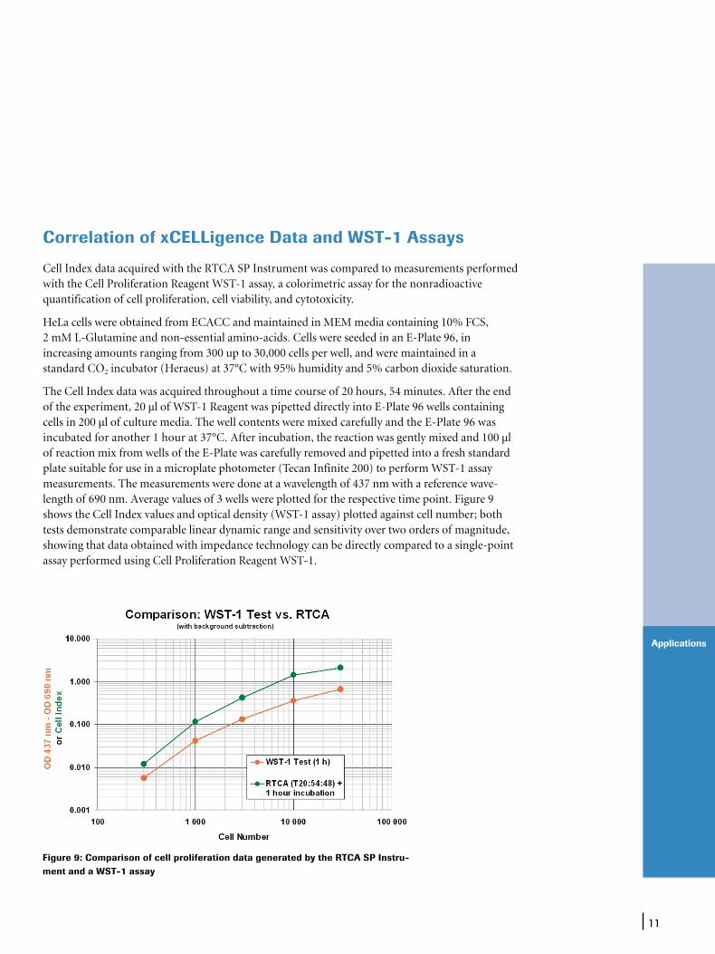
Cell Index data acquired with the RTCA SP Instrument was compared to measurements performed with the Cell Proliferation Reagent WST-1 assay, a colorimetric assay for the non ra dio active quantification of cell proliferation, cell viability, and cytotoxicity.
HeLa cells were obtained from ECACC and maintained in MEM media containing 10% FCS, 2 mM L-Glutamine and non-essential amino-acids. Cells were seeded in an E-Plate 96, in increasing amounts ranging from 300 up to 30,000 cells per well, and were maintained in a standard CO2 incubator (Heraeus) at 37°C with 95% humidity and 5% carbon dioxide saturation.
The Cell Index data was acquired throughout a time course of 20 hours, 54 minutes. After the end of the experiment, 20 µl of WST-1 Reagent was pipetted directly into E-Plate 96 wells containing cells in 200 µl of culture media. The well contents were mixed carefully and the E-Plate 96 was incubated for another 1 hour at 37°C. After incubation, the reaction was gently mixed and 100 µl of reaction mix from wells of the E-Plate was carefully removed and pipetted into a fresh standard plate suitable for use in a microplate photometer (Tecan Infinite 200) to perform WST-1 assay measurements. The measurements were done at a wavelength of 437 nm with a reference wave-length of 690 nm. Average values of 3 wells were plotted for the respective time point. Figure 9 shows the Cell Index values and optical density (WST-1 assay) plotted against cell number; both tests demonstrate comparable linear dynamic range and sensitivity over two orders of magnitude, showing that data obtained with impedance technology can be directly compared to a single-point assay performed using Cell Proliferation Reagent WST-1.
Figure 9: Comparison of cell proliferation data generated by the RTCA SP Instru-ment and a WST-1 assay
12
Applications
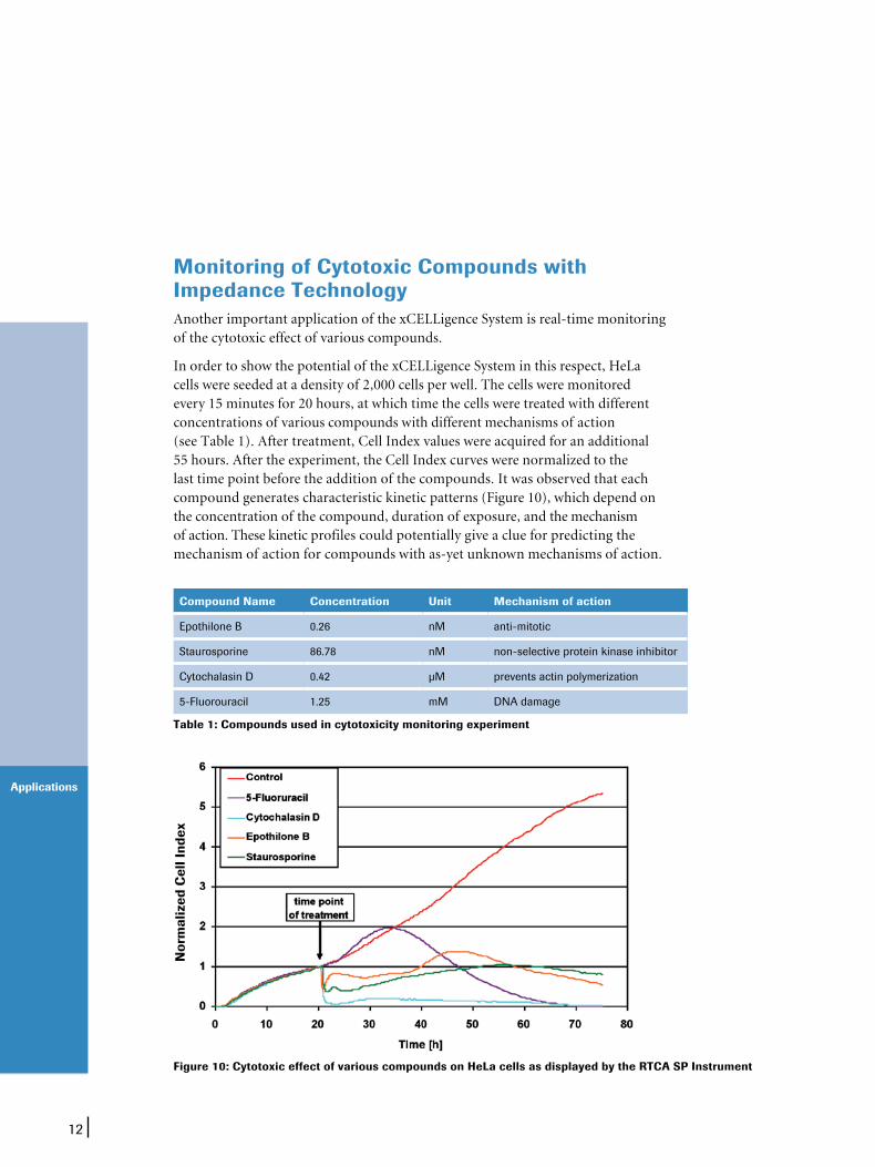
Compound Name Concentration Unit Mechanism of action
Epothilone B 0.26 nM anti-mitotic
Staurosporine 86.78 nM non-selective protein kinase inhibitor
Cytochalasin D 0.42 µM prevents actin polymerization
5-Fluorouracil 1.25 mM DNA damage
Table 1: Compounds used in cytotoxicity monitoring experiment
Figure 10: Cytotoxic effect of various compounds on HeLa cells as displayed by the RTCA SP Instrument
Nor
mal
ized
Cel
l Ind
ex
Another important application of the xCELLigence System is real-time monitoring of the cytotoxic effect of various compounds.
In order to show the potential of the xCELLigence System in this respect, HeLa cells were seeded at a density of 2,000 cells per well. The cells were monitored every 15 minutes for 20 hours, at which time the cells were treated with different concentrations of various compounds with different mechanisms of action (see Table 1). After treatment, Cell Index values were acquired for an additional 55 hours. After the experiment, the Cell Index curves were normalized to the last time point before the addition of the compounds. It was observed that each compound generates characteristic kinetic patterns (Figure 10), which depend on the concentration of the compound, duration of exposure, and the mechanism of action. These kinetic profiles could potentially give a clue for predicting the mechanism of action for compounds with as-yet unknown mechanisms of action.
Monitoring of Cytotoxic Compounds with Impedance Technology
13
Applications
Calculation of IC50 Values
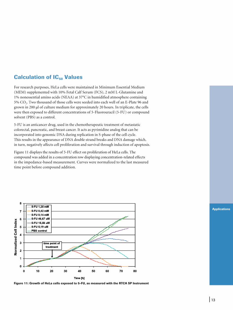
For research purposes, HeLa cells were maintained in Minimum Essential Medium (MEM) supplemented with 10% Fetal Calf Serum (FCS), 2 mM L-Glutamine and 1% nonessential amino acids (NEAA) at 37°C in humidified atmosphere containing 5% CO2. Two thousand of those cells were seeded into each well of an E-Plate 96 and grown in 200 µl of culture medium for approximately 20 hours. In triplicate, the cells were then exposed to different concentrations of 5-Fluorouracil (5-FU) or compound solvent (PBS) as a control.
5-FU is an anticancer drug, used in the chemotherapeutic treatment of metastatic colorectal, pancreatic, and breast cancer. It acts as pyrimidine analog that can be incorporated into genomic DNA during replication in S-phase of the cell cycle. This results in the appearance of DNA double strand breaks and DNA damage which, in turn, negatively affects cell proliferation and survival through induction of apoptosis.
Figure 11 displays the results of 5-FU effect on proliferation of HeLa cells. The compound was added in a concentration row displaying concentration-related effects in the impedance-based measurement. Curves were normalized to the last measured time point before compound addition.
Figure 11: Growth of HeLa cells exposed to 5-FU, as measured with the RTCA SP Instrument
Nor
mal
ized
Cel
l Ind
ex

14
Applications
Three different wells within the same E-Plate 96 were analyzed utilizing the RTCA Software. Standard deviation of the Cell Index observed for the triplicated treatment of HeLa cells in serial dilution of 5-FU indicates a low variability within the respective exposures (Figure 12). Therefore, the effect of the compound can be correlated to the specific dosage via the respective Cell Index curve.
Quantification via the RTCA Software allowed the plotting of a sigmoidal dose-response curve (Figure 13, left panel) and calculation of a half inhibitory concentration (IC50) of 40 µM (Figure 13, right panel) in this specific experiment.
Figure 12: Standard deviation of results of 5-FU addition to HeLa cells
Figure 13: Dose-response curve and IC50 value calculation at 70 hours and 21 minutes
Nor
mal
ized
Cel
l Ind
ex
15
Q and A
xCELLigence System Q and A
Technology
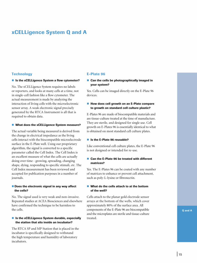
Is the xCELLigence System a flow cytometer?
No. The xCELLigence System requires no labels or reporters, and looks at many cells at a time, not in single-cell fashion like a flow cytometer. The actual measurement is made by analyzing the interaction of living cells with the microelectronic sensor array. A weak electronic signal precisely generated by the RTCA Instrument is all that is required to obtain data.
What does the xCELLigence System measure?
The actual variable being measured is derived from the change in electrical impedance as the living cells interact with the biocompatible microelectrode surface in the E-Plate well. Using our proprietary algorithm, the signal is converted to a specific parameter called the Cell Index. The Cell Index is an excellent measure of what the cells are actually doing over time - growing, spreading, changing shape, dying, responding to specific stimuli, etc. The Cell Index measurement has been reviewed and accepted for publication purposes in a number of journals. Does the electronic signal in any way affect
the cells?
No. The signal used is very weak and non-invasive. Repeated studies at ACEA Biosciences and elsewhere have confirmed the technique to be harmless to the cells.
Is the xCELLigence System durable, especially the station that sits inside an incubator?
The RTCA SP and MP Station that is placed in the incubator is specifically designed to withstand the high temperature and humidity of laboratory incubators.
E-Plate 96
Can the cells be photographically imaged in your system?
Yes. Cells can be imaged directly on the E-Plate 96 devices.
How does cell growth on an E-Plate compare to growth on standard cell culture plastic?
E-Plates 96 are made of biocompatible materials and are tissue-culture treated at the time of manufacture. They are sterile, and designed for single use. Cell growth on E-Plates 96 is essentially identical to what is obtained on most standard cell culture plates.
Is the E-Plate 96 reusable?
Like conventional cell culture plates, the E-Plate 96 is not designed or intended for re-use. Can the E-Plate 96 be treated with different
matrices?
Yes. The E-Plates 96 can be coated with any number of matrices to enhance or prevent cell attachment, such as poly-L-lysine or fibronectin.
What do the cells attach to at the bottom of the well?
Cells attach to the planar gold electrode sensor arrays at the bottom of the wells, which cover approximately 80% of the surface area. All components of the E-Plate 96 are biocompatible and the microplates are sterile and tissue-culture treated.
16
Q and A
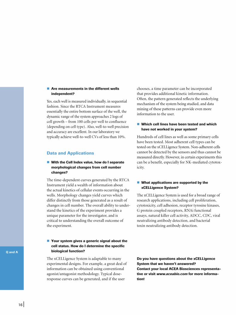
Are measurements in the different wells independent?
Yes, each well is measured individually, in sequential fashion. Since the RTCA Instrument measures essentially the entire bottom surface of the well, the dynamic range of the system approaches 2 logs of cell growth – from 100 cells per well to confluence (depending on cell type). Also, well-to-well precision and accuracy are excellent. In our laboratory we typically achieve well-to-well CVs of less than 10%.
Data and Applications
With the Cell Index value, how do I separate morphological changes from cell number changes?
The time-dependent curves generated by the RTCA Instrument yield a wealth of information about the actual kinetics of cellular events occurring in the wells. Morphology changes yield curves which differ distinctly from those generated as a result of changes in cell number. The overall ability to under-stand the kinetics of the experiment provides a unique parameter for the investigator, and is critical to understanding the overall outcome of the experiment.
Your system gives a generic signal about the
cell status. How do I determine the specific biological function?
The xCELLigence System is adaptable to many experimental designs. For example, a great deal of information can be obtained using conventional agonist/antagonist methodology. Typical dose-response curves can be generated, and if the user
chooses, a time parameter can be incorporated that provides additional kinetic information. Often, the pattern generated reflects the underlying mechanism of the system being studied, and data mining of these patterns can provide even more information to the user. Which cell lines have been tested and which
have not worked in your system?
Hundreds of cell lines as well as some primary cells have been tested. Most adherent cell types can be tested on the xCELLigence System. Non-adherent cells cannot be detected by the sensors and thus cannot be measured directly. However, in certain experiments this can be a benefit, especially for NK-mediated cytotox-icity.
What applications are supported by the xCELLigence System?
The xCELLigence System is used for a broad range of research applications, including cell proliferation, cytotoxicity, cell adhesion, receptor tyrosine kinases, G protein coupled receptors, RNAi functional assays, natural killer cell activity, ADCC, CDC, viral neutralizing antibody detection, and bacterial toxin neutralizing antibody detection.
Do you have questions about the xCELLigence System that we haven’t answered? Contact your local ACEA Biosciences representa-tive or visit www.aceabio.com for more informa-tion!

17
Additional Information
Whether you’d like to access up-to-date technical information on real-time cellular analysis, watch videos from the research community, or contact a sales specialist, this comprehensive site is your ideal destination.
Technology: View an animated introduction to impedance-based measurement.
Applications: See how the xCELLigence System can benefit your research field.
The ACEA Biosciences Site – www.aceabio.com Constantly updated multimedia information, at your fingertips
Systems: Download product literature and get in touch with an ACEA sales representatives.
Support: View up-to-date Frequently Asked Questions.
Multimedia: Watch streaming videos of talks and presentations about impedance-based cell analysis. Literature and References: Browse a growing body of research publications.
18
Additional Information
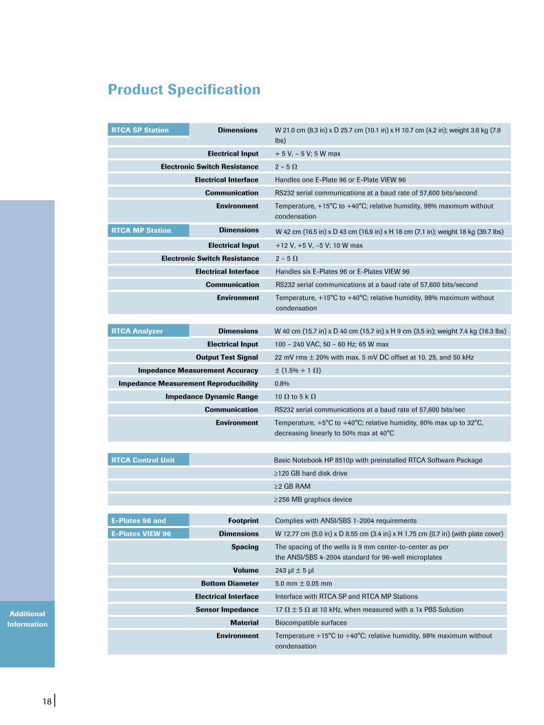
Dimensions W 21.0 cm (8.3 in) x D 25.7 cm (10.1 in) x H 10.7 cm (4.2 in); weight 3.6 kg (7.9 lbs)
Electrical Input + 5 V, – 5 V; 5 W max
Electronic Switch Resistance 2 – 5 V
Electrical Interface Handles one E-Plate 96 or E-Plate VIEW 96
Communication RS232 serial communications at a baud rate of 57,600 bits/second
Environment Temperature, +15°C to +40°C; relative humidity, 98% maximum without condensation
RTCA SP Station
Product Specification
W 42 cm (16.5 in) x D 43 cm (16.9 in) x H 18 cm (7.1 in); weight 18 kg (39.7 lbs)
Electrical Input +12 V, +5 V, –5 V; 10 W max
Electronic Switch Resistance 2 – 5 V
Electrical Interface Handles six E-Plates 96 or E-Plates VIEW 96
Communication RS232 serial communications at a baud rate of 57,600 bits/second
Environment Temperature, +15°C to +40°C; relative humidity, 98% maximum without condensation
Dimensions W 40 cm (15.7 in) x D 40 cm (15.7 in) x H 9 cm (3.5 in); weight 7.4 kg (16.3 lbs)
Electrical Input 100 – 240 VAC, 50 – 60 Hz; 65 W max
Output Test Signal 22 mV rms ± 20% with max. 5 mV DC offset at 10, 25, and 50 kHz
Impedance Measurement Accuracy ± (1.5% + 1 V)
Impedance Measurement Reproducibility 0.8%
Impedance Dynamic Range 10 V to 5 k V
Communication RS232 serial communications at a baud rate of 57,600 bits/sec
Environment Temperature, +5°C to +40°C; relative humidity, 80% max up to 32°C, decreasing linearly to 50% max at 40°C
RTCA Analyzer
Basic Notebook HP 8510p with preinstalled RTCA Software Package
≥120 GB hard disk drive
≥2 GB RAM
≥256 MB graphics device
RTCA Control Unit
Footprint Complies with ANSI/SBS 1-2004 requirements
E-Plates VIEW 96 Dimensions W 12.77 cm (5.0 in) x D 8.55 cm (3.4 in) x H 1.75 cm (0.7 in) (with plate cover)
Spacing The spacing of the wells is 9 mm center-to-center as per the ANSI/SBS 4-2004 standard for 96-well microplates
Volume 243 µl ± 5 µl
Bottom Diameter 5.0 mm ± 0.05 mm
Electrical Interface Interface with RTCA SP and RTCA MP Stations
Sensor Impedance 17 V ± 5 V at 10 kHz, when measured with a 1x PBS Solution
Material Biocompatible surfaces
Environment Temperature +15°C to +40°C; relative humidity, 98% maximum without condensation
E-Plates 96 and
RTCA MP Station Dimensions

19
Additional Information
Product Pack Size Cat. No.
xCELLigence RTCA SP Instrument RTCA Analyzer RTCA SP Station RTCA Control Unit
1 Bundled Package1 Instrument1 Instrument1 Notebook PC
00380601030052289720010522905700105454417001
xCELLigence RTCA MP Instrument RTCA Analyzer RTCA MP Station RTCA Control Unit
1 Bundled Package1 Instrument1 Instrument1 Notebook PC
00380601040052289720010533162500105454417001
E-Plates 96 6 plates 6 x 6 plates
05232368001 05232376001
E-Plates VIEW 96 6 plates 6 x 6 plates
06472451001 06472460001
Contact your local ACEA representative or call 866 308 2232 for more information, or to order.
Ordering Information
Learn more about the enabling technology of the xCELLigence System and its broad range of applications at www.aceabio.com
XCELLIGENCE, E-PLATE, and ACEA BIOSCIENCES are registered trademarks of ACEA Biosciences, Inc. in the US and other countries.
All other product names and trademarks are the property of their respective owners.
For life science research only. Not for use in diagnostic procedures.
License DisclaimersLicense Disclaimer information is subject to change or amendment.
Published by
ACEA Biosciences, Inc.
6779 Mesa Ridge Road Ste. 100
San Diego, CA 92121
U.S.A.
www.aceabio.com
© 2013 ACEA Biosciences, Inc.
All rights reserved.