the wrist & hand - msk masters · the wrist & hand 1st cmc joint longitudinal reciprocal...
TRANSCRIPT

12/13/15
1
The Wrist & Hand

12/13/15
2
The Wrist & Hand Median Nerve Anatomy
Median nerve is NOT midline/center of the wrist, but found toward the Radial margin of the volar wrist
Radius Ulna
The Wrist & Hand Palmar Transverse: Median Nerve Localization
Ask the patient to slowly flex and extend the thumb to activate the FPL
(Flexor Pollicis Longus)
The hyper-echoic FPL tendon is seen pushing the hypo-echoic, ovoid nerve
superficially and right.
FPL

12/13/15
3
The Wrist & Hand Median Nerve Cross- Sectional Area
Wrist to Forearm Ratio : Step One
Identify the hypoechoic nerve at the Carpal Tunnel entry….
Scaphoid and Pisiform are bony landmarks
Scph
P
Lunate
The Wrist & Hand Median Nerve Cross- Sectional Area
Wrist to Forearm Ratio : Step One
Ellipitical measurement yields x-sectional value
Do not compress nerve ! Reduce probe pressure
Irregular contours can be traced manually
12/13/15
4
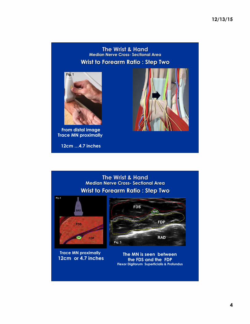
The Wrist & Hand Median Nerve Cross- Sectional Area
Wrist to Forearm Ratio : Step Two
From distal image Trace MN proximally
12cm …4.7 inches
The Wrist & Hand Median Nerve Cross- Sectional Area
Wrist to Forearm Ratio : Step Two
Trace MN proximally 12cm or 4.7 inches
FDS
The MN is seen between the FDS and the FDP
Flexor Digitorum Superficialis & Profundus
FDP
N

12/13/15
5
The Wrist & Hand Median Nerve Cross- Sectional Area
Wrist to Forearm Ratio Calculation
Distal 17mm
Source: Clinical Neurophysiology 2008; 119:1353-1357 (DOI:10.1016/j.clinph.2008.01.101 )
Prox 7mm
17mm
7mm
Ratio = 2.4
> 1.4 positive for
Carpal Tunnel
The Wrist & Hand Palmar Longitudinal
Slightly off midline toward Radial margin
PL
FR
Palmaris Longus (PL) passes superficial to the Flexor Retinaculum (FR).
The most superficial structure of the volar wrist. Absent in 20 % of population
12/13/15
6
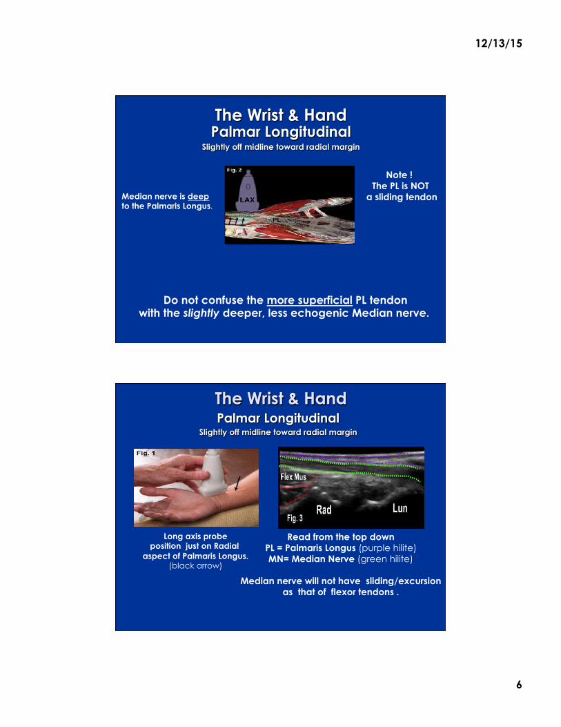
The Wrist & Hand Palmar Longitudinal
Slightly off midline toward radial margin
Median nerve is deep to the Palmaris Longus.
Note ! The PL is NOT
a sliding tendon
Do not confuse the more superficial PL tendon with the slightly deeper, less echogenic Median nerve.
FR
PL
The Wrist & Hand Palmar Longitudinal
Slightly off midline toward radial margin
Read from the top down PL = Palmaris Longus (purple hilite) MN= Median Nerve (green hilite)
Median nerve will not have sliding/excursion
as that of flexor tendons .
Long axis probe position just on Radial
aspect of Palmaris Longus. (black arrow)
PL MN

12/13/15
7
The Wrist & Hand Median Nerve Has little or NO EXCURSION !
The Wrist & Hand Ulnar Nerve Transverse In Guyon�s Canal
Ulnar Nerve is adjacent to the Ulnar Artery and superficial to Flexor Retinaculum Probe is moved in short axis plane to
medial/ulnar side of palmar wrist.
P
FR P

12/13/15
8
The Wrist & Hand Ulnar Nerve Transverse In Guyon�s Canal
Ulnar Nerve can be identified using color flow or doppler
PIS FDMB
Pis : Pisiform Red Hilite : Ulnar Artery (pulsatile, non-compressible)
Yellow Hilite : Ulnar Nerve FDMB = Flexor Digiti Minimi Brevis
The Wrist & Hand Ulnar Nerve Transverse In Guyon�s Canal
Ulnar Nerve is adjacent the artery and superficial to Flexor Retinaculum
Bony landmark is the Pisiform Ulnar
PIS Scaph

12/13/15
9
Dupuytren�s Contracture
Can be treated with injectable collagenase clostridium histolyticum (Hurst L, NEJM 2009) with resolution of contracture in 64% c/w 7% in placebo group.
2 cases of tendon rupture out of 203 patients, local inflammatory response in 30%.
Product known as Xiaflex
Normal SAX
PA= Palmar Aponeurosis (black hilite) PB = Palmaris Brevis (blue hilite)
Palmar Fascia (green hilite)
Abnormal LAX
Dupuytren's contracture is an idiopathic, benign proliferative disorder that results in fibrous tissue deposition in the
Palmar Aponeurosis of the hand. It occurs in the fibro-fatty layer between the skin and deep palmar structures,
resulting in 1st… the formation of nodules, that over time, 2ndly… develop into longitudinal cords.
PB PA
4th or 5th MPJ
Normal Anatomy Red Bracket = Volar Plate Yellow Bracket = Palmaris
Green Bracket = Fibro-fatty Layer Blue Hilite = Fibrous deposit interface
The Wrist & Hand Dupuytren�s Contracture
Hypoechoic… or possibly hyperechoic subcutaneous nodules…
In a “string of pearls” configuration
Long Axis Palmar View
PB PA
LAX
4th or 5th MPJ
Can be treated with injectable collagenase clostridium histolyticum (Hurst L, NEJM 2009) with resolution of contracture
Product known as Xiaflex
5 4 3 21

12/13/15
10
The Wrist & Hand Dorsal Transverse
* Two key dorsum structures *
1. Non Osseous = Extensor Retinaculum
A Horizontal, Linear, Hyperechoic band The ER defines the dorsal anatomy .
Forming 6 synovial compartments by means of radial and ulnar attachments.
Radial Side
Ulnar Side
The Wrist & Hand Dorsal Transverse
* Two key dorsum structures *
2. Osseous: Lister�s Tubercle
(at distal Radius)
Serves as landmark dividing Compartment 2 from Compartment 3
LT
Uln
Scph Lun
TriQ
Lister’s Tubercle = black hilite

12/13/15
11
The Wrist & Hand First 3 Dorsal Compartments
I = Extensor Pollicis Brevis & Abductor Pollicis Longus
II = Extensor Carpi Radialis Longus & Brevis
III = Extensor Pollicis Longus
The Wrist & Hand First 3 Dorsal Compartments
I = Extensor Pollicis Brevis & Abductor Pollicis Longus
Mid-Supination/Pronation to expose Radial margin
Radius
APL EPB
12/13/15
12
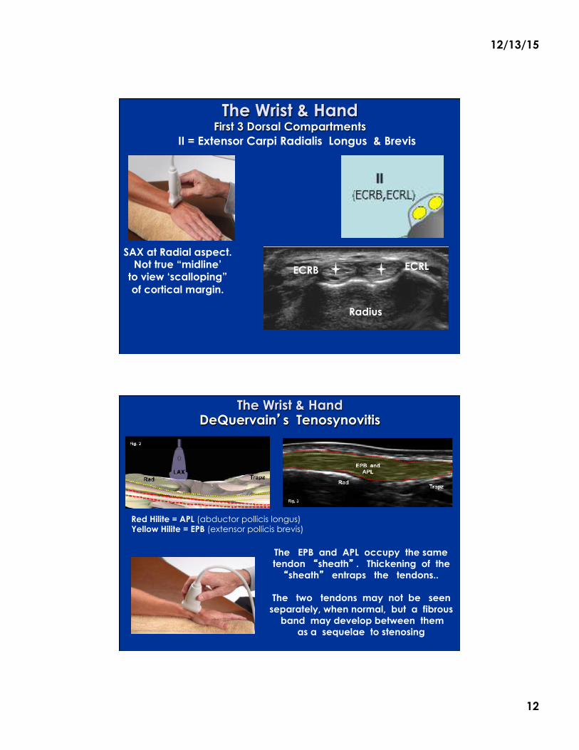
The Wrist & Hand First 3 Dorsal Compartments
SAX at Radial aspect. Not true “midline’
to view ‘scalloping” of cortical margin.
II = Extensor Carpi Radialis Longus & Brevis
Radius
ECRB ECRL
The Wrist & Hand DeQuervain�s Tenosynovitis
The EPB and APL occupy the same tendon �sheath� . Thickening of the �sheath� entraps the tendons..
The two tendons may not be seen
separately, when normal, but a fibrous band may develop between them
as a sequelae to stenosing
Rad
Scaph
Red Hilite = APL (abductor pollicis longus) Yellow Hilite = EPB (extensor pollicis brevis)

12/13/15
13
The Wrist & Hand DeQuervain�s Tenosynovitis
Transverse/Short Axis View
Purple Hilite = Ext. Retinaculum Red Hilite= EPB
Yellow Hilite = APL = Cephalic vein * = Radial Artery
£
Mid-Supination/Pronation
De Quervain’s Injection Out of Plane Approach
Dissecting the two tendons
Radius
APL EPB
A perpendicular view may reveal or confirm
thickening of the sheath or fluid within it.
Note : Some normal
physiologic fluid is expected.
Significant amounts of fluid would produce a �halo�

12/13/15
14
The Wrist & Hand Dorsal Longitudinal
Extensor retinaculum is superficial to tendons.
With Stand off Extensor tendon fibrillar pattern
is seen more clearly
The Wrist & Hand Dorsal Metacarpal-Phalangeal Longitudinal
The extensor tendon is MOST superficial.
Longitudinal probe position across the MCP joint.
Supporting the joint
on palmar side helps with flexion and
distraction for dynamic imaging
Purple = anatomic neck of MC Yellow = synovial membrane
Lite Blue = synovial fluid Red = capsule
Green = tendon

12/13/15
15
The Wrist & Hand Dorsal Metacarpal-Phalangeal Longitudinal
The extensor tendon is MOST superficial.
Acoustic standoff helps with probe contact
Purple = anatomic neck of MC Yellow = synovial membrane
Lite Blue = synovial fluid Red = capsule
Green = tendon
The Wrist & Hand Dorsal MCP Longitudinal
Normal smooth, intact cortical outline
1.Cortical erosion.No distinct anechoic cartilage margin
2. Distended joint capsule and synovial thickening
3. Poorly visualized extensor tendon
1
2
3

12/13/15
16
The Wrist & Hand Synovial Thickening Assessment
Grade 1: Synovium in “triangle” between the bones Grade 2: Synovium extending proximally over the Metacarpal Head
Grade 3: Synovium extending to metaphysis of Metacarpal
Grade 1
The Wrist & Hand Palmar MCP Longitudinal View
*= Volar plate (thickened portion of capsule) 1 = Flexor tendons/ A1 Pulley
The A1 pulley is a very thin, anechoic line above the tendon
Not the optimal view to see the pulley
* 1

12/13/15
17
The Hand & Wrist Palmar Transverse MCP
The A1-2 Pulley
The Annular Ligaments/Pulley system forms a fibro-osseous tunnel through which pass the deep and superficial flexor tendons of the fingers (FT). They provide strategic constraints to the flexor tendons and prevent
“bowstringing”during flexion/extension of the digits.
1 1 2
Transverse probe position yields a cross sectional
view of the pulley-tendon complex.
The Wrist & Hand Palmar Transverse MCP
The A1-2 Pulley “trigger finger”
3
2
1
4
PP= Proximal Phalanx
VP/purple hilite = Volar Plate FT/red hilite = Flexor tendons (FDS and FDP)
Green Hilite = Annular Ligament/Pulley
The Pulley ligament is a slim, uniform “anechoic arch” surrounding the VP and FT.
Clinical Significance : Common site of �trigger finger�.
A thickened A1-2 Pulley or a nodule on the tendon will catch causing
momentary locking of the finger in flexion.
PP

12/13/15
18
The Wrist & Hand Palmar Transverse MCP
The A1-2 Pulley “trigger finger”
PP= Proximal Phalanx
VP/purple hilite = Volar Plate FT/red hilite = Flexor tendons (FDS and FDP)
Green Hilite = Annular Ligament/Pulley
The Pulley ligament is a slim, uniform “anechoic arch” surrounding the VP and FT.
Clinical Significance : Common site of �trigger finger�.
A thickened A1-2 Pulley or a nodule on the tendon will catch causing
momentary locking of the finger in flexion.
PP PP
The Wrist & Hand 1st CMC Joint Longitudinal
Navigation 3rd Joint Space From Radius
Radius Scaphoid Trapezium

12/13/15
19
The Wrist & Hand 1st CMC Joint Longitudinal
Reciprocal reception between 1st Metacarpal and Trapezium The 3rd Joint Space From Radius
Long axis probe
position, kept at
radial aspect of
the joint
FIRST identify the distal radius Count 3 joint spaces to correctly
identify the 1st CMC.
1. Radio-Scaphoid 2. Scaphoid-Trapezium
3. Trapezium-Metacarpal
Trap
Meta R
S
The Wrist & Hand Thenar Longitudinal Views
Ulnar Side
The collateral ligament is deep to the Adductor Pollicis
tendon/aponeurosis (purple hilite)
1
2
The Collateral ligament (1) AND it�s
accessory portion cross over the joint
space.
The aponeurosis is more hyperechoic
interface SUPERFICIAL to UCL.
Long axis probe on ulnar/medial side
of the thumb
Adductor
UCL

12/13/15
20
The Wrist & Hand Thenar Longitudinal Views
Ulnar Collateral Ligament
The collateral ligament
Is deep to the Adductor
Pollicis tendon/
aponeurosis
1
2
The Collateral ligament (1) AND it�s
accessory portion cross over the joint
space.
The aponeurosis is more hyperechoic
interface SUPERFICIAL to UCL.
Long axis probe on ulnar/medial side
of the thumb
Adductor
UCL
1st Metar Prox Phlx
The Wrist & Hand Gamekeeper�s or Skier�s Thumb, Stener Lesion
Severe hyper-abduction injury
The ligament is often torn from the bone, and avulsion
fractures can be seen on ultrasound. Check continuity of met head cortex.
Stener lesion includes UCL tear PLUS
Aponeurosis detachment from 1st MP jt. Mechanism of Injury
Falling forward and catching the thumb will hyper-aBduct
the 1st Metacarpal joint
1st Met Head

12/13/15
21
The Wrist & Hand TFCC: Triangular Fibrocartilage Complex
The Triangular Fibrocartilage Complex is a term used to describe the various structures
suspending the distal Radius and the Ulnar Carpus…
Aka… the anatomic collection connecting the lower arm to the hand.
On the Ulnar side of the wrist, the intra-articular disc thickens, making it somewhat visible to insonation,
…and prone to impaction type injury .
The fibrocartilage/meniscus is supported by a meniscal “homologue”.
Homologue means a “double or duplicate”.
The Wrist & Hand TFCC: Triangular Fibrocartilage Complex
Deep Intra-articular TFC Disc Green Hilite
Meniscal Homologue
Purple Hilite and * the �duplicate��attaching
the disc to Triquetrum.
Homologue, referred to as A “sling or leash”
Superficial UCL >>>>

12/13/15
22
The Wrist & Hand TFCC: Triangular Fibrocartilage Complex
Long Axis probe at the distal Ulna active Radial deviation
TFC images most reliably visualize the TFC Homologue
Ulna
Triquetrum
Lunate
The Wrist & Hand TFCC: Triangular Fibrocartilage Complex
Identifying the Homologue as separate from the fibrocartilage is helpful, and may display tears not involving the fibrocartilage.
Also, ultrasound has known limitations in completely evaluating fibrocartilage in general.

12/13/15
23
Thank You !