the value of tc-99m-tetrofosmin scintigraphy in the...
TRANSCRIPT

Original Article 443Vol. 17, No. 6, 2003
Annals of Nuclear Medicine Vol. 17, No. 6, 443–449, 2003
ORIGINAL ARTICLE
Received February 1, 2003, revision accepted April 21, 2003.For reprint contact: Dr. Zeynep Yapar, Çukurova Üniversitesi
Tıp Fakültesi, Nükleer Tıp Anabilim Dalı, Balcalı, Adana,TURKEY.
INTRODUCTION
ALTHOUGH the prognosis of primary malignant bone tu-mors has improved dramatically since the addition ofchemotherapy to surgery, systemic relapses still occur in40% to 50% of cases.1 Multidrug resistance (MDR), a
phenomenon in which malignant cells demonstrate resis-tance to multiple chemotherapeutic agents, is believed tobe a factor contributing poor outcome for the majority ofthe patients.
MDR has been defined to be a multifactorial phenome-non which may include overexpression of drug resistanceproteins, such as MDR1 P-glycoprotein (Pgp), MDR-associated protein (MRP1) and lung resistance protein(LRP).2 Among these proteins, Pgp has been shownto be the most commonly expressed marker and Pgpexpression correlates with resistance to chemotherapy in
The value of Tc-99m-tetrofosmin scintigraphy in the assessmentof P-glycoprotein in patients with malignant bone and soft-tissue tumors
Zeynep YAPAR,* Mustafa KIBAR,* A. Fuat YAPAR,** Aysun UGUZ,***Serdar OZBARLAS**** and Gulfiliz GONLUSEN***
*Department of Nuclear Medicine, Cukurova University Medical School, Adana, Turkey**Department of Nuclear Medicine, Baskent University Medical School-Adana Hospital, Adana, Turkey
***Department of Pathology, Cukurova University Medical School, Adana, Turkey****Department of Orthopaedics, Cukurova University Medical School, Adana, Turkey
P-glycoprotein (Pgp) overexpression has been shown to be correlated with resistance to chemo-therapy in patients with malignant bone and soft-tissue tumors. The aim of our study was toinvestigate the role of 99mTc-tetrofosmin as a functional imaging agent reflecting Pgp expressionin these tumors. Methods: Twenty eight patients with various malignant bone and soft-tissue tumorswere studied. Radionuclide angiography with 99mTc-tetrofosmin was done first and planar imageswere acquired at 15 min and 90 min postinjection. Vascular phase was evaluated visually ondynamic images, metabolic state was evaluated both visually and quantitatively on planar images.Quantitative analysis was performed by the calculation of tetrofosmin uptake in the lesion againstbackground and percent washout rate (WR%) of the tracer. Immunohistochemical analysis of Pgpwas performed on biopsy specimens and the degree of expression was graded from 0 to 3. Results:There was a positive correlation between the Pgp score and the washout rate of tetrofosmin (r = 0.73,p = 0.000). The mean washout rate of tetrofosmin from the lesions with Pgp expression (31.81 ±6.72) was found to be significantly higher than those of without Pgp expression (21 ± 3.49) (p =0.000). No statistically significant correlation was found between 15 min and 90 min uptake ratios(UR) of tetrofosmin and Pgp score (r = −0.10, p = 0.6 and r = −0.21, p = 0.2, respectively). Whenthe cut-off value of 24.5 (according to ROC-analysis) for the washout rate was used to discriminatethe lesions with and without Pgp expression, the test yielded a sensitivity value of 87.5% with aspecificity of 100%. Conclusions: In malignant bone and soft-tissue tumors, 99mTc-tetrofosminuptake were not related to Pgp overexpression. Pgp overexpression was found to be correlated withthe washout rate of the tracer. 99mTc-tetrofosmin scintigraphy with washout analysis may not onlybe a useful method for evaluating Pgp overexpression but also its function.
Key words: Tc-99m-tetrofosmin, P-glycoprotein, malignant bone and soft-tissue tumors

Annals of Nuclear Medicine444 Zeynep Yapar, Mustafa Kibar, A. Fuat Yapar, et al
osteosarcoma patients.2–4 In soft-tissue sarcomas, Pgpexpression has also been reported to be a prognosticindicator correlating with a poor outcome.5,6
Pgp is a 170-kD transmembrane glycoprotein whichacts as an adenosine triphosphate-dependent efflux pumpto reduce the intracellular accumulation of many chemo-therapeutic drugs, including doxorubicin which is themost effective agent for the treatment of osteosarcoma.799mTc-tetrofosmin (99mTc-1,2-bis[bis(2-ethoxyethyl)phosphino]ethane) is an agent developed for myocardialperfusion imaging and has been shown to accumulate inviable tumor tissue.8–14 The functional characteristics ofthis agent are similar to those of 99mTc-sestamibi.15 Like
sestamibi, tetrofosmin has also been suggested to be apotent agent for the prediction of multi drug resistance(MDR) in various tumour cell lines.16–19
The aim of our study was to examine the role of 99mTc-tetrofosmin as a functional imaging agent reflecting Pgpexpression in malignant bone and soft-tissue tumors. Forthis purpose, the uptake and washout kinetics of 99mTc-tetrofosmin was compared with the degree of Pgp expres-sion of the biopsy specimens.
Table 1 Scintigraphic findings with 99mTc-tetrofosmin and P-glycoprotein expression
Tc-99m tetrofosminPatient Age/
Diagnosis Lesion sitePgp
Visual Washout Uptake ratio/visual uptakeno. sex score
perfusion rate (%) 15 min 90 min
Patients without tetrofosmin uptake on 15-min image
1. 18/F Metastatic Ewing’s sarcoma Right scapula 0 0 — — —2. 50/M Metastatic indifferentiated Left femur 2 0 — — —
carcinomaPatients with tetrofosmin uptake on 15-min image
3. 20/M Fibrosarcoma Left thigh 0 2 18 1.73 (1) 1.41 (0)4. 21/F Ewing’s sarcoma Right calcaneus 0 2 24 8.92 (3) 6.77 (3)5. 22/F Synovial sarcoma Right cruris 0 1 21 7.07 (3) 5.56 (3)6. 51/M Non-Hodgkin’s lymphoma Left calcaneus 0 2 25 4.01 (3) 3.0 (3)7. 74/M Lymphoma Left humerus 0 2 14 2.59 (2) 2.22 (2)8. 34/F Osteosarcoma Right femur 0 2 23 2.45 (3) 1.87 (2)9. 50/M Myxoid liposarcoma Right buttock 1 2 20 1.93 (2) 1.54 (2)
10. 37/M Metastatic undifferentiated Left femur 1 2 23 4.32 (3) 3.32 (3) carcinoma
11. 2.5/M Ewing’s sarcoma Left tibia 1 2 18 2.66 (3) 2.18 (3)12. 20/M Osteosarcoma Left femur 1 2 24 3.35 (3) 2.54 (2)13. 9/F Osteosarcoma Left femur 2 2 30 3.27 (3) 2.27 (2)14. 13/M Osteosarcoma Left femur 2 2 33 2.29 (3) 1.52 (2)15. 19/F Fibrosarcoma Right foot 2 2 14* 4.51 (3) 3.85 (3)16. 19/F Metastatic renal cell Right iliac 3 2 34 4.39 (3) 2.86 (2)
carcinoma17. 50/F Metastatic infiltrative ductal Left tibia 3 1 27 6.79 (3) 4.89 (2)
carcinoma (breast)18. 38/F Osteosarcoma Right femur 3 1 26 1.53 (2) 1.13 (1)19. 18/M Osteosarcoma Right tibia 3 2 30 5.56 (3) 3.88 (2)20. 60/M Renal cell sarcoma Left femur 3 2 48 3.45 (3) 1.77 (1)21. 27/M Osteosarcoma Right femur 3 1 37 1.38 (1) 0.86 (0)22. 69/M Metastatic transitional cell Right femur 3 2 22* 3.34 (3) 2.61 (2)
carcinoma (bladder)23. 78/M Round cell liposarcoma Left cruris 3 2 35 4.0 (3) 2.61 (2)24. 10/M Ewing’s sarcoma Right tibia 3 2 28 3.84 (3) 2.75 (2)25. 7/F Malignant fibrous Right femur 3 2 42 2.19 (3) 1.25 (1)
histiocytoma26. 18/M Osteosarcoma Right femur 3 1 35 2.08 (2) 1.34 (1)27. 50/F Malignant fibrous Left femur 3 1 31 2.18 (2) 1.51 (1)
histiocytoma28. 45/F Ewing’s sarcoma Right humerus 3 2 27 3.96 (3) 2.86 (3)
*Discordant washout ratios with respect to Pgp score when the cut-off value of 25.5 was used

Original Article 445Vol. 17, No. 6, 2003
MATERIALS AND METHODS
PatientsTwenty eight patients (12 F, mean age: 27.66 ± 15.19; 16M, mean age: 37.34 ± 24.53; age range: 2.5 to 78 y) withvarious malignant bone and soft tissue tumors were in-cluded in the study. All patients were examined scintig-raphically before biopsy. The diagnoses were provenpathologically in specimens obtained by biopsy and/orsurgery, Eight patients had osteosarcoma, 5 Ewing’ssarcoma, 2 malignant fibrous histiocytoma, 2 fibrosar-coma, 2 liposarcoma, 2 lymphoma, 1 synovial sarcoma,and 6 bone metastatic tumors (Table 1).
Each patient underwent 99mTc methylene diphosphonate(MDP) three-phase bone scanning as a routine procedure.At least two days later, dynamic and static 99mTc-tetro-fosmin scans were obtained.
99mTc-Tetrofosmin ImagingThe adults received intravenous injection of 370–600MBq 99mTc-tetrofosmin. This activity was reduced in thechildren. The limbs in question were examined with alarge field of view gamma camera (Camstar, GE MedicalSystems) equipped with a low-energy all-purpose colli-mator at most adequate projection in each patient. Datawere obtained every 2 sec for 60 sec for radionuclideangiography (128 × 128 matrix). Then, planar 5-min99mTc-tetrofosmin images (256 × 256 matrix) were ob-tained 15 and 90 min after radionuclide administration.
Data AnalysisRadionuclide angiography was visually evaluated by twoblinded observers and the degree of perfusion increase
was classified into four grades: a) 0 = no increase; b) 1 =mild increase; c) 2 = moderate increase; d) 3 = markedincrease. Static 99mTc-tetrofosmin images were evaluatedvisually and quantitatively. In visual analysis, two blindedobservers evaluated the degree of radionuclide uptakeusing a four-grade scoring system: a) 0 = backgroundactivity; b) 1 = slight increase in uptake; c) 2 = moderateuptake; d) 3 = strong uptake.
In quantitative analysis, a manual region of interest(ROI) was set on the lesion (L) and a symmetrical ROIwas set on the contralateral normal area (B) on 15 minimages. Identical ROIs were applied to 90 min images.The uptake ratio (UR) of both 15 and 90 min images wascalculated by dividing the count density of the lesion bythat of the background ROI. After decay correction of themean counts in the ROIs drawan on the 90 min images, thewashout rate (WR%) of 99mTc-tetrofosmin from the le-sion was determined using the following formula:
WR%/90 min =(L/B) 15 min − (L/B) 90 min
× 100(L/B) 15 min
Detection of P-Glycoprotein ExpressionThe lesions were resected by open biopsy to obtainhistopathological diagnosis. Immunohistochemical(IHC) staining was performed according to the standardstreptoavidin-biotin method. The primary antibody ap-plied for P-glycoprotein was “monoclonal antibody PGP(MDR)” (Immunotech company, France, Ready for use,No. 2142).
Sections of tissue were fixed in formaldehyde solutionand embedded in paraffin, then were cut out and mountedon silane-coated glass slides. The sections were depar-affinized with xylene for 30 min and washed in 90% etha-nol twice. After inhibition of endogenous peroxidase with30% H2O2/methanol and nonspecific binding of antibodyby “ultratech HRP (AEC) kit, protein blocking agent.”Ultratech kit is suitable for use with all primary antibodiesderived from rabbit, mouse, rat or guinea pig.
The sections were then incubated with primary anti-body 5–10 min at room temperature which binds tospecific tissue antigens. Color was developed with AECsystem, and nuclear staining was done with Meyer’shematoxylin. Tissue sections from a normal kidney wereused for control staining because the reactivity of proxi-mal tubules have been shown to be different from that ofglomeruli, so that results of Pgp immunostaining werepositive in proximal tubules and negative in glomeruli. Inthe sections of the lesions, the degree of Pgp staining wasscored from 0–3 based on the distribution of positivity ofimmunostaining of the plasma membrane and the Golgiregion as follows: a) Score 0 when no stain was observed;b) Score 1 when less than 10% of the cells were stained;c) Score 2 when 10% to 49% of the cells were stained; andd) Score 3 when 50% or more of the cells were stained.The scores of 0 and 1 were considered as negative, 2 and
Fig. 1 Significant difference in tetrofosmin washout wasobserved between patients with Pgp scores 0–1 and 2–3 (p =0.000). Tumors with high Pgp expression demonstrated highertetrofosmin washout from lesions. A positive correlation wasfound between washout rate and Pgp expression (r = 0.73, p =0.000).
Annals of Nuclear Medicine446 Zeynep Yapar, Mustafa Kibar, A. Fuat Yapar, et al
3 were considered as positive for Pgp expression.
Statistical AnalysisThe values were presented as mean ± SD. The Mann-Whitney test was used to determine the differences be-tween the tumors with and without Pgp. P < 0.05 wasconsidered as significant. The correlation between Pgplevels and tetrofosmin results were analyzed by Spearman’srank correlation coefficient . For determining the sensitiv-ity and specificity values of tetrofosmin washout ratio in
assessing Pgp positivity, the cut-off value of 24.50 wasdetermined according to the ROC-analysis.
RESULTS
All patients’ data are listed in Table 1. When the scores of0–1 were considered as negative, and 2–3 were consid-ered as positive for Pgp expression, Pgp positivity wasdetected in 17 patients (Pgp scores were 3 and 2 in 13 and4 patients, respectively). In 11 patients, Pgp expressionwas not significant (Score 1 in 4, score 0 in 7 patients).
There were only two lesions (2/28, 7%) withouttetrofosmin uptake. Visually, 26 out of 28 lesions (93%),
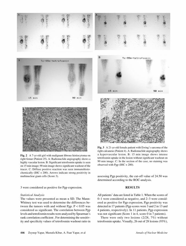
Fig. 2 A 7-yr-old girl with malignant fibrous histiocytoma onright femur (Patient 25). A: Radionuclide angiography shows ahighly vascular lesion. B: Significant tetrofosmin uptake is seenon 15 min image; 90 min image shows significant washout of thetracer. C: Diffuse positive reaction was seen immunohisto-chemically (IHC × 200). Arrows indicate strong positivity inmultinuclear giant cells (Score 3).
Fig. 3 A 21-yr-old female patient with Ewing’s sarcoma of theright calcaneus (Patient 4). A: Radionuclide angiography showsa hypervascular lesion. B: 15 min image shows intensetetrofosmin uptake in the lesion without significant washout on90 min image. C: In the section of the case, no staining wasobserved with Pgp (IHC × 200).

Original Article 447Vol. 17, No. 6, 2003
with or without Pgp expression showed tetrofosmin accu-mulation. Among them, nineteen lesions (19/26) showedstrongly (grade 3), five lesions (5/26) showed moderately(grade 2) and two lesions (2/26) showed slightly (grade 1)increased tetrofosmin uptakes on 15 min images. On 90min images, 17 lesions (17/26, 65%) showed tetrofosminwashout in any degree, while tetrofosmin uptake gradepersisted in 9 lesions (9/26, 35%). Washout group (17lesions) consisted of 14 lesions with Pgp expression(82%) and 3 lesions without Pgp expression (17%).Persistence group (9 lesions) consisted of 7 (77%) and 2(23%) lesions without and with Pgp expression, respec-tively. In other words, washout group mostly includedlesions with Pgp expression (82%), while the lesionswithout Pgp expression mostly showed persistent activity(77%).
Except two lesions which did not accumulatetetrofosmin, all lesions showed increased perfusion onradionuclide angiography. Both Pgp-positive and Pgp-negative tumors showed hypervascularization.
In quantitative analysis, positive correlation betweenthe Pgp score and the washout rate of tetrofosmin wasdetected (r = 0.73, p = 0.000). The relationship betweenthe Pgp score and the tetrofosmin washout rate is shownon Figure 1. No statistically significant correlations werefound between 15 min and 90 min uptake ratios (UR) oftetrofosmin and Pgp score (r = −0.10, p = 0.6 and r = −0.21,p = 0.2).
When the 15 and 90 min mean URs of lesions with Pgpexpression (3.42 ± 1.47 and 2.37 ± 1.14) were comparedwith those of lesions without Pgp expression (3.90 ± 2.34and 3.04 ± 1.77), the differences between the groups werenot significant (p = 0.7 and p = 0.3, respectively). Themean washout rate of tetrofosmin from the lesions with
Pgp expression (31.81 ± 6.72) was found to be signifi-cantly higher than those of without Pgp expression (21 ±3.49) (p = 0.000). Representative cases are shown onFigures 2 and 3.
When the cut-off value of 24.5 (according to ROC-analysis) for the washout rate was used to discriminate thelesions with and without Pgp expression, the test yieldeda sensitivity value of 87.5% with a specificity of 100%(Fig. 4). There were two cases with discordant results(Table 1, patients with 15 and 22 numbers).
DISCUSSION
The results of the present study demonstrated that thewashout of 99mTc-tetrofosmin in malignant bone and soft-tissue tumors was correlated with Pgp overexpression.Although the 15 and 90 min mean URs of the lesionswithout Pgp expression was slightly higher than those ofthe lesions with Pgp expression the difference was notsignificant. For this reason, washout analysis and delayedimaging in addition to the early imaging seems to benecessary when tetrofosmin is used in the evaluation ofPgp expression in malignant bone and soft-tissue tumors.
Preoperative chemotherapy has changed the prognosisof bone tumors dramatically.20 It can eliminate microme-tastases, induce total necrosis in the primary tumor, re-duce the tumor’s size and vascularity, and thus facilitatesthe surgical excision of the margins safely. Clinicaltrials revealed that Pgp overexpression correlates with apoor chemotherapeutic response and poor prognosis inbone and soft-tissue sarcomas.1,5,6 Thus, there is a realchallenge for the detection of Pgp in these tumors.
Noninvasive detection of Pgp uses lipophilic cationiccompounds characterized as substrates for MDR1 Pgp.Among these compounds, 99mTc-sestamibi and 99mTc-tetrofosmin are nonmetabolizable radiopharmaceuticalswith monocationic charges, like most chemotherapeuticagents in the MDR phenotype.21,22 Cellular uptake ofthese tracers depends on series negative transmembraneand mitochondrial potentials,23 and they can accumulateto a much higher degree in malignant cells due to differ-ences in mitochondrial density and membrane polariza-tion.
99mTc-tetrofosmin has been validated as a transportsubstrate for Pgp in a variety of MDR human cells.16,18,24
Also in vivo studies have shown that 99mTc-tetrofosminimaging has been a potential tool for understanding Pgpexpression in lung cancer,25 malignant lymphoma26 andbreast cance.27 From a review of the literature, no reportshave been published previously concerning the relation-ship between Pgp expression and cellular kinetics oftetrofosmin in musculoskeletal tumors. Previous experi-ence has focused on 99mTc-sestamibi and proved that thisagent has been a substrate for Pgp in these tumors.28,29
According to our results, 99mTc-tetrofosmin imaging maybe useful in bone and soft-tissue sarcomas as well, when
Fig. 4 According to ROC analysis, cut-off value for washoutrate was determined as 24.5.
Area under curve: 0.90 (ci: 0.79–1.02)Sensitivity: 82.4%Specificity: 99%

Annals of Nuclear Medicine448 Zeynep Yapar, Mustafa Kibar, A. Fuat Yapar, et al
efflux rate of the tracer is used as an indicator of Pgpexpression. Prior data have shown an inverse relationshipbetween levels of Pgp and the magnitude of tetrofosminuptake in malignant lymphoma, breast and lung can-cers.26,27,30 Contrasting with prior data, tetrofosminaccumulated in most musculoskeletal sarcomas even withhigh Pgp expression (Table 1) in our study. Such contrast-ing points for sestamibi were also raised in the publisheddata and attributed to the presence of subsets of cellssimultaneously expressing varying and multiple resis-tance mechanisms, or poor penetration of tracer becauseof reduced blood flow in tumors undergoing necrosis.2 Inaccordance with our findings, Taki et al.28 and Burak etal.29 did not find significant correlations between tumorto background URs and Pgp expression using 99mTc-sestamibi, and washout analysis was recommended as aconclusion.
The method has failed to detect Pgp expression in twolesions (patient Nos. 15 and 22) when the cut-off value oftetrofosmin was considered as 25.5%. Although theimmunostaining of Pgp was strong, the washout oftetrofosmin from these lesions was slower than the thresh-old level, suggesting a negative result. Suboptimal func-tional capacity of the Pgp efflux pump was thought to bethe possible mechanism to explain the discordant findingsfor these two lesions. Discordant results might also beexplained by the heterogeneous distribution of Pgp intumors, in which a small biopsy specimen might notalways represent whole tumor Pgp expression. However,any patient with heterogeneous tetrofosmin washout fromthe lesion was not observed in our study. Burak et al.29
have advised the application of small standard ROIs usingsingle-photon emission tomography in determining re-gional washout rates because of the possible insufficiencyof planar imaging.
The optimum time for delayed imaging regarding 99mTc-sestamibi is under debate in the literature. Taki et al.28
preferred a delay of 3 hr for late images. On the other hand,Burak et al.29 showed that 1-hr late imaging could also beused to define the clearance of sestamibi. Thus, the timingof the late images was planned as 90 min after injection incurrent study. Our results revealed that 90 min delay afterinjection could be sufficient to demonstrate the differentwashout patterns of tetrofosmin between Pgp positive andPgp negative tumors. However, complete washout in 90min was observed in only one lesion (1/16) among lesionswith Pgp expression. This ratio is very low when com-pared to the ratio obtained from the study in which 3 hrdelayed imaging was used with sestamibi by Taki et al.(7/15).28 From this point of view, it could be argued thatlater imaging times at least 120 min after injection maybe preferred if the images are to be analyzed visually.
Doxorubicin is a chemotherapeutic agent which isincluded in therapy protocols in malignant bone and soft-tissue tumors, and is generally effective. It has beenshown to be a transport substrate for Pgp.7 Functional
identification of Pgp before initiation of chemotherapymight provide important information and might be valu-able in management of patients with malignant bone andsoft-tissue tumors. If modulation of Pgp becomes feasiblein clinical practice, 99mTc-tetrofosmin scintigraphy wouldbe useful as a Pgp function monitoring method.
CONCLUSIONS
According to our results, 99mTc-tetrofosmin uptake inbone and soft-tissue tumors were not related to Pgp overex-pression. Pgp overexpression was found to be correlatingto the washout rate of the tracer. 99mTc-tetrofosminscintigraphy with washout analysis may not only be auseful method for evaluating Pgp overexpression but alsoits function. Tumor imaging with 99mTc-tetrofosmin maybe a promising clinical tool in monitoring patients withmalignant bone and soft-tissue tumors. But further pro-spective studies are needed to evaluate the prognosis andevent-free survival of patients.
REFERENCES
1. Baldini N, Scotlandi K, Barbanti-Brodano G, Manara MC,Maurici D, Bacci G, et al. Expression of P-glycoprotein inhigh-grade osteosarcomas in relation to clinical outcome. NEngl J Med 1995; 333: 1380–1385.
2. Kostakoglu L. Multidrug Resistance. Part B: Other Tumours.In: Khalkhali I, Maublant JC, Goldsmith SJ, eds. NuclearOncology-Diagnosis & Therapy. Philadelphia; LippincottWilliams & Wilkins, 2001: 73–82.
3. Baldini N, Scotlandi K, Serra M, Picci P, Bacci G, Sottili S,et al. P-glycoprotein expression in osteosarcoma: a basis forrisk-adapted adjuvant chemotherapy. J Orthop Res 1999;17: 629–632.
4. Kusuzaki K, Hirata M, Takeshita H, Murata H, HashiguchiS, Ashihara T, et al. Relationship between P-glycoproteinpositivity, doxorubicin binding ability and histologic re-sponse to preoperative chemotherapy in osteosarcoma.Cancer Lett 1999; 26: 203–208.
5. Levine EA, Holzmayer T, Bacus S, Mechetner E, Mera I,Bolliger C, et al. Evaluation of newer prognostic markersfor adult soft tissue sarcomas. J Clin Oncol 1997; 15: 3249–3257.
6. Chan HS, Thorner PS, Haddad G, Ling V. Immunohisto-chemical detection of P-glycoprotein: prognostic correla-tion in soft-tissue sarcoma of childhood. J Clin Oncol 1990;8: 689–704.
7. Gottesman MM, Pastan I. Biochemistry of multidrug resis-tance mediated by the multidrug transporter. Annu RevBiochem 1993; 62: 385–427.
8. Kostakoglu L, Uysal U, Özyar E, Demirkazik FB, HayranM, Atahan L, et al. A comparative study of technetium-99msestamibi and technetium-99m tetrofosmin single-photonemission tomography in the detection of nasopharyngealcarcinoma. Eur J Nucl Med 1997; 24: 621–628.
9. Obwegeser I, Berghammer P, Rodrigues M, et al. A head-to-head comparison between technetium-99m-tetrofosminand technetium-99m-MIBI scintigraphy to evaluate suspi-

Original Article 449Vol. 17, No. 6, 2003
cious breast lesions. Eur J Nucl Med 1999; 26: 1553–1559.10. Takekawa H, Shinano H, Tsukamoto E, Koseki Y, Ikeno T,
Miller F, et al. Technetium-99m-tetrofosmin imaging oflung cancer: relationship with histopathology. Ann NuclMed 1999; 13 (2): 71–75.
11. Hashimoto T, Takahashi K, Goto M, Endo H, Kono T,Nishiyama H, et al. Detection of malignant thymoma inprimary tumour and metastatic lesions using 99mTc-tetrofosmin scintigraphy. Radiot Med 2001; 19 (3): 169–172.
12. Adalet I, Kocak M, Oguz H, Alagol F, Cantez S. Determi-nation of medullary thyroid carcinoma metastases by 201Tl99mTc(V)DMSA, 99mTc-MIBI and 99mTc-tetrofosmin. NuclMed Commun 1999; 20 (4): 353–359.
13. Yapar Z, Kibar M, Ozbarlas S, Yapar AF, Uguz A,Zorludemir S. 99mTc-tetrofosmin scintigraphy in musculo-skeletal tumors: the relationship between P-glycoproteinexpression and tetrofosmin uptake in malignant lesions.Nucl Med Commun 2002; 23 (10): 991–1000.
14. Soricelli A, Cuocolo A, Varrone A, Discepolo A, TedeschiE, Mainenti PP, et al. Technetiun-99m-tetrofosmin uptakein brain tumours by SPECT: comparison with thallium-201imaging. J Nucl Med 1998; 39: 802–806.
15. Arbab AS, Koizumi K, Toyama K, Araki T. Uptake oftechnetium-99m-tetrofosmin, technetiun-99m-MIBI andthallium-201 in tumour cell lines. J Nucl Med 1996; 37:1551–1556.
16. Ballinger JR, Bannerman J, Boxen I, Firby P, Hartman NG,Moore MJ. Technetium-99m-tetrofosmin as a substrate forP-glycoprotein: in vitro studies in multidrug-resistant breasttumour cells. J Nucl Med 1996; 37 (9): 1578–1582.
17. Perek N, Prevot N, Koumanov F, Frere D, Sabido O,Beauchesne P. Involvement of the glutathione S-conjugatecompounds and the MRP protein in Tc-99m-tetrofosminand Tc-99m-sestamibi uptake glioma cell lines. Nucl MedBiol 2000; 27 (3): 299–307.
18. Muzzammil T, Moore MJ, Ballinger JR. In vitro compari-son of sestamibi, tetrofosmin, and furifosmin as agents forfunctional imaging of multidrug resistance in tumours.Cancer Biother Radiopharm 2000; 15 (4): 339–346.
19. Utsunomiya K, Ballinger JR, Piquette-Miller M, RauthAM, Tang W, Su ZF. Comparison of the accumulation andefflux kinetics of technetium-99m sestamibi and techne-tium-99m tetrofosmin in an MRP-expressing tumour cellline. Eur J Nucl Med 2000; 27: 1786–1792.
20. Waxman AD, Abdel-Dayem HM. Primary Bone Tumors:Thallium-201, Technetium-99m-sestamibi, and Fluorine-18-deoxyglucose. In: Kbalkhali I, Maublant JC, Goldsmith
SJ, eds. Nuclear Oncology-Diagnosis & Therapy. Philadel-phia; Lippincott Williams & Wilkins, 2001: 509–524.
21. Higley B, Smith FW, Smith T, Gemmell HG, Das Gupta P,Gvozdanovic DV, et al. Technetium-99m-1,2-bis [bis (2-ethoxyethyl) phosphino] ethane: human biodistribution,dosimetry and safety of a new myocardial perfusion imag-ing agent. J Nucl Med 1993; 34: 30–38.
22. Rossetti C, Vanoli G, Paganelli G, Kwiatkowski M, Zito F,Colombo F, et al. Human biodistribution, dosimetry andclinical use of technetium(III)-99m-Q12. J Nucl Med 1994;35: 1571–1580.
23. Rodrigues M, Chehne F, Kalinowska W, Zielinski C,Sinzinger H. Comparative 99mTc-MIBI, 99mTc-tetrofosminand 99mTc-furifosmin uptake in human soft tissue sarcomacell lines. Eur J Nucl Med 2000; 27: 1839–1843.
24. Crankshaw C, Marmion M, Luker G, Rao V, Dahlheimer J,Burleigh BD, et al. Novel Tc(III)-Q-complexes for func-tional imaging of the multidrug resistance (MDR1) P-glycoprotein. J Nucl Med 1998; 39: 77–86.
25. Kao CH, Hsieh JF, Tsai SC, Ho YJ, ChangLai SP, Lee JK.Paclitaxel-based chemotherapy for non-small cell lung can-cer: predicting the response with 99mTc-tetrofosmin chestimaging. J Nucl Med 2002; 42: 17–20.
26. Shiau YC, Tsai SC, Wang JJ, Ho YJ, Ho ST, Kao CH.Predicting chemotherapy response and comparing with P-glycoprotein expression using technetium-99m tetrofosminscan in untreated malignant lymphomas. Cancer Lett 2001;170: 139–146.
27. Liu TJ, Tsai SC, Ho YJ, Sun SS, Kao CH. Comparison of theexpression of P-glycoprotein Ki-67, and P-53 to techne-tium-99m tetrofosmin mammoscintigraphic findings. Can-cer Invest 2002; 20 (2): 199–205.
28. Taki J, Sumiya H, Asada N, Ueda Y, Tsuchiya H, TonamiN. Assessment of P-glycoprotein in patients with malignantbone and soft-tissue tumors using technetium-99m-MIBIscintigraphy. J Nucl Med 1998; 39: 1179–1184.
29. Burak Z, Ersoy O, Moretti JL, Erinc R, Ozcan Z, Dirlik A,et al. The role of 99mTc-MIBI scintigraphy in the assessmentof MDR1 overexpression in patients with musculoskeletalsarcomas: comparison with therapy response. Eur J NuclMed 2001; 28: 1341–1350.
30. Shiau YC, Tsai SC, Wang JJ, Ho YJ, Ho ST, Kao CH. Topredict chemotherapy response using technetium-99mtetrofosmin and compare with p-glycoprotein and multidrugresistance related protein-1 expression in patients withuntreated small cell lung cancer. Cancer Lett 2001; 169:181–188.