technetlum-99m tetrofosmin: a new myocardial perfusion...
TRANSCRIPT
CONTINUING EDUCATION
Technetlum-99m Tetrofosmin: A New Myocardial Perfusion Agent
Stephanie Jones and Robert C. Hendel
Departments of Nuclear Medicine and Medicine, Northwestern University Medical School, Chicago, Illinois
This is the fifth article in a series of five on new radiopharmaceuticals. Upon completion, the technologist will be able to (1) list the characteristics of tetrofosmin, (2) describe the clinical images, and (3) compare tetrofosmin to other myocardial perfusion agents.
J Nucl Med Technol1993; 21:191-195
Myocardial perfusion imaging is an established technique for the detection and assessment of coronary artery disease. Although electrocardiographic exercise testing is a useful method, improved sensitivity and specificity are present when exercise is combined with perfusion imaging. At present, the most frequently used perfusion agent is thallium-201 e01TI). However, despite the wide acceptance of thallium as a perfusion agent, it is far from an ideal tracer, possessing significant limitations, such as low-energy photons (69-83 keY), which are easily attenuated by soft tissue and may result in artifact production and decreased image resolution. Additionally, the relatively long half-life (73 hr) limits the dosage that can be given to the patient, leading to reduced photon flux and lower count statistics.
Technetium-99m (99mTc) agents have many advantages, including a higher photon energy (140 keY), which is optimal for gamma camera imaging. The short physical half-life ( =6 hr) permits a larger dose to be administered, which results in greater photon flux. Furthermore technetium can be produced on site by a generator, which makes it readily available and relatively inexpensive.
CHARACTERISTICS OF TETROFOSMIN
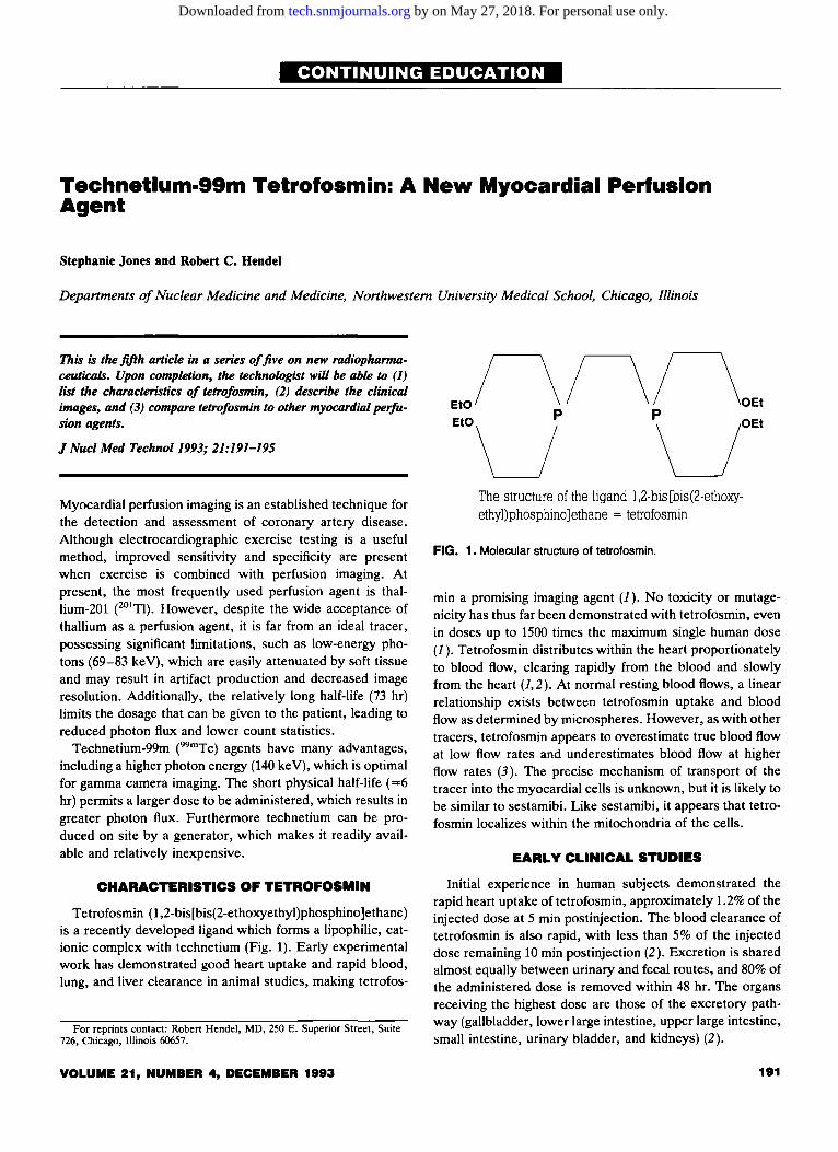
Tetrofosmin (1 ,2-bis[bis(2-ethoxyethyl)phosphino ]ethane) is a recently developed ligand which forms a lipophilic, cationic complex with technetium (Fig. 1). Early experimental work has demonstrated good heart uptake and rapid blood, lung, and liver clearance in animal studies, making tetrofos-
For reprints contact: Robert Hendel, MD, 250 E. Superior Street, Suite 726, Chicago, Illinois 60657.
VOLUME 21, NUMBER 4, DECEMBER 1993
The structure of the ligand l ,2-bis[bis(2-ethoxyethyl)phosphino]ethane = tetrofosmin
FIG. 1. Molecular structure of tetrofosmin.
min a promising imaging agent (1 ). No toxicity or mutagenicity has thus far been demonstrated with tetrofosmin, even in doses up to 1500 times the maximum single human dose (1 ). Tetrofosmin distributes within the heart proportionately to blood flow, clearing rapidly from the blood and slowly from the heart (1,2). At normal resting blood flows, a linear relationship exists between tetrofosmin uptake and blood flow as determined by microspheres. However, as with other tracers, tetrofosmin appears to overestimate true blood flow at low flow rates and underestimates blood flow at higher flow rates (3). The precise mechanism of transport of the tracer into the myocardial cells is unknown, but it is likely to be similar to sestamibi. Like sestamibi, it appears that tetrofosmin localizes within the mitochondria of the cells.
EARLY CLINICAL STUDIES
Initial experience in human subjects demonstrated the rapid heart uptake of tetrofosmin, approximately 1.2% of the injected dose at 5 min postinjection. The blood clearance of tetrofosmin is also rapid, with less than 5% of the injected dose remaining 10 min postinjection (2). Excretion is shared almost equally between urinary and fecal routes, and 80% of the administered dose is removed within 48 hr. The organs receiving the highest dose are those of the excretory pathway (gallbladder, lower large intestine, upper large intestine, small intestine, urinary bladder, and kidneys) (2).
191
by on May 27, 2018. For personal use only. tech.snmjournals.org Downloaded from
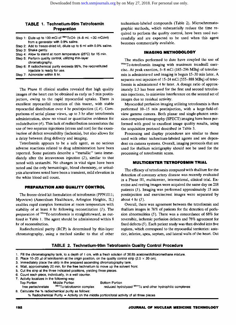
TABLE 1. Technetlum-99m Tetrofosmln Preparation
Step 1: Elute up to 100 mCi of 99"'Tc04- (4-8 ml; <30 mCi/ml) from a generator with 0.9% saline.
Step 2: Add to freeze-dried kit, dilute up to 6 ml with 0.9% saline. Step 3: Shake gently. Step 4: Allow to stand at room temperature (25°C} for 15 min. Step 5: Perform quality control, utilizing thin-layer
chromatography. Step 6: If radiochemical purity exceeds 95%, the reconstituted
injectate is ready for use. Step 7: Administer within 8 hr.
The Phase II clinical studies revealed that high quality images of the heart can be obtained as early as 5 min postinjection, owing to the rapid myocardial uptake. There is excellent myocardial retention of this tracer, with stable myocardial distribution over 4 hr postinjection {1,4). Comparisons of serial planar views, up to 3 hr after tetrofosmin administration, show no visual or quantitative evidence for redistribution (4). This lack of redistribution necessitates the use of two separate injections (stress and rest) for the examination of defect reversibility (ischemia), but also allows for a d~Iay between drug delivery and imaging.
Tetrofosmin appears to be a safe agent, as no serious adverse reactions related to drug administration have been reported. Some patients describe a "metallic" taste immediately after the intravenous injection (2), similar to that noted with sestamibi. No changes in vital signs have been noted and the only hematologic, blood chemistry, or urinalysis alterations noted have been a transient, mild elevation in the white blood cell count.
PREPARATION AND QUALITY CONTROL
The freeze-dried kit formulation oftetrofosmin (PPN1011; Myoview) (Amersham Healthcare, Arlington Heights, IL) enables rapid complex formation at room temperature with stability of at least 8 hr following reconstitution (J ). The preparation of 99mTc-tetrofosmin is straightforward, as outlined in Table 1. The agent should be administered within 8 hr of reconstitution.
Radiochemical purity (RCP) is determined by thin-layer chromatography, using a method similar to that of other
technetium-labeled compounds (Table 2). Microchromatographic methods, which substantially reduce the time required to perform the quality control, have been used successfully and are expected to be used when this agent becomes commercially available.
IMAGING METHODOLOGY
The studies performed to date have coupled the use of 99mTc-tetrofosmin imaging with maximum treadmill exercise. At peak exertion, 5-8 mCi (185-296 MBq) of tetrofosmin is administered and imaging is begun 15-30 min later. A separate rest injection of 15-24 mCi (555-888 MBq) oftetrofosmin is administered 4 hr later. A dosage ratio of approximately 1:3 has been used for the first and second tetrofosmin injections, to minimize interference on the second set of images due to residual activity.
Myocardial perfusion imaging utilizing tetrofosmin is then performed 10-15 min postinjection, with a large-field-ofview gamma camera. Both planar and single-photon emission computed tomography (SPECT) imaging have been performed with good to excellent image quality results, using the acquisition protocol described in Table 3.
Processing and display procedures are similar to those used with other technetium-labeled agents and are dependent on camera systems. Overall, imaging protocols that are used for thallium scintigraphy should not be used for the processing of tetrofosmin scans.
MULTICENTER TETROFOSMIN TRIAL
The efficacy of tetrofosmin compared with thallium for the detection of coronary artery disease was recently evaluated in a Phase III, multicenter, international, clinical trial. Exercise and resting images were acquired the same day on 218 patients (5 ). Imaging was performed approximately 15 min postinjection and exercise/rest images were separated by about 4 hr (5).
Overall, there was agreement between the tetrofosmin and thallium images in 76% of patients for the detection of perfusion abnormalities (5). There was a concordance of 68% for reversible, ischemic perfusion defects and 78% agreement for fixed defects (5). Each patient study was then divided into five regions, which correspond to the myocardial territories: anterior, inferior, apex, septum, and lateral walls of the heart. Out
TABLE 2. Technetium-99m Tetrofosmin Quality Control Procedure
1. Fill the chromatography tank, to a depth of 1 em, with a fresh solution of 36:65 acetone/dichloromethane mixture. 2. Place 10-20 ILl of tetrofosmin at the origin position, on the quality control strip (2.0 x 20 em). 3. Immediately place the strip in the prepared ascending chromatography tank. 4. Wait, approximately 20 min, for the free technetium to move up the solvent front. 5. Cut the strip at the three indicated positions, yielding three pieces. 6. Count each piece, individually, in a well counter. 7. Activity localizes in the following way:
Top Portion Middle Portion Bottom Portion free pertechnetate 99"'Tc-tetrofosmin complex reduced hydrolyzed 99"'Tc and other hydrophilic complexes
8. Calculate the % radiochemical purity as follows: % Radiochemical Purity = Activity on the middle portion/total activity of all three pieces
192 JOURNAL OF NUCLEAR MEDICINE TECHNOLOGY
by on May 27, 2018. For personal use only. tech.snmjournals.org Downloaded from
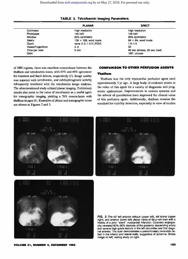
TABLE 3. Tetrofosmln Imaging Parameters
Collimator Photopeak Window
PLANAR
High resolution 140 keV 20% symmetric
SPECT
High resolution 140 keV 20% symmetric
Matrix Zoom Views/Projections Time per view Orbit
128 x 128, word mode none (1.2-1.5 if LFOV) 3-4
64 x 64, word mode 1.0-1.5 32
5 min
of 1080 regions, there was excellent concordance between the thallium and tetrofosmin scans, with 83% and 88% agreement for transient and fixed defects, respectively (5). Image quality was superior with tetrofosmin, and subdiaphragmatic activity infrequently interfered with the tetrofosmin image analysis. The aforementioned study utilized planar imaging. Preliminary results also point to the value of tetrofosmin as a useful agent for tomographic imaging, yielding a 76% concordance with thallium images (6). Examples of planar and tomographic scans are shown in Figures 2 and 3.
VOLUME 21, NUMBER 4, DECEMBER 1993
40 sec (stress), 25 sec (rest) 180°, circular
COMPARISON TO OTHER PERFUSION AGENTS
Thallium
Thallium was the only myocardial perfusion agent until approximately 3 yr ago. A large body of evidence points to the value of this agent for a variety of diagnostic and prognostic applications. Improvements in camera systems and the advent of quantitation have improved the clinical value of this perfusion agent. Additionally, thallium remains the standard for viability detection, especially in view of studies
FIG. 2. The 40° left anterior oblique (upper left), left lateral (upper right), and anterior (lower left) planar views of 56-yr-old man with a history of a prior "silent" myocardial infarction. Coronary angiography revealed 80o/o-90% stenosis of the posterior descending artery and several high-grade lesions in the left circumflex and first diagonal arteries. The scan demonstrates a predominately reversible defect in the inferior and lateral walls, suggestive of ischemia. Stress image on left; resting study on right.
193
by on May 27, 2018. For personal use only. tech.snmjournals.org Downloaded from
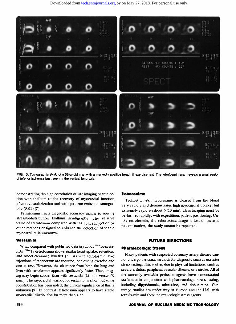
FIG. 3. Tomographic study of a 58-yr-old man with a markedly positive treadmill exercise test. The tetrofosmin scan reveals a small region of inferior ischemia best seen in the vertical long axis.
demonstrating the high correlation of late imaging or reinjection with thallium to the recovery of myocardial function after revascularization and with positron emission tomography (PET) (7).
Tetrofosmin has a diagnostic accuracy similar to routine stress/redistribution thallium scintigraphy. The relative value of tetrofosmin compared with thallium reinjection or other methods designed to enhance the detection of viable myocardium is unknown.
Sestamlbl
When compared with published data (8) about 99mTc-sestamibi, 99mTc-tetrofosmin shows similar heart uptake, retention, and blood clearance kinetics (1 ). As with tetrofosmin, two injections of technetium are required, one during exercise and one at rest. However, the clearance from both the lung and liver with tetrofosmin appears significantly faster. Thus, imaging may begin sooner than with sestamibi (15 min. versus 60 min.). The myocardial washout of sestamibi is slow, but some redistribution has been noted; the clinical significance of this is unknown (9). In contrast, tetrofosmin appears to have stable myocardial distribution for more than 4 hr.
194
Teboroxlme
Technetium-99m teboroxime is cleared from the blood very rapidly and demonstrates high myocardial uptake, but extremely rapid washout ( < 10 min). Thus imaging must be performed rapidly, with expeditious patient positioning. Unlike tetrofosmin, if a teboroxime image is lost or there is patient motion, the study cannot be repeated.
FUTURE DIRECTIONS
Pharmacologic Stress
Many patients with suspected coronary artery disease cannot undergo the usual methods for diagnosis, such as exercise stress testing. This is often due to physical limitations, such as severe arthritis, peripheral vascular disease, or a stroke. All of the currently available perfusion agents have demonstrated usefulness in conjunction with pharmacologic stress testing, including dipyridamole, adenosine, and dobutamine. Currently, studies are under way in Europe and the U.S. with tetrofosmin and these pharmacologic stress agents.
JOURNAL OF NUCLEAR MEDICINE TECHNOLOGY
by on May 27, 2018. For personal use only. tech.snmjournals.org Downloaded from
Myocardial VIability
The assessment of myocardial viability is currently one of the most active areas of investigation in nuclear cardiology. Thallium, with late (24-hr) imaging or following reinjection, has shown promise and provides much of the metabolic information obtained with PET. The most useful method for this goal is still not known. Studies to determine viability with sestamibi have demonstrated conflicting results. Thus, the usefulness of this agent for the accurate assessment of viability is not clear. No viability studies have been completed to date with tetrofosmin. A quantitative approach and comparison to a normal data file is likely the most useful method. However, we must await the results of trials for the detection of myocardial viability with tetrofosmin.
Acute Cardiac Syndromes
Studies with sestamibi have demonstrated the change in myocardial perfusion before and after thrombolytic therapy for an acute myocardial infarction and may be a promising tool for assessing the efficacy of such therapy (10). Additionally, patients presenting with chest pain may have the diagnosis of myocardial ischemia made when sestamibi is injected during the acute episode of pain and then imaged several hours later (11 ). Studies are being planned for the utilization of tetrofosmin due to its similarity to sestamibi.
CONCLUSION
Technetium-99m tetrofosmin is a promising new myocardial perfusion agent, with the inherent imaging superiority of a technetium agent. Studies have demonstrated the safety and efficacy of myocardial perfusion imaging with this agent for the detection of coronary artery disease. The lack of redistribution and rapid clearance from noncardiac tissues allow for flexibility in imaging, which may be started within 15 min of tracer injection. The image quality associated with tetrofosmin is excellent. Studies are currently under way to determine the impact of this agent on departmental throughput, as well as with applications using pharmacologic stress and in acute cardiac syndromes.
VOLUME 21, NUMBER 4, DECEMBER 1993
ACKNOWLEDGMENTS
The authors wish to thank Michael McMahon and the staff of the Radionuclide Core Laboratory at the Yale University School of Medicine for their preparation of the perfusion images.
REFERENCES
I. Kelly JD, Forster AM, Higley B, et al. Technetium-99m-tetrofosmin as a
new radiopharmaceutical for myocardial perfusion imaging. J Nucl Med 1993;34:222-227.
2. Higley B, Smith FW, Smith T, et al. Technetium-99m-1,2-bis[bis(2-ethox
yethyl)phosphino]ethane: human biodistribution, dosimetry and safety of
a new myocardial perfusion imaging agent. J Nucl Med 1993;34:30-38. 3. Sinusas AJ, Shi OX, Saltzberg MT, et al. Technetium-99m tetrofosmin for
assessment of myocardial perfusion: initial distribution and clearance, relationship to blood flow. (Abstract.) J Nucl Med 1992;33:993.
4. Jain D, Wackers FJTh, McMahon M, et al. Is there any redistribution
with Tc-99m-tetrofosmin imaging: a quantitative study using serial planar imaging. (Abstract.) Circulation 1992;86(Suppl 1):1-46.
5. The Tetrofosmin Study Group. Comparative myocardial perfusion imaging with Tc-99m tetrofosmin and thallium-201: results of phase-III inter
national trial. (Abstract.) Circulation 1992;86(Suppl 1):1-506. 6. Sasaki Y, Nishikawa J, Ohtake T, et al. Clinical evaluation of myocardial
SPECT using a new technetium-99m diphosphine agent (PPN.1011). (Ab
stract.) J Nuc/ Med 1992;33:875. 7. Dilsizian V, Rocco TP, Freedman NM, et al. Enhanced detection of
ischemic but viable myocardium by the reinjection of thallium after stress
redistribution imaging. N Eng/ J Med 1990;323:141-146.
8. Wackers FJTh, Berman DS, Maddahi J, et al. Technetium-99m hexakis 2-methoxyisobutyl isonitrile: human biodistribution, dosimetry, safety,
and preliminary comparison to thallium-201 for myocardial perfusion imaging. J Nucl Med 1989;30:301-311.
9. Taillefer R, Primeau M, Costi P, et al. Technetium-99m-sestamibi myocardial perfusion imaging in detection of coronary artery disease; com
parison between initial (1-hour) and delayed (3-hour) postexercise images.
J Nuc/ Med 1991;32:1961-1965. 10. Gibbons RJ, Verani MS, Behrenbeck T, et al. Feasibility of tomographic
99mTc-hexakis-2-methoxy-2-methylpropyl-isonitrile imaging for the assessment of myocardial area at risk and the effect of treatment in acute
myocardial infarction. Circulation 1989;80:1277-1286.
11. Bilodeau L, Theroux P, Gregoire J, et al. Technetium-99m sestamibi
tomography in patients with spontaneous chest pain: correlations with clinical, electrocardiographic and angiographic findings. JAm Col/ Cardio/1991;18:1684-1691.
195
by on May 27, 2018. For personal use only. tech.snmjournals.org Downloaded from
1993;21:191-195.J. Nucl. Med. Technol. Stephanie Jones and Robert C. Hendel Technetium-99m Tetrofosmin: A New Myocardial Perfusion Agent
http://tech.snmjournals.org/content/21/4/191This article and updated information are available at:
http://tech.snmjournals.org/site/subscriptions/online.xhtml
Information about subscriptions to JNMT can be found at:
http://tech.snmjournals.org/site/misc/permission.xhtmlInformation about reproducing figures, tables, or other portions of this article can be found online at:
(Print ISSN: 0091-4916, Online ISSN: 1535-5675)1850 Samuel Morse Drive, Reston, VA 20190.SNMMI | Society of Nuclear Medicine and Molecular Imaging
is published quarterly.Journal of Nuclear Medicine Technology
© Copyright 1993 SNMMI; all rights reserved.
by on May 27, 2018. For personal use only. tech.snmjournals.org Downloaded from