the value of diagnostic whole-body scanning and serum thyroglobulin in the presence of elevated...
TRANSCRIPT
ORIGINAL STUDIES, REVIEWS,AND SCHOLARLY DIALOG
THYROID CANCER AND NODULES
The Value of Diagnostic Whole-Body Scanningand Serum Thyroglobulin in the Presence of Elevated
Serum Thyrotropin During Follow-Up of Anti-ThyroglobulinAntibody–Positive Patients with Differentiated
Thyroid Carcinoma Who Appeared to Be Free of DiseaseAfter Total Thyroidectomy and Radioactive Iodine Ablation
Pedro Weslley Rosario,1,2 Augusto Flavio Campos Mineiro Filho,1 Rafela Xavier Lacerda,1
Davi Alves dos Santos,1 and Maria Regina Calsolari 2
Background: In the presence of anti-thyroglobulin antibodies (TgAb), serum thyroglobulin (Tg) might be un-derestimated. Therefore, the American Thyroid Association does not recommend serum Tg after thyroid hormonewithdrawal or recombinant human thyrotropin administration (stimulated Tg) and diagnostic whole-bodyscanning (DxWBS) in TgAb-positive patients who have serum Tg values while on thyroxine (Tg-on-T4) of< 1 ng/mL. The objective of this study was to determine, in patients with differentiated thyroid cancer (DTC)who appeared to be free of disease after surgery and ablative treatment, but who had positive serum TgAb, thevalue of performing DxWBS and obtaining serum Tg under stimulated Tg conditions.Methods: There were 121 women and 15 men in the study. By selection criteria, all of them had total thyroid-ectomy with apparent complete tumor resection, remnant ablation with 131I (1.1–5.5 GBq), and a post–131Itherapy WBS that were negative for ectopic 131I uptake. On assessment 8–12 months after 131I ablation, theirclinical exam needed to be normal, their Tg-on-T4 needed to be < 1 ng/mL, and the test for TgAb needed to bepositive. Stimulated Tg, neck ultrasound (US), and DxWBS were obtained from all patients. Patients withstimulated Tg > 1 ng/mL without disease on US and DxWBS were evaluated by other imaging methods.Results: In 10 (7.3%) patients, stimulated Tg was > 1 ng/mL. The DxWBS revealed metastases in two of thesepatients, and other imaging methods showed disease in three others. Stimulated Tg was < 1 ng/mL in 126patients. DxWBS revealed metastases in three of these patients, and US detected lymph node metastases in fourwith a negative DxWBS. Tg stimulation combined with DxWBS revealed evidence for disease in 13 (9.5%)patients. When excluding patients with a positive US, DxWBS revealed metastases in four patients, and stim-ulated Tg of > 1 ng/mL led to detection of persistent disease by other imaging methods in two more patients.Conclusions: Performing stimulated Tg and DxWBS at the same time seems to be useful after initial therapy inDTC patients with TgAb who do not otherwise appear to have persistent disease, even when US is negative.
Introduction
Serum thyroglobulin (Tg) is the most common follow-uptest in patients with well-differentiated thyroid cancer
(DTC). After total thyroidectomy followed by ablation ofremnant tissue with 131I, *95% of patients without anti-Tgantibodies (TgAb) have serum Tg < 1 ng/mL at a time whenthey are taking levo-thyroxine (L-T4) therapy (Tg-on-T4) (1,2).In these patients, repeat measurement of serum Tg whenserum thyroid stimulating hormone (TSH) concentrations aregreater than 30 mIU/L, achieved by either thyroid hormone
withdrawal or administration of recombinant human TSH(rhTSH), referred to here as ‘‘stimulated Tg,’’ is recom-mended. Stimulated Tg is sometimes combined with whole-body scanning (WBS) by using a low activity of 131I or 123I(diagnostic WBS [DxWBS]) (3–5). In about 20% of patientswhose serum Tg-on-T4 is < 1 ng/mL, stimulated Tg valuesincrease to > 1 ng/mL (1,6–8) and, if a DxWBS is performed,there is the occasional revelation of ectopic uptake due tometastases (8–10).
In the presence of TgAb, which occurs in up to 25% ofpatients with DTC (11), serum Tg concentrations might be
1Postgraduate Program and 2Endocrinology Service, Santa Casa de Belo Horizonte, Belo Horizonte, Minas Gerais, Brazil.
THYROIDVolume 22, Number 2, 2012ª Mary Ann Liebert, Inc.DOI: 10.1089/thy.2011.0020
113
underestimated when measured by immunometric assays(11,12), currently the most widely used Tg assays. Therefore,adopting the proposal of Mazzaferri (13), the AmericanThyroid Association (4) does not recommend stimulated Tg(or DxWBS) in patients who have Tg-on-T4 values of < 1 ng/mL if they have TgAb. Almost all follow-up studies of patientswith DTC exclude those with TgAb. In the few that do notmake this exclusion, the number of patients is small (8). Wesought, therefore, to determine in patients with DTC whoappeared to be free of disease after surgery and ablativetreatment with radioactive iodine, but who had positive testsfor serum TgAb, the value during follow-up of performingDxWBS and of obtaining stimulated Tg.
Patients and Methods
The study was approved by the Research Ethics Committeeof our institution. A total of 136 consecutive patients (121 wo-men and 15 men; age: 13–74 years, mean: 49 years) with DTC(papillary cancer in 125 and follicular cancer in 11) seen at ourinstitution (Santa Casa de Belo Horizonte), who met the fol-lowing criteria, were selected for the study. They included thosewho had undergone total thyroidectomy followed by remnantablation with 131I (1.1–5.5 GBq [30–150 mCi]), those in whomapparently complete tumor resection at surgery was achieved,and those in whom post 131I-therapy WBS (RxWBS) showed noectopic 131I uptake. To be eligible, patients also had to have anormal clinical exam, Tg-on-T4 of < 1 ng/mL and a positive testfor TgAb on assessment at 8–12 months (mean: 10 months) after131I ablation. The tumor-node-metastasis (TNM) stage (14) ofpatients in the study was T1N0, T1N1, T2N0, T2N1, T3N0, andT3N1 in 28, 15, 23, 20, 25, and 25 patients, respectively.
Stimulated Tg (measured after L-T4 withdrawal for 4weeks or administration of rhTSH), neck ultrasound (US), andDxWBS were obtained from all patients. Patients with stim-ulated Tg > 1 ng/mL without disease on US and DxWBS wereevaluated by other imaging methods (chest and mediastinalcomputed tomography [CT], WBS with 99mTc-MIBI, RxWBS,and fluorodeoxyglucose positron emission tomography[FDG-PET]).
Tg and TgAb measurement
Tg was measured by a radioimmunometric assay (ELSA-hTG; CIS Bio International) with a functional sensitivity of0.8 ng/mL. TgAb were determined by a chemiluminescentassay (Immulite; Diagnostic Products Corp.), with a referencevalue of up to 40 IU/mL.
Imaging methods
WBS was performed with a tracer (185 MBq [5 mCi]) ortherapeutic (1.1–5.5 GBq [30–150 mCi]) activity of 131I after T4withdrawal for 4 weeks or administration of rhTSH and alow-iodine diet during the 10 days preceding the exam.Anterior and posterior whole-body images were obtained 3(DxWBS) or 7 (RxWBS) days after iodine administration. Ul-trasonography was performed with a linear, multifrequency10-MHz transducer. All suspected lesions apparent on USscans (15,16) were initially evaluated by US-guided fine-needle aspiration biopsy. CT was performed on 5-mm thicksequential sections. 99mTc-MIBI scans were performed duringT4 therapy by using a tracer dose of 720–925 MBq, and whole-
body images were obtained during the early (20 minutes) andlate period (6 hours). FDG-PET was carried out after stimu-lation with rhTSH.
Results
TgAb titers ranged from 65 to 2845 IU/mL. Stimulated Tgwas > 1 ng/mL in 10/136 patients (7.3%). DxWBS revealedmetastases in two of these patients (pulmonary and medias-tinal). In eight patients with negative DxWBS, other imagingmethods showed lymph node metastases in one, pulmonarymetastases in one, and bone metastases in another patient.Five patients had no apparent disease, but their stimulated Tgwas > 1 ng/mL.
Stimulated Tg was < 1 ng/mL in 126 patients. DxWBS re-vealed metastases in three of these patients (pulmonary me-tastases in one and lymph node metastases in two). US detectedlymph node metastases in 4 of the 123 patients with a negativeDxWBS. The remaining 119 patients had no apparent DTC.
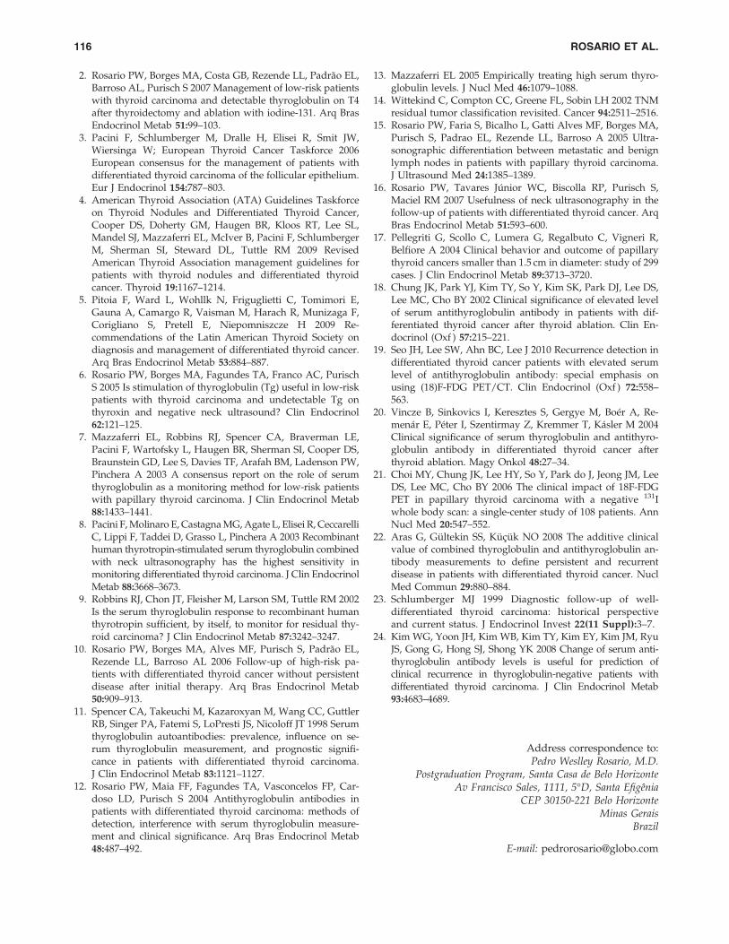
Table 1 presents information for TgAb titers, stimulated Tgvalues, and imaging studies in patients with stimulated Tg> 1 ng/mL and patients with stimulated Tg< 1 ng/mL whoseimaging studies were consistent with metastatic DTC. Five(41.6%) of the 12 patients with apparent metastases as judgedby imaging studies had stimulated Tg > 1 ng/mL. Regardingthe imaging methods, five patients had a positive DxWBS, USrevealed lymph node metastases in six (50%), and both examswere negative in two patients but DTC was detected by FDG-PET. Therefore, determination of stimulated Tg combinedwith DxWBS changed patient management in 13 of 136 patients(9.5%) and detected metastases in 8 (6%). Even when excludingpatients with a positive neck US (n = 6), DxWBS revealed me-tastases in four patients, and stimulated Tg values of > 1 ng/mL led to the detection of disease by other imaging methods intwo additional cases and to a different follow-up than wouldhave otherwise been performed in five other cases.
Discussion
Up to 25% of patients with DTC have positive tests for TgAb(11). Unfortunately, the presence of these antibodies in thecirculation may underestimate serum Tg levels as measured byimmunometric assays (11,12). Two management approachesare recommended in the literature for patients with positivetests for TgAb who do not have apparent disease based on theimpression of complete tumor resection at initial thyroid sur-gery, an RxWBS without metastases, a normal clinical exam,and Tg-on-T4 values of < 1 ng/mL when their assessment iscomplete after initial surgery and radioactive iodine treatment.The recent North American guidelines and an invited pro-spective (4,13) does not recommend stimulated Tg andDxWBS, whereas some investigators propose the measure-ment of stimulated Tg and DxWBS (3,5,8,17,18). Knowledgeabout the proportion of patients with DTC who have no ap-parent residual disease after initial treatment (complete tumorresection at initial thyroid surgery, an RxWBS without metas-tases, a normal clinical exam, and Tg-on-T4 values of < 1 ng/mL), who have stimulated Tg values of > 1 ng/mL, and inwhom metastases are revealed on imaging studies is funda-mental to define the value of obtaining stimulated Tg.
In the current study, 5/12 (41.6%) patients with apparentdisease based on imaging studies had stimulated Tg > 1 ng/mL, demonstrating that even in the presence of TgAb, some
114 ROSARIO ET AL.
patients with metastases have detectable Tg. Notably, TgAbdo not always sufficiently interfere with serum Tg so that thelatter becomes undetectable (12,19,20), especially in cases inwhich this protein is abundantly secreted such as patientswith distant metastases (12,20). In the current study, a rele-vant proportion of patients with Tg-on-T4 < 1 ng/mL (7.3%)converted to levels > 1 ng/mL after TSH stimulation despitethe presence of circulating TgAb. Concordantly, stimulatedTg levels > 1 ng/mL were observed in 7/48 (14.6%) patientswith Tg-on-T4 < 1 ng/mL in previous studies (8,17). There-fore, measurement of stimulated Tg seem to be useful duringthe first year after initial therapy in patients with DTC whohave no apparent residual disease after initial treatment andTg-on-T4 < 1 ng/mL even in the presence of TgAb.
The positive predictive value (PPV) of detectable levels ofTg was 50% in the current study, and this value could be evenunderestimated considering that recurrences may be identi-fied during long-term follow-up. Other studies also demon-strated the elevated PPV of Tg in patients with circulatingTgAb (17,19–21). In patients with stimulated Tg > 1 ng/mLafter TSH stimulation in the presence of circulating TgAbwithout disease on US and DxWBS, other imaging methods(CT and FDG-PET) are indicated. When metastases are notreadily detected, it is recommended that these patients bemaintained under TSH suppression, Tg and neck US moni-toring be performed more frequently, and a new Tg stimu-lation and DxWBS be performed after 1–2 years because of thehigher risk of subsequent detection of metastases (3).
Employing the same preparation as that used for themeasurement of stimulated Tg, DxWBS revealed ectopic up-take (metastases) in 5/136 (3.7%) patients, with neck USshowing no anomalies in 4. Other studies also demonstratedthe value of DxWBS in patients with TgAb (8,18,20,22). Onelimitation of DxWBS, the so-called stunning effect, can beprevented with the use of 123I or of a tracer activity that doesnot exceed 2 mCi 131I (4,5).
Taken together, the results showed that measurement ofstimulated Tg combined with DxWBS was clearly useful in
13/136 (9.6%) patients with Tg-on-T4 < 1 ng/mL and positiveTgAb on assessment after initial therapy (positive DxWBSand/or stimulated Tg > 1 ng/mL). Even when excludingcases with positive US, these procedures changed patientmanagement in 11/130 (8.5%) cases. If Tg stimulation andDxWBS were not performed as proposed by some investiga-tors (4,13), the diagnosis would not have been establishedinitially in 6/12 patients with metastases, including 4 withdistant metastases. In these cases, the diagnosis would only beestablished if TgAb titers doubled or Tg-on-T4 would even-tually become detectable (4,13), or disease would becomeclinically apparent, indicating progression, with the risk of apoor prognosis and lower chance of cure (23). One limitation,that is, the need for L-T4 withdrawal (associated with iatro-genic hypothyroidism), can be resolved by preparation withrhTSH. If Tg continues to be < 1 ng/mL after TSH stimulationand US and DxWBS are negative, then subsequent follow-upcan be done by periodic evaluation of Tg-on-T4, TgAb, andneck US (4,13). The last method is able to detect most cases oflate recurrence (24). If US does not reveal anomalies but TgAbtiters progressively increase, other imaging methods, espe-cially CT and FDG-PET (18,19), are indicated.
In conclusion, measurement of stimulated Tg and DxWBS(using the same preparation) seems to be useful during thefirst year after initial therapy in patients with TgAb withoutapparent disease and Tg-on-T4 < 1 ng/mL, even when neckUS is negative.
Disclosure Statement
The authors declare that no competing financial interestsexist.
References
1. Cailleux AF, Baudin E, Travagli JP, Ricard M, SchlumbergerM 2000 Is diagnostic iodine-131 scanning useful after totalthyroid ablation for differentiated thyroid cancer? J ClinEndocrinol Metab 85:175–178.
Table 1. Results of the Patients with Stimulated Thyroglobulin > 1 ng/ml or Apparent Disease
PatientTgAb
(IU/mL)StimulatedTg (ng/mL) DxWBS Neck US
Other positiveimaging methods
1 65 2.1 Negative Negative2 102 2.2 Negative Negative3 98 2.8 Negative Negative4 225 3 Negative Negative5 72 4.2 Negative Negative6 102 3.4 Negative Positive (LN)7 821 8.5 Positive (mediastinal LN) Negative8 78 34 Positive (lung) Negative9 501 72 Negative Negative Chest CT (lung)
10 81 104.1 Negative Negative PET/CT (bone)11 2854 < 1 Positive (lung) Negative12 112 < 1 Positive (LN) Positive (LN)13 87 < 1 Positive (LN) Negative14 100 < 1 Negative Positive (LN)15 77 < 1 Negative Positive (LN)16 211 < 1 Negative Positive (LN)17 987 < 1 Negative Positive (LN)
US, ultra-sonography; CT, computed tomography; DxWBS, diagnostic whole body scanning; PET, positron emission tomography; LN,lymph nodes; Tg, thyroglobulin; TgAb, anti-Tg antibodies.
DIAGNOSTIC WBS AND STIMULATED TG IN PATIENTS WITH TGAB 115
2. Rosario PW, Borges MA, Costa GB, Rezende LL, Padrao EL,Barroso AL, Purisch S 2007 Management of low-risk patientswith thyroid carcinoma and detectable thyroglobulin on T4after thyroidectomy and ablation with iodine-131. Arq BrasEndocrinol Metab 51:99–103.
3. Pacini F, Schlumberger M, Dralle H, Elisei R, Smit JW,Wiersinga W; European Thyroid Cancer Taskforce 2006European consensus for the management of patients withdifferentiated thyroid carcinoma of the follicular epithelium.Eur J Endocrinol 154:787–803.
4. American Thyroid Association (ATA) Guidelines Taskforceon Thyroid Nodules and Differentiated Thyroid Cancer,Cooper DS, Doherty GM, Haugen BR, Kloos RT, Lee SL,Mandel SJ, Mazzaferri EL, McIver B, Pacini F, SchlumbergerM, Sherman SI, Steward DL, Tuttle RM 2009 RevisedAmerican Thyroid Association management guidelines forpatients with thyroid nodules and differentiated thyroidcancer. Thyroid 19:1167–1214.
5. Pitoia F, Ward L, Wohllk N, Friguglietti C, Tomimori E,Gauna A, Camargo R, Vaisman M, Harach R, Munizaga F,Corigliano S, Pretell E, Niepomniszcze H 2009 Re-commendations of the Latin American Thyroid Society ondiagnosis and management of differentiated thyroid cancer.Arq Bras Endocrinol Metab 53:884–887.
6. Rosario PW, Borges MA, Fagundes TA, Franco AC, PurischS 2005 Is stimulation of thyroglobulin (Tg) useful in low-riskpatients with thyroid carcinoma and undetectable Tg onthyroxin and negative neck ultrasound? Clin Endocrinol62:121–125.
7. Mazzaferri EL, Robbins RJ, Spencer CA, Braverman LE,Pacini F, Wartofsky L, Haugen BR, Sherman SI, Cooper DS,Braunstein GD, Lee S, Davies TF, Arafah BM, Ladenson PW,Pinchera A 2003 A consensus report on the role of serumthyroglobulin as a monitoring method for low-risk patientswith papillary thyroid carcinoma. J Clin Endocrinol Metab88:1433–1441.
8. Pacini F, Molinaro E, Castagna MG, Agate L, Elisei R, CeccarelliC, Lippi F, Taddei D, Grasso L, Pinchera A 2003 Recombinanthuman thyrotropin-stimulated serum thyroglobulin combinedwith neck ultrasonography has the highest sensitivity inmonitoring differentiated thyroid carcinoma. J Clin EndocrinolMetab 88:3668–3673.
9. Robbins RJ, Chon JT, Fleisher M, Larson SM, Tuttle RM 2002Is the serum thyroglobulin response to recombinant humanthyrotropin sufficient, by itself, to monitor for residual thy-roid carcinoma? J Clin Endocrinol Metab 87:3242–3247.
10. Rosario PW, Borges MA, Alves MF, Purisch S, Padrao EL,Rezende LL, Barroso AL 2006 Follow-up of high-risk pa-tients with differentiated thyroid cancer without persistentdisease after initial therapy. Arq Bras Endocrinol Metab50:909–913.
11. Spencer CA, Takeuchi M, Kazaroxyan M, Wang CC, GuttlerRB, Singer PA, Fatemi S, LoPresti JS, Nicoloff JT 1998 Serumthyroglobulin autoantibodies: prevalence, influence on se-rum thyroglobulin measurement, and prognostic signifi-cance in patients with differentiated thyroid carcinoma.J Clin Endocrinol Metab 83:1121–1127.
12. Rosario PW, Maia FF, Fagundes TA, Vasconcelos FP, Car-doso LD, Purisch S 2004 Antithyroglobulin antibodies inpatients with differentiated thyroid carcinoma: methods ofdetection, interference with serum thyroglobulin measure-ment and clinical significance. Arq Bras Endocrinol Metab48:487–492.
13. Mazzaferri EL 2005 Empirically treating high serum thyro-globulin levels. J Nucl Med 46:1079–1088.
14. Wittekind C, Compton CC, Greene FL, Sobin LH 2002 TNMresidual tumor classification revisited. Cancer 94:2511–2516.
15. Rosario PW, Faria S, Bicalho L, Gatti Alves MF, Borges MA,Purisch S, Padrao EL, Rezende LL, Barroso A 2005 Ultra-sonographic differentiation between metastatic and benignlymph nodes in patients with papillary thyroid carcinoma.J Ultrasound Med 24:1385–1389.
16. Rosario PW, Tavares Junior WC, Biscolla RP, Purisch S,Maciel RM 2007 Usefulness of neck ultrasonography in thefollow-up of patients with differentiated thyroid cancer. ArqBras Endocrinol Metab 51:593–600.
17. Pellegriti G, Scollo C, Lumera G, Regalbuto C, Vigneri R,Belfiore A 2004 Clinical behavior and outcome of papillarythyroid cancers smaller than 1.5 cm in diameter: study of 299cases. J Clin Endocrinol Metab 89:3713–3720.
18. Chung JK, Park YJ, Kim TY, So Y, Kim SK, Park DJ, Lee DS,Lee MC, Cho BY 2002 Clinical significance of elevated levelof serum antithyroglobulin antibody in patients with dif-ferentiated thyroid cancer after thyroid ablation. Clin En-docrinol (Oxf ) 57:215–221.
19. Seo JH, Lee SW, Ahn BC, Lee J 2010 Recurrence detection indifferentiated thyroid cancer patients with elevated serumlevel of antithyroglobulin antibody: special emphasis onusing (18)F-FDG PET/CT. Clin Endocrinol (Oxf ) 72:558–563.
20. Vincze B, Sinkovics I, Keresztes S, Gergye M, Boer A, Re-menar E, Peter I, Szentirmay Z, Kremmer T, Kasler M 2004Clinical significance of serum thyroglobulin and antithyro-globulin antibody in differentiated thyroid cancer afterthyroid ablation. Magy Onkol 48:27–34.
21. Choi MY, Chung JK, Lee HY, So Y, Park do J, Jeong JM, LeeDS, Lee MC, Cho BY 2006 The clinical impact of 18F-FDGPET in papillary thyroid carcinoma with a negative 131Iwhole body scan: a single-center study of 108 patients. AnnNucl Med 20:547–552.
22. Aras G, Gultekin SS, Kucuk NO 2008 The additive clinicalvalue of combined thyroglobulin and antithyroglobulin an-tibody measurements to define persistent and recurrentdisease in patients with differentiated thyroid cancer. NuclMed Commun 29:880–884.
23. Schlumberger MJ 1999 Diagnostic follow-up of well-differentiated thyroid carcinoma: historical perspectiveand current status. J Endocrinol Invest 22(11 Suppl):3–7.
24. Kim WG, Yoon JH, Kim WB, Kim TY, Kim EY, Kim JM, RyuJS, Gong G, Hong SJ, Shong YK 2008 Change of serum anti-thyroglobulin antibody levels is useful for prediction ofclinical recurrence in thyroglobulin-negative patients withdifferentiated thyroid carcinoma. J Clin Endocrinol Metab93:4683–4689.
Address correspondence to:Pedro Weslley Rosario, M.D.
Postgraduation Program, Santa Casa de Belo HorizonteAv Francisco Sales, 1111, 5�D, Santa Efigenia
CEP 30150-221 Belo HorizonteMinas Gerais
Brazil
E-mail: [email protected]
116 ROSARIO ET AL.