bihormonal regulation of the thyrotropin-releasing hormone
TRANSCRIPT

Bihormonal Regulation of the Thyrotropin-Releasing
Hormone Receptor in Mouse
Pituitary Thyrotropic Tumor Cells in Culture
MARVIN C. GERSHENGORN, Endocrine Division, Department of Medicine, New YorkUniversity Medical Center, New York 10016
A B S T R A C T Receptors for thyrotropin-releasing hor-mone (TRH) are present on mouse pituitary thyro-tropic tumor cells. Incubation of thyrotropes with 100nM TRH or 4 nM L-triiodothyronine (T3) for 48 h de-creased the number ofTRH receptors to -50 and 20%of control, respectively. There was no effect on theequilibrium dissociation constant which was 3-5 nM.The depletion in the number ofavailable TRH receptorswas time- and dose-dependent. TRH, 100 nM, de-creased the receptor number to 70% after 24 h, 50%after 48 h, and 45% ofcontrol after 72 h. T3, 4 nM, decreasedthe receptor number to 52% after 24 h, 20% after 48 h,and 17% of control after 72 h. After 48 h, half-maximaldepletion occurred with 1-2 nM TRH and -0.15 nMT3. Incubation with 100 nM TRH and 4 nM T3 causeda significantly greater reduction in the receptor levelthan either hormone alone. The decrease in the re-
ceptor level was reversible within 72 h after removalof TRH, 100 nM, but was only partially reversed, from20 to 40% of control, after removal of T3, 4 nM, after120 h. By regulating the number of available TRH re-
ceptors on the thyrotrope, TRH and T3 interact to con-
trol thyrotropin release.
INTRODUCTION
A number of studies have demonstrated that thyro-tropin-releasing hormone (TRH)1 stimulates synthesisand release of thyrotropin (TSH) and prolactin in vitro(1-8), and release of TSH and prolactin in intact ani-mals (9-11). TRH has been shown to bind to receptorson the plasma membrane of cells derived from whole
This work was presented in part at the National Meetingof the American Federation for Clinical Research, San Fran-cisco, Calif., May 1978.Receivedfor publication 10 April 1978 and in revisedform
13 July 1978.' Abbreviations used in this paper: T3, L-triiodothyronine;
TRH, thyrotropin-releasing hormone; TSH, thyrotropin.
J. Clin. Invest. ©D The American Society for Clinical InvestigatVolume 62 November 1978 937-943
anterior pituitary (12), to mouse pituitary thyrotropictumor cells that produce TSH but not prolactin (13-15),and to growth hormone (GH) cells, clonal strains ofrat pituitary tumor cells that produce prolactin andgrowth hormone but not TSH (16, 17). Recent studiesindicate that the number of plasma membrane receptorsfor a variety of hormones (18-20), including TRH (21-23), can be modulated, and it has been suggested thatthis may serve as an important site of regulation of hor-mone action.
In this report we describe modulation of the level ofTRH receptors but not their affinity for TRH in sus-pension cultures of mouse pituitary thyrotropic tumorcells which we have shown to be comprised only ofthyrotropes (24). These experiments demonstrate time-and dose-dependent regulation of the thyrotrope re-ceptor for TRH by TRH and L-triiodothyronine (T3).The TRH receptor may be an important site for regula-tion of TSH release.
METHODS
Dulbecco's modified Eagle's medium was obtained fromGrand Island Biological Co., Grand Island, N. Y. Hypothyroidcalfserum was purchased from Rockland Farms, Gilbertsville,Pa. The calf serum was shown to contain <5 ng T3/dl and<0.1 ,ug L-thyroxine/dl. [3H]TRH (L-[2,3-3H]proline, 40 Ci/mmol or L-[2,3,4,5-3H]proline, 115 Ci/mmol) was from NewEngland Nuclear, Boston, Mass. and unlabeled TRH was fromBeckman, Instruments, Inc., Fullerton, Calif. [125I]T3 (1,090,uCi/,ug) was from New England Nuclear and unlabeled T3was from Calbiochem, San Diego, Calif.
Short-term suspension cultures of thyrotropes were es-tablished with cells derived from mouse pituitary thyrotropictumors by selective attachment techniques from primary cul-tures comprised of several cell types (25). TSH-producingcells were grown in medium supplemented with 10% hypo-thyroid calf serum at 37°C in a humidified atmosphere at 5%C02-95% air. These cells have been shown to produce TSHat a constant rate for up to 9 days in culture (24).Binding of TRH to thyrotropes was performed in fresh me-
dium without serum containing [3H]TRH at 37°C in a shakingwater bath (-100 oscillations/min). Experiments were carried
'ion, Inc., 0021-9738/78/1101-937 $1.00 937

out in 0.5 ml of medium in 12 x 75 polypropylene test tubeswith 0.1 to 0.5 x 10⁶ cells that were all derived from a singletumor. After incubation the cells were centrifuged at 650 gfor 5 min at 4°C, the medium was removed and the cellswere washed three times with 1 ml of 0.1 M NaCl, 0.05 MNa phosphate, pH 7.5. The cell pellets were dissolved in0.4 N NaOH; aliquots were used to measure radioactivityby liquid scintillation counting and for measurement of cellprotein by the method of Lowry et al. (26) using bovine serumalbumin as standard. Nonspecific binding, that is, [3H]TRHbound in the presence of a 200-fold molar excess of un-labeled TRH, was subtracted from each value. Nonspecificbinding represented <8% of the total bound radioactivity.
Binding of [3H]TRH to thyrotropes was determined afterculture for 48 h in the presence of 4 nM T3 or 100 nM TRHand compared to controls. For these experiments, 1-2 x 10⁶cells were incubated in 3 ml ofmedium containing 10% hypo-thyroid calf serum in 25 cm2 flasks (Falcon Plastics, Div. ofBioQuest, Oxnard, Calif.). Cells were usually used between6 and 9 days in culture, a time during which there was littlechange in cell number. After 48 h, the cells were collected,centrifuged at 650 g for 5 min, and washed three times with2 ml of medium at 37°C. After the second wash, the cellswere kept suspended in medium at 37°C in a shaking waterbath for 15 min to increase dissociation of TRH from thosecells exposed to TRH. Binding of [3H]TRH to equal aliquotsof cells from each flask was performed as above.The number of available TRH receptors and "basal" and
TRH-stimulated TSH production were measured in cells ex-posed to varying concentrations of T3 or TRH for 48 h. Forthese experiments, cells were incubated in medium supple-mented with 10% hypothyroid calf serum. After 48 h, the cellswere collected, washed as above, and divided into two ali-quots. One aliquot was incubated in serum-free medium con-taining 20-40 nM [3H]TRH for 90 min to estimate the num-ber of available TRH receptors. The other ali(luot was cul-tured again, in duplicate tubes, in serum-containing medium,in the presence of 10 nM TRH, or in its absence for 24 h.TSH in the medium was measured by a heterologous, double-antibody radioimmunoassay using a pool ofmedium from cul-tures of mouse thyrotropic tumor cells containing 0.54 ,ieq ofrat TSH/ml as standard (10, 25).Nuclear binding of 125I-T3 was measured after incubation
in medium containing 10% hypothyroid calf serum at 370Cfor 2.5 h as described (25).
Statistical analysis was performed by unpaired t test.
RESULTS
Binding ofTRH to thyrotropic tumor cells. [3H]TRHadded to the medium of thyrotropic tumor cell culturesbound rapidly to the cells. The specific binding was47% of maximum after 10 min, 59% after 15 min, 89%after 30 min, maximum after 60 min, and remainedconstant for longer than 3 h. Therefore, in all otherbinding studies incubation with [3H]TRH was for 90or 120 min. To demonstrate reversibility of binding,aliquots of cells from a single culture were incubatedwith 25 nM [3H]TRH for 90 min, the cells were washed,and the amount of specifically bound radioactivity wasmeasured in triplicate (Fig. 1, zero time). The remain-ing cell aliquots were resuspended in medium con-taining the same concentration of [3H]TRH, unlabeledTRH, or no TRH and the amount of [3H]TRH bound
0C)
0.-
-0cm
ICK
Ir
0 30 60 90 120Minutes
FIGURE 1 Reversibility of [3H]TRH binding. Identical sam-ples of cells from a single culture were incubated with25 nM [3H]TRH. After 90 min (zero time) the cells were washed,resuspended in fresh medium with 25 nM [3H]TRH (controlcells), with no added TRH (0), or with 10 nM unlabeled TRH(C) and the amount of [3H]TRH bound measured at the timesindicated. Control cells bound 0.60 pmol [3H]TRH per milli-gram protein. The points represent the mean of duplicate val-ues whose average variation was ± 13%.
to the cells was measured at 30-min intervals for 2 h.The amount of bound [3H]TRH remained constant inthe cells re-exposed to [3H]TRH. In contrast, as shownin Fig. 1, the radioactivity rapidly dissociated fromcells exposed to no TRH or unlabeled TRH. The rateconstant of dissociation of [3H]TRH from cells wasgreater in the presence of unlabeled TRH, 2.3 x 10⁻⁴/s,than in its absence, 1.3 x 10⁻⁴/s. The 3H radioactivitywhich dissociated from the cells was able to bind toother cells to the same extent as authentic [3H]TRH sug-gesting that it was unaltered [3H]TRH.The characteristics of binding ofTRH to its receptors
at equilibrium were defined by incubating cells withincreasing concentrations of [3H]TRH, up to 100 nM,for 90 min. Fig. 2 shows the analysis of [3H]TRH bind-ing to thyrotropes by the method of Scatchard (27).A linear plot of the data was found consistent with asingle class of noninteracting binding sites. The dis-sociation constant was 3-5 nM. There were 1.1 pmol[3H]TRH bound/mg cell protein at saturation, cor-responding to 99,000 binding sites/cell.Binding of TRH to TRH and T3-treated cells. Be-
fore study of the binding of [3H]TRH to cells exposedto TRH or T3, it was necessary to show that these hor-mones did not interfere with the subse(luent bindingreaction under the experimental conditions employed.Binding of [3H]TRH, 25 nM, to control cells was per-formed in triplicate in the presence of 10 nM T3 and com-pared to that in medium alone (control). The amount of[3H]TRH bound in the presence of T3 was 94±12%of control (mean+SD). In a parallel set of experiments,the effect of preincubation with TRH for 90 min wasdetermined. Equal aliquots of cells, in triplicate, wereincubated with 100 nM unlabeled TRH, 25 nM [3H]TRH,or medium alone (control). The cells were washed three
938 M. C. Gershengorin

5
4
0;O 3
-3c~ 2
4* Control
_ TRH (1OOnM)
-0 Ts (4nM)
0
0.5 1.0 1.5 2.0B (pM)
2.5
FIGURE 2 [3H]TRH binding to cells incubated with TRH orT3 for 48 h. Equal portions of cells were incubated with me-dium alone, 100 nM TRH, or 4 nM T3. After 48 h the cellswere washed and samples containing an average of 27 ,ugof protein were incubated with [3H]TRH, 1 to 100 nM, for90 min after which bound [3H]TRH was measured. Therewas no difference in the amount of cell protein in the threecultures. The amount of [3H]TRH bound at saturation was1.1 pmol/mg protein in control cells, 0.55 pmol/mg proteinin TRH-treated cells, and 0.22 pmol/mg protein in T3-treated cells. The points represent the mean of duplicate de-terminations whose average variation was ±12%.
times as described in Methods. After washing, theamount of [3H]TRH bound was decreased to 63+5%of the initial level. Binding of [3H]TRH, 25 nM, for90 min was then performed with the washed cells. Thecells previously exposed to unlabeled TRH bound93+±3% of the amount of [3H]TRH bound by controls.Therefore, it was possible to accurately measure [3H]TRHbinding to cells exposed to T3 or TRH.Because TRH may be degraded during incubation,
its stability was determined by incubating 20 nM- [3H]-TRH at 37°C in medium containing 10% hypothyroid calfserum. After 48 h, the incubated [3H]TRH was ableto bind specifically to thyrotropes to the same extentas fresh [3H]TRH (104±3% ofcontrol). Therefore, therewas virtually no degradation of TRH in the mediumunder the conditions of incubation.
Cells from a single suspension culture were incubatedwith 100 nM TRH or 4 nM T3 for 48 h; control cultureswere incubated with no added hormone. After 48 h,the cells were washed and binding of [3H]TRH wasstudied. There was no difference in binding affinityof the TRH receptor after incubation with TRH or T3as compared to controls (Fig. 2). In contrast, there wasa marked decrease in the number of available receptorsto 50 and 22% of control after a 48-h exposure to TRHand T3, respectively. When cells were incubated with100 nM TRH plus 4 nM T3, the number of availablereceptors decreased further to 12% of control (P < 0.01).The time-course of depletion of available TRH re-
ceptors was studied in cells from a single tumor ex-posed to 100 mM TRH or 4 nM T3. The number ofTRH receptors was estimated after 24, 48, and 72 hby incubating the cells with 25 nM [3H]TRH, a con-
centration sufficient to saturate 83-89% of the recep-tors. The number ofreceptors available to bind [3H]TRHwas 70% of control after 24 h (P <0.05), decreasedfurther to 50% after 48 h (P < 0.01), and to 45% after72 h (P < 0.01 vs. control; P > 0.1 vs. 48 h) exposureto 100 nM TRH. The depletion ofTRH receptors afterincubation with 4 nM T3 was more profound. After24 h there were 52% (P < 0.001) of the control numberof receptors, after 48 h the level decreased further to20% (P < 0.001) and after 72 h was 17% of control (P< 0.001 vs. control; P > 0.1 vs. 48 h).As shown in Fig. 3, depletion of the number of avail-
able TRH receptors was dose dependent for TRH andT3. After 48 h, there were 79% of the control level ofreceptors present in cells exposed to 1 nM TRH (P< 0.05),65% in cells exposed to 10 nM TRH and 55% incells exposed to 100 nM TRH or greater; half-maximaldepletion occurred at -1-2 nM TRH. T3, 0.1 nM, low-ered the number ofreceptors to 62% ofcontrol (P < 0.05)and maximal inhibition was achieved with T3 concen-trations >4 nM; half-maximal inhibition occurred at=0.15 nM T3.To determine the reversibility of the loss of TRH
receptors, cells from a single tumor were incubatedwith 100 nM TRH and 4 nM T3 for 48 h which reducedthe TRH receptor number to 52 and 21%, respectively.After 48 h, the cells were washed three times, resus-pended in fresh TRH- and T3-free medium and in-cubated for up to 120 h. During this experiment cellprotein increased <20%. After removal of TRH, pro-gressive repletion of the TRH receptor level occurred.After 24 h the level had returned to 65% of control(P < 0.05), after 48 h to 74% of control, and by 72 h
lo y
.0 75-JRH
W> 50-j
_L T,2) 5025
0 0.1 0.2 0.6 4 10T3 (nM)
FIGURE 3 Dose-response effect ofTRH or T3 on the numberofTRH receptors. Equal aliquots of cells were incubated withmedium alone (control cells), with TRH, 1 to 100 nM, or withT3 0.1-10 nM. After 48 h the cells were washed, incubatedwith 40 nM [3H]TRH and bound [3H]TRH measured after120 min. There was -65 ,ug of cell protein in each culture.The points represent the mean of duplicate determinationswhose average variation was ±+6%. Control cells bound 0.91pmol [3H]TRH /mg protein. Half-maximal receptor depletionoccurred at -1-2 nM TRH and 0.15 nM T3.
Regulation of Thyrotrope Thyrotropin-Releasing Hormone Receptor
'*,% o--
% 0-41%
939
to control levels. In contrast, there was a much moredelayed repletion of TRH receptor number after re-moval of T3. There was no change in the number ofreceptors after 24 h (P > 0.1), an increase to 30% ofcontrol after 48 h (P < 0.05), followed by a gradual riseto only 40% of control after 120 h.To determine whether the differences in the rates
of repletion of the TRH receptor after removal ofTRHand T3 from the medium was a result, in part, of dif-ferences in the amount of residual hormone remain-ing bound to the respective receptor, the time-courseof disappearance of [3H]TRH from the cell was com-pared to the disappearance of 125I-T3 from the nucleus.Cells from a single tumor were incubated in mediumsupplemented with 10% hypothyroid calf serum and 5nM or 100 nM [3H]TRH or 0.2 nM or 4 nM 125I-T3for 2.5 h at 37°C. After 2.5 h, the cells were washedthree times with medium, resuspended in TRH- andT3-free medium and incubated again at 37°C. As shown
Nl
0
0
0
z
mS
:z
A
7RH (IOOnM)
101-
6
0 30 60 90 120 150Minutes
15 210 1 2 5Hours
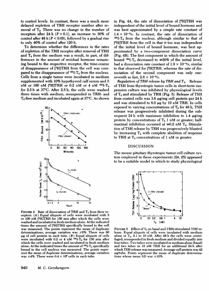
FIGURE 4 Rate of dissociation ofTRH and T, from their re-ceptors. (A) Equal aliquots of cells were incubated with 5or 100 nM [3H]TRH for 150 min after which the cells werewashed and incubated in fresh medium alone. At the indicatedtimes the amount of [3H]TRH specifically bound to the cellwas measured. The points represent the mean of duplicatedeterminations; average variation was +6%. There was 60,ug of cell protein in each tube. (B) Equal aliquots of cellswere incubated with 0.2 or 4 nM 125I-T3 for 150 min afterwhich the cells were washed and incubated in fresh mediumalone. At the indicated times the amount of 125I-T3 specificallybound to the cell nucleus was measured. The points repre-sent the mean of duplicate determinations; average variationwas +4%. There were 0.4 x 10⁶ cells in each tube.
in Fig. 4A, the rate of dissociation of [3H]TRH wasindependent of the initial level of bound hormone andwas best approximated by a simple rate constant of1.4 x 10-4/s. In contrast, the rate of dissociation of125I-T3 from the nucleus, although similar to that of[3H]TRH from the cell in that it too was independentof the initial level of bound hormone, was best ap-proximated by a two-component dissociation curve(Fig. 4B). The first component in which the amount ofbound I251-T3 decreased to -50% of the initial level,had a dissociation rate constant of 1.9 x 10⁻⁴/s, similarto that observed for [3H]TRH, whereas the rate of dis-sociation of the second component was only one-seventh as fast, 2.6 x 10⁻⁵/s.Regulation ofTRH release by TRH and T3. Release
ofTSH from thyrotropic tumor cells in short-term sus-pension culture was inhibited by physiological levelsof T3 and stimulated by TRH (Fig. 5). Release ofTSHfrom control cells was 3.6 ,.g/mg cell protein per 24 hand was stimulated to 6.0 ,ug by 10 nM TRH. In cellsexposed to varying concentrations of T3 for 48 h, TSHrelease was progressively inhibited during the sub-sequent 24 h with maximum inhibition to 1.4 ,ug/mgprotein by concentrations of T3 1 nM or greater; half-maximal inhibition occurred at =0.2- nM T3. Stimula-tion ofTSH release by TRH was progressively bluntedby increasing T3 with complete abolition of responseto TRH at T3 concentrations of 1 nM or greater.
DISCUSSION
The mouse pituitary thyrotropic tumor cell culture sys-tem employed in these experiments (24, 25) appearedto be a suitable model in which to study physiological
te5 \
+ TRH WM
".4me
o o.1 0.2 0.6 1 4 10T3 (nM)
FIGURE 5 Effect ofT3 on basal and TRH-stimulated TSH re-lease. Equal aliquots of cells were incubated with mediumalone or T3, 0.1 to 10 nM. After 48 h the cells were centri-fuged, resuspended in fresh medium and divided equally intofour tubes. Two tubes were incubated in medium alone (basal)and two tubes in 10 nM TRH for an additional 24 h afterwhich TSH release was measured. Average cell protein was 42,ug/tube. Points represent the mean of duplicate determina-tions whose mean SD was +10%.
940 M. C. Gershengorn

regulation of thyrotrope function. In these cells, re-lease ofTSH was inhibited in a dose-dependent fashionby T3; half-maximal inhibition occurred at a total me-dium T3 concentration of _0.2 nM which correspondsto a concentration of unbound or free T3 within thephysiological range (25). Moreover, TRH stimulated re-lease of TSH and this stimulation could be progres-sively blunted by increasing concentrations ofT3 until,at T3 levels of 1 nM or greater, the effect of TRH wascompletely abolished. This interaction between TRHand T3 is quantitatively very similar to that observedin humans (28, 29).
Putative TRH receptors on mouse thyrotropic tumorcells were originally described by Grant et al. (13, 15)and Eddy et al. (14). The characteristics of the inter-action of TRH with its receptors described by Grantand his colleagues differ from those reported here. Theyconcluded, from Scatchard analysis of their data, thatthere were two receptors for TRH with apparent equi-librium dissociation constants of 20 and _500 nM (15).Subtle differences in the kinetics of [3H]TRH displace-ment by a series of TRH analogues were interpretedas further evidence in support of two binding sites.The number of TRH receptors per cell could not becalculated from the data presented in their report. Incontrast, we have observed only a single class of bind-ing sites for TRH, i.e., a Scatchard plot of our bind-ing data was linear, using up to 100 nM [3H]TRH, withan apparent dissociation constant of3-5 nM. At satura-tion in control cultures there were 99,000 moleculesof TRH bound per cell. Although we are unable tooffer a conclusive explanation for the differences inbinding characteristics ofTRH to these cells observedin these two studies, there were major differences inthe conditions under which the binding experimentswere performed, in that Grant et al. (13, 15) employedhalf-isotonic medium at 0°C, whereas we used the morephysiological conditions of incubation in isotonic me-dium at 370C.A second aspect of the binding of TRH to control
thyrotropes is of interest. We observed an increasedrate of dissociation of [3H]TRH from its receptor whencells with previously bound [3H]TRH were incubatedin the presence of unlabeled TRH (kd = 2.3 x 10⁻⁴/s)as compared to incubation of fresh medium alone (kd= 1.3 x 10⁻⁴/s). This enhanced dissociation of [3H]TRHfrom its binding site in the presence of unlabeled TRHmay be a result ofprevention ofreassociation of [3H]TRH.DeMeyts et al. (30, 31), using insulin binding tolymphocytes as a model, suggested that enhanced dis-sociation of labeled ligand in the presence ofunlabeledligand is consistant also with negatively cooperativesite-site interactions within a homogeneous populationof binding sites. However, the model of site-site inter-action proposed by DeMeyts et al. is usually associatedwith a concave upward, curvilinear Scatchard plot of
the equilibrium binding date, whereas our data fit bestwith a linear Scatchard plot. Moreover, because therewas no difference in the rates ofdissociation of[H]TRHfrom its receptor after binding at medium concentra-tions of 5 and 100 nM, levels that would occupy _60and 95% of the receptors, there appeared to be no site-site interactions among TRH receptors. Recently, Polletet al. (32) presented a very interesting re-analysis ofthe binding of insulin to lymphocytes which sug-gested also that interactions within a group of homo-geneous binding sites was not the explanation for theincreased rate of dissociation observed in the presenceof unlabeled ligand. At the present time, the explana-tion for this phenomenon remains unclear.The data presented here demonstrated that the num-
ber of receptors for TRH on TSH-producing cells, butnot their affinity for TRH, was regulated by TRH andT3. The depletion of the TRH receptor number wasshown to be time- and dose-dependent. After 48 h ofexposure, half-maximal receptor depletion occurred at1-2 nM TRH and -0.15 nM T3. These findings withTRH are similar to those reported by Hinkle and Tash-jian (21) in the GH3 clonal strain ofprolactin and growthhormone producing rat pituitary tumor cells and withT3 by Perrone and Hinkle (22) in GH3 cells and byDeLean et al. (23) in homogenates of whole anteriorpituitary glands after in vivo administration of L-thy-roxine to hypothyroid rats.The time-course of repletion of the TRH receptor
number was very different after cells were exposed tomaximally effective doses ofTRH or T3. After removalof TRH the receptor number had returned to controllevels by 72 h, whereas after removal of T3 there wasonly a very gradual rise up to 40% of the control levelafter 120 h. To determine whether the more prolonged ef-fect ofT3 compared to TRH could be a result ofthe amountofhormone remaining bound to its respective receptor,after their removal from the incubation medium, wecompared the rate of disappearance of TRH from thecell to that of T3 from the nucleus. We have dem-onstrated (25) putative nuclear receptors for thyroidhormones in these cells. The half-life of the receptor-TRH complex was _85 min. Therefore, after 9 h therewas virtually no TRH remaining on the receptor. T3disappearance from its nuclear receptor was muchslower. After a rapid decline to -50% of its initial levelwithin 1.5 h, there was a more gradual decrease ofbound T3 (t,/2 = 7 h). Therefore, even 12 h after ex-posure to 4 nM T3 there was still as much T3 associatedwith the nuclear receptor as was initially present afterincubation with 0.2 nM T3, a level which yields a half-maximal T3 effect. The more prolonged binding of T3to its receptor after removal of T3 from the mediummay be a result of the high concentration of T3 foundin the cytoplasm of the cell, a compartment with whichthe nucleus equilibrates. It seems, therefore, that the
Regulation of Thyrotrope Thyrotropin-Releasing Hormone Receptor 941

differences in the residual hormone bound after re-moval ofTRH and T3 may account, to some extent, forthe different durations oftheir affects on TRH receptornumber. However, although there was still a profoundeffect on the number of TRH receptors 120 h after re-moval of T3 or >72 h after virtually no T3 would bebound to its receptor, T3 must induce a long-lived mes-sage for this effect.Modulation of the number of TRH receptors may
be an important mechanism by which thyrotropesautoregulate their sensitivity to TRH and control TSHrelease. For example, in the intact animal an increasein the circulating level of thyroid hormones would de-crease the number of TRH receptors. Thyrotropeswould then be less sensitive to TRH, TSH release woulddecline, thyroidal secretion of T3 and thyroxine woulddiminish, and the level of circulating thyroid hormoneswould return towards normal. Because the receptorsfor several other polypeptide hormones have beenshown also to be regulated under physiological condi-tions, it has been suggested that modulation ofthe num-ber of hormone receptors might be a common mecha-nism by which endocrine target cells autoregulate theirsensitivity to tropic hormones (18-23).
ACKNOWLEDGMENTSThe author wishes to thank Mrs. Bernice E. Maucus-Samuelsand Ms. Elizabeth Geras for their expert technical assistanceand Ms. Patricia Wilson for her secretarial assistance.
This work was supported in part by grants AM20249 andS07RRO5399 from U. S. Public Health Service.
REFERENCES
1. Schally, A. V., and T. W. Redding. 1965. In vitro studieswith thyrotropin releasing factor. Proc. Soc. Expt. Biol.Med. 126: 320-325.
2. Wilber, J. F., and R. D. Utiger. 1967. In vitro studies onmechanism of action of thyrotropin releasing factor. Proc.Soc. Expt. Biol. Med. 127: 488-490.
3. Vale, W., R. Burgus, and R. Guillemin. 1968. On the mech-anism of action of TRH: effects of cycloheximide andactinomycin on the release of TSH stimulated in vitroby TRF and its inhibition by thyroxine. Neuroendocrinol-ogy. 3: 34-46.
4. Wilber, J. F. 1971. Stimulation of '4C-glucosamine and'4C-alanine incorporation into thyrotropin by syntheticthyrotropin-releasing hormone. Endocrinology. 89: 873-877.
5. Tashjian, A. H., Jr., N. J. Barowsky, and D. K. Jensen.1971. Thyrotropin releasing hormone: direct evidence forstimulation of prolactin production by pituitary cells inculture. Biochem. Biophys. Res. Commun. 43: 516-523.
6. May, P. B., and R. K. Donabedian. 1973. Thyrotropinreleasing hormnone mediated thyroid-stimulating hormonerelease from human anterior pituitary tissue in vitro J.Clin. Endocrinol. Metab. 36: 605-607.
7. Eto, S., and N. Fleischer. 1976. Regulation of thyrotropinrelease and production in monolayer cultures of trans-plantable TSH-producing mouse tumors. Endocrinology.98: 114-122.
8. Blackman, M. R., M. C. Gershengorn, and B. D. Wein-traub. 1978. Excess production of free alpha subunitsby mouse pituitary thyrotropic tumor cells in vitro. Endo-crinology. 102: 499-508.
9. Bowers, C. Y., A. V. Schally, G. A. Reynolds, and W. D.Hawley. 1967. Interactions of L-thyroxine or L-triiodo-thyronine and thyrotropin-releasing factor on the releaseand synthesis of thyrotropin from the anterior pituitarygland of mice. Endocrinology. 81: 741-747.
10. Jacobs, L. S., P. J. Snyder, J. F. Wilber, R. D. Utiger,and W. H. Daughaday. 1971. Increased serum prolactinafter administration ofsynthetic thyrotropin releasing hor-mone in man. J. Clin. Endocrinol. Metab. 33: 996-998.
11. Gaul, C., A. J. Kastin, and A. V. Schally. 1972. ClinicalExperience with hypothalamic releasing hormones. part1. Thyrotropin-releasing hormone. Recent Prog. Horm.Res. 28: 173-200.
12. Labrie, F., N. Barden, G. Poirier, and A. DeLean. 1972.Binding of thyrotropin releasing hormone to plasmamembranes ofbovine anterior pituitary gland. Proc. Natl.Acad. Sci. U. S. A. 69: 282-287.
13. Grant, C., W. Vale, and R. Guillemin. 1972. Interactionof thyrotropin releasing factor with membrane receptorsof pituitary cells. Biochem. Biophys. Res. Commun. 46:28-34.
14. Eddy, L. S., J. M. Hershman, R. E. Taylor, Jr., and S. B.Baker. 1973. Binding ofthyrotropin releasing hormone bythyrotropin-secreting cells. Biochem. Biophys. Res.Commun. 54: 140-146.
15. Grant, G., W. Vale, and R. Guillemin. 1973. Character-istics of the pituitary binding sites for thyrotropin-re-leasing factor. Endocrinology. 92: 1629-1633.
16. Hinkle, P. M., and A. H. Tashjian, Jr. 1973. Receptorsfor thyrotropin-releasing hormone in prolactin-producingrat pituitary cells in culture. J. Biol. Chem. 248: 6180-6186.
17. Hinkle, P. M., E. L. Woroch, and A. H. Tashjian, Jr. 1974.Receptor-binding affinities and biological activities ofanalogs of thyrotropin-releasing hormone in prolactin-producing pituitary cells in culture. J. Biol. Chem. 249:3085-3090.
18. Gavin, J. R., J. Roth, D. M. Neville, P. DeMeyts, andD. N. Buell. 1974. Insulin-dependent regulation ofinsulinreceptor concentrations: a direct demonstration in cellculture. Proc. Natl. Acad. Sci. U. S. A. 71: 84-88.
19. Posner, B. I., P. A. Kelly, and H. G. Friesen. 1974. In-duction of a lactogonic receptor in rat liver: influenceofestrogen and the pituitary. Proc. Natl. Acad. Sci. U. S. A.71: 2407-2410.
20. Mukherjee, C., M. G. Caron, and R. J. Lefkowitz. 1975.Catecholamine-induced subsensitivity of adenylateeyslase associated with loss off3-adrenergic receptor bind-ing sites. Proc. Nalt. Acad. Sci. U. S. A. 72: 1845-1949.
21. Hinkle, P. M., and A. H. Tashjian, Jr. 1975. Thyrotropin-releasing hormone regulates the number of its own re-ceptors in the GH3 strain of pituitary cells in culture.Biochemistry. 14: 3845-3851.
22. Perrone, M. H., and P. M. Hinkle. 1978. Regulation ofpituitary receptors for thyrotropin-releasing hormone bythyroid hormones. J. Biol. Chem. 253: 5168-5173.
23. DeLean, A., L. Ferland, J. Drouin, P. A. Kelly, and F.Labrie. 1977. Modulation of pituitary thyrotropin releas-ing hormone receptor levels by estrogens and thyroid hor-mones. Endocrinology. 100: 1496-1504.
24. Gershengom, M. C., M. Cohen, and S. T. Hoffstein. 1978.Cellular heterogeneity in primary monolayer cultures ofmouse pituitary thyrotropic tumors: separation of thyro-tropes. Endocrinology. 103: 648-651.
942 M. C. Gershengorn

25. Gershengorn, M. C. 1978. Regulation of thyrotropin pro-duction by mouse pituitary thyrotropic tumor cells in vitroby physiological levels of thyroid hormones. Endocrinol-ogy. 102: 1122-1128.
26. Lowry, 0. H., N. J. Rosebrough, A. L. Farr, and R. J. Randall.1951. Protein measurement with the Folin phenol reagent.J. Biol. Chem. 193: 265-275.
27. Scatchard, G. 1949. The attractions of proteins for smallmolecules and ions. Ann. N. Y. Acad. Sci. 51: 660-672.
28. Snyder, P. J., and R. D. Utiger. 1972. Inhibition of thyro-tropin response to thyrotropin-releasing hormone by smallquantities of thyroid hormones.J. Clin. Invest. 51: 2077-2084.
29. Shenkman, L., T. Mitsuma, and C. S. Hollander. 1973.Modulation of pituitary responsiveness to thyrotropin-re-
leasing hormone by triiodothyronine. J. Clin. Invest. 52:205-209.
30. DeMeyts, P., J. Roth, D. M. Neville, Jr., J. R. Gavin, III,and M. A. Lesniak. 1973. Insulin interactions with itsreceptors: experimental evidence for negative cooperativ-ity. Biochem. Biophys. Res. Commun. 55: 134-161.
31. DeMeyts, P., A. R. Bianco, and J. Roth. 1976. Site-siteinteractions among insulin receptors. Characterization ofthe negative cooperativity. J. Biol. Chem. 251: 1877-1888.
32. Pollet, R. J., M. L. Standaert, and B. A. Haase. 1977. In-sulin binding to the human lymphocyte receptor. Evalua-tion of the negative cooperativity model. J. Biol. Chem.252: 5828-5834.
Regulation of Thyrotrope Thyrotropin-Releasing Hormone Receptor 943