the terrestrial algae of glerardalur akureyri,...
TRANSCRIPT

ACTA BOT. ISL. 5: 3-60, 1978.
The terrestrial algae of Glerardalur
Akureyri, Iceland
PAUL A. BROADY
Department of Plant Biology, University of Newcastle upon Tyne,Newcastle upon Tyne, NEl 7RU, United Kingdom
ABSTRACT: 200 taxa of algae were recovered from cultures of 24 "terrestrial" and "hydro-terrestrial ll soil and vegetation samples from Glerardalur, northern Iceland. 22 of the samples were collected at heights ofbetween 500 and 1300 m. The algae were divided between the classes asfollows: Cyanophyceae 49, Cryptophyceae one, Dinophyceae one, Chrysophyceae seven, Xanthophyceae 12, Eustigmatophyceae three, Bacillariophyceae45, Euglenophyceae one, Chlorophyceae 56 and Zygophyceae 25 taxa. Shortdescriptions and figures of 184 of these are given. 100 taxa are identified to the species level and a total of 176 to the genus level. Two newvarieties are described. It is emphasized that much descriptive work onisolates in unialgal culture must be performed before the terrestrial flora is fully known.
The terrestrial algae of Iceland are incompletely known despitethe extensive survey of PETERSEN (1928a and 1928b), in which over150 wide-ranging terrestrial samples were examined, and the intensive survey of the new volcanic island, surtsey, reported bySCHWABE (1970), BEHRE and SCHWABE (1970) and SCHWABE and BEHRE(1972), in which a long term study produced much valuable information regarding the colonization of the new substrata. PETERSEN'sstudies (1928a) were limited by the lack of utilization of culturetechniques and only forms readily observable by microscopic examination of preserved samples were noted, although in an appendixa short list of algae subsequently recovered in cultures of 18soil samples points to the potential of this technique, The details of this work are recorded by PETERSEN (1928b). Terrestrialalgae were first reported on Surtsey by MAGUIRE (1968), SCHWABE(1970) summarizes the results of the examination of 212 cultures.BEHRE and SCHWABE (1970) present detailed descriptions of the terrestrial algae of Surtsey from enrichment and agar cultures andprobably recovered the majority of algae present at that time.The continuing colonization of the island is described by SCHWABEand BEHRE (1972). BROCK (1973) regarded the blue-green algae asunimportant primary colonizers. Nitrogen fixation by the terres-

4 ACTA BOTANICA ISLANDICA NO. 5
trial algae is reported by HENRIKSSON and others (1972) and HENRIKSSON and HENRIKSSON (1974) whilst SCHWABE (1974) describes thespecies involved and their ecology. CASTENHOLZ (1972) reports onthe occurrence of a thermophilic cyanophycean on hot moist ash butsuch substrata are not dealt with in the present study. RODGERSand HENRIKSSON (1976) describe associations between terrestrialcyanophyceans and moss which could have importance in the colonization of Surtsey. Further information on the terrestrial algaeof mainland Iceland is limited. 180 taxa of diatoms from "terrestrial" and "swamp" samples are included by FOGED (1974) in a widesurvey of fresh water diatoms. HALLGRIMSSON (1974) reports on theterrestrial algal genus Trentepohlia in Iceland.
The present study was undertaken in order to extend the worksummarized above which is particularly sparse for mainland Iceland.The collection of samples and the microscooic examination of enrichment cultures and"agar cultures was performed between June andSeptember 1977 whilst the author was based at the Museum of NaturalHistory, Akureyri. The survey was limited in that only algae insoil and amongst vegetation were included, epilithic forms were notsought, and only samples from one region were examined, these mostly being from relatively high altitude. However, it is hoped thatby the use of culture techniques a large proportion of the algaein the area studied will have been recovered.
GENERAL FEATURES OF THE AREA STUDIED
Glerardalur is a wide glacial valley stretching for about 17 km immediately behind and to the south-west of Akureyri in northern Iceland. It lies at about 65° 35' N, 18° 10' Wand is 100 km south ofthe Arctic Circle. The valley is enclosed by high mountains exceptat the north-east where it opens out onto the coastal lowlands bordering Eyjafjor6ur at a height of ca. 150 m. At its head the baseof the valley is at an altitude of ca. 800 m. The surrounding mountains attain a maximum height of 1538 m whilst most peaks are over1100 m. The rock chiefly comprises basalts and liparite which decay to form raw mineral soils of ca. pH 5-6. Several small glaciers are present on the higher slopes as well as small permanentsnow fields (Fig. 1).
At Akureyri the generally oceanic Icelandic climate, with coolsummers and relatively warm winters, takes on a comparatively continental character. Table I summarizes data collected for 19661975 at Akureyri. Above 600 rn, where most of the samples weretaken, conditions are more severe with lower temperatures, higherprecipitation and longer lying snow cover. Precipitation is lowcompared with southern Iceland but the high relative humidity prevents rapid evaporation. In summer the generally high moisturecontent of the ground is also maintained by water from the meltingof late-lying snowdrifts. However, the surfaces of freely-drainedslopes not under such an influence can become dry during periodswithout precipitation. Above 600 m there may be extensive snowcover by late September and this usually persists until Mayor Junewhen rapid melting occurs. Sheltered snow accumulations may remainthroughout summer. In the lowland regions in winter thaws oftenoccur up to ca.350 m but rarely above this altitude.

1979 P.A. BROADY: TERRESTRIAL ALGAE OF GLERARDALUR 5
TABLE I: Meteorological data collected at Akureyri for 1966-19751 )
Means~
mH Ul
Mean monthly temp. ('C) 2) Q) Ul~ Q)~
P<~:>i~
0 ~ UlS U u~ .r< C1JQ), • 0\0 dP 'd+J8~ P::~ ,,- ::s .>:
J F M A M J J A S 0 N D 0 o 0~ M -
Ul u
-2.0 -2.4 -1. 7 1.6 5.2 8.7 10.0 9.8 6.8 2.5 -0.7 -2.1 3.0 84 39 6.2
1) Abstracted from Arsyfirlit sami6 a Ve6urstofunni.2) Calculated from temperature readings every three hours.3) Percentage number of days per year with snow cover at Akureyri.
The vegetation of Iceland is generally of a subarctic type and become~ increasingly arctic at higher elevations (GRONTVED, '1942). InGlerardalur the vegetation becomes clearly arctic above 600 m. TheBetula pubescens limit for this region of Iceland is ca. 450-500 m.
In summer sheep graze throughout Glerardalur although most tendto remain on the lower slopes and in the base of the valley wherethe pasture is richest.
SITES EXAMINED FOR ALGAE
A total of 24 samples were removed from Glerardalur or on the mopesand mountains bordering the valley (see Fig. 1 for the approximate locations).These laried from being raw mineral soils devoid of vegetation tosoils and vegetation from areas of complete cover of bryophytes orphanerogams. The moisture content varied from damp to very wet butcare was taken to ensure that no samples were removed from trulyaquatic habitats. Some of the bryophyte samples in particular werealmost water saturated and could be termed "hydro-terrestrial".The samples were removed in mid-June when extensive snow driftsstill remained from winter although 1977 was a rather exceptionalyear in this respect. By late July the large majority of these hadmelted and the soils and vegetation were generally drier than atthe time of sampling.
1. Rhacomitriwn lanuginoswn from the summit immediately north of the mountainKerling; 1300 m altitude.
2. Polytrichwn sp. growing sparsely in a bright red mineral soil below thesummit; moss and soil sampled; 1290 m.
3. Yellowish raw mineral soil devoid of macroscopic vegetation; 1250 m.
4. Reddish-brown raw mineral soil devoid of macroscopic vegetation, the dominant soil type covering large areas of the slopes; 1210 m.
5. Soil from below a small growth of Saxifraga oppositifolia, scatteredgrowths of which occur on more stable areas of the slopes; 900 m.
6. Rhacomitriwn canescens and Drepanocladus sp. below a snow drift and affected by water seepage; 850 m.
7. Polytrichwn sp. and hepatics below a mixed cover of monocotyledons and di-

6 ACTA BOTANICA ISLANDICA NO. 5
N1
cotyledons, damp, with some sheep droppings present, 750 m.
8. Pohlia wahlenbergii in a bright green flush where spring water emergesfrom the hillside, 750 m.
9. Andreaea rupestris, small cushion removed from a rock face, probably proneto desiccation, 730 m.
10. Catoscopium hypnoides from the relatively dry summit of a hummock of theformation known as "thufur", 700 m.
11. Raw mineral soil from a frost sorted polygon devoid of macroscopic vegetation, 1300 m.
12. Kiaeria sp. and Rhacomitrium canescens from a damp situation between hummocks of "thufur"; 700 m.
13. Carex sp. and Sphagnum sp. on wet hummocky ground affected by water seepage; 700 m.
Contour Iines (m)
Rivers and streams
Steep rock faces
Glacier
Town
5 km
ZO°
Fig. 1. Map of sampling locations. The inset shows the position of Akureyri (A) in northern Iceland.
1979 P. A. BROADY: TERRESTRIAL ALGAE OF GLERARDALUR 7
14. CaZZiergon sarmentosum from a flat but water-flushed area, reddish irondeposits on the surface; 600 m.
15. RhytidiadeZphus squarrosus, soil and other bryophytes in the understoreyof grass and dicotyledonous pasture, damp, sheep droppings present; 400m.
16. Sphagnum teres and soil from the understorey of a grass and dicotyledonous pasture, damp; 220 m.
17. Rhaeomitrium sp. from a small cushion on gently sloping ground generallydevoid of macroscopic vegetation; 1300 m.
18. Brown, raw mineral soil devoid of macroscopic vegetation; 1300 m.
19. DrepanoeZadus sp. in a moist situation below an extensive snow drift,with Carex sp. and other bryophytes; 900 m.
20. Soil from below Carex sp. and dicotyledons, slightly damp, sheep droppings present.
21. Peaty eroded soil devoid of vegetation cover but surrounded by dicotyledonous vegetation; 700 m.
22. Soil from below an extensive area of Empetrum nigrum, damp; 650 m.
23. PohZia wahZenbergii from a bright green flush where spring water emergesfrom the hillside; 600 m.
24. Soil from below a dense cover of grasses and dicotyledons, damp; 500 m.
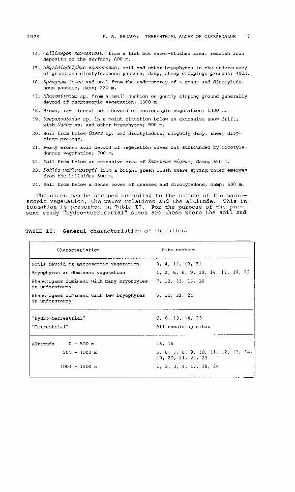
The sites can be grouped according to the nature of the macroscopic vegetation, the water relations and the altitude. This information is presented in Table 11. For the purpose of the present study "hydro-terrestrial" sites are those where the soil and
TABLE 11: General characteristics of the sites.
Characteristics Site numbers
Soils devoid of macroscopic vegetation 3, 4, 11, 18, 21
Bryophytes as d6minant vegetation 1, 2, 6, 8, 9, 10, 14, 17, 19, 23
Phanerogams dominant with many bryophytes 7, 12, 13, 15, 16in understorey
Phanerogams dominant with few bryophytes 5, 20, 22, 24in understorey
"Hydro-terrestrial" 6, 8, 13, 14, 23
"Terrestrialll All remaining sites
Altitude 0 - 500 m 15, 16
501 - 1000 m 5, 6, 7, 8, 9, 10, 11, 12, 13, 14,19, 20, 21, 22, 23
1001 - 1500 m 1, 2, 3, 4, 17, 18, 24
8 ACTA BOTANICA ISLANDICA NO. 5
vegetation approached water saturation but were not covered by water, they were in areas of groundwater seepage and poor drainage."Terrestrial" sites are those which did not approach water saturation and were not affected by seepage of groundwater.
METHODS AND MATERIALS
a. S a m pI in g. Normal precautions were taken to avoid contamination of sample material. Vegetation, litter and soil wereremoved down to the depth below which it was thought no lightcould penetrate, e.g. the mineral soils were sampled to a depthof ca. 1 cm but below phanerogamic vegetation the looser, lessdense moss, litter and soil was sampled to a depth of ca. 5 cm.A total surface area of ca. 100 cm 2 was removed each site.
b. E x a m i n a t ion 0 f s a m pIe s. The methods used largelyfollowed those of BROADY (l977b). First, fresh sample material wasmicroscopically examined. Secondly, two types of culture were setup within 24 hours of sampling, namely moist plate enrichment cultures (LUND, 1945), in which the sample was kept moist using halfstrength Bold's modified Bristol's medium (BBM) (CHANTANACHAT andBOLD, 1962), and Petri plate cultures using full strength BBM solidified with 2.5% agar. About one gram of sample material was spreadover the latter. Cultures were incubated under constant day-lightfluorescent tube illumination at room temperature (ca. 20-25°C.).Each moist plate enrichment culture was microscopically examinedfour or more times over a period of two months. Algal growths appearing as colonies on the agar cultures were removed for microscopic identification and used to innoculate unialgal cultures. Material from fresh samples and moist plate enrichment cultures wastreated with concentrated sulphuric acid in order to remove organicmaterial prior to making permanent microscopic preparations forexamination of diatoms. The diatoms, however, remain incompletelystudied as time was not available for the examination of all samples.
ALGAE RECOVERED
In the following section the algae are first listed according tothe general classification of BOURRELLY (1966, 1968, 1970) althoughthe Euchlorophyceae and Ulothricophyceae of BOURRELLY (1966) arecombined into the Chlorophyceae as described by ROUND (1973). Thealgae have been identified as far as available literature and timeallowed. Of the 200 taxa recovered from the samples 100 are identified to species or variety, 176 to genus, species or variety,eight are compared with described genera, their exact generic position remains doubtful, and 16 remain unidentified. Eleven of thelatter are unicellular free-living members of the Chlorococcales ofwhich cultures have been obtained. It is hoped that these will beidentified to a higher level after further examination. It is apparent that much critical work, especially the examination of algaein unialgal cultures, remains to be performed before the terrestrial flora is fully known.
In the descriptive section the algae are listed alphabeticallywithin their orders. Below the name is given the figure referencefollowed by a list of the sites from which the alga was recoveredand finally a reference to the literature from which the identification was made. The Fritsch Collection of algal drawings lodged
1979 P. A. BROADY: TERRESTRIAL ALGAE OF GLERARDALUR 9
at the Freshwater Biological Association, Arnbleside, Cumbria, United Kingdom, was also of great help with the identifications. References to other recoveries of the alga from "terrestrial" and"hydro-terrestrial" sites in Iceland are given after each description. In several of the diatom figures not all striae and punctaeare illustrated, however, the regions they occupy on the valvesare delineated e.g. Fig. 7.12.
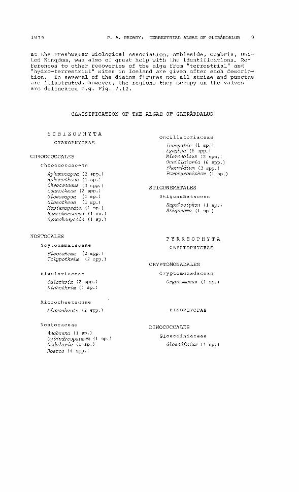
CLASSIFICATION OF THE ALGAE OF GLERARDALUR
SCHIZOPHYTA
CYANOPHYCEAE
CHROOCOCCALES
Chroococcaceae
Aphanocapsa (2 spp.)Aphanothece (1 sp.)Chroococcus (2 spp.)Cyanothece (2 spp.)Gloeocapsa (1 sp.)Gloeothece (1 sp.)Merismopedia (1 sp.)Synechococcus (1 sp.)Synechocystis (1 sp.)
NOSTOCALES
Scytonemataceae
Osci11atoriaceae
Isocystis (1 sp.)Lyngbya (6 spp.)Microcoleus (2 spp.)Oscillatoria (6 spp.)Phormidium (3 spp.)Porphyrosiphon (1 sp.)
STIGONEMATALES
Stigonemataceae
Hapalosiphon (1 sp.)Stigonema (1 sp.)
PYRRHOPHYTA
CRYPTOPHYCEAEPlectonemaTolypothrix
(2 spp.)(2 spp.)
CRYPTOMONADALES
Rivulariaceae
Calothrix (2 spp.)Dichothrix (1 sp.)
Microchaetaceae
Microchaete (2 spp.)
Nostocaceae
Anabaena (1 sp.)Cylindrospermum (1 sp.)NoduZaria (1 sp.)Nostoc (4 spp.)
Cryptomonadaceae
Cryptomonas (1 sp.)
DINOPHYCEAE
DINOCOCCALES
Gloeodiniaceae
Gloeodinium (1 sp.)
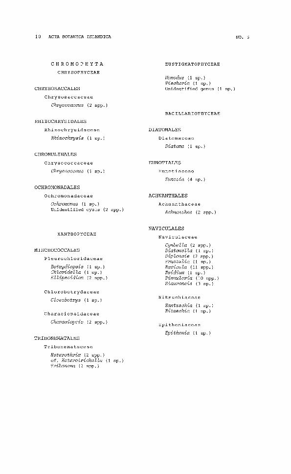
10 ACTA BOTANICA ISLANDICA
CHROMOPHYTA EUSTIGMATOPHYCEAE
NO. 5
CHRYSOPHYCEAE
CHRYSOSACCALES
Chrysosaccaceae
Chrysosaccus (2 spp.)
RHIZOCHRYSIDALES
Rhizochrysidaceae
Rhizochrysis (1 sp.)
CHROMULINALES
Chrysococcaceae
Chrysococcus (1 sp.)
OCHROMONADALES
Ochromonadaceae
Ochromonas (1 sp.)Unidentified cysts (2 spp.)
XANTHOPYCEAE
MISCHOCOCCALES
Pleurochloridaceae
Botrydiopsis (1 sp.)Ch loride lla (1 sp.)Ellipsoidion (2 spp.)
Chlorobotrydaceae
Gloeobotrys (1 sp.)
Characiopsidaceae
Characiopsis (2 spp.)
TRIBONEMATALES
Tribonemataceae
Heterothrix (2 spp.)cf. Heterotrichella (1 sp.)Tribonema (2 spp.)
Monodus (1 sp.)Vischeria (1 sp.)Unidentified genus (1 sp.)
BACILLARIOPHYCEAE
DIATOMALES
Diatomaceae
Diatoma (1 sp.)
EUNOTIALES
Eunotiaceae
Eunotia (4 sp.)
ACHNANTHALES
Achnanthaceae
Achnanthes (2 spp.)
NAVICULALES
Naviculaceae
Cymbella (2 spp.)Diatomella (1 sp.)Diploneis (2 spp.)Frustulia (1 sp.)Navicula (11 spp.)Neidiwn (1 sp.)Pinnularia (10 spp.)Stauroneis (3 sp.)
Nitzschiaceae
Hantzschia (1 sp.)Nitzschia (1 sp.)
Epithemiaceae
Epithemia (1 sp.)

1979
EUGLENOPHYTA
EUGLENOPHYCEAE
EUGLENALES
Euglenaceae
EugZena (1 sp.)
CHLOROPHYTA
CHLOROPHYCEAE
VOLVOCALES
R. A. BROADY: TERRESTRIAL ALGAE OF GLERARDALUR
PZanktosphaereZZa (1 sp.)ScotieZZa (1 sp.)
Radiococcaceae
Sphaerocystis (3 spp.)
Dictyosphaeriaceae
Dictyosphaerium (1 sp.)
ULOTHRICALES
Ulothricaceae
ChZorhormidium (3 spp.)Fottea (1 sp.)Heterothrichopsis (1 sp.)Stichococcus (2 sp.)Unidentified genus (1 sp.)
11
Unidentified family (1 sp.)
Chlamydomonadaceae
Carteria (2 spp.)ChZamydomonas (4 spp.)ChZoromonas (1 sp.)
TETRASPORALES
Gloeocystaceae
Asterococcus (1 sp.)Chlamydomonas (2 spp.)
Hypnomonadaceae
Hypnomonas (1 sp.)
CHLOROCOCCALES
Unidentified sphericalunicells (11 spp.)
Chlorococcaceae
Characium (1 sp.)FemandineZZa (1 sp.)Kentrosphaera (1 sp.)RhopaZocystis (1 sp.)cf. RhopaZocystis (1 sp.)cf. Spongiococcum (1 sp.)
Oocystaceae
ChZoreZZa (2 spp.)cf. JaagiochZoreZZa (1 sp.)Murie ZZa (1 sp.)cf. Oocystis (1 sp.)
CHAETOPHORALES
Chlorosarcinaceae
ChZorosarcinopsis (2 spp.)cf. PseudendocZoniopsis (1 sp.)
Chaetophoraceae
c f. Desmococcus (1 sp.)cf. Gongrosira (1 sp.)Microthamnion (2 spp.)
ZYGOPHYCEAE
ZYGNEMATALES
Mesotaeniaceae
CyZindrocystis (1 sp.)Mesotaenium (2 spp.)Netrium (1 sp.)
Desmidiaceae
Actinotaenium (1 sp.)cf. Actinotaenium (1 sp.)CZosterium (1 sp.)Cosmarium (9 spp.)Euastrum (4 spp.)SpondyZosium (1 sp.)Staurastrum (1 sp.)Tetmemorus (1 sp.)

12 ACTA BOTANICA 18LANDICA
DESCRIPTIONS OF THE ALGAE
CYANOPHYCEAE
CHROOCOCCALES
NO. 5
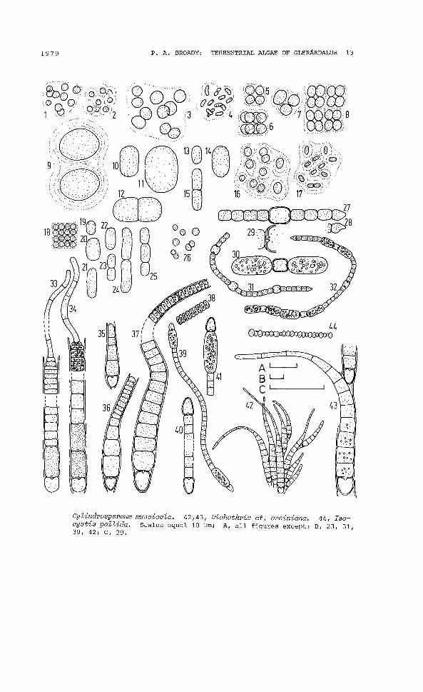
APHANOCAPSA ELACHISTA West and West(Fig. 2.1,2; 82, 6, 7, 10, 11, 14-16, 19, 22-24; GEITLER, 1932)
Colonies small, approximately spherical; cells blue-green, 1.8-3~m diameter, irregularly arranged throughout hyaline mucilagewhich is usually homogeneous (Fig. 2.1) but may have a faint stratification around each cell (Fig. 2.2).
APHANOCAPSA GREVILLEI (Hass.) Rabenh.(Fig. 2.3; 86, 12, 16, 19; GEITLER, 1932)
Cells blue-green, 3.5-5 ~m diameter, single or in pairs throughouthyaline, homogeneous mucilage.
BEHRE and SCHWABE (1970), SCHWABE and BEHRE (1972).
APHANOTHECE CLATHRATA West and West f.(Fig. 2.4; 87, 21; GEITLER, 1932)
Cells pale blue-green, ellipsoidal to cylindrical, often slightlycurved, 1-3.5 ~m long by 0.75 ~m wide, irregularly arrangedthroughout hyaline homogeneous mucilage.
A. cZathrata has somewhat longer cells of 3.5-4.5 ~m.
CHROOCOCCUS MINOR (Kuetz.) Naeg.(Fig. 2.5-8; 83, 11, 13, 14, 16, 17, 21; GEITLER, 1932)
Cells blue-green, spherical to ellipsoidal, 3-5 ~m diameter, incolonies of 1-4, rarely more, cells; occasionally remaining inMerismopedia-like aggregates (Fig. 2.8).
CHROOCOCCUS TURGIDUS (Kuetz.) Naeg.(Fig. 2.9; 814; GEITLER, 1932)
Cells pale blue-green, ellipsoidal, 14-16 ~m by 11-12.5 ~m, inpairs in stratified hyaline mucilage.
PETER8EN (1928a).
Fig. 2. 1, 2, Aphanocapsa e Zachista. 3, A. gl'evi ZZei. 4, AphanothececZathrata f. 5-8, Chroococcus minor. 9, C. turgidus. 10-12, Cyanothece aeruginosa. 13-15, C. cedrorum. 16, GZoeocapsa geZatinosa.17, GZoeothece sp. 1. 18, Merismopedia tenuissima. 19-25, Synechococcus eZongatus. 26, Synechocystis minuscuZa. 27-32, Anabaena osciZZarioides f. 33, 34, CaZothrix sirrruZans f. 35-38, C. eZenkinii. 39-41,
1979 P. A. BROADY: TERRESTRIAL ALGAE OF GLERARDALUR 13
Cylindrospermum muscicola. 42,43, Dichothrix cf. orsiniana. 44, Isocystis pallida. Scales equal 10 ~m; A, all figures except: B, 23, 31,39, 42; C, 29.

14 ACTA BOTANICA 18LANDICA NO. 5
CYANOTHECE AERUGINOSA (Naeg.) Kom.8yn. Syneohooooeus aeruginosa Naeg.(Fig. 2.10 -12; 82, 9, 18, 19; KOMAREK, 1976)
Cells bright blue-green, broadly ellipsoidal to short cylindrical,11-34 ~m long by 8-30 ~m wide.
PETER8EN (1928a and 1928b);
CYANOTHECE CEDRORUM (Sauv.) Kom.8yn. Syneohooooous oedrorum 8auv.(Fig. 2.13-15; 83, 5; KOMAREK, 1976)
Cells blue-green, broadly ellipsoidal to short cylindrical, 6.511 ~m long by 3.5-7.5 ~m wide.
GLOEOCAPSA GELATINOSA Kuetz.(Fig. 2.16; 8 21; GEITLER, 1932)
Cells blue-green, spherical to ellipsoidal, 2-3.5 ~m diameter,single and in pairs throughout hyaline, clearly stratified mucilage.
GLOEOTHECE sp. 1(Fig. 2.17; 811, 17)
Cells pale blue-green, cylindrical, 2-3 ~ long by 1 ~m wide, irregularly distributed throughout hyaline, faintly stratified mucilage.
MERISMOPEDIA TENUISSIMA Lemm.(Fig. 2.18; 83, 13; GEITLER, 1932)
Cells blue-green, spherical, 2 ~ diameter, in groups of multiplesof four throughout small colonies.
SYNECHOCOCCUS ELONGATUS Naeg.(Fig. 2.19-25; 811; KOMAREK, 1976)
Cells blue-green, ellipsoidal to cylindrical, 5-12 ~ long by 3-3.5~m wide, single, in pairs and occasionally in short chains.
SYNECHOCYSTIS MINUSCULA Vor.(Fig. 2.26; 813; GEITLER, 1932)
Cells pale blue-green, spherical, 1.5-2.5 ~m diameter.
NOSTOCALES
ANABAENA OSCILLARIOIDES Bory f.(Fig. 2.27-32; 82-6, 8, 11, 13, 14, 16, 19, 20; GEITLER, 1932)
Trichomes blue-green, 3.5-6.5 ~m wide, with or without a thinsheath, occasionally forming Microcoleus-like strands with severaltrichomes inside a single sheath; cells barrel-shaped, 2.5-6.5 ~m
1979 P. A. BROADY: TERRESTRIAL ALGAE OF GLERARDALUR 15
long, terminal cell conical (Fig. 2.27, 28); heterocysts slightlylarger than vegetative cells, 6-7.5 ~m wide by 8-9.5 ~m long (Fig.2.27, 29, 31); akinetes ellipsoidal to cylindrical, 4.5-7.5 ~m wideby 6-17 ~ long, usually produced on either side of the heterocysts,singly or in short chains (Fig. 2.30, 32) but occasionally betweenvegetative cells.
PETERSEN (1928a) records A. oscillarioides var. tenuis Lemm.
CALOTHRIX SIMULANS Gardner f.(Fig. 2.33, 34; S8, 14; GEITLER, 1932)
Trichomes blue-green, up to 200 ~m long, in a thin, hyaline, closefitting sheath, width at base 5-7 ~m, tapering suddenly at apex toa colourless hair 1.5-2.5 ~m wide, slightly constricted at transverse walls except in region of terminal hair; cells usually shorter than wide, except along hair, though occasionally longer atbase (Fig. 2.34), 2-5 (-14)~ long; heterocyst single, basal, samewidth as vegetative cells; akinetes single, or a pair, immediatelyabove heterocyst, same width as cells, 9-14 ~ long.
CALOTHRIX ELENKINII Kossink.(Fig. 2.35-38; S3, 4, 6, 13, 14, 16, 20; COCKE, 1967)
Trichomes blue-green, up to 225 ~m long, in a thin, hyaline, closefitting sheath which reaches to the end of the trichome, width atthe somewhat bulbous base 6-10 ~m, at apex 3-5 ~m, no long terminalhair, slightly constricted at transverse walls; cells usually shorter than wide, except at base where sometimes slightly longer, terminal cells often very short, discoidal and short lengths of thesereleased as motile hormogones (Fig. 2.37, 38), immature trichomes(Fig. 2.35) without these; heterocyst basal, rarely two, narrowerthan basal vegetative cells.
CYLINDROSPERMUM MUSCICOLA Kuetz.(Fig. 2.39-41; S9, 11, 13, 14, 19; GEITLER, 1932)
Trichomes blue-green, 2.5-4 ~ wide, slightly constricted at transverse walls; cells 2.5-5 (-13) ~m long, usually about as long aswide; heterocysts terminal, slightly longer than vegetative cells;akinetes adjacent to heterocyst (Fig. 2.41), ellipsoidal to cylindrical, up to 16 ~m long and 5 ~m wide but mature akinetes probably not observed.
PETERSEN (1928a).
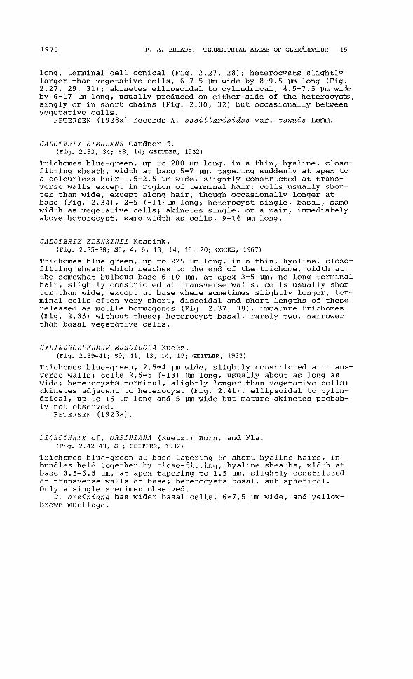
DICHOTHRIX cf. ORSINIANA (Kuetz.) Born. and Fla.(Fig. 2.42-43; S6; GEITLER, 1932)
Trichomes blue-green at base tapering to short hyaline hairs, inbundles held together by close-fitting, hyaline sheaths, width atbase 3.5-6.5 ~m, at apex tapering to 1.5 ~m, slightly constrictedat transverse walls at base; heterocysts basal, sub-spherical.Only a single specimen observed.
D. orsiniana has wider basal cells, 6--7.5 ~m wide, and yellowbrown mucilage.

16 ACTA BOTANICA 18LANDICA NO. 5
ISOCYSTIS PALLIDA Vor.(Fig. 2.44; 87,11,13,14,16,17; KULLBERG, 1971)
Trichomes pale blue-green, sheathless; cells globular and ellipsoidal, 1-2 ~m wide, often of different widths along the same trichome.
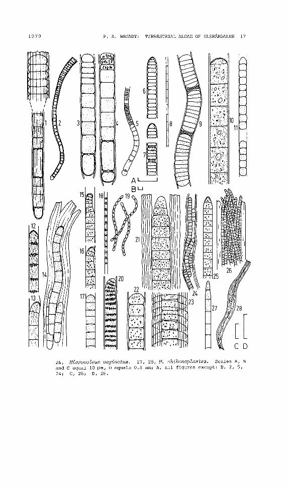
LYNGBYA AERUGINEO-COERULEA (Kuetz.) Gom.(Fig. 3.12-14; 82, 3, 6, 11, 16, 20; GEITLER, 1932)
Trichomes greyish blue-green, 3-6.5 ~m wide, not constricted attransverse walls, usually with a thin hyaline sheath around singletrichomes but growths removed from a drying moist plate enrichmentculture had thickened stratified sheaths containing from one tothree parallel trichomes (Fig. 3.14); cells 3-9.5 ~m long, with orwithout granules along transverse walls, terminal cell with calyptra in mature trichomes.
The growth form in which up to three trichomes are enclosed ina single sheath resembles Schizothrix, however, because the dominant growth form was of single trichomes in individual sheaths thisalga has been assigned to Lyngbya.
LYNGBYA sp. 4(Fig. 3.6, 7; 82, 11, 21)
Trichomes blue-green, 4-5 vm wide, slightly attenuated terminally,in a thin hyaline sheath which encloses the terminal cell; cellsshorter than wide, 1-2.5 ~m long, granular; separation discs formed(Fig. 3.7).
LYNGBYA sp. 6(Fig. 3.8; 82-4, 7, 9, 11, 16-18, 20, 21)
Trichomes pale blue-green, 0.5-0.8 vm wide in a very thin sheath,transverse walls faint; cells 3-5 ~m long.
LYNGBYA sp. 7(Fig. 3.9; 82-4, 18)
Trichomes pale blue-green, 6-8 vm wide, in a thin hyaline sheath,faintly constricted at transverse walls, readily fragmenting intoshort lengths up to 50 ~m long by the production of bi-concave separation discs.
LYNGBYA sp. 8(Fig. 3.10; 87, 14, 16)
Trichomes blue-green, 8-11 ~m wide, in readily visible hyalinesheath ca. 1 ~m wide; cells 5-18 ~m long with granular contents
Fig. 3. 1,2, Microchaete striatula. 3-5, M. cf. investiens. 6,7, Lyngbyasp. 4. 8, L. Sp. 6. 9, L. Sp. 7. 10, L. sp. 8. 11, L. Sp. 9.12-14, L. aerugineo-coeru lea. 15-1 7, Phormidium autumnale. 18, 19 ,P. frigidum. 20, P. uncinatum. 21-24, Porphyrosiphon notarisii. 25,
1979 P. A. BROADY, TERRESTRIAL ALGAE OF GLERARDALUR 17
~.
~/I.~.~
~~..'. ;~:. 24
mill~.• :~.'=i.•.{1 " J:l23• ,'f
·,:·,.°'0'~'. '0', .. ~
26, Miaroaoleus vaginatus. 27, 28, M. ahthonoplastes. Scales A, Band C equal 10 pm, D equals 0.5 mm; A, all figures except: B, 2, 5,24; C, 26; D, 28.

18 ACTA BOTANICA ISLANDICA NO. 5
and readily visible polyhedral bodies; terminal cell rounded andsometimes with calyptra; separation discs formed.
LYNGBYA sp. 9( Fig. 3. 11; S6, 11, 18, 21)
Trichomes blue-green, 3.5-4 ~ wide, constricted at transversewalls, in a thin hyaline sheath; cells 3-6 ym long; terminal cellconical.
MICROCHAETE cf. INVESTIENS Fremy(Fig. 3.3-5; S16; GEITLER, 1932)
Trichomes blue-green, up to 180 ym long, in a hyaline sheath up to3 ym thick; cells 6-8 ym wide, at base longer than wide, up to 13ym long, at apex shorter than wide and often granular; motile hormogones released (Fig. 3.5); heterocyst basal, usually sub-spherical, a second adjacent, cylindrical heterocyst sometimes present(Fig. 3.4).
M. investiens forms large akinetes which were not observed inthe present specimens.
MICROCHAETE STRIATULA Hy.Syn. Leptobasis striatula (Hy.) Elenk.(Fig. 3.1,2; S 13, 14, 16; GEITLER, 1932)
Trichomes blue-green, width at base 3-3.5 ym, at apex 8-11 ym, constricted at transverse walls at apex but only occasionally at base,in an obvious hyaline sheath down which parallel striations areoften visible, especially at apex; cells 3-16 ym long, longer atbase than at apex; terminal, subspherical, heterocyst always present, also occasionally intercalary cylindrical heterocysts; motilehormogones released. Immature filaments in which the difference inwidth between apex and base has not developed often resemble M. cf.investiens.
MICROCOLEUS CHTHONOPLASTES Thur.(Fig. 3.27, 28; Sll, 21; GEITLER, 1932)
Plant mass consisting of unbranched bundles of trichomes numberingthree to over 40 enclosed within a common thick, hyaline sheath,total width up to 70 ym and length up to 6 mm (Fig. 3.28); individual trichomes (Fig. 3.27) blue-green, 2-3.5 ym wide, slightly attenuated a~ apices, slightly constricted at transverse walls; cells4.5-9.5 ym long, slightly granular.
MICROCOLEUS VAGINATUS (Vauch.) Gom.(Fig. 3.25, 26, S21; GEITLER, 1932)
Plant mass similar to M. chthonoplastes (Fig. 3.28); individual trichomes (Fig. 3.25) blue-green, 4-5 ym wide, not constricted attransverse walls, slightly attenuated; cells 3-6 ym long with granular contents; terminal cell with calyptra.
1979 P. A. BROADY: TERRESTRIAL ALGAE OF GLERARDALUR 19
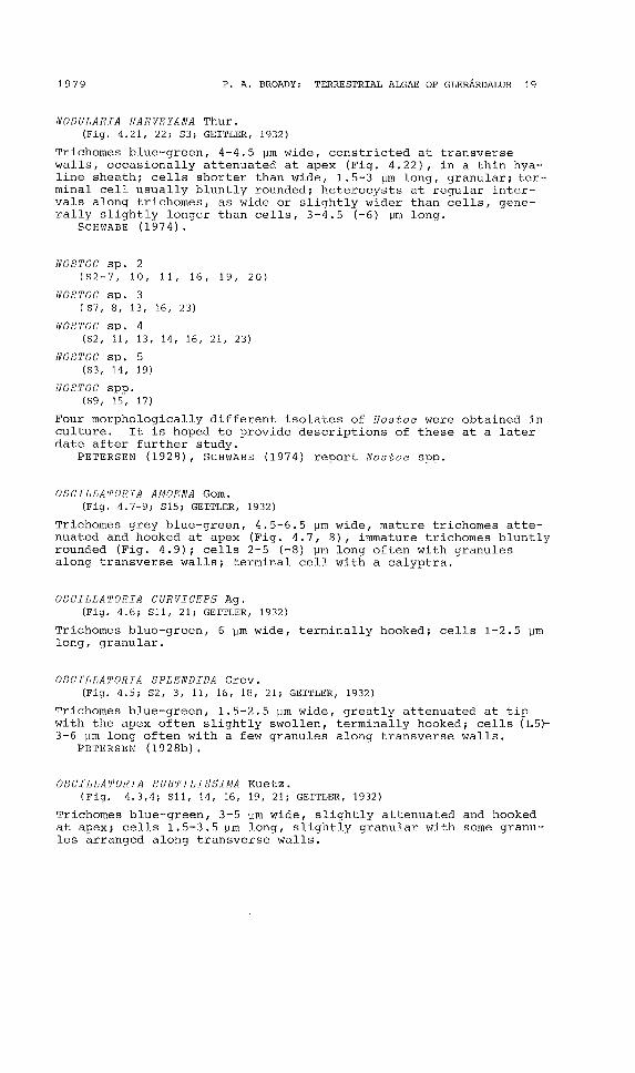
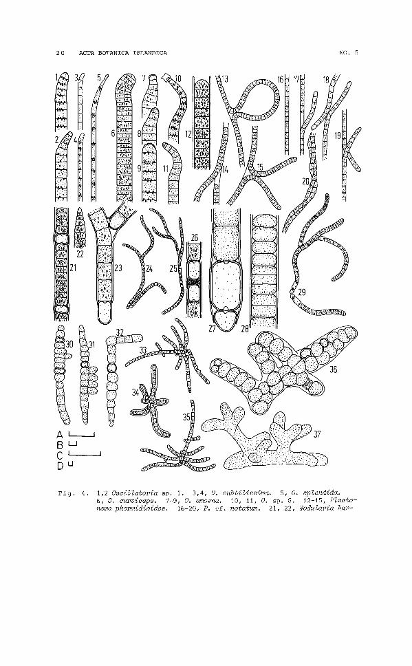
NODULARIA HARVEYANA Thur.(Fig. 4.21, 22; 53; GEITLER, 1932)
Trichomes blue'-green, 4-4.5 IJm \vide, constricted at transversewalls, occasionally attenuated at apex (Fig. 4.22), in a thin hyaline sheath; cells shorter than wide, 1.5-3 IJm long, granular; terminal cell usually bluntly rounded; heterocysts at regular intervals along trichomes, as wide or slightly wider than cells, generally slightly longer than cells, 3-4.5 (-6) IJm long.
SCHWABE (1974).
NOSTOC sp. 2(52-7, 10, 11, 16, 19, 20)
NOSTOC sp. 3(87, 8, 13, 16, 23)
NOSTOC sp. 4(82, 11, 13, 14, 16, 21, 23)
NOSTOC sp. 5(83, 14, 19)
NOSTOC spp.(89, 15, 17)
Four morphologically different isolates of Nostoc were obtained inculture. It is hoped to provide descriptions of these at a laterdate after further study.
PETER8EN (1928), SCHWABE (1974) report Nostoc spp.
OSCILLATORIA AUOENA Gom.(Fig. 4.7-9; 815; GEITLER, 1932)
Trichomes grey blue-green, 4.5-6.5 IJm wide, mature trichomes attenuated and hooked at apex (Fig. 4.7, 8), i~mature trichornes bluntlyrounded (Fig. 4.9); cells 2-5 (-8) IJm long often with granulesalong transverse walls; terminal cell with a calyptra.
OSCILLATORIA CURVICEPS Ag.(Fig. 4.6; 811, 21; GEITLER, 1932)
Trichomes blue-green, 6 IJm wide, terminally hooked; cells 1-2.5 IJrnlong, granular.
OSCILLATORIA SPLENDIDA Grev.(Fig. 4.5; 82, 3, 11, 16, 18, 21; GEITLER, 1932)
Trichomes blue-green, 1.5-2.5 IJrn wide, greatly attenuated at tipwi th the apex often slightly swollen, terminally hooked; cells (1.5)3-6 IJm long often with a few granules along transverse walls.
PETER8EN (1928b).
OSCILLATORIA SUBTILISSIMA Kuetz.(Fig. 4.3,4; 811, 14, 16, 19, 21; GEITLER, 1932)
Trichomes blue-green, 3-5 IJm wide, slightly attenuated and hookedat apex; cells 1. 5-3.5 IJm long, slightly granular with some granu"les arranged along transverse walls.
20 ACTA BOTANICA ISLANDICA NO. 5
A L-.-.JBUCo u
Fig. 4. 1,2 Oscillatoria Sp. 1. 3,4, O. subtilissima. 5, O. splendida.6, O. curviceps. 7-9, O. amoena. 10, 11, O. sp. 6. 12-15, Plectonema phormidioides. 16-20, P. cf. notatum. 21, 22, Nodularia har-
1979 P. A. BROADY: TERRESTRIAL ALGAE OF GLERARDALUR 21
OSCILLATORIA sp. 1.(Fig. 4.1, 2; S2-6, 8, 11, 15, 16, 19, 21)
Trichomes blue-green, 3-5 Wm wide, attenuated slightly and hookedat apex; cells 1.5-3.5 Will long, slightly granular with some granules arranged along transverse walls.
OSCILLATORIA sp. 6.(Fig. 4.10, 11; S4, 18)
Trichomes blue-green, 3.5'-5 Will wide, attenuated and often markedlyhooked at apex; cells 1.5-5 Will long, often with granules alongtransverse walls; terminal cell often attached to the empty remainsof a single cell (Fig. 4.10) probably the results of the fragmentation 0= a longer trichome at the position of a dead cell, otherterminal cells with a calyptra.
PHORMIDIUM AUTUMNALE (Ag.) Gom.(Fig. 3.15-17; S2, 4,6,11,15,17,19-21,24; GEITLER, 1932)
Trichomes blue-green, 3.5-5 wm wide, often vlith slight terminalhook (Fig. 3.15, 16) though this is lacking in immature trichomes(Fig. 3.17), enclosed in a thin hyaline sheath; cells 2-5 Will long,often granular; terminal cell with calyptra in mature trichomes.
PETERSEN (1928a and 1928b), SCHNABE (1970), BEHRE and SCHWABE(1970) .
PHORMIDIUM FRIGIDUN Fritsch(Fig. 3.18, 19; S3-5, 7, 8, 11, 13, 14, 16-21; GEITLER, 1932)
Trichomes blue-green, 1-2 Wm wide, generally short and readilyfragmented, either in a thin hyaline sheath (Fig. 3.18) or in entangled masses embedded in mucilage in which individual sheathsnot visible (Fig. 3.19), also occurring as free motile trichomeslacking sheath; cells 1.5-3 wm long, apparently joined by shortgelatinous pads, often with a single granule at both apices; terminal cell broadly rounded and often with a pair of apical granules.
The trichomes which lack a sheath closely resemble Pseudanabaena catenata Lauterb. SCHWABE (1970) records the presence of Pseudanabaena cf. catenata on Surtsey and BEHRE and SCHWABE (1970) describe what appears to be a similar alga to the present specimensas a life-form of Schizothrix lardacea (ees.) Gom. In the presentspecimens the Schizothrix-like, Microcoleus-like and Plectonemalike stages described by those authors were not recorded. However,it is possible that the former is also a life-form of S. lardacea.
PHORMIDIUM UNCINATUM Gom.(Fig. 3.20; S20; GEITLER, 1932)
Trichomes blue-green, 5·-7 Will wide, slightly attenuated towards apex,
veyana. 23-26, Tolypothrix tenuis f. terrestris. 27-29, T. lanata.30-35, Hapalosiphon hibernicum. 36, 37, Stigonema sp. 1. Scalesequal 10 wm; A, all figures except: B, 13-15, 33-35; C, 24, 25, 29;D, 30-32, 36, 37.

22 ACTA BOTANICA 18LANDICA NO. 5
often slightly terminally hooked, in a readily visible hyalinsheath although sheathless hormogones are released; cells 2-3.5 ~m
long, often very granular with granules along transverse walls,apical cell often without granulation.
PLECTONEMA PHROMIDIOIDES Hansg.(Fig. 4.12-15; 89, 10, 16; GEITLER, 1932)
Trichomes blue-green, 6-7 ~ wide, in a thin hyaline sheath, withoccasional false-branching (Fig. 4.13-15); cells 3-6 ~ long, granular, transverse walls not clear.
PLECTONEMA cf. NOTATUM Schmidle(Fig. 4.16-20; 83-7, 8-11, 13-21; GEITLER, 1932)
Trichomes blue-green, 1"1.5 ~m wide, in a thin hyaline sheath. Thedegree of bending and false-branching of trichomes varies with culture conditions and age of culture. In young moist plate enrichment cultures trichomes were mostly straight or slightly flexuouswith rare false-branching. In young BBM agar cultures trichomeswere very flexuous and false-branching rare. On an old, dryingmoist plate enrichment culture trichomes were more twisted andfalse-branching was frequent, occasionally two trichomes lay sideby side within a single sheath (Fig. 4.20). Cells 1.5-3 ~m long,occasionally granular.
P. notatum has trichomes 1.7-2 ~m wide. Plectonema sp. B described by BEHRE and SCWABE (1970) is of similar size to the present specimens.
PORPHYROSIPHON NOTARISII Kuetz.(Fig. 3.21-24; 511; COCKE, 1967)
Trichomes bright blue-green, 8-11 ~m wide, constricted at transverse walls, terminally attenuated in mature specimens (Fig. 3.21),but not in immature ones (Fig. ':.22); sheath up to 13 ~m thick,hyaline, clearly stratified anf occasionally with ring-like striations (Fig. 3.23), enclosing fom one to three parallel trichomes(Fig. 3.24); cells 3-8 ~m lone granular.
P. notarisii is usually dE ;cribed as having bright red sheaths,but where the plants are not subjected to strong light the sheathsmay be hyaline (DROUET, 1938) as in the ice1andic specimens.
TOLYPOTHRIX TENUIS f. TERRESTRIS Boye Pet.(Fig. 4.23-26; 52-6, 11, 14, 16, 19-21; PETER5EN, 1923)
Trichomes blue-green, 4-7.5 ~ wide, in a close-fitting, thin hyaline sheath; cells 5-11 ~ long; heterocysts 8-12.5 ~m long, atbases of false-branches and occasionally intercalary, in the latter case two heterocysts are often on either side of a separationdisc (Fig. 4.25, 26) and fragmentation occurs here (Fig. 4.24).
PETER8EN (1928a), SCWABE (1974).
TOLYPOTHRIX LANATA Lemm.(Fig. 4.27-29; 514, 16; GEITLER, 1932)
Few specimens observed; trichomes blue-green, 9-11 (-16) ~m wide
1979 P. A. BROADY: TERRESTRIAL ALGAE OF GLERARDALUR 23
in a thin, close-fitting, hyaline sheath up to 2 ~m thick, constricted at transverse walls particularly towards the apices; cellsshorter at apices, 4.5-8 ~m long (Fig. 4.28), than towards basesof branches, 8-19 ~m long; heterocysts single at bases of branches,double at base of whole plant mass with penultimate cylindricalheterocyst (Fig. 4.27), four adjacent intercalary heterocysts (Fig.4.29) observed on one occasion with fragmentation apparently occurring at the mid-point.
STIGONEMATALES
HAPALOSIPHON HIBERNICUM West and West(Fig. 4.30-35; S13; GEITLER, 1932)
Trichomes blue-green, 4.5-8 ~m wide, in a thin sheath, hyaline inyoung specimens (Fig. 4.30-32) but reddish-brown in old (Fig. 4.3335); branches frequent; cells 3.5-6.5 ~m long; heterocysts similarsize to vegetative cells.
SCHWABE (1974).
STIGONEMA sp. 1(Fig. 4.36/ 37; S10)
Few specimens observed; cells 7-15 ~ wide, granular; branchesfrequent; sheath hyaline; principal axis biseriate with uniseriatebranches.
CRYPTOPHYCEAE
CRYPTOMONADALES
CRYPTOMONAS cf. CYLINDRICA Ehrenb.(Fig. 5.1-4; S13, 14; JAVORNICKY, 1967)
Cells narrowly ellipsoidal to cylindrical, 6-9 Vm wide by 14-19 ~
long, in transverse section spherical or slightly laterally flattened (Fig. 5.3); apex obliquely cut at entrance of gullet with twoslightly unequal flagella inserted subapically; two, golden-brown,parietal plate-like chromatophores almost completely line wall(dorsal view, Fig. 5.2), inner surface of chromatophores sometimeslined with large granules (Fig. 5.4); gullet lined with rows oftrichocysts (Fig. 5.1); contractile vacuole apical; two large ellipsoidal Maupas corpuscules often present (Fig. 5.1, 4).

24 ACTA BOTANICA 18LANDICA
DINOPHYCEAE
DINOCOCCALES
NO. 5
GLOEODINIUM MONTANUM Klebs(Fig. 5.5, 6; 82, 6, 10, 12, 13, 14, 15, 16; 8CHILLER, 1937)
Cells usually non-motile, spherical to broadly ellipsoidal, up to30 ~m diameter, enclosed in hyaline mucilage (Fig. 5.5); chromatophores numerous, golden-brown, parietal; reproduction by autospores with the sporangium wall splitting irregularly and the remainsoften visible; zoospores also formed (Fig. 5.6) but only one specimen observed, this possessed a pyrenoid-like body and numerous discoidal, parietal chromatophores.
CHRY80PHYCEAE
CHRYSOSACCALES
CHRYSOSACCUS EPILITHICUS Starmach(Fig. 5.7; 821; 8TARMACH, 1966)
Only one colony of three cells embedded in homogeneous hyalinemucilage observed; cells naked, subspherical, 8-12.5 ~m in diameter, with two or three contractile vacuoles and a single, parietal, lobed, golden-brown chromatophore.
CHRYSOSACCUS SPHAERICUS Bourr.(Fig. 5.8, 9; 821; BOURELLY, 1957)
Colonies consisting of four to eight cells embedded in homogeneous hyaline mucilage; cells naked, spherical, 6-8 \lm diameter,with disc-like, parietal, golden-brown chromatophores.
RHIZOCHRYSIDALES
RHIZOCHRYSIS sp. 1(Fig. 5.10; 810, 13, 14; BOURELLY, 1968)
Cells 3.5 ~m diameter with fine rhizopodia, a single, goldenbrown, band-like chromatophore and one or two indistinct contractile vacuoles.
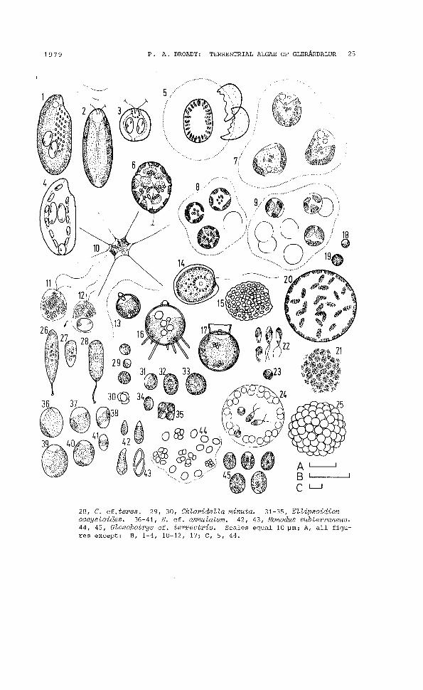
Fig. 5. 1-4, Cryptomonas cf. cylindrica. 5,6, Gloeodinium montanum. 7, Chrysosaccus epilithicus. 8,9, C. sphaericus. 10, Rhizochrysis sp. 1113, Ochromonas sp. 1. 14, 15, Chrysococcus sp. 1. 16, Cyst 2. 17,Cyst 1. 18-25, Botrydiopsis sp. 1. 26, 27, Characiopsis cf. minuta.
1979 P. A. BROADY: TERRESTRIAL ALGAE OF GLEAARDALUR 25
......... ,
, , .
..~/
/ri~)\lS:(.~:.'
8 o 9ifj::@ (1\' ).~ !
\. Q\' :i ~) <. ~ / 18'., ~ .' .'\ ') - ~.ii @
"..' ··0···········19 ...,.................... .
......
, ,
5
28, C. cf. teres. 29, 30, Chloridella minuta. 31-35, Ellipsoidionoocystoides. 36-41, E. cf. annulatum. 42, 43, Monodus subterraneus.44, 45, Gloeobotrys cf. terrestris. Scales equal 10 ~m; A, all figures except: B, 1-4, 10-12, 17; C, 5, 44.
26 ACTA BOTANICA ISLANDICA
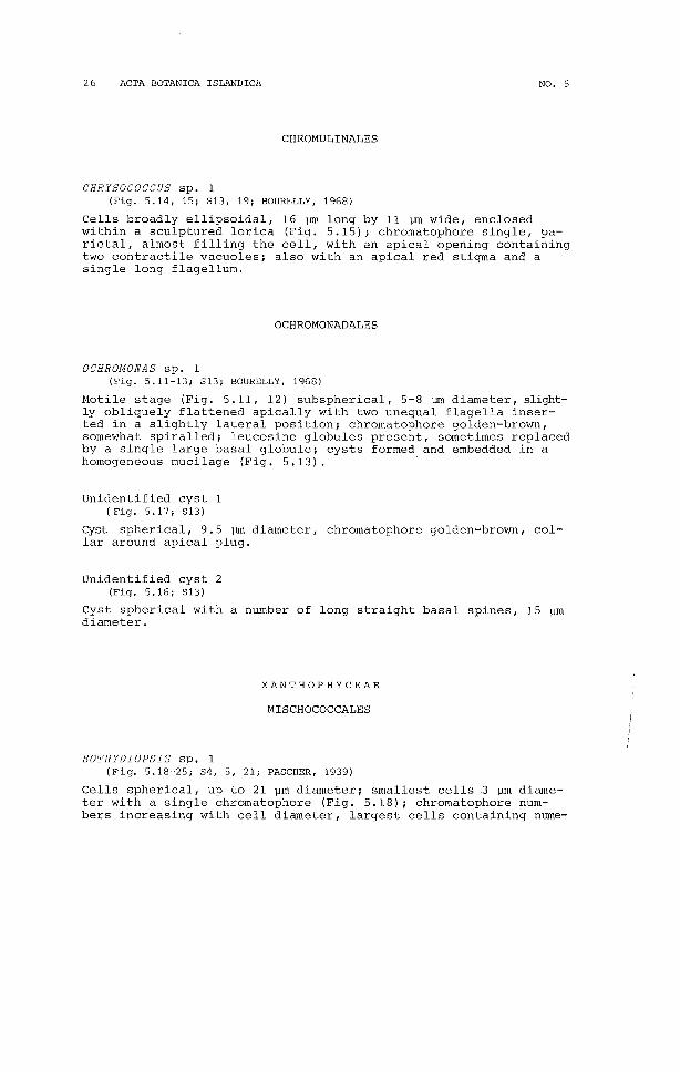
CHROMULINALES
NO. 5
CHRYSOCOCCUS sp. 1(Fig. 5.14, 15; S13, 19; BOURELLY, 1968)
Cells broadly ellipsoidal, 16 ~ long by 11 ~ wide, enclosedwithin a sculptured lorica (Fig. 5.15); chromatophore single, parietal, almost filling the cell, with an apical opening containingtwo contractile vacuoles; also with an apical red stigma and asingle long flagellum.
OCHROMONADALES
OCHROMONAS sp. 1(Fig. 5.11-13; S13; BOURELLY, 1968)
Motile stage (Fig. 5.11, 12) subspherical, 5-8 ~ diameter, slightly obliquely flattened apically with two unequal flagella inserted in a slightly lateral position; chromatophore golden-brown,somewhat spiralled; leucosine globules present, sometimes replacedby a single large basal globule; cysts formed and embedded in ahomogeneous mucilage (Fig. 5.13).
Unidentified cyst 1(Fig. 5.17; S13)
Cyst spherical, 9.5 ~ diameter, chromatophore golden-brown, collar around apical plug.
Unidentified cyst 2(Fig. 5.16; s13)
Cyst spherical with a number of long straight basal spines, 15 ~m
diameter.
XANTHOPHYCEAE
MISCHOCOCCALES
BOTRYDIOPSIS sp. 1(Fig. 5.18-25; S4, 5, 21; PASCHER, 1939)
Cells spherical, up to 21 ~m diameter; smallest cells .3 ~m diameter with a single chromatophore (Fig. 5.18); chromatophore numbers increasing with cell diameter, largest cells containing nume-

1979 P. A. BROADY: TERRESTRIAL ALGAE OF GLERARDALUR 27
rous parietal and some internal chromatophores (Fig. 5.2'0), in surface view these a!Jpear as polygonal plates (Fig. 5.21); reproduction by numerous aplanospores, which remain in close aggregates onrelease from the sporangium (Fig. 5.25), and zoospores (Fig. 5.22)which are only slightly unequally biflagellate, naked, and whichrapidly become spherical on quiescence (Fig. 5.23), the singlechromatophore contains a variably positioned orange stigma whichis usually in the posterior half of the cell; spore release is bygelatinization of the sporangium wall but many often become caughtin the mucilage and lose their motility (Fig. 5.24).
PETERSEN (1928a) records B. arhiza Borzi and PETERS EN (1928b)B. minor Schmidle.
CHARACIOPSIS cf. MINUTA Lemm.(Fig. 5.26, 27; S13, 14; PASCHER, 1939)
Cells 10-25 ~m long by 4-5 ~ wide with a short stalk; chromatophore parietal; young cells initially without a stalk (Fig. 5.27).
C. minuta possesses cells with more acute apices.
CHARACIOPSIS cf. TERES Pa.(Fig. 5.28; S2, 11; PASCHER, 1939)
Few specimens observed; cells cylindrical, 11-20 ~m long by 4.5~ wide, attached to substratum by thin stalk ca.7.5 ~m long withterminal button; several parietal chromatophores.
C. teres has generally larger cells, 19-30 ~ long by 8-14 ~m
wide.
CHLORIDELLA MINUTA Gayral and Mazancourt(Fig. 5.29, 30; S4, 5, 6, 8, 12, 15, 21; GAYRAL and MAZANCOURT, 1958)
Cells spherical to subspherical, 1.5-5 ~ diameter; chromatophoreparietal; reproduction by formation of four or eight autospores(Fig. 5.30).
ELLIPSOIDION cf. ANNULATUM Pa.(Fig. 5.36-41; S18; PASCHER, 1939)
Mature cells broadly ellipsoidal, up to 12.5 ~m long by 10 ~m wide,smallest cells narrowly ellipsoidal to almost cylindrical, 6 ~m
long by 3 ~m wide; chromatophore parietal, plate-like, lobed, orband-like; reproduction by formation of eight autospores.
E. annulatum has contractile vacuoles in young cells and thechromatophore is more regularly band-shaped and equatorial.
ELLIPSOIDION OOCYSTOIDES Pa.(Fig. 5.31-35, S2, 4-10, 12, 15, 17, 18, 20, 24; PASCHER, 1939)
Cells broadly ellipsoidal to spherical, 4.5-8 ~ diameter, withone parietal chromatophore in young cells, usually two, but up tofour, in mature cells; reproduction by formation of two, four oreight autospores.

28 ACTA BOTANICA 15LANDICA NO. 5
GLOEOBOTRYS cf. TERRESTRIS Reisigl(Fig. 5.44, 45; 54-8, 12, 13, 15, 19, 20, 22; REI5IGL, 1964)
Colonies containing cells in groups of four or eight or irregularly arranged throughout homogeneous, hyaline mucilage (Fig. 5.44);cells ellipsoidal, up to 9.5 ~m long by 8 ~m wide, smallest spores2 ~m in diameter; chromatophore parietal, broadly lobed; reproduction by formation of four or eight spores released by gelatinization of sporangium wall.
G. terrestris has one or two chromatophores. G. coenococcoidesFott is similar in the possession of a single chromatophore but hasspherical cells and stratified mucilage.
HETEROTHRIX EXILIS Pa. f.(Fig. 6.1, 52, 6, 20, 24; PA5CHER, 1939)
Filaments readily fragmented but usually more than eight cells inlength; cells 3-3.5 ~m wide, slightly swollen at centre, 6-13 ~m
long, vlith one or two, rarely three chromatophores.SCHWABE and BEHRE (1972).
HETEROTHRIX DEBILIS Pa. f.(Fig. 6.2, 58; PA5CHER, 1939)
Filaments readily fragmented, from nine to more than 20 cells lonfficells 6-6. 5 ~ wide by 4.5-9. 5~ long, with one to four chromatophores.
cf. HETEROTRICHELLA GRACILIS Reisigl(Fig. 6.5-9; 59, 17; REI5IGL, 1964)
Filaments short, flexuous, redily fragmented, many single cells(Fig. 6.9); cells cylindrical, straight or curved, occasionallyslightly attenuated at one apex, 3-5 ~ wide by 8-35 ~ long;chromatophore parietal stretching the length of the cell and covering one half or more of the wall; nucleus central; many small oilglobules often present.
H. gracilis has several similarities to this alga, namely theshort fragmenting filaments and the large range in cell lengthwith many curved cells. However, the cells of H. gracilis arenarrower and more markedly attenuated at one pole.
TRIBONEMA VULGARE Pa.(Fig. 6.3; 52, 4, PA5CHER, 1939)
Filaments long, 8 ~ wide, with cells slightly swollen at centre;cells 9-18 ~m long containing several parietal, plate-like chromatophores; cells fragmenting into "H"-shaped fragments.
Fig. 6. 1, Heterothrix exilis f. 2, H. debilis f. 3, Tribonema vulgaris.4, T. minus. 5-9, cf. Heterotrichella gracilis. 10, Vischeria puntata. 11-18, Unidentified species of the Eustigmatophyceae. 19,Diatoma hiemale var. maior novo var. 20, Eunotia exigua var. compacta. 21, E. gracilis. 22-24, E. praerupta. 25-27, E. praerupta cf.var. muscicola. 28-31, E. repens var. arcuata. 32, 33, Achnantheslanceolata f. rostrata. 34, 35, A. montana. 36, Diatomella balfouri-
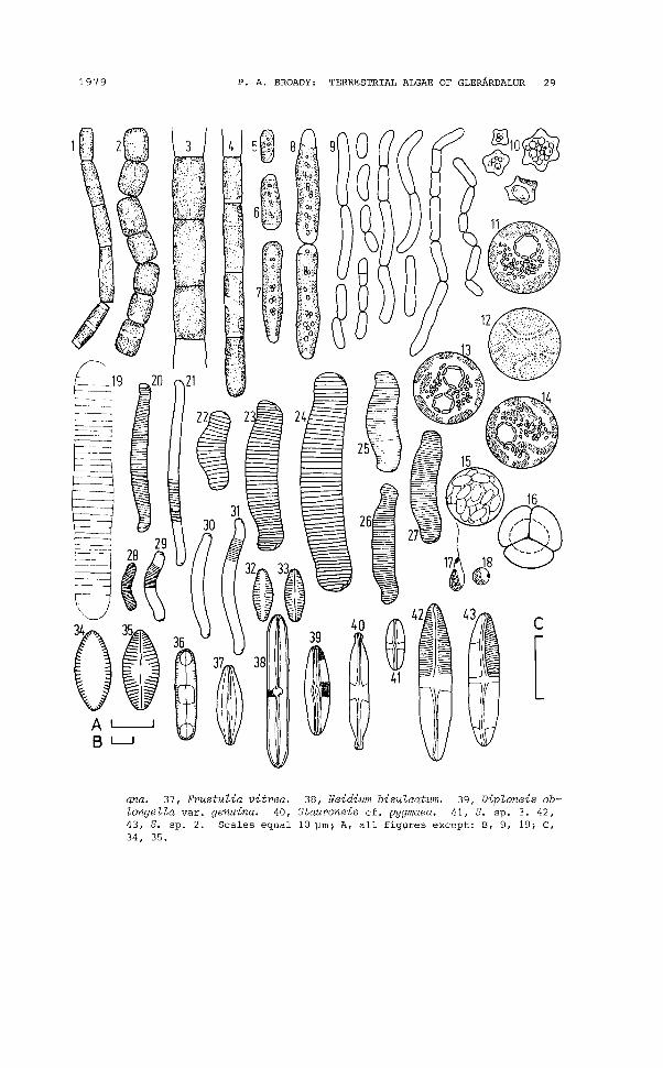
1979 P. A. BROADY: TERRESTRIAL ALGAE OF GLERARDALUR 29
4 5~ 8
619
40 ~ 42
41
@.. &.· •. 10~.·.. ·@~~~
c
[
ana. 37, Frustulia vitrea. 38, Neidium bisulcatum. 39, Diploneis oblongella var. genuina. 40, Stauroneis cf. pygmaea. 41, S. sp. 3. 42,43, S. sp. 2. Scales equal 10 ~m; A, all figures except: B, 9, 19; C,34, 35.

30 ACTA BOTANICA I8LANDICA NO. 5
TRIBONEMA MINUS Hazen(Fig. 6.4; 820, 24; PA8CHER, 1939)
Filaments long, 4.5 vm wide, with cells barely swollen at centre;cells 11-24 vm long, containing two or three chromatophores; "H"shaped walls sometimes clearly visible in healthy material.
EU8TIGMATOPHYCEAE
MONODUS SUBTERRANEUS Boye Pet.(Fig. 5.42, 43; 81-24; PETER8EN, 1932a)
Cells pyriform, sometimes slightly curved, 4-11 vm long by 1.5-4~
wide; chromatophore parietal, plate-like; reproduction by formation of two or four autospores.
WHITTLE and CA88ELTON (1975) put forward strong evidence on thebasis of pigment composition for the transfer of M BubterraneuBfrom the Xanthophyceae to the Eustigmatophyceae.
SCHWABE and BEHRE (1972).
VISCHERIA PUNCTA TA Visch.(Fig. 6.10; 82, 3, 6, 10; PA8CHER, 1939)
Cells irregularly star-shaped with a warty wall, 3.5-9.5 ~m diameter; all specimens observed had a high oil content and thechromatophore was only occasionally visible.
This alga was transferred from the Xanthophyceae to the Eustigmatophyceae by HIBBERD and LEEDALE (1972).
Unidentified sp.(Fig. 6.11-18; 82, 5, 9, 10, 13, 20, 21)
Cells spherical up to 24 ~m diameter, smallest cells 5 ~m diameter; chromatophore yellow-green, parietal, fissured (Fig. 6.12,surface view) and in larger cells with inward projections (Fig.6.13, 14), no starch revealed on treatment with Lugol-s iodine;pyrenoid polygonal, usually single, occasionally two, surroundedby large granules, often adpressed to the inner surface of, thoughnot embedded in, the chromatophore; vegetative division occurringwith formation of two or four cells (Fig. 6.16); zoospores (Fig.6.17) naked, becoming spherical on quiescence (Fig. 6.18), uniflagellate with a single basal chromatophore and an anterior orange stigma independent of the chromatophore, formed in sporangiacontaining 16 (?) spores (Fig. 6.15); aplanospores also formed.
This alga has been placed in the Eustigmatophyceae because itpossesses several of the features by which the class has been distinguished by HIBBERD and LEEDALE (1972), namely: zoospores witha single chromatophore, single flagellum and an anterior stigmaremote from the chromatophore; pyrenoid only in vegetative cells,attached to the inner surface of the chromatophore, polygonal andsurrounded by large flattened vesicles; vegetative cell coccoid.It is hoped to make more complete observations on cultures beforepresenting a formal description of this probably new member of theclass.
1979 P. A. BROADY: TERRESTRIAL ALGAE OF GLERARDALUR 31
BACILLARIOPHYCEAE
DIATOMALES
DIATOMA HIEMALE var. MAIOR novo var.(Fig. 6.19; S24)
Only two specimens observed; frustules lightly silicified withsepta faintly visible and striae not resolved; valves 125 ~ longby 17 ~m wide, with a slight central swelling; septa 3.5 in 10 ~.
D. hiemale var. anceps (Ehrenb.) A. Cl. has the largest frustules of described varieties, 12-100 ~ long by 4-7 ~ wide. Thepresent variety has longer and wider valves and lacks the capitate,attenuated apices of D. hiemale var. anceps. The density of septa lies in the range given for D. hiemale (Lyngb.) Reib. by CLEVEEULER (l953b).
EUNOTIALES
EUNOTIA EXIGUA var. COMPACTA Rust.(Fig. 6.20; S13; HUSTEDT, 1930)
Valves 23-47 ~m long by 4 ~m wide, with obviously curved dorsalmargin, barely curved ventral margin and markedly capitate apices;striae 20 in 10 ~m.
PETERSEN (1928b), FOGED (1974) and SCHWABE (1970) all record E.exigua but this possesses valves with a greater curvature than thevariety.
EUNOTIA GRACILIS (Ehrenb.) Rabenh.(Fig. 6.21; S14, 16; HUSTEDT, 1959-1962)
Valves 54-90 ~m long by 4.5-5 ~m wide, slightly curved, with parallel margins; striae 12-13 in 10 ~m.
PETERSEN (1928a and 1928b), FOGED (1974) .
EUNOTIA PRAERUPTA Ehrenb.(Fig. 6.22-24; S16, 24; HUSTEDT, 1959-1962)
Valves 19-53 ~m long by 7.5-11 ~m wide; striae 10-13 in 10 ~m.
FOGED (1974).
EUNOTIA PRAERUPTA cf. var. MUSCICOLA Boye Pet.(Fig. 6.25-27; S2; HUSTEDT, 1959-1962).
Valves 24-30 ]lll long by 5-7 ~ wide; striae 15-17 in 10 ~.
This alga appears to lie somewhere in between E. praerupta var.muscicola and E. septentrionalis Oestrup, the former has 6-12 striae in 10 ~ and the latter 16-19. The shape of the former is mostsimilar to the present specimens with the ventral edge straight atthe poles and then becoming suddenly concave (Fig. 6.25, 27) al-
32 ACTA BOTANICA ISLANDICA NO. 5
similar to the present specimens with the ventral edge straight atthe poles and then becoming suddenly concave (Fig. 6.25, 27) although this was not obvious in all specimens (Fig. 6.26).
PETERSEN (1928a)records E. praerupta var. muscicola and FOGED(1974) E. septentrionalis.
EUNOTIA REPENS var. ARCUATA (Naeg.) A.CI.(Fig. 6.28-31; S7, 16, 24; CLEVE-EULER, 1953a)
Valves 8-33~ long by 2-3 ~ wide, curved with parallel margins;striae 25 in 10 ~.
Similar to E. lunaris (Ehrenb.) Hust. which, however, has fewerstriae, 12-20 in 10~. The present specimens had a smallerlength range than that given for E. repens var. arcuata by CLEVEEULER (1953a) of 20-50 ~.
ACHNANTHALES
ACHNANTHES LANCEOLATA f. ROSTRATA (Oestrup) Hust.(Fig. 6.32, 33; S7; HUSTEDT, 1959-1962)
Valves 12 ~ long by 5 ~ wide, ellipsoidal with slightly attenuated apices; striae 16 in 10 ~, slightly fewer at centre.
The rostrate apices in the present specimens are not so obviousas those illustrated by HUSTEDT (1959-1962).
FOGED (1974).
ACHNANTHES MONTANA Krasske(Fig. 6.34, 35; S4; HUSTEDT, 1959-1962)
Valves 13 ~ long by 6 ~~ wide, ellipsoidal; striae 20 in 10 ~.
HUSTEDT (1959-1962) illustrates a clear, axial area on thearaphid valve which is narrower than that of the present specimens.
NAVICULALES
CYMBELLA sp. 1(Fig. 7.35, 36; S24)
Valves 17-22 ~ long by 5 ~ wide, ellipsoidal with attenuated,rostrate apices; striae radiate 13 in 10 ~; clear, unilateral,central area visible on occasional valves (Fig. 6.36) and a single, large, unilateral punctum always present.
CYMBELLA sp. 2(Fig. 7.33, 34; S14, 16(24)
Valves 19-33 ~m long by 3-5 ~m wide; striae 12-15 in 10 ~m.
1979 P. A. BROADY: TERRESTRIAL ALGAE OF GLERARDALUR 33
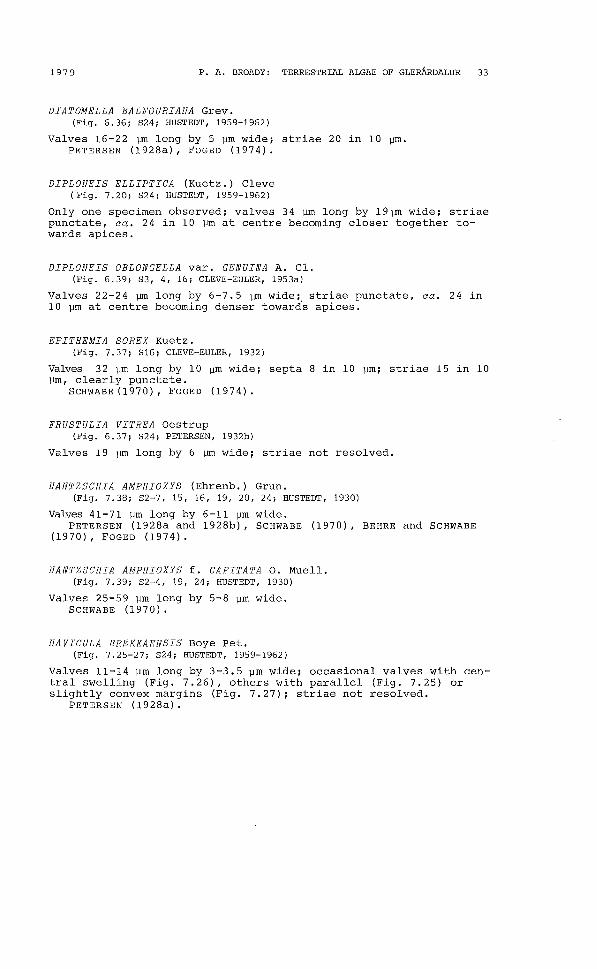
DIATOMELLA BALFOURIANA Grev.(Fig. 6.36; S24; HUSTEDT, 1959-1962)
Valves 16-22 ~ long by 5 vm wide; striae 20 in 10 Vm.PETERSEN (l928a), FOGED (1974).
DIPLONEIS ELLIPTICA (Kuetz.) Cleve(Fig. 7.20; S24; HUSTEDT, 1959-1962)
Only one specimen observed; valves 34 vm long by 19~ wide; striaepunctate, ca. 24 in 10 vm at centre becoming closer together towards apices.
DIPLONEIS OBLONGELLA var. GENUINA A. Cl.(Fig. 6.39; S3, 4, 16; CLEVE-EULER, 1953a)
Valves 22-24 vm long by 6-7.5 vm wide; striae punctate, ca. 24 in10 vm at centre becoming denser towards apices.
EPITHEMIA SOREX Kuetz.(Fig. 7.37; S16; CLEVE-EULER, 1932)
Valves 32 vm long by 10 Vm wide; septa 8 in 10 Vm; striae 15 in 10vm, clearly punctate.
SCHWABE(1970), FOGED (1974).
FRUSTULIA VITREA Oestrup(Fig. 6.37; S24; PETERSEN, 1932b)
Valves 19 vm long by 6 vm wide; striae not resolved.
HANTZSCHIA AMPHIOXYS (Ehrenb.) Grun.(Fig. 7.38; S2-7, 15, 16, 19,20,24; HUSTEDT, 1930)
Valves 41-71 vm long by 6-11 vm wide.PETERSEN (1928a and 1928b), SCHWABE (1970), BEHRE and SCHWABE
(1970), FOGED (1974).
HANTZSCHIA AMPHIOXYS f. CAPITATA O. Muell.(Fig. 7.39; S2-4, 19, 24; HUSTEDT, 1930)
Valves 25-59 vm long by 5-8 vm wide.SCHWABE (1970).
NAVICULA BREKKAENSIS Boye Pet.(Fig. 7.25-27; S24; HUSTEDT, 1959-1962)
Valves 11-14 vm long by 3-3.5 Vm wide; occasional valves with central swelling (Fig. 7.26), others with parallel (Fig. 7.25) orslightly convex margins (Fig. 7.27); striae not resolved.
PETERSEN (1928a).

34 ACTA BOTANICA ISLANDICA NO. 5
24
23
e
11
40
109 .8
32
20
31
o·
5
.......... " ...... "'v_39
14o
Fig. 7. 1, Pinnularia borealiB. 2, P. borealiB f. lanceolata. 3, P. divergenB var. elliptica. 4, P. divergentiBBima. 5, P. sp. 4. 6, 7, P.parva var. minuta. 8, 9, P. Btomatophora. 10, P. Bubcapitata var.hilBeana. 11, P. Bublanceolata f. 12, P. viridiB var. intermedia f.13, P. globicepB var. krookei. 14, Navicula sp. 9. 15, 16, N. contenta. 17-19, N. contenta cf. var. cruciata. 20, DiploneiB elliptica. 21, Navicula grimmei. 22, N. mutica var. cohnii. 23, N. Beminulum var.genuina. 24, N. BubtiliBBima. 25-27, N. brekkaenBiB.28, N. sp. 2. 29, N. sp.3. 30, N. Sp. 4. 31, 32, N. Sp. 6. 33,34, Cymbella sp. 2. 35, 36, C. Sp. 1. 37, Epithemia Borex. 38,HantzBchia amphioxyB. 39, H. amphioxyB f. capitata. 40, NitzBchiafruBtulum var. perpuBilla. Scales equal 10 ~m; A, all figures except: B, 3, 8, 12; C, 20.
1979 P. A. BROADY: TERRESTRIAL ALGAE OF GLERARDALUR 35
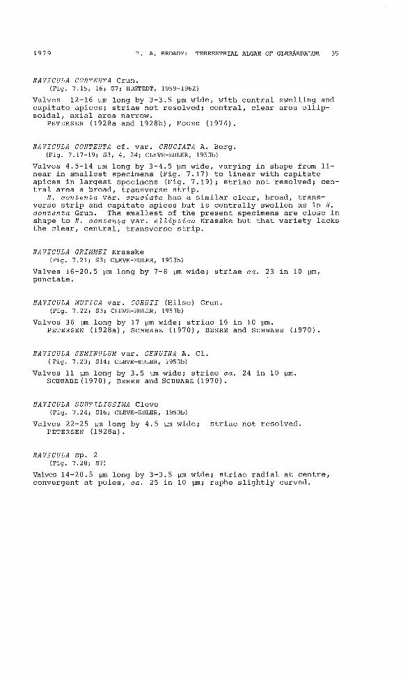
NAVICULA CONTENTA Grun.(Fig. 7.15, 16; S7; HUSTEDT, 1959-1962)
Valves 12-16 Vm long by 3-3.5 vm wide, with central swelling andcapitate apices; striae not resolved; central, clear area ellipsoidal, axial area narrow.
PETERSEN (1928a and 1928b), FOGED (1974).
NAVICULA CONTENTA cf. var. CRUCIATA A. Berg.(Fig. 7.17-19; S3, 4, 24; CLEVE-EULER, 1953b)
Valves 4.5-14 vm long by 3-4.5 vm wide, varying in shape from linear in smallest specimens (Fig. 7.17) to linear with capitateapices in largest specimens (Fig. 7.19); striae not resolved; central area a broad, transverse strip.
N. contenta var. cruciata has a similar clear, broad, transverse strip and capitate apices but is centrally swollen as in N.contenta Grun. The smallest of the present specimens are close inshape to N. conten~a var. eZZiptica Krasske but that variety lacksthe clear, central, transverse strip.
NAVICULA GRIMMEI Krasske(Fig. 7.21; S3; CLEVE-EULER, 1953b)
Valves 16-20.5 vm long by 7-8 vm wide; striae ca. 23 in 10 Vm,punctate.
NAVICULA MUTICA var. COHNII (Hilse) Grun.(Fig. 7.22; S3; CLEVE-EULER, 1953b)
Valves 36 ~ long by 17 Vm wide; striae 16 in 10 Vm.PETERS EN (1928a), SCHWABE (1970), BEHRE and SCHWABE (1970).
NAVICULA SEMINULUM var. GENUINA A. Cl.(Fig. 7.23; S14; CLEVE-EULER, 1953b)
Valves 11 vm long by 3.5 VIDwide; striae ca. 24 in 10 vm.SCHWABE (1970), BEHRE and SCHWABE (1970).
NAVICULA SUBTILISSIMA Cleve(Fig. 7.24; S16; CLEVE-EULER, 1953b)
Valves 22-25 vm long by 4.5 VID wide; striae not resolved.PETERSEN (1928a).
NAVICULA sp. 2(Fig. 7.28; S7)
Valves 14-20.5 vm long by 3-3.5 vm wide; striae radial at centre,convergent at poles, ca. 25 in 10 VIDi raphe slightly curved.
36 ACTA BOTANICA 18LANDICA NO. 5
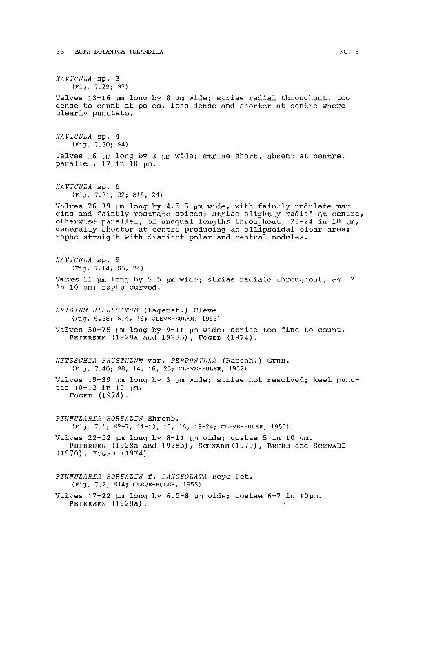
NA VICULA sp. 3(Fig. 7.29; 82)
Valves 13-16 ~ long by 8 ~m wide; striae radial throughout, toodense to count at poles, less dense and shorter at centre whereclearly punctate.
NA VICULA sp. 4(Fig. 7.30; 84)
Valves 16 ~m long by 3 ~m wide; striae short, absent at centre,parallel, 17 in 10 ~m.
NA VICULA sp. 6(Fig. 7.31, 32; 816, 24)
Valves 26-39 ~m long by 4.5-5 ~m wide, with faintly undulate margins and faintly rostrate apices; strtae slightly radial at centre,otherwise parallel, of unequal lengths throughout, 20-24 in 10 ~m,
generally shorter at centre producing an ellipsoidal clear area;raphe straight with distinct polar and central nodules.
NA VICULA sp. 9(Fig. 7.14; 83, 24)
Valves 11 ~m long by 5.5 ~m wide; striae radiate throughout, ca. 25in 10 ~m; raphe curved.
NEIDIUM BISULCATUM (Lagerst.) Cleve(Fig. 6.38; 814, 16; CLEVE-EULER, 1955)
Valves 50-75 ~m long by 9-11 ~m wide; striae too fine to count.PETER8EN (l928a and 1928b) , FOGED (1974).
NITZSCHIA FRUSTULUM var. PERPUSILLA (Rabenh.) Grun.(Fig. 7.40; 88, 14, 16, 23; CLEVE-EULER, 1952)
Valves 19-39 ~m long by 3 ~m wide; striae not resolved; keel punctae 10-12 in 10 ~.
FOGED (1974).
PINNULARIA BOREALIS Ehrenb.(Fig. 7.1; 82-7, 11-13, 15, 16, 18-24; CLEVE-EULER, 1955)
Valves 22-52 ~ long by 8-11 ~ wide; costae 5 in 10 ~m.
PETER8EN (1928a and 1928b), SCHWABE (1970), BEHRE and SCHWABE(1970), FOGED (1974).
PINNULARIA BOREALIS f. LANCEOLATA Boye Pet.(Fig. 7.2; 814; CLEVE-EULER, 1955)
Valves 17-22 ~ long by 6.5-8 ~m wide; costae 6-7 in 10~m.
PETER8EN (1928a).

1979 P. A. BROADY, TERRE8TRIAL ALGAE OF GLERARDALUR 37
PINNULARIA DIVERGENS var. ELLIPTICA Grun.(Fig. 7.3; 814; CLEVE-EULER, 1955)
Valves 118-160 ~m long by 25-31 ~m wide; costae 7-8 in 10 ~m.
FOGED (1974).
PINNULARIA DIVERGENTISSIMA (Grun.) Cleve(Fig. 7.4; 814; CLEVE-EULER, 1955)
Valves 30 ~m long by 5 ~m wide; costae 14 in 10 ~m.
FOGED (1974).
PINNULARIA GLOBICEPS var. KROOKEI Grun.(Fig. 7.13; 814; CLEVE-EULER, 1955)
Valves 19-28 ~m long by 5 ~m wide; costae 16 in 10 ~m at centre,more at apices.
PETER8EN (1928a and 1928b).
PINNULARIA PARVA var. MINUTA aestrup(Fig. 7.6, 7; 82; PETER8EN, 1928)
Valves 14-22 ~m long by 4.5-5 ~m wide; costae 9-10 in 10 ~m; thereare either no central costae (Fig. 7.6) or a short unilateral central costa (Fig. 7.7).
PETER8EN (1928a and 1928b)
PINNULARIA STOMATOPHORA Grun.(Fig. 7.8, 9; 816; CLEVE-EULER, 1955)
Valves 71-101 ~m lo~g by 8-11 ~m wide; costae 12-14 in 10 ~m.
PETER8EN (1928a and 1928b), FOGED (1974).
PINNULARIA SUBCAPITATA var. HILSEANA (Jan.) a.M.(Fig. 7.10; 84, 7, 14, 16, 24; CLEVE-EULER, 1955)
Valves 28-38 ~m long by 4-5 ~m wide; costae 10-13 in 10 ~m.
PINNULARIA SUBLANCEOLATA (Boye Pet.) A. Cl. f.8yn. PinnuZaria subcapitata var. subZanceoZata Boye Pet.(Fig. 7.11; 82, 4, 16; CLEVE-EULER, 1955)
Valves 22-38 ~ long by 3.5-5 ~m wide; costae 8-12 in 10 ~m.
P. subZanceoZata has slightly more costae/ 11-14 in 10 ~m/
faintly attenuated poles and is only 17-22 ~m long.
PINNULARIA VIRIDIS var. INTERMEDIA Cleve f.(Fig. 7.12; 814, 16, 24; CLEVE-EULER, 1955)
Valves 66-159 ~m long by 10-20 ~m wide; costae 8-10 in 10 ~.
The apices of the present specimens are somewhat less broadlyrounded than those of P. viridis var. intermedia.
FOGED (1974).
38 ACTA BOTANICA 18LANDICA NO. 5
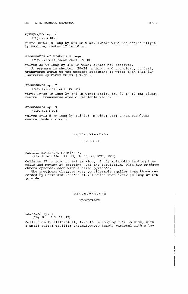
PINNULARIA sp. 4(Fig. 7.5; 816)
Valves 38-53 ~ long by 5-8 ~m wide, linear with the centre slightly swollen; costae 12 in 10 ~m.
STAURONEIS cf.PYGMAEA Krieger(Fig. 6.40; 84; CLEVE-EULER, 1953b)
Valves 28 ~ long by 4.5 ~ wide; striae not resolved.S. pygmaea is shorter, 20-24 ~ long, and the clear, central,
transverse strip of the present specimens is wider than that illustrated by CLEVE-EuLER (1953b).
STAURONEIS sp. 2(Fig. 6.42, 43; 82-4, 16, 24)
Valves 19-38 ~ long by 5-8 ~ wide; striae ca. 20 in 10 ~m; clear,central, transverse area of variable width.
STAURONEIS sp. 3(Fig. 6.41; 824)
Valves 8-12.5 ~m long by 3.5-4.5 ~m wide; striae not resolved;central nodule clear.
EUGLENOPHYCEAE
EUGLENALES
EUGLENA MUTABILIS Schmitz f.(Fig. 8.1-4; 82-4, 11, 13, 16, if, 21; ETTL, 1960)
Cells ca.27 ~ long by 2-4 ~ Hide, highly metabolic lacking flagella and moving by creeping (?er the substratum, with two or threechromatophores, each with a naked pyrenoid.
The specimens observed were considerably smaller than those recorded by BEHRE and SCHWABE (1970) which were 50-60 ~m long by 6-8~ wide.
CHLOROPHYCEAE
VOLVOCALES
CARTERIA sp. 1(Fig. 8.5; 813, 14, 19)
Cells broadly ellipsoidal, 12.5-16 ~ long by 7-12 ~ wide, witha small apical papilla; chromatophore thick, parietal with a la-
1979
teral pyrenoid.
P. A. BROADY: TERRESTRIAL ALGAE OF GLERARDALUR 39
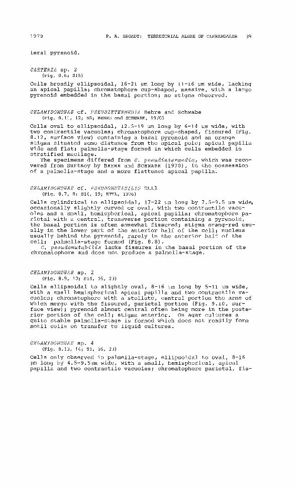
CARTERIA sp. 2(Fig. 8.6; S15)
Cells broadly ellipsoidal, 16-21 vm long by 11-16 vm wide, lackingan apical papilla; chromatophore cup-shaped, massive, with a largepyrenoid embedded in the basal portion; no stigma observed.
CHLAMYDOMONAS cf. PSEUDINTERMEDIA Behre and Schwabe(Fig. 8.11, 12; S8; BEHRE and SCHWABE, 1970)
Cells oval to ellipsoidal, 12.5-19 vm long by 6-14 vm wide, withtwo contractile vacuoles; chromatophore cup-shaped, fissured (Fig.8.12, surface view) containing a basal pyrenoid and an orangestigma situated some distance from the apical pole; apical papillawide and flat; palmella-stage formed in which cells embedded instratified mucilage.
The specimens differed from C. pseudintermedia, which was recovered from Surtsey by BEHRE and SCHWABE (1970), in the possessionof a palmella-stage and a more flattened apical papilla.
CHLAMYDOMONAS cf. PSEUDOMUTABILIS Ettl(Fig. 8.7, 8; S14, 19; ETTL, 1976)
Cells cylindrical to ellipsoidal, 17-22 vm long by 7.5-9.5 vm wide,occasionally slightly curved or oval, with two contractile vacuoles and a small, hemispherical, apical papilla; chromatophore parietal with a central, transverse portion containing a pyrenoid,the basal portion is often somewhat fissured; stigma orang-red usually in the lower part of the anterior half of the cell; nucleususually behind the pyrenoid, rarely in the anterior half of thecell; palmella-stage formed (Fig. 8.8).
C. pseudomutabiZis lacks fissures in the basal portion of thechromatophore and does not produce a palmella-stage.
CHLAMYDOMONAS sp. 2(Fig. 8.9, 10; S14, 16, 23)
Cells ellipsoidal to slightly oval, 8-16 vm long by 5-11 vm wide,with a small hemispherical apical papilla and two contractile vacuoles; chromatophore with a stellate, central portion the arms ofwhich merge with the fissured, parietal portion (Fig. 9.10, surface view); pyrenoid almost central often being more in the posterior portion of the cell; stigma anterior. On agar cultures aquite stable palmella-stage is formed which does not readily formmotil cells on transfer to liquid cultures.
CHLAMYDOMONAS sp. 4(Fig. 8.13, 14; S3, 16, 23)
Cells only observed in palmella-stage, ellipsoidal to oval, 8-16vm long by 4.5-9.5vrn wide, with a small, hemispherical, apicalpapilla and two contractile vacuoles; chromatophore parietal, fis-

40 ACTA BOTANICA ISLANDICA
1~
....;
A L-....J
BLJ
NO. 5
Fig. 8. 1-4, Euglena mutabiZis f. 5, Carteria sp. 1. 6, C. sp. 2. 7,8,Chlamydomonas cf. pseudomutabilis. 9, 10, C. sp. 2. 11, 12, C. cf.pseudintermedia. 13, 14, C. Sp. 4. 15, 16, Chloromonas cf. clath-
1979 P. A. BROADY: TERRESTRIAL ALGAE OF GLERARDALUR 41
sured, containing usually a single but up to three pyrenoids; nostigma observed.
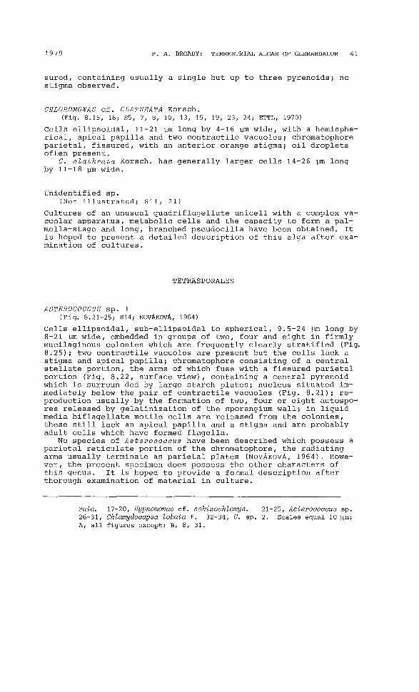
CHLOROMONAS cf. CLATHRATA Korsch.(Fig. 8.15, 16; SS, 7, 8, 10, 13, 15, 19, 23, 24; ETTL, 1970)
Cells ellipsoidal, 11-21 vm long by 4-16 urn wide, with a hemispherical, apical papilla and two contractile vacuoles; chromatophoreparietal, fissured, with an anterior orange stigma; oil dropletsoften present.
C. clathrata Korsch. has generally larger cells 14-26 vm longby 11-18 Vm wide.
Unidentified sp.(Not illustrated; Sll, 21)
Cultures of an unusual quadriflagellate unicell with a complex vacuolar apparatus, metabolic cells and the capacity to form a palmella-stage and long, branched pseudocilia have been obtained. Itis hoped to present a detailed description of this alga after examination of cultures.
TETRASPORALES
ASTEROCOCCUS sp. 1(Fig. 8.21-25; S14; NovAKovA, 1964)
Cells ellipsoidal, sub-ellipsoidal to spherical, 9.5-24 vm long by8-21 urn wide, embedded in groups of two, four and eight in firmlymucilaginous colonies which are frequently clearly stratified (Fig.8.25); two contractile vacuoles are present but the cells lack astigma and apical papilla; chromatophore consisting of a centralstellate portion, the arms of which fuse with a fissured parietalportion (Fig. 8.22, surface view), containing a central pyrenoidwhich is surroun ded by large starch plates; nucleus situated immediately below the pair of contractile vacuoles (Fig. 8.21); reproduction usually by the formation of two, four or eight autospores released by gelatinization of the sporangium wall; in liquidmedia biflagellate motile cells are released from the colonies,these still lack an apical papilla and a stigma and are probablyadult cells which have formed flagella.
No species of Asterococcus have been described which possess aparietal reticulate portion of the chromatophore, the radiatingarms usually terminate as parietal plates (NovAKovA, 1964). However, the present specimen does possess the other characters ofthis genus. It is hoped to provide a formal description afterthorough examination of material in culture.
rata. 17-20, Hypnomonas cf. schizochlamys. 21-25, Asterococcus sp.26-31, ChZamydocapsa lobata f. 32-34, C. sp. 2. Scales equal 10 Vm;A, all figures except: B, 8, 31.

42 ACTA BOTANICA ISLANDICA NO. 5
CHLAMYDOCAPSA LOBATA mh. f.(Fig. 8.26-31; S6-8, 12, 13, 16, 19, BROADY, 1977a)
Adult cells spherical, up to 16 ym diameter, young cells ellipsoidal and sub-sphaerical, from 7.5 Hm long by 4.5 ym wide, embedded in faintly stratified, mucilaginous colonies (Fig. 8.31) ingroups of two, four, eight and 16 or irregularly arranged; chromatophore parietal, deeply lobed, containing a prominent pyrenoidaurrounded by small starch grains in a thickened portion oppositewhich are two permanent contractile vacuoles (Fig. 8.26), in youngercells these tend to lie to one side (Fig. 8.28, 29); reproductionby formation of two, four, eight or 16 autospores or similar numbers of biflagellate, tunicate zoospores (Fig. 8.30) which possessa lateral chromatophore and a bar-shaped stigma.
The zoospores of C. tobata possess a small apical papilla, notobserved in the present specimens, and a smaller stigma.
CHLAMYDOCAPSA sp. 2(Fig. 8.32-34; S22)
Cells arranged in groups of four, eight and 16 in faintly stratified, mucilaginous colonies, the stratifications are limited tosingle lines around cell groups; adult cells broadly ellipsoidal,oval, sub-spherical or spherical, up to 22 ym by 19 ym, youngcells ellipsoidal or slightly oval, from 8 ym by 5 ym; chromatophore parietal, thick, covering most of wall except for apicalopening containing two contractile vacuoles, deeply fissured although these often do not pass completely trhough the chromatophore, surrounded by numerous small starch grains; reproductionby formation of four, eight or 16 autospores or zoospores; zoospores (Fig. 8.34) of~en oval, but also ellipsoidal, with a faintspot-like stigma.
Unfortunately a culture of this alga was lost and a more complete description cannot be made. It is very similar to the previous alga in its major features.
HYPNOMONAS cf. SCHIZOCHLAMYS Korsch.(Fig. 8.17-20; S13; KORSCHIKOFF, 1953)
Cells spherical to ellipsoidal, up to 16 ym diameter; chromatophore single, pareital, deeply lobed containing a prominent pyrenoid or two pyrenoids (Fig. 8.18); a pair of permanent contractilevacuoles present between chromatophore lobes; reproduction by fouror eight autospores and two, four, eight or 16 zoospores; zoospores tunicate, biflagellate, 8 ym long by 3.5 ym wide, often slightly curved.
The zoospores of H. schizochtamys are oval with a small apicalpapilla.
CHLOROCOCCALES
CHARACIUM sp. 1(Fig. 9.1, 2, S21; BOURRELLY, 1966)
Cells ca. ellipsoidal with a short stalk and terminal button-like
1979 P. A. BROADY: TERRESTRIAL ALGAE OF GLERARDALUR 43
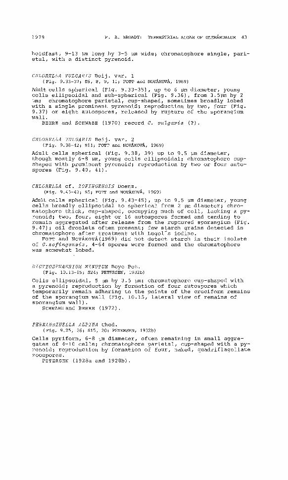
holdfast, 9-13 ~m long by 3-5 ~m wide; chromatophore single, parietal, with a distinct pyrenoid.
CHLORELLA VULGARIS Beij. var. 1(Fig. 9.33-37; SS, 8, 9, 11; FOTT and NovAKOVA, 1969)
Adult cells spherical (Fig. 9.33-35), up to 6 ~m diameter, youngcells ellipsoidal and sub-spherical (Fig. 9.36), from 3.5~ by 2~; chromatophore parietal, cup-shaped, sometimes broadly lobedwith a single prominent pyrenoid; reproduction by two, four (Fig.9.37) or eight autospores, released by rupture of the sporangiumwall.
BEHRE and SCHWABE (1970) record C. vuZgaris (?).
CHLORELLA VULGARIS Beij. var. 2(Fig. 9.38-42; Sll; FOTT and NOVAKovA, 1969)
Adult cells spherical (Fig. 9.38, 39) up to 9.5 ~m diameter,though mostly 6-8 ~m, young cells ellipsoidal; chromatophore cupshaped with prominent pyrenoid; reproduction by two or four autospores (Fig. 9.40, 41).
CHLORELLA cf. ZOFINGENSIS Doenz.(Fig. 9.43-47; SS; FOTT and NOVAKOVA, 1969)
Adult cells spherical (Fig. 9.43-45), up to 9. 5 ~m diameter, youngcells broadly ellipsoidal to spherical from 2 ~ diameter; chromatophore thick, cup-shaped, occupying much of cell, lacking a pyrenoid; two, four, eight or 16 autospores formed and tending toremain aggregated after release from the ruptured sporangium (Fig.9.47); oil droplets often present; few starch grains detected inchromatophore after treatment with Lugol-s iodine.
FOTT and NOVAKovA(1969) did not detect starch in their isolateof C.zofingensis, 4-64 spores were formed and the chromatophorewas somewhat lobed.
DICTYOSPHAERIUM MINUTUM Boye Pet.(Fig. 10.13-15; S24; PETERSEN, 1932b)
Cells ellipsoidal, 5 ~ by 3.5 ~m; chromatophore cup-shaped witha pyrenoid; reproduction by formation of four autospores whichtemporarily remain adhering to the points of the cruciform remainsof the sporangium wall (Fig. 10.15, lateral view of remains ofsporangium wall).
SCHWABEand BEHRE (1972).
FERNANDINELLA ALPINA Chod.(Fig. 9.25, 26; S15, 20; PETERSEN, 1932b)
Cells pyriform, 6-8 ~m diameter, often remaining in small aggregates of 4-10 cells; chromatophore parietal, cup-shaped with a pyrenoid; reproduction by formation of four, naked, quadriflagellatezoospores.
PETERS EN (1928a and 1928b).

44 ACTA BOTANICA ISLANDICA NO. 5
~1 2
Fig. 9.t1~ 2, Characium Sp. 1 3-z-s ef "' • -6 ef S 'd' . ove-z-fera. 17-2 ' . pong-z-ococcum-z-nella alpina. 27-32 4Kee~. Rhopalocystis euc~: 7-16, Rhopalocys, n rosphaera bristolae -z-~. 25, 26, Fernan-• 3-37, Chlorella

1979 P. A. BROADY: TERRESTRIAL ALGAE OF GLERARDALUR 45
cf. JAAGIOCHLORELLA GEOMETRICA Reisigl(Fig. 9.48-50; 517, 18, 24; REISIGL, 1964)
Cells broadly ellipsoidal, up to 17.5 ~m long by 14 pm wide, youngest cells from 4.5 pm by 3 ~m; chromatophore cup-shaped, lobed,sub-parietal in its basal portion (Fig. 9.48) with a faint pyrenoid lacking a starch sheath, but many starch grains throughoutchromatophore; reproduction by formation of two to many (ca. 30)aplanospores which are closely packed in the sporangia and assumepolygonal shapes (Fig. 9.50).
The polygonal spores in the sporangia and the sub-parietalchromatophore are similar characters to those of J. geometrica butthe 'cells of that species are spherical and small, only ca. 4 pmdiameter.
KENTROSPHAERA BRISTOLAE G. M. Srn.(Fig. 9.27-32; 520; SMITH, 1933)
Cells spherical, ellipsoidal and irregularly shaped, 16-140 pm butmostly 30-60 pm long, often with an apical wall thickening whichis often faintly lamellate, occasionally more thickenings (Fig. 9.31); chromatophore axial with numerous radiating lobes which spreadto form irregular plates against the cell wall (Fig. 9.29, surfaceview) pyrenoid usually single, central, with a thick starch sheath,occasionally two or three pyrenoids present; spore formation notobserved.
MURIELLA cf. TERRESTRIS Boye Pet.(Fig. 10.1-8; 59; PETERSEN, 1932a)
Cells spherical to sub-spherical, up to 14.5 pm diameter but mostly 8-11 pm, occasionally with small papilla-like wall thickenings(Fig. 10.1), smallest cells 5 pm diameter; chromatophore single inrecently released aplanospores (Fig. 10.6), several in largestcells (Fig. 10.3-5); pyrenoids absent; starch grains scatteredthroughout chromatophores; reproduction by formation of two, four,eight and 16 aplanospores released by rupture of sporangium wall.
M. terrestris has smaller cells, 3-7 pm diameter, and does notpossess the small papillae seen in the present specimens.
SCHWABE (1970), BEHRE and SCHWABE (1970) record M. terrestris.
cf. OOCYSTIS sp.(Fig. 10.9-12; 513; BOURRELLY, 1966)
Cells ellipsoidal but often with a greater curvature on one side(Fig. 10.10), 9-14 pm long by 3.5-8 pm wide, with slightly thickened apical walls; chromatophore parietal, often lobed, containingfrom one to three pyrenoids; reproduction by formation of two,four or eight autospores released by rupture of the sporangium
vulgaris var. 1. 38-42, C. vulgaris var. 2. 43-47, C. cf. zOfingensis. 48-50, cf. Jaagiochlorella geometrica. 51-55, Planktosphaerella terrestris. Scales equal 10 pm; A, 1, 2, 7-24, 33-47;B, 27-29, 51-55; C, 3, 4, 25, 26, 48-50, D, 5, 6, 30-32.

46 ACTA BOTANICA ISLANDICA NO. 5
wall (Fig. 10.12).Oocystis spp. are often characterized by the autospores remai
ning within a greatly expanded sporangium wall before their eventual release, this was not observed in the present specimens.However, cell shape, the thickened apices, and reproduction byautospores are characteristic of that genus.
PLANKTOSPHAERELLA TERRESTRIS Reisigl(Fig. 9.51-55; S5-7, 22; REISIGL, 1964)
Cells spherical up to 13 ~m diameter, smallest released sporesfrom 3 ~ diameter; chromatophores parietal, each with a singlepyrenoid, numerous in adult cells (Fig. 9.51, 52), single in smallest spores (Fig. 9.55); reproduction by formation of two, four,eight, 16 or 32 autospores.
cf. RHOPALOCYSTIS CUCUMIS Reisigl( Fig. 9.17-24; S21; REISIGL, 1964)
Cells ca. pyriform, 13-18 ~ long by 6.5-11 ~ wide, often slightly curved at the narrow apex (Fig. 9.18); chromatophore parietal,often appearing bilobed, with a single pyrenoid; oil globulesoften present; reproduction by spherical aplanospores (Fig. 9.19-22), from two to 32 per sporangium, or by zoospores (Fig. 9.23, 24),sporangia containing numerous spores; zoospores equally biflagellate, spherical to pyriform, ca. 4.5 ~ in diameter, releasedthrough an apical rupture.
R. cucumis has similarly shaped cells and reproduction. Thecells, however, are up to 30 ~ long. BOURRELLY (1966) statesthat after zoosporulation in RhopaZocystis a portion of the sporangium remains in the base of the otherwise emptied cell. REISIGL (1964) does not describe this for R. cucumis nor was it observed in the present specimen. There would appear to be sometaxonomic confusion here (cf. the species described below).
RHOPALOCYSTIS cf. OLEIFERA Schuss.(Fig. 9.7-16; S2, 4-7, 14, 16, 19, 20; SCHUSSNIG, 1955)
Cells pyriform to ca. cylindrical, often slightly curved, 6-17 ~m
long by 2-8 ~m wide (Fig. 9.7-10), occasionally a small, apicalpapilla is visible (Fig. 9.8 and Fig. 9.16, on a recently settledzoospore); chromatophore parietal with a prominent pyrenoid; reproduction by autospores, only one sporangium observed with two spores (Fig. 9.11), and zoospores which are formed in large numbers(Fig. 9.15) ; after zoosporulation a portion of the mother cell, oroccasionally two separate portions, remains in the base of thesporangium wall (Fig. 9.12-14), this then regrows, if zoosporulation again occurs then the basal remaining portion is surroundedby two aId sporangium walls (Fig. 9.12).
This alga has the characters of RhopaZocystis described by BOU~
RELLY (1966). R. oZeifera is the only species described in whicha portion of the mother cell remains in the base of the old sporangium wall. However, the cells of R. oZeifera are up to 30 ~m
long by 12-14 lJll wide.

1979 P. A. BROADY, TERRESTRIAL ALGAE OF GLERARDALUR 47
SCOTIELLA cf. OOCYSTIFORMIS Lund(Fig. 10.16-22; 52, 3, 6, 7, 9-11, 13, 20-22, 24; LUND, 1957)
Cells ellipsoidal to broadly fusiform, 9-22 ~m long by 5-16 ~m
wide; in cultures the cell wall thickenings are often not visible(Fig. 10.16) or only faintly visible as small polar thickenings(Fig. 10.18), occasional cells have more readily visible ridges(Fig. 10.20) which in apical view are slightly spiralled (Fig. 10.21); chromatophore parietal, single in young cells, occasionallyfissured (Fig. 10.17) and in older cultures fragmenting into numerous portions (Fig. 10.19); pyrenoid single, prominent; reproduction by four or eight autospores (Fig. 10.22).
S. oocystiformis has similarly sized cells with ridges whichare often not detectable. However, two to sixteen autospores areformed and in apical view the cells have 8-10 angles. Further observations of cultures are necessary before the present alga canbe confidently assigned to this species.
SPHAEROCYSTIS cf. SIGNIENSIS mho(Fig. 10.31-33; 58; BROADY, 1976)
cells in groups of two and four or irregularly arranged throughouthomogeneous, mucilaginous colonies (Fig. 10.31); adult cells spherical, 7.5-9.5 ~ diameter, chromatophore cup-shaped and lobedwith a basal pyrenoid surrounded by large starch grains; oil droplets often present; reproduction by ellipsoidal to subsphaericalautospores, two or four released by rupture (Fig. 10.33) or gelatinization of sporangium wall.
S. signiensis has a larger size range with adult cells up to15 ~m diameter, the colonial mucilage is sometimes faintly stratified and the pyrenoid is surrounded by small starch grains.
SPHAEROCYSTIS sp. 1(Fig. 10.23-26; SS)
Cells in groups of four and eight or irregularly arranged throughout homogeneous, mucilaginous colonies; adult cells broadly ellipsoidal (Fig. 10.24, 25), up to 11 ~m long by 9.5 ~m wide, with alobed, parietal chromatophore containing and indistinct pyrenoidlacking a starch sheath; starch grains revealed in the chromatophore using Lugol-s iodine; oil globules often present; reproduction by ellipsoidal autospores from 6.5 ~ long by 5 ~ wide (Fig.10.26), released by partial gelatinization and rupture of sporangium wall.
This alga is in many respects similar to the three species ofSphaerocystis described by BROADY (1976) but cannot be assignedto any of these.
SPHAEROCYSTIS sp. 2(Fig. 10.27-30; 52, 6, 7, 9, 10, 16, 20)
Cells in groups of four and eight and irregularly arranged throughout homogeneous, mucilaginous colonies; adult cells broadly ellipsoidal and spherical, up to 9.5 ~ long by 6.5 ~ wide, with a cupshaped chromatophore occupying about half the wall and containinga basal pyrenoid surrounded by small starch grains; oil globules
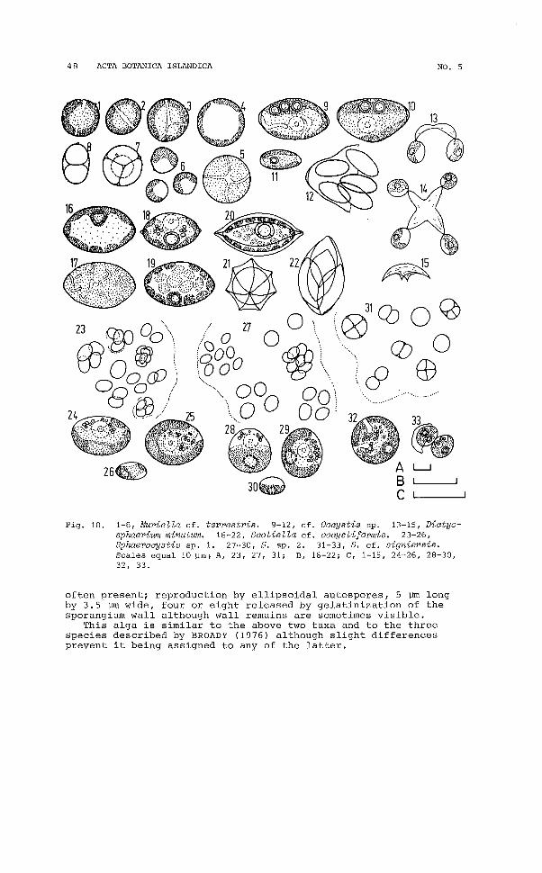
48 ACTA BOTANICA ISLANDICA NO. 5
L..J
.......
31Q) 0 ~
..~ 0 0
\..... & EB................
'G}27 0
(,'0& 0 cCih/tJOO W
..... \ 000 00\..,0 00
29
Fig. 10. 1-8, Muriella cf. terrestris. 9-12, cf. Oocystis sp. 13-15, Dictyosphaerium minutum. 16-22, Scotiella cf. oocystiformis. 23-26,Sphaerocystis sp. 1. 27-30, S. Sp. 2. 31-33, S. cf. signiensis.Scales equal 10 ~m; A, 23, 27, 31; B, 16-22; C, 1-15, 24-26, 28-30,32, 33.
often present; reproduction by ellipsoidal autospores, 5 ~ longby 3.5 ~ wide, four or eight released by gelatinization of thesporangium wall although wall remains are sometimes visible.
This alga is similar to the above two taxa and to the threespecies described by BROADY (1976) although slight differencesprevent it being assigned to any of the latter.

1979 P. A. BROADY: TERRESTRIAL ALGAE OF GLERARDALUR 49
cf. SPONGIOCOCCUM sp.(Fig. 9.3-6; S28; BOURRELLY, 1966)
Cells spherical, up to 47 vm diameter, with a thickened wall 1.56.5 vm wide, often with irregular wall thickenings (Fig. 9.5, 6)or occasionally with a regularly much thickened wall (Fig. 9.4);chromatophore sponge-like with an irregularly shaped central pyrenoid possessing a thick starch sheath; aplanospore productionobserved in a sporangium 84 ym diameter with spores varying indiameter from 11 to 50 vrn.
In the absence of cultures in which life-history could be studies it is impossible to assign this alga to either of the two similar genera Spongiococcum and Spongiochloris which can only beseparated on the basis of the presence or absence of a cell wallaround the zoospores.
Unidentified spp.
Eleven free-living, spherical members of the Chlorococcales havebeen isolated into culture and recognized as different taxa. Theyinclude species of Chlorococcum and other as yet undeterminedgenera. It is hoped to present descriptions of these after exa- emination of their morphologies and life-histories.
ULOTHRICALES
CHLORHORMIDIUM DISSECTUM (Chod.) Fott f.Syn. Hormidium dissectum (Gay) Chad.(Fig. 11.1-4; S2, 5, 6, 9, 10, 12, 15, 18; PRINZ, 1964)
Filaments short, readily fragmented, from two to 14 cells inlength (Fig. 11.3, 4); cells 6 vrn wide by 8-22 ym long.
The width range of C. dissectum is 7-9 vrn, slightly more thanthe present specimens.
CHLORHORMIDIUM FLACCIDUM (A. Br.) FottSyn. Hormidium flaccidum A. Br.(Fig. 11.5, 6; S2, 15, 17, 20, 21; PRINZ, 1964)
Filaments long, 7.5-9.5 vrn Wide; cells 12-27 ym long.PETERSEN (1928b), SCHWABE and BEHRE (1972).
CHLORHORMIDIUM SUBTILE (Herr.) FottSyn. Hormidium subtile (Kuetz.) Heer.(Fig. 11.7, 8; S15, 24; PRINZ, 1964)
Filaments from two to more than 40 cells long, width 3-4ym; cells6-14.5 ym long; small grains of starch around the pyrenoid; oilglobules often present.
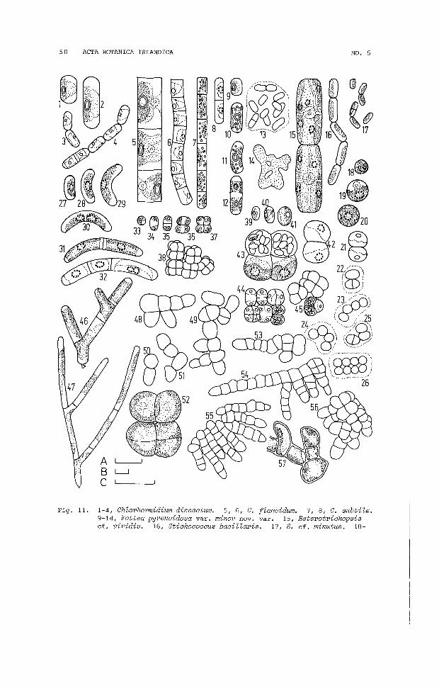
50 ACTA BOTANICA ISLANDICA NO. 5
Fig. 11. 1-4, Chlorhormidium dissectum. 5, 6, C. flaccidum. 7, 8, C. subtile.9-14, Fottea pyrenoidosa var. minor novo var. 15, Heterotriohopsiscf. viridis. 16, Stiohoooocus baoillaris. 17, S. cf. minutus. 18-

1979 P. A. BROADY: TERRESTRIAL ALGAE OF GLERARDALUR 51
FOTTEA PYRENOIDOSA var. MINOR novo var.(Fig. 11.9-14; s19)
Irregular colonies (Fig. 11.14, from a young agar culture) containing cells distributed irregularly throughout homogeneous mucilage (Fig. 11.13); cells sylindrical, straight or occasionallyslightly curved (Fig. 11.11), 4.5 ~m wide by 6-11 ~m long; chromatophore single, occasionally two in dividing cells (Fig. 11.12),parietal, plate-like, occupying more than half the wall and containing a single prominent pyrenoid, rarely two; oil globulesoften present; transverse cell division produces two daughtercells which remain temporarily united.
F. pyrenoidosa Broady (1976) differs in its larger size withcells 6 ~m wide by 6-18 ~m long, and in its possession of cellswhich are usually slightly curved.
HETEROTRICHOPSIS cf. VIRIDIS Iyengar and Kanthamma(Fig. 11.15; S2, 11; IYENGAR and KATHAMMA, 1941)
Cells single or in pairs, cylindrical, barely swollen centrallyand occasionally slightly curved, 10-11 ~m wide by 25-45 ~m long,containing two to four plate-like, parietal chromatophores eachwith one or two pyrenoids surrounded by large starch grains; nucleus central; transverse cell division occurs with the formationof two daughter cells.
H. viridis has smaller cells, 6-8 ~m wide by 16-33 ~m long andalso forms aplanospores, the structure of the vegetative cells isotherwise similar to the present specimens.
STICHOCOCCUS BACILLARIS Naeg. sens. ampl.(Sl, 2, 4, 6-8, 10, 12, 13, 15, 16, 19, 20, 22; PRINZ, 1964)
PETERS EN (1928a), BEHRE and SCHWABE (1970)
STICHOCOCCUS BACILLARIS ~aeg. sens. strict.(Fig. 11.16; S24; PRINZ, 1964)
Filaments of up to 22 loosely connected cells; cells 3-3.5 ~m
wide by 5-11 ~m long; chromatophore single, parietal.
STICHOCOCCUS cf. MINUTUS Grintzesco and Peterfi(Fig. 11.17; S24; PRINZ, 1964)
Filaments rarely as many as four cells long, cells usually singleor in pairs; cells 1.5-2.5 ~m wide by 3-6.5 ~m long, chromatophoresingle, parietal.
S. minutus has cells 2.5-3 ~m wide.
26, cf. PseudendocZoniopsis sp. 27-32, Unidentified species of theUlothricaceae. 33-38, ChZorosarcinopsis sp. 1. 39-45, C. sp. 2.46, Microthamnion kuetzingianum. 47, M. strictissimum f. 48-52,cf. Gongrosira terricoZa. 53-57, cf. Desmococcus sp. Scales A and
. B equal 10 ~m, C equals 1 mm; A, all figures except: B, 3, 4, 6,13, 21-26, 44, 45, 48-51, 53-56; C, 14.
52 ACTA BOTANICA 18LANDICA NO. 5
Unidentified genus of the Ulothricaceae(Fig. 11.27-32; 812)
Cells usually single, though occasionally in short filaments oftwo or three cells' (Fig. 11.31, 32), cylindrical, more or lesslunate, 4-5 ym wide by 9.5-24 ym long; chromatophore single, witha central pyrenoid surrounded by small starch grains; transversecell division.
The cells resemble those of Fottea pyrenoidosa mho in generalshape but lack that alga-s mucilaginous envelopes. Chodatia Kol,a doubtful member of the Chlorophyta (BOURRELLY, 1966), is similar in shape and lacks mucilage. However, it also lacks a pyrenoid. Unfortunately no cultures of this alga were obtained andit must remain undetermined.
CHAETOPHORALES
CHLOROSARCINOPSIS sp. 1(Fig. 11.33-38; 86, 15, 19; BOURRELLY, 1966)
Cells single, in pairs, fours, cubical aggregates of eight cellsor larger, readily fragmented cubical aggregates (Fig. 11.38);cells 3-5 ym diameter with a cup-shaped chromatophore containinga small pyrenoid; transverse cell division (Fig. 11.33-37).
Further observation of cultured material is required.
CHLOROSARCINOPSIS sp. 2(Fig. 11.39-45; 81, 9, 17, 22; BOURRELLY, 1966)
Cells up to 11 ym diameter, single, in pairs, cubical or tetrahedral packets (Fig. 11.45) and larger, readily fragmented cubical aggregates (Fig. 11.44); chromatophore cup-shaped with a somewhat irregular border, containing a pyrenoid surrounded by smallstarch grains (Fig. 11.43); aplanospore formation with two, four,eight or larger numbers of ellipsoidal to short cylindrical spores(Fig. 11.39, 40), from 4.5 ym long by 2 ym wide, being formed insingle cells and cells in aggregates (Fig. 11.43-45).
Further observation of cultured material is required.
cf. DESMOCOCCUS sp.(Fig. 11.53-57; 820, 22; BOURRELLY, 1966)
Colonies on agar consisting of a central mass of cells in more orless cubical aggregates (Fig. 11.56) with a peripheral growth ofrichly branching filaments (Fig. 11.53-55); cells 5-7 ym wide andup to 10 ym long, with a parietal chromatophore and a faint pyrenoid (Fig. 11.57), often packed with oil globules.
Spore formation was not observed and hence it is not possibleto confidently assign this alga to a genus. On vegetative characteristics it appears close to Desmococcus Brand.
1979 P. A. BROADY; TERRESTRIAL ALGAE OF GLERARDALUR 53
cf. GONGROSIRA TERRICOLA Bristol(Fig. 11.48-52; S12, 14; LUND, 1947)
Thalli small, consisting of a few loosely joined cells, oftenbranched or in small cubical packets; cells 11-16 ~m in diameterwith a parietal chromatophore containing a faint pyrenoid (Fig.11.52) .
The features of the vegetative cells are similar to those ofG. terricola described by LUND (1947). However, in the absenceof spore formation no confident identification can be made.
MICROTHAMNION KUETZINGIANUM Naeg.(Fig. 11.46; S2, 7, 11-13, 16-19; PRINZ, 1964)
Thalli consisting of richly branched filaments; cells 3-5 ~m wideand up to 15 ~m long; chromatophore parietal lacking a pyrenoid;small holdfast often distinguishable on basal cell of small thalli, as shown in figure.
PETERS EN (1928a and 1928b).
MICROTHAMNION STRICTISSIMUM Rabenh. f.(Fig. 11.47; S7, 13, 17, 18; PRINZ, 1964)
Thalli of generally similar form to the above species; cells 1.52.0 ~m wide by 14-33 ~m long.
This species has a somewhat narrower width range than that given for M. strictissimum by PRINZ (1964) of 3-4 ~m. However, bothspecies have similar long cells.
cf. PSEUDENDOCLONIOPSIS sp.(Fig. 11.18-26; S21; BOURRELLY, 1966)
Cells spherical to ellipsoidal, 5-8 ~m in diameter, embedded insoft, homogeneous mucilage; chromatophore parietal, cup-shapedwith a lobed border and a basal pyrenoid surrounded by smallstarch grains (Fig. 11.18-20); vegetative cell division resultsin groups of two, four or eight cells being formed, the four cellgroups may open out to form short chains of loosely adhering cells(Fig. 11.23, 24); colonies from agar plate cultures when transferred to aqueous microscopic preparations split into separateaggregates of two to eight cells when pressure is applied to thecoverglass, a thin mucilage layer around each group is revealed byindian ink.
BOURRELLY (1966) provides a similar description for vegetativecells of Pseudendocloniopsis Vischer, however, spore formation asdescribed by him was not observed in the present specimens.

54 ACTA BOTANICA ISLANDICA
ZYGOPHYCEAE
ZYGNEMATALES
NO. 5
ACTINOTAENIUM CUCURBITA (Breb.) Teil.(Fig. 12.6; S14, 16; TElLING, 1954)
Cells in frontal view 41-64 ~m long by 19-27 ~m wide.
ACTINOTAENIUM CUCURBITA var. ATTENUATUM Teil f.(Fig. 12.7; S2, 4, 6, 18, 19, 21; TElLING, 1954)
Cells in frontal view 27-38 ~m long by 11-16 ~m wide.A. cucurbita var. attenuatum possesses somewhat wider cells
15-23 ~m wide.
cf. ACTINOTAENIUM PINICOLUM Rosa(Fig. 12.8-13; S15; ROSA, 1959)
Cells cylindrical, 6.5-14 ~ long by 3.5-6.5 ~m wide, single or intemprary pairs after cell division; chromatophore single, axial,or" occasionally double in dividing cells (Fig. 12.10), with a central pyrenoid surrounded by starch grains; wall appears to be composed of two halves although this is only apparent in occasionalcells which may have a slight central bUlge (Fig. 12.13) or aslight depression. A cell was observed in which division appearedto have been abortive (Fig. 12.12) and one half of one of the pairof new cells had failed to form, the remaining half of the original mother cell adhered to the single completely-formed daughtercell.
Unfortunately only a few spec)mens of this curious algae wereobserved in mixed cultures. It ppears to be a member of the Desmidiaceae due to the cell wall 1 "ing composed of two halves andthe possession of an axial chrc .atophore. However, there is nomarked median constriction and ,ygote formation was not observed.A. pinicoZum has a similar chr( ~atophore and similarly shapedcells although a faint median constriction is generally visibleand the apices are less broadly rounded, the wall is punctate andthe cells considerably larger being 25-29 ~ long by 7.5-9 ~m wide.
CLOSTERIUM PUSILLUM var. MAJOR Racib.(Fig. 12.14; S14; WEST and WEST, 1904)
Only two specimens observed, 102-110 ~m long by 15-16 ~ wide.WEST and WEST (1904) describe a smaller size range, 102-110 ~
long by 15-16 ~ wide.
Fig. 12. I, CyZindrocystis brebissonii. 2, Mesotaenium endZicherianum var.grande. 3, M. macrococeum. 4, Netrium digitus. 5, N. digitus var.curtum. 6, Actinotaenium cucurbita. 7, A. cueurbita var. attenuatum.8-13, cf. A. pinicoZum. 14, CZosterium pusiZZum var. major. 15, 16,
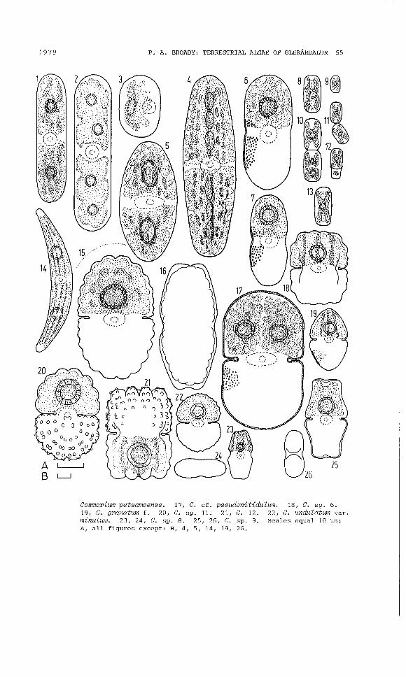
1979 P. A. BROADY: TERRESTRIAL ALGAE OF GLERARDALUR 55
Cosmarium petsamoense. 17, C. cf. pseudonitidulum. 18, C. sp. 6.19, C. granatum f. 20, C. Sp. 11. 21, C. 12. 22, C. undulatum var.minutum. 23, 24, C. Sp. 8. 25, 26, C. Sp. 9. Scales equal 10 ~m;
A, all figures except: B, 4, 5, 14, 19, 26.
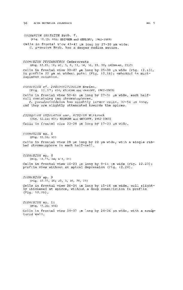
56 ACTA BOTANICA 18LANDICA NO. 5
COSMARIUM GRANATUM Breb. f.(Fig. 12.19; 816; KRIEGER and GERLOFF, 1962-1969)
Cells in frontal view 43-47 ~m long by 27-30 ~m wide.C. granatum Breb. has a deeper median suture.
COSMARIUM PETSAMOENSE Cedercreutz(Fig. 12.15, 16; 82, 3, 6, 11, 14, 16, 19, 20; GRONBLAD, 1933)
Cells in frontal view 39-47 ~m long by 25-30 ~m wide (Fig. 12.15),in profile 22 ~m at widest point (Fig. 12.16); embedded in mucilaginous colonies.
COSMARIUM cf. PSEUDONITIDULUM Nordst.(Fig. 12. 17; 814; KRIEGER and GERLOFF, 1962-1969)
Cells in frontal view 50-61 ~m long by 27-31 ~m wide, each halfcell containing two chromatophores.
C. pseudonitidulum has slightly larger cells, 37-54 ~m long,and they are slightly attenuated towards the apices.
COSMARIUM UNDULATUM var. MINUTUM Wittrock(Fig. 12.22; 814; KRIEGER and GERLOFF, 1962-1969)
Cells in frontal view 22-28 ~m long by 17-23 ~m wide.
COSMARIUM sp. 6(Fig. 12.18; 82)
Cells in frontal view 28 ~m long by 22 ~m wide, with a single ribbed chromatophore in each half-cell.
COSMARIUM sp. 8(Fig. 12.23, 24; 811, 21)
Cells in frontal view 16-23 ~m long by 9-11 ~m wide (Fig. 12.23);profile view without an apical depression (Fig. 12.24).
COSMARIUM sp. 9(Fig. 12.25, 26; 82, 3, 16, 20, 21)
Cells in frontal view 26-34 ~m long by 15-16 ~m wide, wall slightly thickened at apices, without a deep constriction in profile(Fig. 12.26).
COSMARIUM sp. 11(Fig. 12.20; 816)
Cells in frontal view 34-37 ~m long by 24-26 ~m wide, with a scu4rtured wall.

1979 P. A. BROADY: TERRESTRIAL ALGAE OF GLERARDALUR 57
oo
IlClO 000 0.. ' ~ .." ... ,
'11111!~ABL-I
CDu
Fig. 13. 1, Euastrum dubium f. 2, E. sp. 2. 3, E. oblongum. 4, E. sp. 4.5, Tetmemorus laevis. 6, Spondylosium cf. planum. Scales equal 10llffi; A, 1, 2, 4; B, 5; C, 6; D, 3.
COSMARIUM sp. 12(Fig. 12.21; S2, 20)
Cells in frontal view 31-36 vm long by 24-25 vm wide, with a scu4rtured wall, often occurring in short chains.
CYLINDROCYSTIS BREBISSONII (Ralfs) De Bary(Fig. 12.1; S2-4, 11-13, 16-19, 21; KRIEGER, 1937)
Cells cylindrical, 41-53 Vm long by 11-19 Vm wide.
EUASTRUM DUBIUM Naeg. f.(Fig. 13,1; S16; KRIEGER, 1937)
Cells in frontal view 27-28 vm long by 16-17.5 Vm wide.E. dubium has slightly wider cells, 17-26 vm wide.
EUASTRUM OBLONGUM (Grev.) Ralfs(Fig. 13.3; S14; KRIEGER, 1937)
Cells in frontal view 149-162 vm long by 69-72 vm wide; chromatophores not clearly observed.
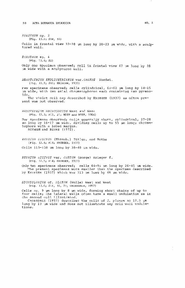
58 ACTA BOTANICA ISLANDICA NO. 5
EUASTRUM sp. 2(Fig. 13.2; S14, 16)
Cells in frontal view 33-38 ~ long by 20-23 ~ wide, with a scu4rtured wall.
EUASTRUM sp. 4(Fig. 13.4; S2)
Only one specimen observed; cell in frontal view 47 ~ long by 28~ wide with a sculptured wall.
MESOTAENIUM ENDLICHERIANUM var.GRANDE Nordst.(Fig. 12.2; S16; KRIEGER, 1937)
Few specimens observed; cells cylindrical, 61-62 ~m long by 14-15vm wide, with two axial chromatophores each containing two pyrenoids.
The violet cell sap described by KRIEGER (1937) as often present was not observed.
MESOTAENIUM MACROCOCCUM West and West(Fig. 12.3; S12, 21; WEST and WEST, 1904)
Few specimens observed; cells generally short, cylindrical, 27-28~m long by 16-17 vm wide, dividing cells up to 55 vm long; chromatophore with a lobed margin.
SCHWABE and BEHRE (1972).
NETRIUM DIGITUS (Ehrenb.) Itzigs. and Rothe(E'ig. 12.4; S14; KRIEGER, 1937)
Cells 113-138 vm long by 38-40 vm wide.
NETRIUM DIGITUS var. CURTUM (Borge) Krieger f.(Fig. 12.5; S14; KRIEGER, 1937)
Only two specimens observed; cells 64-91 vm long by 26-45 ~m wide.The present specimens were smaller than the specimen described
by KRIEGER (1937) which was 113 ~m long by 46 vm wide.
SPONDYLOSIUM cf. PLANUM (Wolle) West and West(Fig. 13.6; Sll, 16, 21; CROASDALE, 1957)
Cells ca. 9 vm long by 8 vm wide, forming short chains of up tofour cells; the lateral walls often have a small undulation as inthe second cell illustrated.
CROASDALE (1957) describes the cells of S. planum as 12.5 Vmlong by 13 vm wide and does not illustrate any cell wall undulations.

1979 P. A. BROADY: TERRESTRIAL ALGAE OF GLERARDALUR 59
STAURASTRUM sp.(not illustrated; S18)
Only one poor specimen observed; cell with four radiating arms inapical view; wall punctate; in profile 33 ~m long by 23 ~m wide.
TETMEMORUS LAEVIS (Kuetz.) Ralfs(Fig. 13.5; S14; KRIEGER, 1937)
Only two specimens observed; cells in frontal view 74-76 ~m longby 20 ~m wide.
ACKNOWLEDGEMENTS
The author is indebted to H. Hallgrimsson, Museum of Natural History, Akureyri, and Dr. H. Kristinsson, University of Iceland,Reykjavik, for provision of laboratory facilities, invaluablediscussion, help in identification of plants in sample material andhelp in finding accomodation, during his period in Akureyri. Mostof the written work was performed during the tenure of the WilfredHall Fellowship at the University of Newcastle upon Tyne, UnitedKingdom.
REFERENCES
BEHRE, K. & G.H. SCHWABE. 1970. Auf Surtsey, Island, im Sornmer 1968 nachgewiesene nicht marine Algen. Schr. Naturw. Ver. Schlesw.-Holst., Sonderband:31-100.
BOURRELLY, P. 1957. Recherches sur les Chrysophycees, Morphologie, Phylogenie,Systematique. Rev. Algol., Memoire Hors-Serie, No. 1: 412 pp.
BOURRELLY, P. 1966. Les algues d'eau douce. Initiation a la systematique. 1.Les algues vertes. N. Boubee, Paris. 511 pp.
BOURRELLY, P. 1968. Les algues d'eau douce. Initiation a la systematique. 2.Les algues jaunes et brunes Chrysophycees, Pheophycees, Xanthophycees etDiatomees. N. Boubee, Paris. 438 pp.
BOURRELI,Y, P. 1970. Les algues d'eau douce. Initiation a la systematique. 3.Eugleniens, Peridiniens, Algues rouges et Algues bleues. N. Boubee, Paris.512 pp.
BROADY, P.A. 1976. Six new species of terrestrial algae from Signy Island,South Orkney Islands, Antarctica. Br. phycol. J. 11: 387-405.
BROADY, P.A. 1977a. A new genus and two new species of terrestrial chlorophycean algae from Signy Island, South Orkney Islands, Antarctica. Br. phycol.J. 12: 7-15.
BROADY, P.A. 1977bin press. The terrestrial algae of Signy Island, South Orkney Islands. Brit. Antarct. Surv. Sci. Rep., No. 98.
BROCK, T.D. 1973. Primary colonization of Surtsey, with special reference tothe blue-green algae. Oikos 24: 239-243.
CASTENHOLZ, R.W. 1972. The occurrence of the thermophilic blue-green algaMastigoctadus taminosus on Surtsey in 1970. Surtsey Res. Progr. Rep. 6: 1419.
60 ACTA BOTANICA ISLANDICA NO. 5
CHANTANACHAT, S. & H.C. Bold. 1962. Phycological Sutdies. 11. Some algaefrom arid soils. univ. Tex. Publ. No. 6218: 75 pp.
CLEVE-EULER, A. 1952. Die Diatomeen von Schweden und Finnland.raphideen, Kalyptoraphideen. Kungl. Sv. Vetensk. Ak. Handl.,3, 3: 153 pp.
Teil V: EuSer. 4, Bd.
CLEVE-EULER, A. 1953a. Die Diatomeen von Schweden und Finnland. Teil 11:Araphidae, Brachyraphidae. Kungl. Sv. Vetensk. Ak. Handl., Ser. 4, Bd. 4,1: 158 pp.
CLEVE-EULER, A. 1953b. Die Diatommeen von Schweden und Finnland. Teil Ill:Monoraphidae, Biraphidae 1. Kungl. Sv. Vetensk. Ak. Handl., Ser. 4, Bd. 4,5: 254 pp.
CLEVE-EULER, A. 1955. Die Diatomeen von Schweden und Finnland. Teil IV: Biraphidae 2. Kungl. Sv. Vetensk. Ak. Handl., Ser. 4, Bd. 5, 4: 232 pp.
COCKE, E.C. 1967. The Myxophyceae of North Carolina. Winston-Salem, N.C. 206pp.
CROASDALE, H. 1957. Freshwater algae of Alaska I. Some desmids from the interior. Part. 3: Cosmariae concluded. Trans. Am. microsc. Soc. 76: 116-158.
DROUET, F. 1937. The Brazilian Myxophyceae. Am. J. Bot. 24: 598-608.
ETTL, H. 1960. Die Algenflora des Schonhengstes und seiner Umgebung. I. NovaHedwigia 2: 509-544.
ETTL, H. 1970. Die Gattung Chloromonas Gobi emend. Wille. Nova Hedwigia, Beih.34: 283 pp.
ETTL, H. 1976. Die Gattung Chlamydomonas Ehrenberg. Nova Hedwigia, Beih. 49:1122 pp.
FOGED, N. 1974. Freshwater diatoms in Iceland. Bibl. Phycol. 15: 118 pp.
FOTT, B. & M. NovAKOvA. 1969. A monograph of the genus Chlorella the fresh water species. In Studies in Phycology. Fott, B. editor, B.E. Schweizerbart'sche Verlagsbuchhandlung, 10-74.
GAYRAL, P. & J.S. MAZANCOURT. 1958. Algues microscopiques nouvelles provenantd'un sol d'estuaire (Oued Bou Regreb, Maroc). Bull. Soc. Bot. Fr. 105:344-350.
GEITLER, L. 1932. Cyanophyceae. In Kryptogamen-Flora von Deutschland, osterreich und der Schweiz. Rabenhorst, L., editor, Akad. Verlagsgesellschaft,Leipzig, 14: 1196 pp.
GRONBLAD, R. 1933. A contribution to the knowledge of sub-aerial desmids. Soc.Scient. Fenn. Comm. Bioi. 4: 1-7.
GRONTVED, J. 1942. The Pteridophyta and Spermatophyta of Iceland. The Botanyof Iceland 4, 1: 427 pp.
HALLGRIMSSON, Helgi. 1974. Islenzkir loporungar (Trentepohlia). Nattfr. 44:71-79.
HENRIKSSON, L.E. & E. HENRIKSSON. 1974. Studies in the nitrogen cycle of Surtsey in 1972. Surtsey Res. Progr. Rep. 7: 36-44.
HENRIKSSON, E., L.E. HENRIKSSON & B. PEJLER. 1972.green algae on the island of Surtsey, Iceland.66-68.
Nitrogen fixation by blueSurtsey Res. Progr. Rep. 6:
HIBBERD, D.J. & G.F. LEEDALE. 1972. Observations on the Cytology and Ultrastructure of the New Algal Class, Eustigmatophyceae. Ann. Bot. 36: 49-71.
HUSTEDT, F. 1930. Bacillariophyta. In Die Susswasserflora Mitteleuropas.Pascher, A. editor, G. Fischer, Jena, 10: 466 pp.

1979 P.A. BROADY: TERRESTRIAL ALGAE OF GLERARDALUR 61
HUSTEDT, F. 1959-1962. Die Kieselalgen. In Kryptogamen-Flora von Deutschland,osterreich und der Schweiz. Rabenhorst, L., editor, Akad. Verlagsgesellschaft,Leipzig. 7,2: 843 pp. 7,3, Pt. 1: 1-160. 7,3 Pt. 2: 101-348.
IYENGAR, M.O.P. & S. KANTHAMMA. 1941. A note on Hetepothrichopsis vipidis gen.et spec. novo J. Indian Bot. Soc. 20: 105.
JAVORNICKY, P. 1967. Some interesting algal flagellates. Folia Geobot. Phytotax.2: 45-67.
KoMAREK, J. 1976. Taxonomic review of the genera Synechocystis Sauv. 1892, Synechococcus Nag. 1849, and Cyanothece gen. novo (Cyanophyceae). Arch. Protistenk. 118: 119-179.
KORSCHIKOV, A.A. 1953. Viznacnik prisnovodnich vodorostej U.R.S.R. - V. Protococcineae. Science Academy of the U.R.S.R., Kijev. 437 pp.
KRIEGER, W. 1937. Die Desmidiaceen. In Kryptogamen-Flora von Deutschland,osterreich und der Schweiz. Rabenhorst, L., editor, Akad. Verlagsgesellschaft,Leipzig. 13, Sect. 1, No. 1, Pts. 1-4: 712 pp.
KRIEGER, W. & J. GERLOFF. 1962-1969. Die Gattung Cosmarium, Parts 1-3. J.Cramer,Weinheim (1962-1965), Lehre (1969). 410 pp.
KULLBERG, R.G. 1971.Am. microsc. Soc.
Algal distribution in six thermal spring effluents.90: 412-434.
Trans.
LUND, J.W.G. 1945. Observations on soil algae, I. The ecology, size and taxonomy of British soil diatoms. Part 1. New Phytol. 44: 196-219.
LUND, J.W.G. 1947. Observations on soil algae, II. Notes on groups other thansoil diatoms. New Phytol. 46: 35-60.
LUND, J.W.G. 1957. Four new green algae. Rev. Algol. N.S. 3: 26-44.
MAGUIRE, B. 1968. The early development of freshwater biota on Surtsey. Surtsey Res. Progr. Rep. 4: 83-88.
NovAKOvA, M. 1964. Astepococcus Scherffel and SphaepeZZocystis Ettl, two genera of palmelloid green algae. Acta Univ. Carol. (Biol.) 2: 155-166.
PASCHER, A. 1939. Heterokonten. In Kryptogamen-Flora von Deutschland, osterreich und der Schweiz. Rabenhorst, L., editor, Akad. Verlagsgesellschaft,Leipzig. 11: 1092 pp.
PETERSEN, J.B. 1923. The fresh-water Cyanophyceae of Iceland. Bot. Icel. 2.7:251-324.
PETERSEN, J.B. 1928a. The aerial algae of Iceland. Bot. Icel. 2,8: 325-447.
PETERSEN , J.B. 1928b. Alge-Floraen i nogle Jordpr6ver fra Island. Dansk Bot.Ark. 5: 1-23.
PETERSEN, J.B. 1932a. Einige neue Erdalgen. Arch. Protistenk. 76: 395-408.
PETERSEN, J.B. 1932b. The algal vegetation of Hammer Bakker. Bot. Tidsskr. 42:1-48.
PRINZ, H. 1964. Die Chaetophoralen der Binnengewasser. Hydrobiologia 24: 1-376.
REISIGL, H. 1964. Zur Systematik und okologie alpiner Bodenalgen. Ost. Bot. Z.111: 402-499.
RODGERS, G.A. & E. HENRIKSSON. 1976. AssociationsAnabaena vaPiabiZis and Nostoc muscoPum and thewith reference to the colonization of Surtsey.
between the blue-green algaemoss FunaPia hygpometpicaActa Bot. Isl. 4: 10-15.
ROSA, K. 1959. Actinotaenium pinicoZum species nova. Preslia 31: 377-380.

62 ACTA BOTANICA ISLANDICA NO. 5
ROUND, F.E. 1973. The biology of the algae. 2nd Ed. Edward Arnold, London.278 pp.
SCHILLER, J. 1937. Dinoflagellatae. In Kryptogamen-Flora von Deutschland,Osterreich und der Schweiz. Rabenhorst, L., editor. Akad. Verlagsgesellschaft, Leipzig 10, Sect. 3, Pt. 2: 589 pp.
SCHUSSNIG, B. 1955. Eine neue Protococcalen-Gattung. Ost Bot. Z. 102: 444-459.
SCHWABE, G.H. 1970. On the algal settlement in craters on Surtsey during summer 1968. Surtsey Res. Progr. Rep. 5: 68-69.
SCHWABE, G.H. 1974. Nitrogen fixing blue-green algae as pioneer plants onSurtsey 1968-1973. Surtsey Res. Progr. Rep. 7: 22-25.
SCHWABE, G.H. & K. BEHRE. 1972. Algae on Surtsey in 1969-1970. Surtsey Res.Progr. Rep. 6: 85-89.
SMITH, G.M. 1933. The fresh-water algae of the United States. McGraw-Hill,New York and London. 716 pp.
STARMACH, K. 1966. Nowe i rzadsze zlotow ici owce (Chrysophyceae) ine florzeglonow Polsk. Acta Hydrobiol. 8: 5-14.
TEILING, E. 1954. Actinotaenium genus Desmidiacaerum resuscitatum. Bot. Notis.Lund 4: 376-426.
WEST, W. & G.S. WEST. 1904. A monograph of the British Desmidiaceae. Ray Society, London. 1.
WHITTLE, S.J. & P.J. CASSELTON, 1975. The chloroplast pigments of the algalclass Eustigmatophyceae and Xanthophyceae: I. Eustigmatophyceae. Br.Phycol. J. 10: 179-191.
UTDRATTUR
Hofundur dvaldi a Akureyri sumari6 1977 og safna6i pa synum af yfirbor6i jar6
vegs og mosagro6ri i 500-1300 m hre6 yfir sjo i fjollunum vi6 Glerardal(mynd 1).
synin voru fyrst sk06u6 eins og pau kornu fyrir en si6an tekin af peirn deilisyni
til rrektunar jar6vegsporunga. Hvorki voru tekin syni af klettum(steinum) ne ur
varanlegu vatni,en sum voru tekin i mjog blautu landi.
Rrektun p6runganna og smasjarsk06un (greining) for a6 mestu fram i rannsoknastofu
Natturugripasafnsins a Akureyri petta sama sumar.
Vi6 rannsoknina fundust urn 200 ger6ir (taxa) af jar6vegsp6rungum og er 184
peirra lyst i greininni. Af peim eru urn 100 ger6ir grelliHr til tegundar en hinar
yfirleitt til rettkvislar.LYst er tveimur nyjum afbrig6um.
Hreinrrektu6 syni af flestum porungager6unum voru flutt til Bretlands og ver6a
k6nnu6 par nanar a nrestu arum.
Received December 1977