the statistic analysis of gothic arch records with tapping …jpda.dental/pdf/sauto201011.pdf ·...
TRANSCRIPT
1
Original report / Occlusion
The Journal of Clinical Dentistry Vol.29 No.4, 2009 (Japanese)
The purpose of this study was to clarify relation betw en the morphology of
the Gothic Arch (following GoA) and the Tapping Point (following TP) provided by GoA
tracing method with Tapping (following Ta) method whic are using for decision of the
horizontal mandibular position of the edentulous patie t.
161 records which were gotten by the GoA tracing method with Ta method
during 11 years were examined. The evaluations were qu ntitative and morphological
using GoA Score which was contrived newly. These were divided into 4 oups in the
distance among the apex (following Ap) / TP and were statistically analyzed.
A group (0 – 0.9mm) was 40.4% (65 patients), B group (1.0 – 1.9mm) was
29.8% (48 patients), C group (2.0 - mm) was 22.4% (36 patients), D group (TP depction
only) 7.4% (12 patients). The Ap confirmed in TP (in the A group) was 13.0% (21
patients). There were not either of statistical differences in the fro t momentum
and in the lateral momentum. Among each of the groups, there was no statistical
The S tat ist ic Analys is of Gothic Arch Records with Tapping Point when
Taking the Maxillomandibular Regis trat ion for the Complete Denture
- The Re lat ion of the Tracing between the Quantitative Evaluat ion and the
Morphological Evaluat ion by the Gothic Arch Score
Yos hihiro Saito, DDS ., phD.
Kunimino S aito Dental Clinic
4-2-1 Kunimi Aoba-ku S endai-shi, Miyagi
Japan 981-0943
E-mail: [email protected]
U RL : http://kunimino.jp
Key words : Gothic Arch, Tapping point , Apex, Gothic Arch S core
Abstract :
Method:
As result s : ①
②③
④

2
difference in the spread angle from Ap. The average wa 113.54 The
spread angle from TP were 119.51 9.07 in the A group, 121.83 9.16 in the B
group, 138.42 21.06 in the C group. There was a statistically difference in he C
group to the A group and the B group (P<0.05). The GoA Score was 4.2 2.37 in
the A group, 6.01 2.50 in the B group, 7.52 1.93 in the C group. A statistical
difference was admitted among each of the groups. (amo g A - B B – C: P<0.05,
among A – C: P<0.01). About the number of the denture adjustments, total ave age
was 2.38 1.64 times, 2.28 1.64 times in the A group, 2.92 1.78 times in the B
group, 1.83 1.12 times in C group, 1.41 0.76 times in the D group. There was a
statistically difference in the B group to the C group and the D group (P<0.05).
When the distance among Ap/TP increase, there was no change in
the functional limit of the mandibular movement from A . On the other hand, the
movement from TP was using the area of intermediate movement, therefore, the
statistical variance was increasing. Also it found that the GoA Score increased, when
the distance among Ap/TP increase, it was proved the dysfunction of the mandib lar
movement. About the number of the denture adjustments, there was statistical
different on the B group, but clinically, there were o ly a few difference with the other
group. At the end, GoA tracing method with Ta method can diagnose whether or not
that the TP is appropriate to the mandibular position or the denture.
Gothic Arch Tracing (hereafter called GoA) has been br adly accepted clinically until
present since A.Gysi1) advocated it as a mechanical procedure of determining he
horizontal mandibular position of an edentulous jaw. Presently this GoA method is
applied jointly with record of Tapping Point (hereafter called TP) by he Tapping
method (hereafter called Ta), and TP is generally accepted as the position of
maxillomandibular registration.2-8) It is because erroneous registration of
maxillomandibular registration can be minimized better by judging from multiple
ways of examination rather than a single examining procedure, since individual
patient’s mandibular position varies too much widely to determine best position of
± 7.91°. ⑤
± ° ± °
± °
⑥ ±
± ±
?
⑦
± ± ±
± ±
From these result s :
Introduction

3
maxillomandibular registration. 6.9.10) Clinical mandibular manipulation in general
does not leave any record as an index like a traced graphic, and so it is not known
exactly at what point a registration is recorded nor it is not possible to investigate
relations with postoperative prediction. It is therefore significantly useful as a method
of assessing what point of position a clinical operator has made decision to take the
mandibular position when GoA tracing method is used jo ntly together with Ta method
when the maxillomandibular registration is recorded.
Meanwhile, GoA tracing method is an easiest procedure of testing mandibular jaw
functions for clinical application, but it will require ample studies to make an
interpretation of traced graphics in case where typical examples are not developed or
in case where the Apex (hereafter called Ap) does not match with TP. With less study in
this regard, clinical evaluation has not yet been highly appreciated.
Furthermore, cases where GoA drawing is typically defined with clear Ap is lim ed to
the normal condition of temporomandibular joint, and it is already known that these
drawings would demonstrate deviations among patients of jaw functional disorders.
Murakami et al. 3) investigate quantitative evaluations of both GoA and T , and they
suggest that matching of Ap and TP is limited to 24.4%, while the rest of 75.6% is
involved with functional problems, referring to the fact that 9 diffe nt morphological
patterns are involved. Tanaka et al. 11) report that 68.6% of complete denture wearers
demonstrate TMJ internal derangement, mentioning that complete denture patients
are less associated with normal TMJ. And Abe 8) concludes that occlusion of dentures
are unstable when Ap/TP distance is over 2mm in GoA depiction performed in the
passive recording method. And Suzuki et al. 12) report that denture adjustment is more
frequently needed when Ap/TP distance is over 0.6mm. Either report is related with
positional and functional relationship between Ap/TP, and the relation of GoA and TP
is suggested to be valid enough for functional evaluation, but they are not researched
well as far as morphological evaluation is concerned. Suzuki 12) also confirms that any
diagnostic study is not found possible from GoA among reports so far up to the present,
and any sufficient research is not yet seemingly done as far as GoA morphology and the
maxillomandibular registration are concerned.

4
In this study, therefore, quantitative and morphological evaluations are made on GoA
and TP records of 161 subjects obtained at the maxillomandibu ar registration for
complete dentures at the author’s clinics over the period of 11 years. The records are
statistically analyzed in association with frequency o denture adjustment, and, as a
result, clinically significant conclusions have been o ained and herewith will be
reported.
This study is based on the clinical records of “Kunimino Saito Dental Clinic”
performed by the same individuals of a clinical operator (the author) and a dental
technician, and so procedures of denture construction nd methods of
maxillomandibular registration are of consistent technique all throughout the clinical
cases. 14-16) As the author thinks that this information may have no small effect on
research results, procedures of denture construction and method of maxillomandibular
registration will be illustrated here as follows.
Final impressions are taken on the upper and lower residual alveolar ridges using
conventional manners. After working stone models are made, upper and lower ite
rims are made joined together with standard wax rims. 16-17) Before taking an
maxillomandibular registration using GoA tracing device (H-A Gothic Arch Tracer,
Tokyo Shizaisha Co.), a preliminary bite taking is done for fabricating upper and lower
jaw stone models that were to be mounted using a split cast method on an average
value articulator (Handy , Shofu Dental Co.) ( ) GoA Tracer was assembled with
a base plate and the second round of maxillomandibular registration was made
through the GoA tracing by the Active Method and Ta motions in order to verify by way
of the split cast method whether or not any difference was present from the
preliminary registration. And then the mandibular position was finalized for dent re
construction. ( ) (Detailed information will follow later.)
Method
. Procedures of denture construct ion
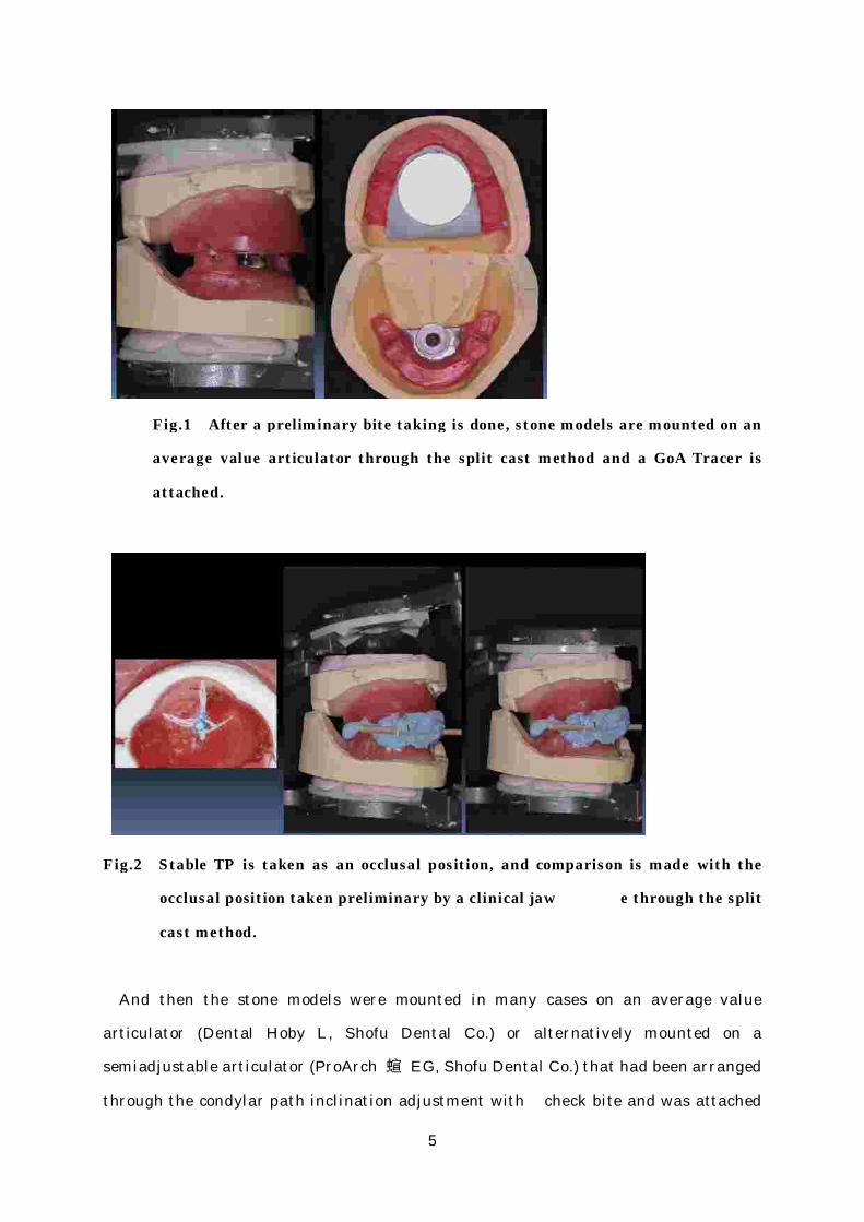
Fig.1
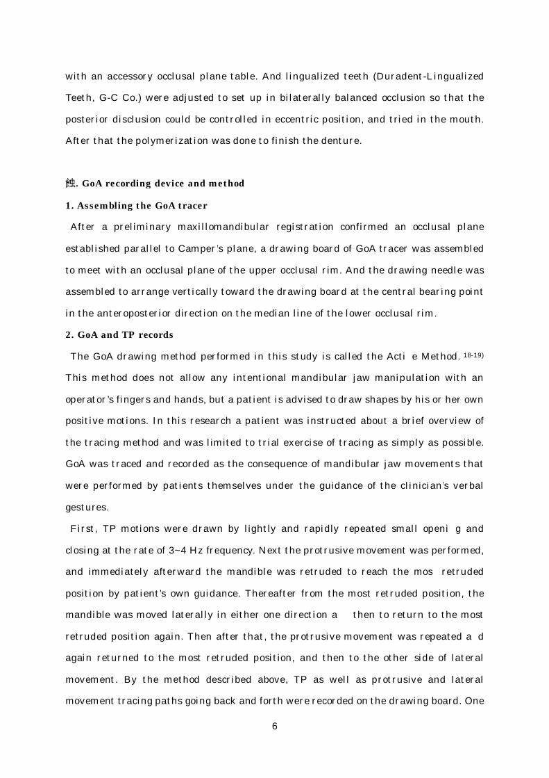
Fig.2
Ⅰ
Ⅱ
5
And then the stone models were mounted in many cases on an average value
articulator (Dental Hoby L, Shofu Dental Co.) or alternatively mounted on a
semiadjustable articulator (ProArch EG, Shofu Dental Co.) that had been arranged
through the condylar path inclination adjustment with check bite and was attached
Fig.1 After a preliminary bit e taking is done, s tone models are mounted on an
average value art iculator through the split cast method and a GoA Tracer is
at tached.
F ig.2 S table TP is taken as an occlusal pos it ion, and comparison is made with the
occlusal posit ion taken preliminary by a clinical jaw e through the split
cast method.
Ⅲ
6
with an accessory occlusal plane table. And lingualized teeth (Duradent-Lingualized
Teeth, G-C Co.) were adjusted to set up in bilaterally balanced occlusion so that the
posterior disclusion could be controlled in eccentric position, and tried in the mouth.
After that the polymerization was done to finish the denture.
After a preliminary maxillomandibular registration confirmed an occlusal plane
established parallel to Camper’s plane, a drawing board of GoA tracer was assembled
to meet with an occlusal plane of the upper occlusal rim. And the drawing needle was
assembled to arrange vertically toward the drawing board at the central bearing point
in the anteroposterior direction on the median line of the lower occlusal rim.
The GoA drawing method performed in this study is called the Acti e Method. 18-19)
This method does not allow any intentional mandibular jaw manipulation with an
operator ’s fingers and hands, but a patient is advised to draw shapes by his or her own
positive motions. In this research a patient was instructed about a brief overview of
the tracing method and was limited to trial exercise of tracing as simply as possible.
GoA was traced and recorded as the consequence of mandibular jaw movements that
were performed by patients themselves under the guidance of the clinician’s verbal
gestures.
First, TP motions were drawn by lightly and rapidly repeated small openi g and
closing at the rate of 3~4 Hz frequency. Next the protrusive movement was performed,
and immediately afterward the mandible was retruded to reach the mos retruded
position by patient’s own guidance. Thereafter from the most retruded position, the
mandible was moved laterally in either one direction a then to return to the most
retruded position again. Then after that, the protrusive movement was repeated a d
again returned to the most retruded position, and then to the other side of lateral
movement. By the method described above, TP as well as protrusive and lateral
movement tracing paths going back and forth were recorded on the drawing board. One
Ⅱ. GoA recording device and method
1. Assembling the GoA tracer
2. GoA and TP records

7
piece of plastic sheet plate at one tracing was used a the drawing board, mounted on
an articulator, and was stored together with patient clinical chart.
TP position which was stable was taken as the maxillomandibular registration.
Reviewing what position in the GoA graphics TP is corresponding or whether or not it
is repeatable and stable, mandibular position is caref lly decided for its final denture
construction. Especially when TP is shown scattered or is not clearly defined of
focusing, points on the protrusive movement path drawn in GoA were taken.
Essential points of maxillomandibular registration by the GoA tracing method are
presented here as follows.
1) The drawing board is to be set up in parallel to the Camper’s plane.
2) Patient’s active records are to be taken by the Active method.
3) Trace practicing is to be limited as least as possible.
4) Tracing path records are to be taken both in going back and forth.
5) Stable TP is to be taken as the position for the maxillomandibular registration.
6) TP on the protrusive movement path is to be taken.
Measurements were made on the distance between Ap and TP, the length of line
segment of tracing paths of GoA on the protrusive, rig t lateral and left lateral
movements as well as the angle between these line segm nts, using 1/20mm reading
caliper and 1/2 degree graduations protractor.
GoA Score evaluation method, which the author has uniquely developed by himself, is
in an object to evaluate GoA shapes as objectively as possible. Components of the
graphics are classified into four groups, and scores are counted by adding each one
point depending on the grade of disorders of each segm nt. And these points are
totaled for evaluating as GoA Scores. These components of the graphics and elements
of count addition will be shown in the followings.
3. Establishment of horizontal mandibular posit ion
.Graphic measurement
.GoA S core evaluat ion method (GoA point rat ing evaluat ion)
Ⅲ
Ⅳ

8
Each line segment under the length of 3mm from the point that is estimated as Ap is
used for evaluation.
In case when Ap is not in a pointed shape but rounded.
In case when lateral movement paths, either side of right or left, are not focused into
Ap. (2 points will be added when both right and left sides bilaterally are involved.)
In case when the protrusive movement is not focused into Ap. (It is understood as only
TP depiction for evaluation, when not defined on either right or left as well as
protrusive path graphics.)
In case when line segments are plural in number.
In case when line segments are curved.
In case when line segments are short. (less than 5mm)
In case when line segments are plural in number.
In case when line segments are curved.
In case when line segments are short. (less than 3.8mm)
In case when line segments are plural in number.
In case when line segments are curved.
In case when line segments are short. (less than 3.8mm)
“Short line segment” is defined from length measurements of total records taking
the length of standard error deducted from mean values as minimum parameter, and
also it should be under 1 SD and under 5mm for the protrusive movement and under
3.8mm for the lateral movement. ( )
1.Ap
2.Protrus ive movement path
3.Right lateral movement path
4.Left lat eral movement path
Table 1
・
・
・
・
・
・
・
・
・
・
・
・

9
Every time when point addition scoring is needed, one point is added as score, and
maximum value should be 3 points as per each component Total scores for these four
different components are evaluated as total GoA Scores to each patient. If GoA tracing
is clear, the score will be zero point, and as the GoA shapes becomes the less clearly
defined, the score will be the higher, and finally the highest scores will become 12
points as in the table shown separately. ( )
Apex Protrus ive
movement path
Right lateral
movement path
Left lat eral
movement path
N ot pointed
but round
Plural line
segments
Plural line
segments
Plural line
segments
Maxillomandibular
regist rat ion
pos it ion
Ap TP
Ap TP
Lateral movement
paths are not
focused
Right side
Left s ide
Line segment
is curved
Line segment
is curved
Line segment
is curved
Ap/TP distance
m
Protrus ive
movement paths
are not focused
Line segment
is under
5.0mm
Line segment
is under
3.8mm
Line segment
is under
3.8mm
0 1 2 3 0 1 2 3 0 1 2 3 0 1 2 3
GoA Score
Total point s
Table 1. GoA S core check chart . Enter total count of each component as to matching
and unmatching of Ap/TP, and Ap/TP distance as GoA S co Evaluate both
going back and forth of movement paths . Count over 3 point s in the Apex
column is taken as Score 3.
Table 2
□ □ □ □
□
□
□
□
□ □ □
□ □ □ □
=
≠
m
※
※

10
From patient’s medical chart, data of patient’s visit frequency for denture adjustment
was extracted.
For statistical analysis, EXCEL and JSTAT softwares were used to test the
significant difference of mean values through calculating of basic statistics, F-test,
Student’s t-test, Pearson’s correlation coefficient and through processing
Kruskal-Wallis test from Scheffe’s method. Especially comparisons were made from the
following two viewpoints.
Total number of subjects and number of males and femal s (male-female ratio), Mean
ages 1 SD value (lowest values – highest values) and mean values 1 SD values as
for ratio of age group.
After basic statistic analysis is processed, subjects were classifie in 4 groups from
distance differences between Ap/TP. Group A defines 0 ~ 0.9mm of Ap/TP distance.
Table 2. Example of GoA S cores
.Denture adjustment frequency
.S tat ist ical analys is
1. Test subject s
2. Analys is of groups of Ap/TP dis tance
Ⅴ
Ⅵ
± ±

11
Group B 1.0 ~ 1.9mm, Group C over 2.0mm, and Group D d ew only TP depiction. And
all measurements, GoA Scores, and denture adjustment frequency were analyzed
statistically.
Number of subjects: 161 subjects
Male-female ratio: 58/103 subjects (36.0/64.0%)
Overall mean ages: 73.8 ±9.6 years (46 – 94 years)
Mean male ages: 72.3 ±10.6 years (48 – 90 years)
Mean female ages: 74.6 ±8.9 years (46 – 94 years)
46 – 59 years: 10.6% (17 subjects)
60 – 69 years: 15.5% (25 subjects)
70 – 79 years: 47.2% (76 subjects)
80 + years: 26.7% (43 subjects)
Among total number of 161 subjects, 149 subjects could be discriminated between Ap
from TP. And only TP depiction was found in 12 subjects.
Mean values were calculated per each measurement exceptionally without Group D as
measurements were not available with this group. And highest GoA Scores showed 12
points in Group of only TP depiction, and so this group was eliminated from the
calculation of mean values. So the highest GoA Scores were 11 points among the rest of
149 subjects combined together of Groups A, B and C. As for the denture adjustment
frequency, records of all 161 subjects throughout the Groups were taken. ( )
Result s
. Regarding subject s
1. Total number and male-female ratio and ages
2. Rat io of age group
. Analys is of groups of Ap/TP distance
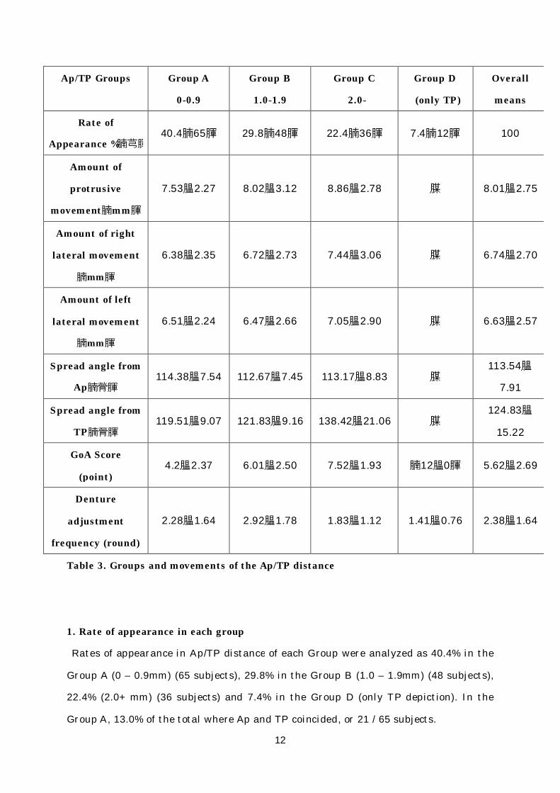
Table 3
Ⅰ
Ⅱ
12
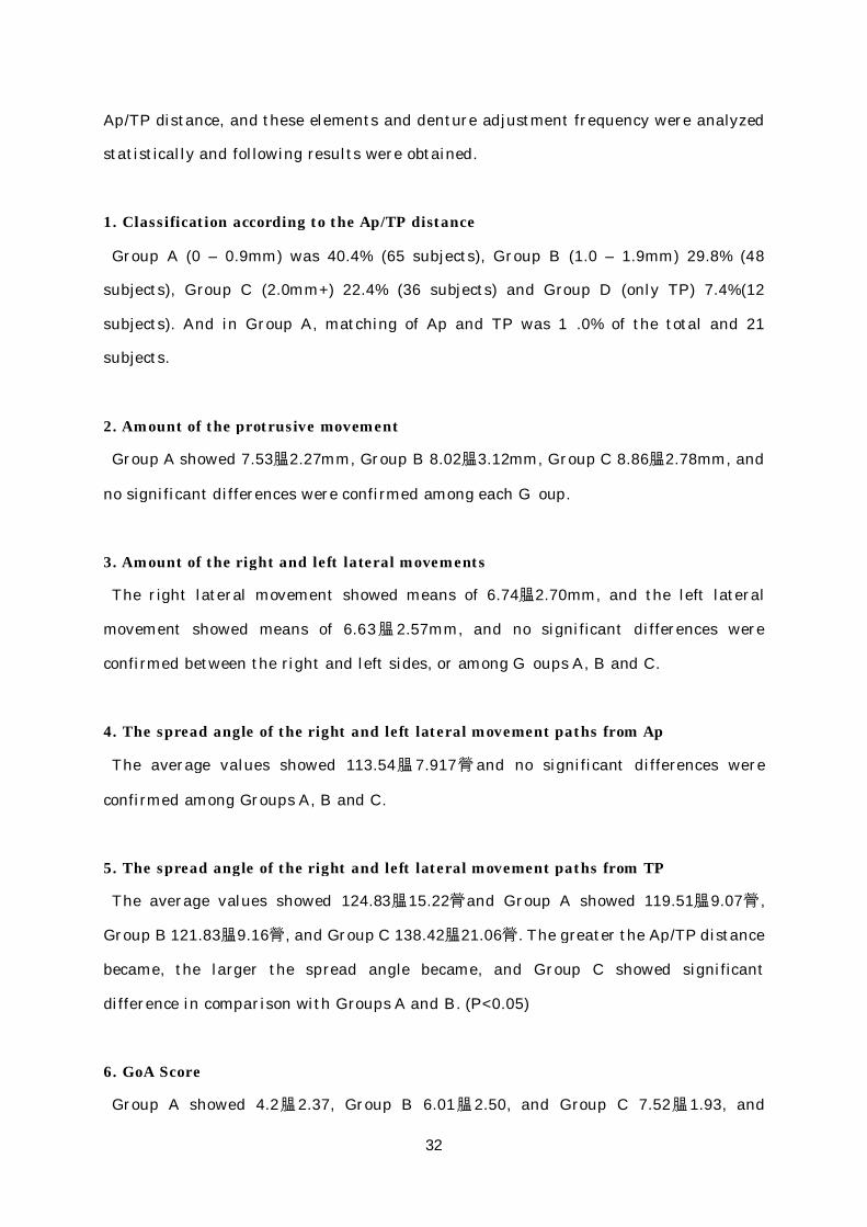
40.4 65 29.8 48 22.4 36 7.4 12 100
7.53 2.27 8.02 3.12 8.86 2.78 8.01 2.75
6.38 2.35 6.72 2.73 7.44 3.06 6.74 2.70
6.51 2.24 6.47 2.66 7.05 2.90 6.63 2.57
114.38 7.54 112.67 7.45 113.17 8.83113.54
7.91
119.51 9.07 121.83 9.16 138.42 21.06124.83
15.22
4.2 2.37 6.01 2.50 7.52 1.93 12 0 5.62 2.69
2.28 1.64 2.92 1.78 1.83 1.12 1.41 0.76 2.38 1.64
Rates of appearance in Ap/TP distance of each Group were analyzed as 40.4% in the
Group A (0 – 0.9mm) (65 subjects), 29.8% in the Group B (1.0 – 1.9mm) (48 subjects),
22.4% (2.0+ mm) (36 subjects) and 7.4% in the Group D (only TP depiction). In the
Group A, 13.0% of the total where Ap and TP coincided, or 21 / 65 subjects.
Ap/TP Groups Group A
0-0.9
Group B
1.0-1.9
Group C
2.0-
Group D
(only TP)
Overall
means
Rate of
Appearance %
Amount of
protrus ive
movement mm
Amount of right
lateral movement
mm
Amount of left
lat eral movement
mm
S pread angle from
Ap
S pread angle from
TP
GoA Score
(point )
Denture
adjustment
frequency (round)
Table 3. Groups and movements of the Ap/TP distance
1. Rate of appearance in each group
(n)
( )
( )
( )
(°)
(°)
( ) ( ) ( ) ( )
± ± ± ― ±
± ± ± ― ±
± ± ± ― ±
± ± ± ―±
± ± ± ―±
± ± ± ( ± ) ±
± ± ± ± ±

13
As for the amount of protrusive movement, 7.53 ±2.27mm was in the Group A, 8.02
±3.12mm in the Group B and 8.86 ±2.28mm in the Group C. And as the Ap/TP distance
became larger, some amount of increasing tendency show d, but there was no
significant difference among groups.
As for the amount of right lateral movement, mean value was 6.74 ±2.70mm. On the
left lateral movement, it was 6.63 ±2.57mm, and there was no significant difference.
Or no significant difference was confirmed among Groups A, B and C.
The spread angle of right and left lateral movement p ths from Ap was in the mean of
113 ±7.91° No significant difference was confirmed among Groups , B and C.
The spread angle of right and left lateral movement paths from Ap was in the mean of
124.83 ±15.22 That of Group A was 119.51 ±9.07 , Group B was 121.83 ±9.16 and
Group C was 138.42 ±21.06 . And as the Ap/TP distance became larger, the spread
angle increased, and Group C showed significantly larger than Group A or B. (P<0.05)
GoA Scores were in the mean value of 5.62 ±2.69 points in the Groups A, B and C
where Ap and TP were clearly defined. As TP was only depicted in the Group D, it
showed 12 ±0 points. That of Group A was 4.2 ±2.37, Group B was 6.01 ±2.50 and Group
C was 7.52 ±1.93, and GoA Scores between Groups A, B and C were not confirmed with
significant difference. (P<0.05 as for Groups A – B and B – C. P<0.01 for Groups A - C)
( )
2. Amount of protrus ive movement
3. Amount of lateral movement
4. S pread angle from Ap
5. Amount of spread angle from TP
6. GoA Score
Fig.3
.
°. ° °
°

14
And also the correlation coefficient of the Ap/TP distance and GoA Scores were found
to be +0.50817, and moderately positive linear correlation was confirmed. ( )
Fig.3 GoA Score according to the Ap/TP distance
F ig.4
Fig.4 Correlation between Ap/TP dis tance and GoA Score

15
As for the denture adjustment frequency, total average requency was 2.38 ±1.64
rounds, Group A average was 2.28 ±1.64 rounds, Group B 2.92 ±1.78 rounds and Group
C 1.83 ±1.12 rounds and Group D 1.41 ±0.76 rounds. Adjustment frequency in the
Group B was significantly higher than Groups C and D. (P<0.05) No significant
difference was confirmed among other Groups. ( )
Totaling 161 pieces of GoA measurements used in this study were all traced graphics
taken clinically at the time of complete denture const uction. When patient’s graphic
was taken originally in the past, data collection was ot intended for processing like
this study but was intended to find out repeatable and stable TP positions in order to
prevent errors from recording final maxillomandibular registration. Now the purpose
of the study this time is to research clinical significance of GoA by analyzing
relationship between GoA and TP.
The maxillomandibular registration in complete denture construction is a procedure to
7. Denture adjustment frequency
Fig.5
Fig.5 Denture adjustment frequency according to Ap/TP distance
Discuss ion

16
find out a single point of TP over the intermaxillary relation s the masticatory
terminal position after the occlusal vertical dimension and the plane a determined.
This TP position must be certainly a consolidated point of the neuromuscular
mechanism and the bilateral status of TMJ that are related with the patient’s own
specific stomatognathic system at this point of moment. And also this point should be
repeatable and consistent within reasonable range of the living body, even though it is
a compromised one when patient’s dental history is taken account.
Meanwhile, as far as Ta method is concerned without using GoA tracing method in
order to fix the maxillomandibular registration, there is no index existing as to how to
correct any error if it happens, because such a Ta method does not permit relations
clearly with the mobility of TMJ in visual form like the GoA method.
For this reason, combined use of the GoA tracing method and TP method is capable of
clarifying the mobility of TMJ in form of traced graphic at this point of moment of an
individual patient, and it is objectively able to know in what way the TP is positioned.
So it is thought effective that the maxillomandibular registration can be processed
more precisely. And even if any error occurs in the maxillomandibular registration, it
is possible to understand in what way errors have been modified and corrected through
the GoA processes. And so the GoA tracing technique is excellent in this regard.
The GoA tracing method is most familiar and simple recording method of jaw
movement for a practitioner, but the interpretation is mostly complicated. There is no
question about a basic and beautiful shape. But when the drawing is in disorderly
manner, sufficient researches have not been conducted so far in the past for reasoning
with excuses that ‘there is no information available’. So for better interpretation of
GoA tracing method, factors of this complicated background will be elaborated and
organized.
GoA tracings are recorded with three points of support. Two points are supported in
the back by TMJ bilaterally, and the recording apex is the front support. The tracing
Ⅰ. Organizing GoA
1. GoA and posterior guidance

17
needle works to record three-dimensional movements of bilateral TMJ onto the
two-dimensional flat plate in certain level. On the other hand, posterior guidance is an
element that defines the jaw movement as TMJ morphological factors.20) And so, GoA
tracings are drawn by the posterior guidance that is guided by the TMJ architectures
including joint ligaments and discs on both sides, and they are expressed in graphics of
movements of bilateral mandible condyles. For this reason, this is thought as one of
recording methods of jaw movements. If any organic change is present, therefore,
including an eccentric displacement of mandible condyle or disc like TMJ internal
derangement, it will become an impaired posterior guidance, and GoA will not be
traced smoothly. As a result, disorderly GoA will be drawn, indicated as a tracing
disorder. In other words, it is known that GoA analysis is a recording method of
mandibular jaw movement that can detect any presence of organic change in the
posterior guidance. 10)
In practical clinics, there is another reason for inhibiting the smooth tracing of GoA
other than organic dysfunctions of the posterior guidance as described above. That is
related with inhibition of smooth working of neuromusc lar mechanism. Furthermore,
this kind of tracing disorders caused from neuromuscular mechanism will be divided
into two groups. First one refers to a case of essential cause like after-effects from
brain damages. ( )
2. GoA and neuromuscular mechanism
Fig.6

18
The next one refers to a problem of tracing the mandibular jaw movement smoothly as
a voluntary movement, even if simply trying to follow an operator ’s request. It may not
be an easy task for an elderly people who often are complete denture wearers to do the
right and left lateral movements voluntarily without lifting up the tracing needle from
the drawing board. In the latter case, it may often be nominated as the tracing disorder
even without any problems involved with TMJ. In this case, a patient is suggested to
do a free moving horizontally forward, backward, right and left from TP. Then, it may
be possible to take the component of involuntary motion into an examination
supplementary.
This kind of involvement of neuromuscular mechanism might have m de the
understanding of GoA more complicated, and so there ma be major reasons of
background that there has not been good enough research in many reports on GoA
reasoning of excuses that “Individual patients variety are many.” 13)
Fig.6 GoA depicted by a patient suffering from part ial paralys is aft er st roke. It was
es t imated that Ap and TP would be matched at first of e construct ion
but aft er the insert ion, the intercuspat ion of art ificial teeth were found in
discrepancy, and remount ing was done aft er new maxillo
regist rat ion taken at the TP pos it ion on the protrus ive movement path.

19
GoA is usually recorded under certain height of occlusal vertical dimension on an
imaginary occlusal plane in parallel to the Camper’s plane. But it is more flattened
with no inclination than the sagittal condylar path inclination of the po terior
guidance, and so the mandible condyle would be forced to rotate in the negative
direction at the protrusive movement. ( ) 21,22)
3. GoA tracing method is a loading t es t
F ig.7,8
Fig.7 When GoA is to be drawn on the board paralle l to Camper’s plane, TMJ is forced
to rotate conversely.
F ig.8 IP: Intercuspal pos ition, EP: Edge-to-edge occlusal pos ition. If the incisal path
angle < the condylar path angle , the movement from EP IP would rotate the

20
From this viewpoint, it would be understood that GoA tracing method has a
component of a loading test. In order to reciprocate this negative rotation, it is
proposed whether the tracing should be initiated under ncreased vertical dimension to
provide the positive rotation, or alternatively the tracing board should be tilted
anteriorly along with about equivalent amount to the sagittal condylar ath like a
supplemental element of anterior guidance. From this measure, ther might be an
improvement depicted on the drawing board. If there happens to be any clicking or
crepitus sounding or any catching, the mandible condyle will detect this problem not to
move smoothly under loaded condition like this. In case the condyle cannot climb over
this drawback, GoA depiction will become extremely small, or even i it can climb over
it, the tracing will become discontinued or will not be coincided on the way going back
and forth. ( )
condyle conversely and would adversely affect TMJ. And so, it would be
preferable if the incisal path angle be larger than the condylar path angle
(quoted from the reference literature number 21 and 22).
F ig.9.10
Fig. 9 If any interference is present on the posterior guidance, smooth
mandibular movement is inhibit ed. The needle will l ift off from the board
when it climbs over and it s drawn line will become a broken line.

21
From research reports so far to the present, there may be grouped about in two
different intended uses of GoA. The first one is to use it to determine preoperative
horizontal mandibular position in relation to the TP d a. The second one is to obtain
functional status of TMJ.
In the latter use, pre- and post-operative comparisons of GoA will be conducted. It is
reported that functional improvement like masticatory fficiency may be confirmed
from changes of GoA graphics depending on consequences of provisio of treatment
denture and other treatment measures. 13,23,24) Even if there may be, however, any
graphic changes confirmed, it is still unknown to what degree of changes some organic
problems in the TMJ area or some muscle tissues problems have been involved.
As described above, GoA tracing method as for recording mandibular jaw m vements
can be obtained from the mandible condyle translation on th posterior guidance. And
Fig.10 If any interference is present on the posterior guidance, and when it cannot
climb over it , the drawn path will become shorter in a unilateral case, as the
movement on the t roubled s ide is controlled and the balancing s ide is largely
affected. In a bilateral case, all the movement paths , protrus ive or lateral,
will become shorter.
4. Intended use of GoA
22
so it is an objective method of graphics to show the d ree of functional status of
mandibular jaw movement. Its drawn graphic is a comprehensive record of patient’s
complicated background including patient individual TMJ organic status,
neuromuscular mechanism and tracing condition. For this reason it is difficult to
appreciate any single significance of graphic, but it s rather helpful for us to obtain
patient special information on TMJ functions by studying to break down the
components. It is not, however, well established on procedures for nalyzing the
components, or yet sufficiently organized currently. In this study, therefore, if
procedure of scoring like GoA Scores this time can be taken as objectively as possible
and can compare the degree of pre- and post-operative improvement in numerical
values, then clinical significance of GoA tracing method may be enhanced.
As described above, GoA tracings that were evaluated i this study were taken in our
practice for determining the maxillomandibular registration of denture construction in
practice, and they were not intended for statistical study or ot based on precise
research protocol. Furthermore, many subjects of them had resorbed residual alveolar
ridges and some of recording bite plates were troubled with problems of stability from
mobility or displacement on the mucosa, and so it is necessary to understand the
measurements roughly as to the line segment length and the angle data. In practical
case, a practitioner needs to recognize that such records might be always accompanied
with problems of bite plate displacement.
In this time of GoA tracing method, following the meth of Nanami’s et al. 5), an
intraoral recording method was taken with the drawing board on the maxilla and t e
needle on the mandible. The author considers that the bite plate would not dislodge
easily with pressure loading in the center of the plate even at eccentric displacement
by setting the tracing needle on the mandible. And it is pointed out that GoA tracer
would block physiological shape of the tongue as its trac r interferes with the anterior
Ⅱ.Discuss ion on method
1. Regarding GoA records that were evaluated
2. Regarding installat ion method of GoA tracing unit
23
portion of tongue space whether or not the needle is set up on either side of upper or
lower device. 6,25) We think it rather beneficial to control the tongue posteriorly to
some degree in order to inhibit the anterior projection of the mandible that occurs
often at the time of maxillomandibular registration of edentulous patient and to
prevent the lift-up of mandibular bite plate as well.
GoA tracing method in this study is called the Active method. 18,19) This method does
not permit the operator ’s manipulation with fingers and hands but a patient’s own
positive actions. The operator ’s manipulation with fingers and hands as called the
Passive method8) is to record the intrinsic range of movement composed with the TMJ
structure. Researches up to the present 18,19,26) report that about 0.6mm~1.0mm of
positional differences of the mandible condyle are measured between the Active and
Passive methods. But in a study like this time where denture is constructed without
using a treatment denture, the Active method is estimated most appropriate in order
to determine the mandibular position which position is repeatable to produce by
patient oneself. And a briefing to patient is made only about the tracin ethod, and a
simple exercise is suggested in order to see mandibular jaw movement more naturally.
An appropriate mandibular jaw position that is acceptable to a patient can be taken
successfully by the guidance of clinician’s verbal gestures.
Even when TP depiction is scattered within some certain range without focusing on a
single point, any point on the protrusive movement path is understood to retain well
balanced with both right and left sides of TMJ status as well as neuromuscular
mechanism, and so they are determined as the position of maxillomandibular
registration. This method is thought appropriate even from the results of denture
adjustment frequency throughout all the cases in this study. So it is significant enough
to trace the protrusive movement paths.
In the beginning of this study, groupings were studied according to the shapes of GoA
tracings, but the whole images were thought difficult to evaluate objectively. So the
four different components of graphics were separated indicating respectively zero
3. Regarding GoA tracing method and pos it ion of maxillomandibular regist rat ion
4. Regarding GoA Scores

24
point for clearly drawn trace, and 1~3 points given on the traces depending on the
degree of disorder so that graphics could be evaluated as objectively as possible. As
this evaluation did not include the Ap/TP distance or the spread angle of right and left
lateral movement paths, it should be noted that significant difference was confirmed in
the relation of these results and GoA Scores.
Among total number of 161 subjects, males belong to 3 .0% (58 subjects) and females
to 64.0% (103 subjects), and females are more in number than males as in 1.78 times.
And mean age was 73.8 9.6 years and no difference between males and females.
Although there were not mentioned in the chapter of res lts here, no significant
difference was confirmed between age differences regarding the amount of protrusive
and right-left lateral movements, the spread angle from Ap or TP, GoA Scores and
denture adjustment frequency. From this fact, therefore, it is known that favorable
prognosis may be predicted with any degree of certainty as far as this procedure of
denture construction is concerned.
1) Regarding amount of movement
The amount of protrusive movement showed 7.53 2.27mm in Group A, 8.02
3.12mm in Group A and 8.86 2.78mm in Group C. And as far as mean values were
concerned, the more anteriorly the TP was positioned, the greater the movement
amount would tend to be. No significant difference was found among each Group. As
long as Ap was well defined, the amount of protrusive movement of the mandible
condyle from Ap did not exhibit any greater change even though TP was positioned
anteriorly. Meanwhile if the maxillomandibular registration is fixed in TP position, the
amount of protrusive movement will become less than in case of that amount from Ap,
and the mobile range in the forward direction will become limited within small area.
And as for the amount of lateral movement, the right lateral movement showed 6.74
2.70mm and the left lateral movement showed 6.63 2.57mm and no significant
Ⅲ. Discuss ion on the result s obtained
1. Regarding pat ient subject s
2. Regarding analysis of Ap/TP distance groups
±
± ±
±
± ±

25
difference was found between the right and left sides or among Groups A, B and C. And
it is known that, even if the Ap/TP distance becomes larger, the lateral movement
amount will not change. Likewise in case of the protrusive movement, the range of
lateral movement from TP is thought to be limited to s e extent.
For this reason, when TP is taken for the maxillomandibular registration as occlusal
position, it is considered to increase the freedom from the posterior direction, or the
freedom in the movement range from Ap to TP. But it is also considered that the
movement amount from TP toward protrusive and lateral directions may be limited.
2) The spread angle of lateral movement path
The spread angle between the right and left lateral movement paths from Ap showed
113.54 7.91 and no significant difference was found among Groups A, B and C.
These values were confirmed consistent approximately with the report from Murakami
et al. 3) When this is put together with the movement amount as described above, it
shows that TMJ natural structures of individual subjects are not different basically
and that functional structures of natural anatomy represent sim larity regardless of
Ap/TP distance.
Meanwhile, the spread angle between the right and lef lateral movement paths from
TP showed 124.83 15.22 at an average rate. Group A showed 119.51 9.07 , Group
B 121.83 9.16 , Group C 138.42 21.06 , and the larger the Ap/TP distance
became, the spread angle showed the greater, and Group C showed significantly larger
than Group A and B (P<0.05), and at the same time it showed larger deviation and
variance. This means that the spread angle of the lateral movemen from TP is
performed in the range of intermediate movement as the mandible condyle is already
positioned in the forward position, and it is thought that it showed rather larger values
at standard deviation as the terminal position of late al movement is located
somewhere within the path of border movement. Therefore the spread angle of lateral
movement path from TP showed largely different behavio s among individuals and so
the variance is thought to become larger. In clinical practice, a check bite is taken and
an articulator is adjusted, and it can be confirmed that the working side lateral
condylar movement (a rear wall) has produced larger values. In pursuit of more precise
± °
± ° ± °
± ° ± °

26
denture construction, it will need to have a semiadjustable articulator coordinated
with this information and to select and setup the artificial te h based on this
adjustment.
3) Regarding GoA Scores
GoA Scores showed 5.62 2.69 in Groups A, B and C at an average rate when Ap and
TP were discriminated. In Group D, only TP was depicted, showing 12 0 points.
Group A showed 4.2 2.37, Group B 6.01 2.50 and Group C 7.52 1.93, and
significant difference was found in GoA Scores in each Group of A, B and C. (P<0.05 as
for A – C and B – C, P<0.01 for A – C)
This shows that GoA Score became higher as the Ap/TP distance was larger, and TP
was positioned more anteriorly. In addition to this, the correlation coefficient of the
Ap/TP distance and GoA Scores were found to be +0.50817 and moderately positive
linear correlation was confirmed. And so, it also suggested that the higher GoA Scores
became, the more possible TP was positioned in the for ard direction.
When GoA Score is the higher as the Ap/TP distance is the larger, it means that the
mandibular movement under the leading role of TMJ will become less smooth
accordingly as TP is positioned in the forward directi n from Ap, and there may be
seemingly any presence of TMJ organic alteration or neuromuscular disharmony.
In the followings, items of clinical observation of GoA Score will be described.
Ap
Consolidated point of the right and left lateral movement paths and protrusive
movement path are determined as Ap. And, since the Active method was adopted, the
most retruded position was demonstrated available for individual patient’s own
neuromuscular mechanism and TMJ status, but it is not definitely the most retruded
position generated by intrinsic architecture of TMJ under the guidance of the Passive
method. In case the shape of Ap is not pointed and is rounded, a presence of immediate
side shift or Bennett movement is known, and, generally speaking, it is owing to
looseness of TMJ 13,23) . The non-pointed Ap is drawn at the time when both the
working and balancing condyles move simultaneously at the initial stage of the lateral
movement. And when the lateral and protrusive paths are not consolidated, causes
±
±
± ± ±
①

27
may be possible in case when neuromuscular mechanism cannot work well to express
with voluntary movement, or alternatively in case when organic alteration of T J is
presented as dysfunction surrounding Ap position.
Protrusive movement path
Normal protrusive movement is to be generated in balanced TMJ motion bilaterally,
and their paths going back and forth should be matched clearly in straight line. In case
protrusive movement paths are in plural lines, according to our observation, these
paths going back and forth are not matched, or initial and terminal points of line
segment are plural in number.
In case these line segments are curved, it is understood that amounts of movement of
the right and left condyle are different. If any grave dysfunction is present in one of
TMJ, only the other side of normal TMJ is mobile and the depicted line will be largely
curved toward the side in dysfunction. This kind of curvature will become an important
index to detect whether or not any disorder of unilateral side of TMJ itself is present in
addition to some problem involved with lateral movement path.( )
Cases where the line segment was short were based on the length less than 5mm after
standard deviation was deducted from total mean values of measurements. Since any
decreased amount of movement caused by aging is not confirmed, it is significantly
important that the length of line segment should be to indicate the degree of recording
disorder.
As described above so far, evaluation of the protrusive movement path will be
expressed as recording disorder when any trouble is present on the movement path of
uni- or bi-lateral TMJ and when any avoidance or interference are underway.
Right and left lateral movement paths
In cases where the line segment is in plural lines, like in the case of protrusive
movement, it is frequent that many paths going back and forth are not matched. And,
in general, cases were seen more often when paths going back return to TP, although
paths going forth were initiated from Ap.
Cases where the line segment was curved were observed when paths going back were
shown consolidated toward TP in mid-course of the way in spite of the forth record
②
③
Fig.9,10

28
showing straight linear border movement path, or alternatively when movement was
inhibited by any TMJ disorder on one side contrary to the movement direction (e.g. left
side TMJ disorder in case of right lateral movement). This might be considered that
the mandible condyle is not able to move on the border vement path for some reason
or other and only is allowed to move in the intermediate moving area.
Cases where the line segment is short were based on the length value less than
3.8mm after standard deviation was deducted from total mean values of measurements.
In general, check bite is taken at about 5mm of eccentric translation amount in order
to adjust the condylar inclination path of an adjustab e articulator, and if the line
segment is short with the minimum movement amount, correct check bite cannot be
taken and so its clinical significance is grave.
4) Denture adjustment frequency
As for denture adjustment frequency, total average show d 2.38 1.64 rounds, Group
A 2.28 1.64 rounds, Group B 2.92 1.78 rounds, Group C 1.83 1.12 rounds and
Group D 1.41 0.76 rounds. Significant difference was found in Group B in relation
with Group C and D respectively. No significant difference was found among other
Groups. It is known from these data that denture adjustment fre uency was highest in
Group B where the Ap/TP distance showed 1.0 – 1.9mm and that it was lowest in
Group C where the distance was over 2mm. So far to the present, Suzuki et al. reports
that denture adjustment frequency increases as the Ap/TP distance is over 0.6mm12),
and Abe reports that the mandibular positions will become unstable in the groups
where the Ap/TP distance is over 2.0mm under the manipulation of the Passive method
8). It is already known that Ap is positioned rather anteriorly in case like this study of
the Active method with patient’s positive tracing than in case of the Passive method
with patient’s passive tracing with the help of an operator. 18,19,26), and so not only the
Ap/TP distance but also its performed method should be taken into account for
studying comparisons. The results obtained, therefore, indicating increased rounds of
denture adjustment frequency, when the Ap/TP distance is positioned more anteriorly
to certain degrees, are thought to be compatible with their results.
In this study, furthermore, denture adjustment frequency decreased in Group C
±
± ± ±
±

29
where the Ap/TP distance was over 2.0mm and in Group D of only TP depiction without
graphic evaluation available. This might mean that GoA Scores are significantly
higher in these Groups and higher of degree of recording disorder, and that the
mandibular position was clearly defined for necessary treatment especially in Group D.
In considering this fact, it is estimated that highly epeatable masticatory terminal
position has been obtained and simultaneously function of movement during
mastication have been controlled. In other words, it is also estimated that, as the TP
depiction is largely positioned in the protrusive dire ion, masticatory functions
involved with TMJ have been controlled and that TP as asticatory terminal position
have been limited to a smaller area.
Denture prognosis will need to be evaluated comprehens vely depending on the
adjustment degree, its frequency as well as its necessary duration, but the position of
maxillomandibular relation will be the greatest factor of all. Suzuki et al. determines
that prognosis would not be good enough for the frequency over 6 rounds and the
duration over a period of one month 12). Someya states that Ta motion is one of clinical
skills to determine the mandibular jaw position and that the denture prognosis will be
influenced when clicking is detected in the way of the movement path o mouth
opening and closing, when any eccentric deflection is present or when any hesitation or
looseness in the Ta motion is present. He also states that, if occlusal relationship is to
be corrected properly, open-close motion with smoothness and without looseness will be
resulted within around one month27). After all, adjustment duration within one month
would be favorably indicated.
In practical cases of Group B (1.0 – 1.9mm) of GoA Scores of 6.04 2.21, it is known
from these results above to have a tendency to increase denture adjustment fre uency
with possible errors raised in the maxillomandibular registration.
As long as Ap and TP are matched, recording of the mand bular position might be an
easy procedure, but this matching of both points were en only in 21 subjects (13.0%)
±
Ⅳ. Regarding clinical s ignificance
1. GoA and maxillomandibular regis trat ion

30
in Group A. It is compatible with the report from Mizokami et al. 28) stating that
clinical cases of Ap and TP matching were found in abo 10%. Meanwhile, even in case
of only TP depiction with 12 subjects (7.4%), it is rather difficult to fi d proper
horizontal mandibular position exceptionally in the point where TP are focused. There
is no other way but to take this position as the horiz ntal mandibular position.
Accordingly the rate of patients where mandibular positions are exactly identifiable i
clinical cases would be thought 20.4% combined together with both groups, or about
two out of ten cases.
Oshima et al. 3) are suggesting that, when Ap is not matched with TP, the mandible
condyle positions are greater in degree of horizontal deviation. Furthermore,
deviations of the mandibular positions are suggested extremely high with lower
consolidation rate of TP.
And also Kobayashi 29) demonstrates that the degree of functional disorders along
the masticatory system will become advanced progressiv y as the degree of
mandibular deviation increases. From these viewpoints, GoA Scores that evaluate GoA
shapes are considered effective for objective evaluation of individual subjects’ organic
status of TMJ as well as the capability of neuromuscular mechanism.
If any occlusal position is deflected in practical cases for denture onstruction,
rearrangement of artificial teeth and denture remaking will have to be needed. In this
case it will be clinically necessary to study how to t ke or change new mandibular
position by understanding graphic status and its relat on with TP by the use of GoA
Scores.
As described above, GoA is drawn with the support of t e tracing needle in the front
and bilateral TMJ in the back. Its graphic is drawn as a tracing graph of the needle a
a result of the simultaneous motion of bilateral TMJ. In other words, GoA is the
movement of the mandible condyle drawn on a plane surf ce under the posterior
guidance. While governed by the neuromuscular mechanism, the drawing is processed
with the reflection of the protrusive movement of the lateral mandible condyle with
similar amount of moving at the same time, with the reflection of the left border
2. Regarding clinical diagnos is

31
movement of the left mandible condyle with smaller motion on the left side and large
scale of motion on the right side, and with the reflection of the right border movement
of the right mandible condyle with smaller motion on the right ide and majority of
motion on the left side.
Consequently if any dysfunction is present on either side of TMJ, ght or left, some
disorder is detected on a drawn line on the opposite direction 25,26) Like this from the
drawn graph, it is possible to judge whether or not any trouble on either side of TMJ
structures are present, and GoA Scores in this study have demonstrated the grade of
abnormality objectively. And the fact that significant relationship has been confirmed
with the Ap/TP distance has made us possible to grasp more precise status of jaw
functions from both of these diagnosis results, and fu hermore it has made us possible
also to judge whether or not a certain result of TP is appropriate to take as the
horizontal mandibular position.
That is to say, GoA drawing method is possible to overv ew the TMJ organic status
and neuromuscular mechanism comprehensively through ob erving the mutual
relationship of the whole overviews of the drawing as well as each component in it. And
also it is a diagnostic method to decide whether or no the results of TP can be taken as
the mandibular position for therapeutic means.
Quantitative and morphological evaluation were processed on the records of GoA and
TP of 161 subjects collected over a period of 11 years when taken at the
maxillomandibular registration for complete denture construction in pra ical cases.
And relations of these evaluations and denture adjustme t frequency were studied and
discussed. As for quantitative study, amount of the protrusive and right-left lateral
movement and the spread angle of lateral movement composed from Ap and TP were
measured. As for morphological study, new development of GoA Score method was
applied. This GoA Score method is to count scores on 4 different components grouped
from GoA, and their total score was taken as scores of morphological evaluation of
relevant graphics to ensure the objectivity. 4 kinds of groups were classified by the
Conclus ion
32
Ap/TP distance, and these elements and denture adjustment frequency were analyzed
statistically and following results were obtained.
Group A (0 – 0.9mm) was 40.4% (65 subjects), Group B (1.0 – 1.9mm) 29.8% (48
subjects), Group C (2.0mm+) 22.4% (36 subjects) and Group D (only TP) 7.4%(12
subjects). And in Group A, matching of Ap and TP was 1 .0% of the total and 21
subjects.
Group A showed 7.53 2.27mm, Group B 8.02 3.12mm, Group C 8.86 2.78mm, and
no significant differences were confirmed among each G oup.
The right lateral movement showed means of 6.74 2.70mm, and the left lateral
movement showed means of 6.63 2.57mm, and no significant differences were
confirmed between the right and left sides, or among G oups A, B and C.
The average values showed 113.54 7.917 and no significant differences were
confirmed among Groups A, B and C.
The average values showed 124.83 15.22 and Group A showed 119.51 9.07 ,
Group B 121.83 9.16 , and Group C 138.42 21.06 . The greater the Ap/TP distance
became, the larger the spread angle became, and Group C showed significant
difference in comparison with Groups A and B. (P<0.05)
Group A showed 4.2 2.37, Group B 6.01 2.50, and Group C 7.52 1.93, and
1. Class ificat ion according to the Ap/TP distance
2. Amount of the protrus ive movement
3. Amount of the right and left lateral movements
4. The spread angle of the right and left lat eral movement paths from Ap
5. The spread angle of the right and left lat eral movement paths from TP
6. GoA Score
± ± ±
±
±
± °
± ° ± °
± ° ± °
± ± ±

33
significant differences of GoA Scores were confirmed among Group A, B and C. (P<0.05
between A – B, and B – C, P<0.01 between A – C)
Total average showed 2.38 1.64 rounds, Group A 2.28 1.64 rounds, Group B 2.92
1.78 rounds, Group C 1.83 1.12 rounds and Group D 1.41 0.76 rounds. Significant
difference was found in Group B in relation with Group C and with Group D. (P<0.05)
No significant difference was found among other Groups.
As mentioned above, it is suggested from the statistic analysis results of GoA
drawing method under the Active method that no alterations have been made for the
movement distance from Ap or the spread angle of later movement nor any difference
from the border movement function of TMJ itself, even when the Ap/TP distance
becomes larger. But from TP, it is suggested that the movement path is taken on the
intermediate movement path and that the spread angle as well as the deviation ar
grown larger significantly. And the larger the Ap/TP distance becomes, the higher the
GoA Score becomes significantly. So the more anteriorly TP is positioned, the more
often the recording disorders are raised, and the more the function is inhibited. On the
other hand, as for denture adjustment frequency, in th group of the Ap/TP distance
with 1.0 – 1.9mm, the frequency increases significantly but in small difference from
the total average. It is therefore clarified that adopting proper TP by the use of the
proposed method as the mandibular position for complete denture construction would
be the means of minimum errors. Accordingly in order to diagnose TP appropriately as
for the position of maxillomandibular registration, procedures of GoA drawing method
(under the Active method) combined with the use of Ta ethod would be thought
extremely significant from clinical practice.
In concluding this article, the author expresses his deep and sincere gratitude for
Mr.Takeo Ohno at Hakusan Dental Laboratory for complete denture constru ion
7. Denture adjustment frequency
Acknowledgements
± ± ±
± ±

34
throughout the cases. Furthermore the author extends his acknowledgements with
heartfelt thanks to Dr.Jiro Abe, President for Japan Dental Association (JDA) for his
supervision through this study.
1) Gysi A.: Kieferbewegung und Zahnform, Handbuch der Zahnheilk nde IV, Scheff,
Urban u. Schwarzenberg, Berlin u. Wien: 1-171,1929
2) Ohshima M, Tanaka A, Kobayashi Y: Clinical study on Go hic Arch tracing method
– Comparison of the mandible condyle position at apex and tapping point of
edentulous patients, Shigaku, 85 (1): 140-153, 1997 (Japanese)
3) Murakami Y, Shiga H, Kobayashi Y: Quantitative evaluation of Gothic Arch and
Tapping Point in edentulous patients, Shigaku, 80 (4): 783-808, 1992 (Japanese)
4) Morita O: Maxillomandibular registration of edentulous jaws, Shigaku, 81 (3):
593-601, 1993 (Japanese)
5) Nanami T, Mizokami T: Gothic Arch drawing method in edentulous clinical cases,
Dental Diamond, 10: 240-245, 1985 (Japanese)
6) Ichikawa T, Kitamura S: Edentulous jaw therapy using a omplete denture – From
viewpoints of oral anatomy, 1st ed.: 80-97, Quintessence, Tokyo, 2004 (Japanese)
7) Omatsu M: Study on clinical procedures of tapping poin drawing method in
maxillomandibular registration of edentulous jaws, the Shikwa Gakuho, 92 (7):
39-51, 1992 (Japanese)
8) Abe J: Predictive diagnostic method of the mandibular ability in edentulous jaws
based on the criteria of hyoid bone position, J the Japan Prosthodontic Society, 44
(2): 323-331, 2000 (Japanese)
9) Fukushima T: Reviewing over Gothic Arches, J Practice in Prosthodontics, 28 (4):
447-466,1995 (Japanese)
10) Ai M, Kawaguchi T: Significance and method of Gothic Arch tracing, J Japan
Dental Association, 38 (8): 789-795,1985 (Japanese)
Tanaka H, Mushimoto E, Chiba M et al.: Clinical characteristics of
Reference
11)

35
temporomandibular joint disorders in complete denture earers – Occurrence
frequency of TMJ internal derangement, J the Japan Prosthodontic Society, 39 (2):
396-405, 1995 (Japanese)
Suzuki K, Shiina N, Hosoi T et al.: A study on the numbers of denture adjustments
in complete denture wearers – Relationship to stability of tapping point, J the
Japan Prosthodontic Society, 45 (1): 106-116, 2001 (Japanese)
13) Suzuki H: Prior to using Gothic Arch, J Practice in Prosthodontics, 28 (2):
127-138,1995 (Japanese)
14) Saito Y: Lingualized artificial teeth and bilateral ba ance for complete denture, J
Dental Diamond, 32 (12): 131-141, 2007 (Japanese)
15) Abe J, Saito Y, Sato K et al: Challenge to lower complete denture suction, J Nippon
Dental Review 67(10):49-89, 2007 (Japanese)
16) Saito Y, Ohno T: Bite rims that control errors made by operators and custom
impression trays that make suction effective: 4-9, G-C Corp, Tokyo, 2007
(Japanese)
17) Ohno T, Shimozawa S: Technical advice on the procedures of making bite plate for
edentulous jaw, based on the accurate measurement. Pra ical method which is
focusing on how to make a good-fitting to dentulous jaw dentition by estimating the
pre-prosthetic oral cavity, J Dental Technology, 35 (2): 200-212,2007 (Japanese)
18) Helkimo M, Ingervall B, Carlsson GE: Comparison of dif erent methods in active
and passive recording of the retruded position of the mandible, Scand J dent Res,
81: 265-271, 1973 (Japanese)
19) Endo Y: Discussion on horizontal mandibular position r ording method using for
complete denture patients – Comparisons of Active and Passive recording methods,
J the Japan Prosthodontic Society, 40 – 95 Special issue: 184,19961 (Japanese)
20) Glossary of Prosthodontic terms edited by the Japan Pr sthodontic Society, 2nd ed.
Ishiyaku, Tokyo, 2004
21) Kawano S, Shiozawa I, Nakano M: The relationship of anterior guidance to
condylar path in protrusive movement, J the Japan Prosthodontic Society, 19:
426-433, 1975 (Japanese)
12)

36
22) Nakano M, Ishikawa T, Ishida O: A clinical occlusion for dental technicians that
stands in patient’s standpoint – A practical theory and articulator handling useful
for laboratory work: J Dental Technology, Extra issue, 46-52, Ishiyaku, Tokyo, 2007
(Japanese)
23) Nagata S: From questions of establishing mandibular po itions in clinical practice
– Clinical statistics of Gothic Arch, J Practice in Prosthodontics, 28 (2):
155-162,1995 (Japanese)
24) Kumagai S: Searching higher efficiency of evaluation o Gothic Arch, J Practice in
Prosthodontics, 28 (4): 147-154,1995 (Japanese)
25) Kodama H: Effect of the tongue space infringing on jaw muscles activity, Shigaku,
65: 1008-1042, 1978 (Japanese)
26) Ono Y, Hasegawa K, Takahashi K et al.: Position of the mandible in retrusive
movement path, Shika Igaku (J Osaka Odontol Soc), 54 (5): 415-422, 1991
27) Someya S: Partial Denture Anatomy, 1st ed. 28-31, Dental Diamond, Tokyo, 1997
(Japanese)
28) Mizokami T, Omatsu M: Gothic Arch tracing method combined with tapping point
recording in clinical cases of edentulous jaws and its advantage, Dental Diamond,
10: 246-257, 1985
29) Kobayashi Y: My way of maxillomandibular registration, J Tokyo Dental
Association, 38: 110-124, 1990