the standardized bone marrow exam: pearls for residents in
TRANSCRIPT
Dr. Clinton Campbell, MD, PhD, FRCPCHematopathologist, Hamilton Health SciencesAssistant Professor, Department of Pathology and Molecular MedicineMcMaster University, Hamilton ONCAP hematopathology review course January 16, [email protected]
The standardized bone marrow exam: pearls for residents in transition to practice

Disclosures• None
• I am not affiliated with the examination committee and am not involved in the exam process

Objectives• Review of standardized (ICSH, CAP, WHO) guidelines for bone
marrow examination
• Review of process and ICSH standardized guidelines for bone marrow IHC
• Discuss key topics important for RCPSC exam preparation and transition to early practice
• Brief overview of QC/QA pertaining to bone marrow

Why standardize bone marrow examination?
• Variation in preparation, processing and reporting of BM specimens
• Lack of uniformity can lead to inconsistencies in disease diagnosis and classification, and thereby affect treatment and clinical outcomes
• In an attempt to standardize the indications for BM examination, the specimens required and report format, the International Council for Standardization in Hematology (ICSH) released consensus guidelines in 2008 for bone marrow examination and in 2015 for bone marrow IHC

ICSH guidelines for the standardization of bone marrowimmunohistochemistryE. E. TORLAKOVIC*, R. K. BRYNES†, E. HYJEK‡, S.-H. LEE§, H. KREIPE¶, M. KREMER**, R. MCKENNA††,Y. SADAHIRA‡‡, A. TZANKOV§§, M. REIS¶¶, A. PORWIT*,***, FOR THE INTERNATIONAL COUNCIL FORSTANDARDIZATION IN HAEMATOLOGY
*Department of LaboratoryHematology, University HealthNetwork, University of Toronto,Toronto, ON, Canada†Department of Pathology, KeckSchool of Medicine, Universityof Southern California, LosAngeles, CA, USA‡Department of Pathology,University of Chicago, Chicago,IL, USA§Department of Haematology, StGeorge Hospital, SEALS Central,Sydney, NSW, Australia¶Department of Pathology,Hannover Medical School,Hannover, Germany**Munich Municipal Hospital,Institute of Pathology, Munich,Germany††Special Hematology,Department of LaboratoryMedicine and Pathology,University of Minnesota,Minneapolis, MN, USA‡‡Department of Pathology,Kawasaki Medical School,Kurashiki, Japan§§Institute of Pathology,University Hospital Basel, Basel,Switzerland¶¶Department of ClinicalPathology, Sunnybrook HealthSciences Centre, Toronto, ON,Canada***Department of Pathology,Karolinska Institute, Stockholm,Sweden
SUMMARY
Bone marrow (BM) tissue biopsy evaluation, including trephine
biopsy and clot section, is an integral part of BM investigation and
is often followed by ancillary studies, in particular immunohisto-
chemistry (IHC). IHC provides in situ coupling of morphological
assessment and immunophenotype. The number of different IHC
tests that can be applied to BM trephine biopsies and the number
of indications for IHC testing is increasing concurrently with the
development of flow cytometry and molecular diagnostic methods.
An international Working Party for the Standardization of Bone
Marrow IHC was formed by the International Council for Standard-
ization in Hematology (ICSH) to prepare a set of guidelines for the
standardization of BM IHC based on currently available published
evidence and modern understanding of quality assurance principles
as applied to IHC in general. The guidelines were discussed at the
ICSH General Assemblies and reviewed by an international panel
of experts to achieve further consensus and represent further
development of the previously published ICSH guidelines for the
standardization of BM specimens handling and reports.
© 2015 John Wiley & Sons Ltd, Int. Jnl. Lab. Hem. 2015, 37, 431–449 431
REVIEW INTERNATIONAL JOURNAL OF LABORATORY HEMATOLOGY
International Journal of Laboratory HematologyThe Official journal of the International Society for Laboratory Hematology
ICSH guidelines for the standardization of bone marrowspecimens and reportsS.-H. LEE*, W. N. ERBER†, A. PORWIT‡, M. TOMONAGA§, L. C. PETERSON– FOR THE INTERNATIONAL COUNCIL
FOR STANDARDIZATION IN HEMATOLOGY
*Department of Haematology,St George Hospital, Sydney, NSW,Australia†Department of Haematology,Addenbrooke’s Hospital,Cambridge, UK‡Department of Pathology,Radiumhemmet, KarolinskaUniversity Hospital, Stockholm,Sweden§Department of Hematology,Nagasaki University Hospital,Nagasaki City, Japan–Department of Pathology,Feinberg Medical School ofNorthwestern University, Chicago,IL, USA
Correspondence:Szu-Hee Lee, Department of Hae-matology, St George Hospital, Kog-arah, Sydney NSW 2217, Australia.Tel.: +61 2 91133426;Fax: +61 2 91133942;E-mail: [email protected]
doi:10.1111/j.1751-553X.2008.01100.x
Received 26 July 2008; acceptedfor publication 13 August 2008
KeywordsStandardization, bone marrow,aspirate, trephine biopsy, report
SUMMARY
The bone marrow examination is an essential investigation for the
diagnosis and management of many disorders of the blood and bone
marrow. The aspirate and trephine biopsy specimens are complemen-
tary and when both are obtained, they provide a comprehensive
evaluation of the bone marrow. The final interpretation requires the
integration of peripheral blood, bone marrow aspirate and trephine
biopsy findings, together with the results of supplementary tests such
as immunophenotyping, cytogenetic analysis and molecular genetic
studies as appropriate, in the context of clinical and other diagnostic
findings. Methods for the preparation, processing and reporting of bone
marrow aspirates and trephine biopsy specimens can vary considerably.
These differences may result in inconsistencies in disease diagnosis or
classification that may affect treatment and clinical outcomes. In
recognition of the need for standardization in this area, an interna-
tional Working Party for the Standardization of Bone Marrow
Specimens and Reports was formed by the International Council for
Standardization in Hematology (ICSH) to prepare a set of guidelines
based on preferred best practices. The guidelines were discussed at the
ICSH General Assemblies and reviewed by an international panel of
experts to achieve further consensus.
REVIEW ARTICLE INTERNATIONAL JOURNAL OF LABORATORY HEMATOLOGY
! 2008 The Authors
Journal compilation ! 2008 Blackwell Publishing Ltd, Int. Jnl. Lab. Hem. 2008, 30, 349–364 349
LEE, S. H. et al. ICSH guidelines for the standardization of bone marrow specimens and reports. International journal of laboratory hematology 30, 349–364 (2008).Torlakovic, E. E. et al. ICSH guidelines for the standardization of bone marrow immunohistochemistry. Int J Lab Hematol 37, 431–449 (2015).
Bedtime reading for the exam
Protocol for the Examination of Specimens From Patients With Hematopoietic Neoplasms Involving the Bone Marrow
Based on AJCC/UICC TNM, 7th Edition Protocol web posting date: June 2012 Procedures • Bone marrow aspiration • Bone marrow core (trephine) biopsy Authors Jerry W. Hussong, MD, DDS, FCAP* Cedars-Sinai Medical Center, Los Angeles, California Daniel A. Arber, MD Stanford University School of Medicine, Stanford, California Kyle T. Bradley MD, MS, FCAP
Emory University Hospital, Atlanta, Georgia Michael S. Brown, MD, FCAP Yellowstone Pathology Institute Inc, Billings, Montana Chung-Che Chang, MD, PhD, FCAP The Methodist Hospital, Houston, Texas Monica E. de Baca, MD, FCAP Physicians Laboratory Ltd, Sioux Falls, South Dakota David W. Ellis, MBBS, FRCPA Flinders Medical Centre, Bedford Park, South Australia Kathryn Foucar, MD, FCAP University of New Mexico, Albuquerque, New Mexico Eric D. Hsi, MD, FCAP Cleveland Clinic Foundation, Cleveland, Ohio Elaine S. Jaffe, MD National Cancer Institute, Bethesda, Maryland Michael Lill, MB, BS, FRACP, FRCPA Cedars-Sinai Medical Center, Los Angeles, California Stephen P. McClure, MD Presbyterian Pathology Group, Charlotte, North Carolina L. Jeffrey Medeiros, MD, FCAP MD Anderson Cancer Center, Houston, Texas Sherrie L. Perkins, MD, PhD, FCAP
University of Utah Health Sciences Center, Salt Lake City, Utah For the Members of the Cancer Committee, College of American Pathologists * Denotes the primary and senior author. All other contributing authors are listed alphabetically.

Conceptual view of bone marrow exam
• Think of variables at each point in process
• What variables could be standardized at each step?
• Senior year: how do the details fit into the big picture?
Clinicalindications
Specimencollection
Specimenprocessing Interpretation Reporting

Conceptual approach in senior year1) Understand basic laboratory setup and workflow as it pertains to
hematopathology
2) Conceptual understanding of each step in bone marrow exam, how validate, maintain, troubleshoot
3) Basics of quality control (QC) and quality assurance (EQA) as it pertains to hematopathology
4) Ask WHY at each step… then ask WHY again…

Organizing information in pathology
Clinical problem
Marrowexamination
Peripheral smear
Aspirate
Core biopsy/IHC
Flow cytometry
Cytogenetics
Molecular testing
Biochemistry
Organizedinformation
(report)
Pathologistreview
Continuousassessment
Information gain

Integration of information• Bone marrow reporting requires integration of a
significant amount of information beyond morphology
• Often done in “real time” with clinical input
• Standardization of methods is essential for a robust and universal interpretation

Why is a bone marrow study performed?
Bone marrow study
AnemiaCytopenias
Unexplainedfever /
wasting
Abnormalperipheral
smear
Staging(lymphoma)
MetastasisStem cell
donorMass lesions
OrganomegalyIron
stores

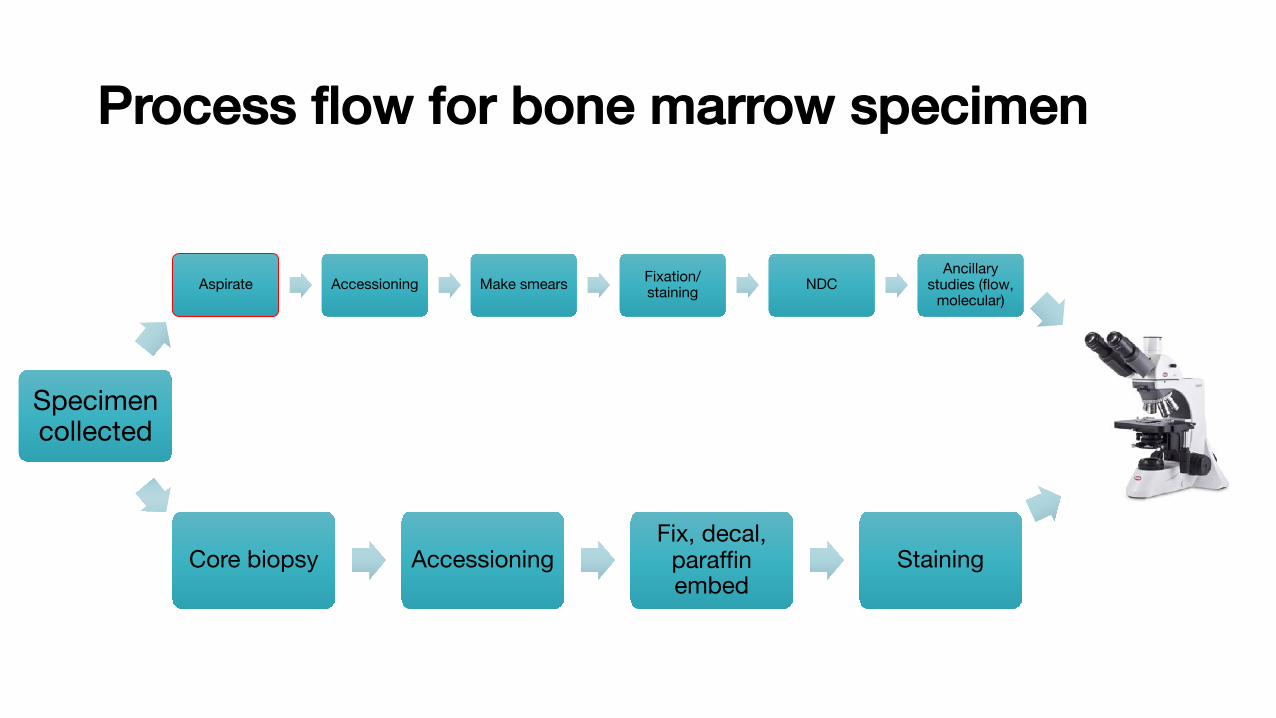
Process flow in the bone marrow exam
Aspirate Accessioning Make smears Staining NDCAncillary
studies (flow, molecular)
Core biopsy AccessioningFix, decal,
paraffin embed
Staining
Specimen collected
Analysis &Reporting

Bone marrow aspirate
Patient consent
Aspirate and biopsy needlesAspirateAseptic techniqueposterior superioriliac spine (PSIS)
• 10 – 20 mL syringe
• Usually no anticoagulant
• Smears may be made at bedside (first draw)
• Aspirate may be placed into EDTA tubes for making smears in lab
• Clot ideally made and placed into fixative at the bedside
• Second draw more diluted (used for ancillary studies)

Bone marrow trephine core biopsy
Core biopsyAseptic techniqueposterior superioriliac spine (PSIS)
• Core usually done after aspirate
• Separate needle
• Same incision
• Placed directly into fixative at bedsidePatient
consentAspirate and biopsy needles

Q: What the components of a bone marrow exam?
Components of bone marrow exam (ICSH 2008):
• Aspirate • Smear (6 slides) (EDTA)• Squash or crush preparation (≥ 2 slides) (EDTA)• Iron stain (on smear or squash preparation)• Particle clot• Flow cytometry (Heparin)• Molecular Biology (EDTA)• Cytogenetics (in tissue culture media or heparin)
• Peripheral blood smear (within 2 days of marrow)• Core biopsy Histology (Fixative, Staining)
• Touch preparation (≥ 2 slides)
Ancillary studies:• additional information to morphology

Q: What the components of a bone marrow exam?

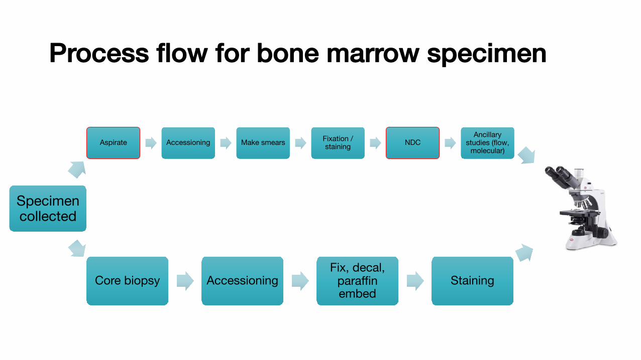
Process flow for bone marrow specimen
Aspirate Accessioning Make smears Fixation/ staining NDC
Ancillary studies (flow,
molecular)
Core biopsy AccessioningFix, decal,
paraffin embed
Staining
Specimen collected
Analysis &Reporting

Q: What is specimen accessioning?
What is accessioning?• Specimen Accessioning and
Processing (Laboratory Receiving) is the process by which specimens are:• received• sorted• and entered into the Laboratory
Information System (LIS)• labelled with barcoded labels
and processed…
Generating accessioning label

The bone marrow aspirate and clot: accessioning
• Be familiar with your LIS; how it works, communicates with instruments, how to validate it etc.
• Use 2 patient identifiers to open a new specimen accession, plus time collected and time received
• Each part of the case (clot, core, aspirate, flow, molecular etc.) usually separately accessioned
• Single piece workflow (only one specimen at a time)
Generating accessioning label
Process flow for bone marrow specimen
Aspirate Accessioning Make smears Fixation/ staining NDC
Ancillary studies (flow,
molecular)
Core biopsy AccessioningFix, decal,
paraffin embed
Staining
Specimen collected
Analysis

Collect into 10-20 mL syringe
• Used to make bedside smears
Transferremaining to EDTA tube
• Used to make smears in lab
Collect intosecond syringe
• Forancillary studies
• Relatively diluted
Transfer remaining to
EDTA and heparin tubes
The bone marrow aspirate:collection
• EDTA for molecular• Heparin for flow cytometry• and cytogenetics
Unstained bedside smear• Ideal method (less artifact)• Takes operator time• Must be done ASAP (CLOTTING)

• EDTA for molecular• Heparin for flow cytometry• and cytogenetics
Why not make all smears in the lab?• EDTA may distort morphology• Smears must be made and stained
within 24 h of collection (ideally in < 4-6 h)
The bone marrow aspirate:collection
Collect into 10-20 mL syringe
• Used to make bedside smears
Transferremaining to EDTA tube
• Used to make smears in lab
Collect intosecond syringe
• Forancillary studies
• Relatively diluted
Transfer remaining to
EDTA and heparin tubes

Process flow for bone marrow specimen
Aspirate Accessioning Make smears Fixation/ staining NDC
Ancillary studies (flow,
molecular)
Core biopsy AccessioningFix, decal,
paraffin embed
Staining
Specimen collected
Analysis

Q: What are some advantages/ disadvantages of a push prep versus a squash prep for morphology?
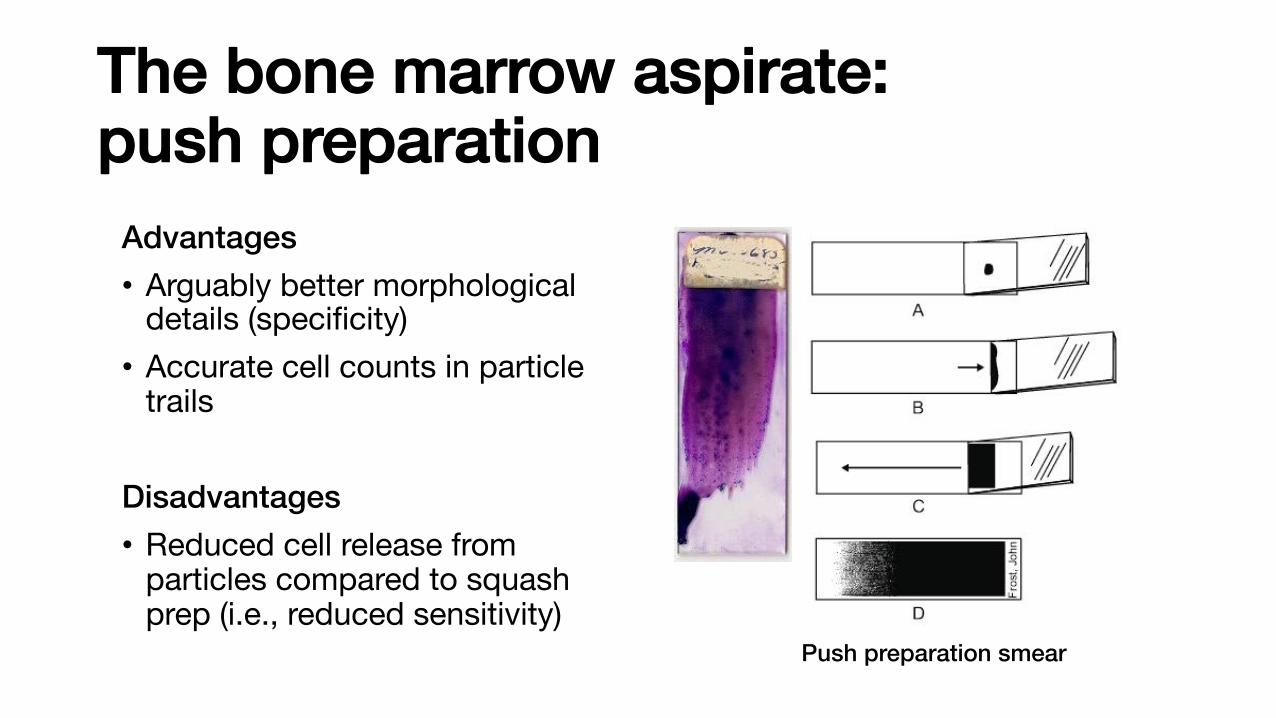
The bone marrow aspirate:push preparation
Advantages• Arguably better morphological
details (specificity)• Accurate cell counts in particle
trails
Disadvantages• Reduced cell release from
particles compared to squash prep (i.e., reduced sensitivity)
Push preparation smear
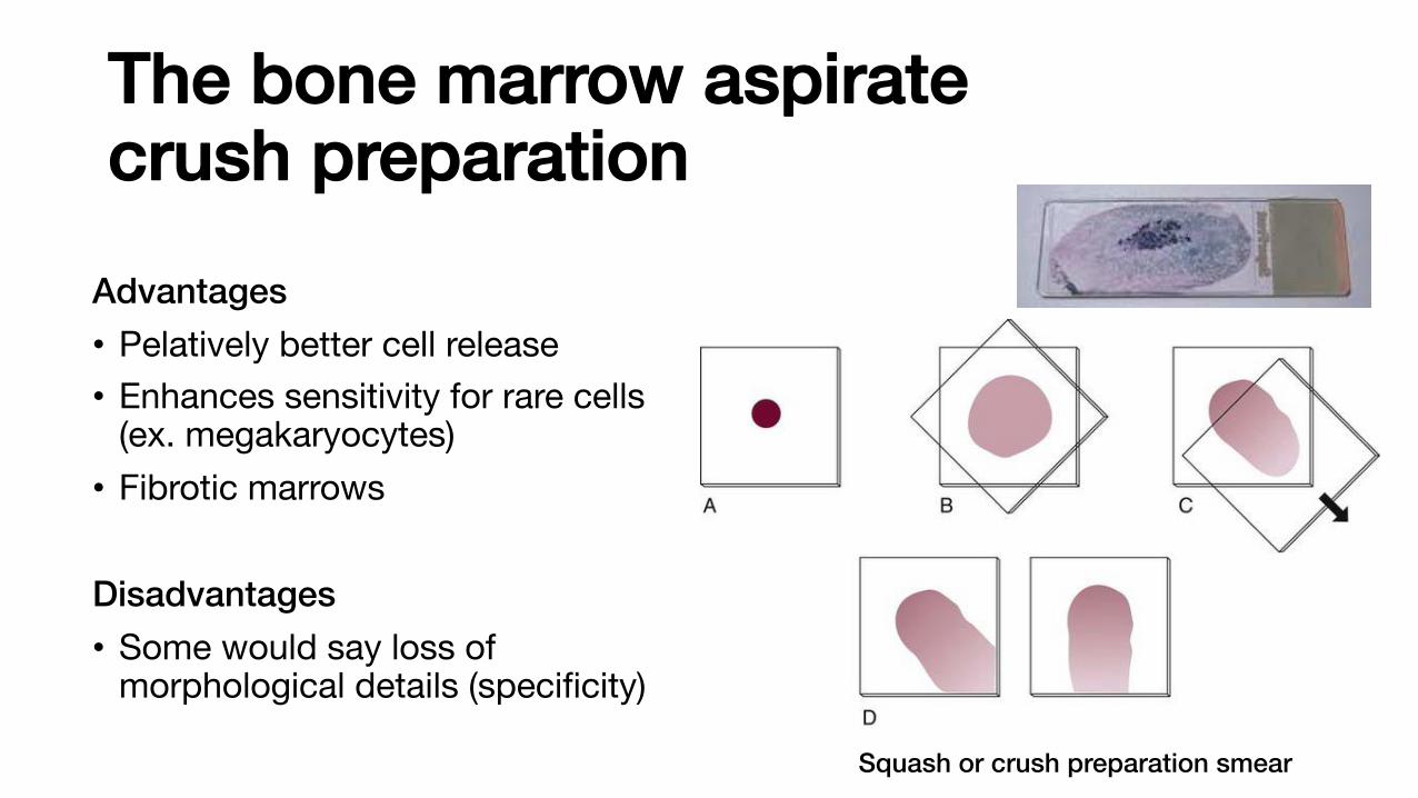
The bone marrow aspiratecrush preparation
Advantages• Pelatively better cell release• Enhances sensitivity for rare cells
(ex. megakaryocytes)• Fibrotic marrows
Disadvantages• Some would say loss of
morphological details (specificity)
Squash or crush preparation smear
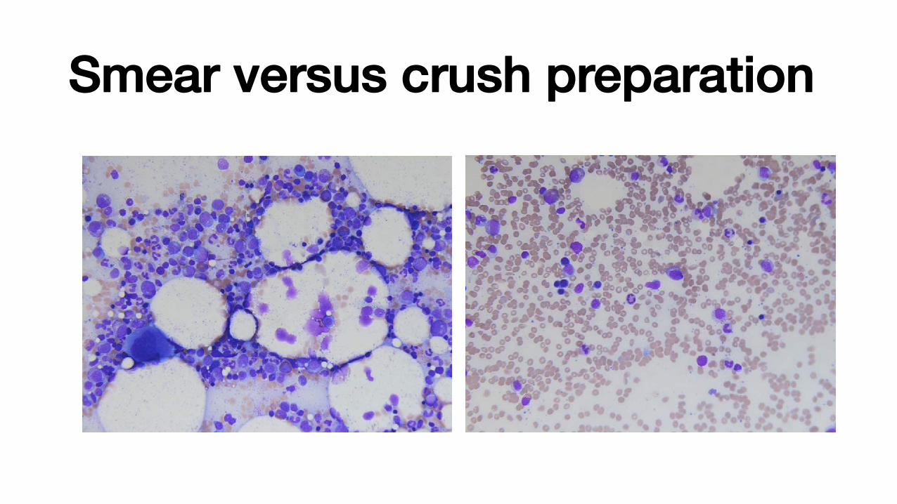
Smear versus crush preparation

Q: What EDTA concentration is recommended by ICSH for morphology?
Q: What is a disadvantage of using heparinized specimens for bone marrow morphology?

Bone marrow aspirate: anticoagulantsEDTA• ICSH recommends the dipotassium
EDTA salt at a concentration of 1.50 ± 0.25 mg/ml• Can distort cellular morphology• Smears must be made and fixed within
24 h of collection (less time ideal)• Should not be used for cell culture
(cytogenetics)
Lavender top tube (EDTA)

Bone marrow aspirate: anticoagulantsHeparin• ICSH recommends sodium
heparin for flow cytometry and cytogenetics• Heparinized samples should not
be used for morphology (staining artifact)• Heparin should not be used for
molecular biology (may interfere with PCR reactions)
Green top tube (heparin)

Process flow for bone marrow specimen
Aspirate Accessioning Make smears Fixation / staining NDC
Ancillary studies (flow,
molecular)
Core biopsy AccessioningFix, decal,
paraffin embed
Staining
Specimen collected
Analysis

Q: What is the ICSH recommended fixative for bone marrow aspirates?

Bone marrow Aspirate:Fixation and Staining
According to ICSH• Air dry and fix in absolute methanol• Dry ~ 20 mins • Acetone-free absolute methanol
(~ 10 min)• Water in methanol can lead to
hydration artifact• Fixation and staining is automated in
most labs nowMedium jar
programmed to prepare and stain a single film per sam-ple. Others can prepare and stain multiple films from asingle blood sample; this is useful for preparing slidesfor teaching large numbers of students. Some systemsapply staining solutions to slides lying horizontally (flat-bed staining), whereas others either immerse a slide orslides in a bath of staining solution (‘dip-and-dunk’ tech-nique) or spray stain onto slides in a cytocentrifuge. Pro-blems include increased background staining, inadequatestaining of neutrophil granules, degranulation of baso-phils and blue or green rather than pink staining oferythrocytes. These problems are usually related to thespecific stains and staining protocols used rather than tothe type of instrument, although flat-bed stainers aremore likely to cause problems with stain deposit. How-ever, as a rule, staining is satisfactory provided that reli-able stains are used and there is careful control of thecycle time and other variables.8 Flat-bed stainers maynot stain an entire film (e.g. a bone marrow film) if thefilm exceeds the standard length.
Rapid Staining Method
Field’s method9,10 was introduced to provide a quickmethod for staining thick films for malaria parasites(see below). With some modifications, it can be usedfairly satisfactorily for the rapid staining of thin films.The stains are available commercially ready for use, orthey can be prepared as follows.
Stains
Stain A (Polychromed Methylene Blue)
Methylene blue 1.3 g
Disodium hydrogen phosphate(Na2HPO4.12H2O)
12.6 g
Potassium dihydrogen phosphate (KH2PO4) 6.25 g
Water 500 ml
A B
C D
Figure 4.2 Blood film appearances following methanol fixation. Photomicrographs of Romanowsky-stained blood films that havebeen fixed in methanol containing: (A) 1% water; (B) 3% water; (C) 4% water; and (D) 10% water. The red cells and leucocytesare well fixed in (A), reasonably well fixed in (B) but badly fixed in (C) and (D).
Practical Haematology
62

Q: What is the principle of a romanowskystain?

Bone marrow Aspirate:Fixation and Staining
Romanowsky staining principle:
• Basic and acid component• Eosin Y ACIDIC
• Stains positively charged material (cytoplasmic granules and proteins, ex. hemoglobin).
• Azure B / methylene blue BASIC• Stains negatively charged material
(nucleic acids).
Marrow aspirate smear
Particles
Example stains:• May-Grunwald-Giemsa (MGG)• Wright-Giemsa (WG)• pH ~ 6.8
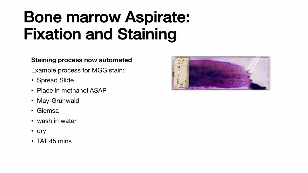
Bone marrow Aspirate:Fixation and Staining
Staining process now automatedExample process for MGG stain:• Spread Slide • Place in methanol ASAP • May-Grunwald• Giemsa• wash in water • dry • TAT 45 mins
Marrow aspirate smear
Particles
Example stains:• May-Grunwald-Giemsa (MGG)• Wright-Giemsa (WG)• pH ~ 6.8

Bone marrow Aspirate:IRON stainPerls’ Stain / Perls’ reaction / Prussian blue reactionThe Perls' reaction occurs in THREE steps, with sequential treatment of marrow aspirate sections:1) 5% hydrochloric acid solution2) 5% ferrocyanide3) counterstain, nuclear fast red or Safranin-O
• Any ferric ion (+3) in the tissue combines with the ferrocyanide and results in the formation of a bright blue pigment called ferric ferrocyanide (Prussian blue).
Storage iron
Bone marrow study:other special stains to knowBasic rule: if it’s on your lab’s hematology staining menu, then you should know HOW it works and at least ONE application…• Periodic acid schiff (PAS)• Gomori Methenamine-Silver (GMS)• Gram• Ziehl–Neelsen stain• Reticulin• Trichrome
Process flow for bone marrow specimen
Aspirate Accessioning Make smears Fixation / staining NDC
Ancillary studies (flow,
molecular)
Core biopsy AccessioningFix, decal,
paraffin embed
Staining
Specimen collected
Analysis
Q: How many cells does ICSH recommend counting on a marrow NDC?
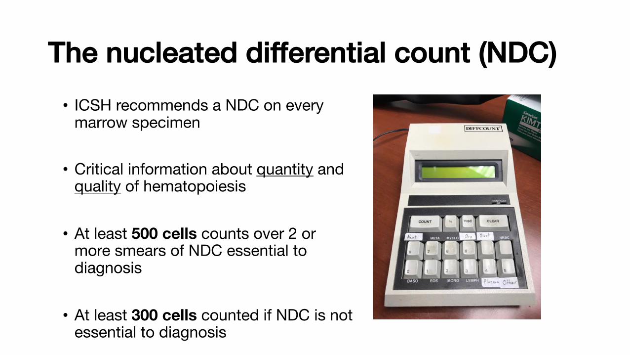
The nucleated differential count (NDC)• ICSH recommends a NDC on every
marrow specimen
• Critical information about quantity and quality of hematopoiesis
• At least 500 cells counts over 2 or more smears of NDC essential to diagnosis
• At least 300 cells counted if NDC is not essential to diagnosis

The nucleated differential count (NDC)Why perform a NDC on every marrow?
1) To assess hematopoietic activity
2) To compare the proportions of the different lineages (M:E ratio)
3) To quantify abnormal cells
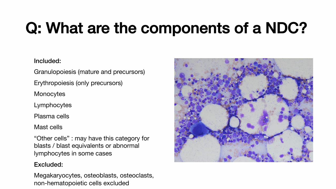
Q: What are the components of a NDC?
Included:Granulopoiesis (mature and precursors)Erythropoiesis (only precursors)Monocytes Lymphocytes Plasma cellsMast cells
“Other cells” : may have this category for blasts / blast equivalents or abnormal lymphocytes in some casesExcluded:Megakaryocytes, osteoblasts, osteoclasts, non-hematopoietic cells excluded
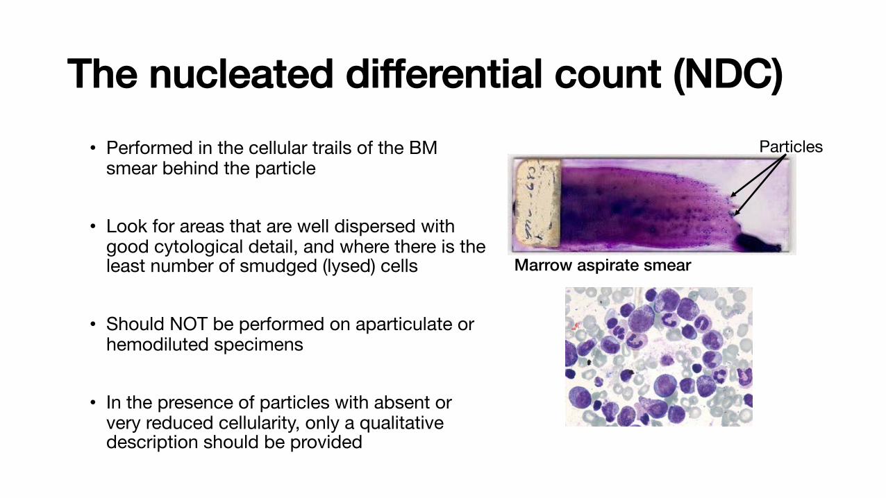
The nucleated differential count (NDC)• Performed in the cellular trails of the BM
smear behind the particle
• Look for areas that are well dispersed with good cytological detail, and where there is the least number of smudged (lysed) cells
• Should NOT be performed on aparticulate or hemodiluted specimens
• In the presence of particles with absent or very reduced cellularity, only a qualitative description should be provided
Marrow aspirate smear
Particles

Q: What are the components of the M:E ratio?
Myeloid : Erythroid ratio• Ratio of all myeloid cells (granulocytes and
monocytes and precursors) to all erythroid precursors
• normally 2-4: 1
Increased M/E:• >5:1• Infection, CML/MPN, erythroid hypoplasia,
inflammation,
Decreased M/E:• <1.2:1• Decreased leukopoiesis or erythroblast proliferation
Marrow aspirate smear
Particles

Process flow for bone marrow specimen
Aspirate Accessioning Make smears Fixation / staining NDC
Ancillary studies (flow,
molecular)
Core biopsy AccessioningFix, decal,
paraffin embed
Staining
Specimen collected
Analysis

Ancillary studies on marrow aspirate • Investigations that add additional
information beyond morphological findings
• ICSH recommends that following as essential components of any bone marrow examination:• Flow cytometry (immunophenotype,
maturational stage, lineage assignment)• Molecular studies (Clonality, mutations ex.
PML-RAR, BCR-ABL1, prognostication)• Cytogenetics (Diagnosis, prognostication)
Peripheralsmear
Aspirate
Core biopsy
Flow cytometry
Cytogenetics
Moleculartesting
Pathologistreview
Continuousassessment/
TRIAGE

Process flow for bone marrow specimen
Aspirate Accessioning Make smears Fixation / staining NDC
Ancillary studies (flow,
molecular)
Core biopsy AccessioningFix, decal,
paraffin embed
Staining
Specimen collected
Analysis

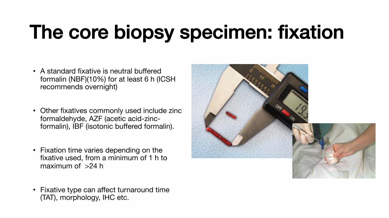
The core biopsy specimen: fixation
• Should be at least 2 cm (in reality it rarely is)
• Placed in fixative at the bedside
• Trephine core specimens may be fixed by a number of different methods
• Fixation methods can significantly affect histomorphology, cytological detail and immunoreactivity
Core biospsy at bedside

Q: What are three possible bone marrow fixatives?
The core biopsy specimen: fixation
• A standard fixative is neutral buffered formalin (NBF)(10%) for at least 6 h (ICSH recommends overnight)
• Other fixatives commonly used include zinc formaldehyde, AZF (acetic acid-zinc-formalin), IBF (isotonic buffered formalin).
• Fixation time varies depending on the fixative used, from a minimum of 1 h to maximum of >24 h
• Fixative type can affect turnaround time (TAT), morphology, IHC etc.
Core biopsy at bedside
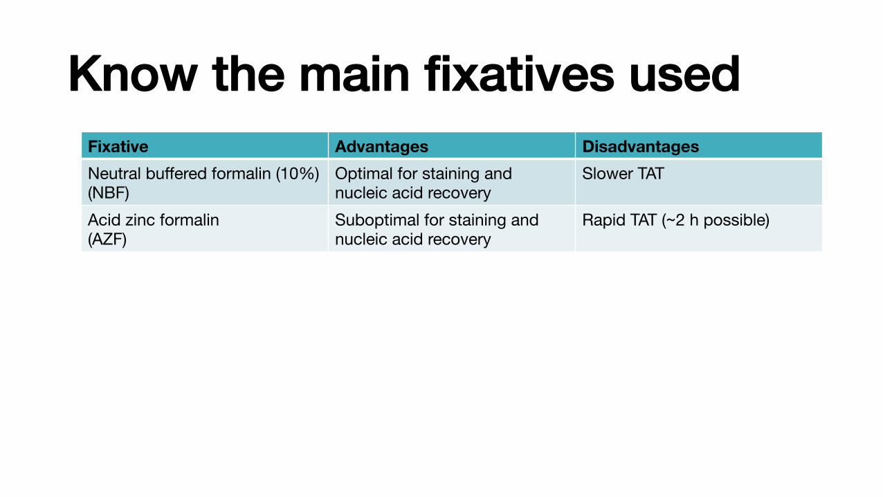
Know the main fixatives usedFixative Advantages DisadvantagesNeutral buffered formalin (10%)(NBF)
Optimal for staining and nucleic acid recovery
Slower TAT
Acid zinc formalin(AZF)
Suboptimal for staining and nucleic acid recovery
Rapid TAT (~2 h possible)
• Over-fixation (ie, more than 24 hours in formalin, more than 4 hours in zinc formalin) should be avoided for optimal immunophenotypic reactivity.
• Fixation type and time can both affect immunoreactivity.
See table in Torlakovic, E. E. et al. ICSH guidelines for the standardization of bone marrow immunohistochemistry. Int J Lab Hematol 37, 431–449 (2015).
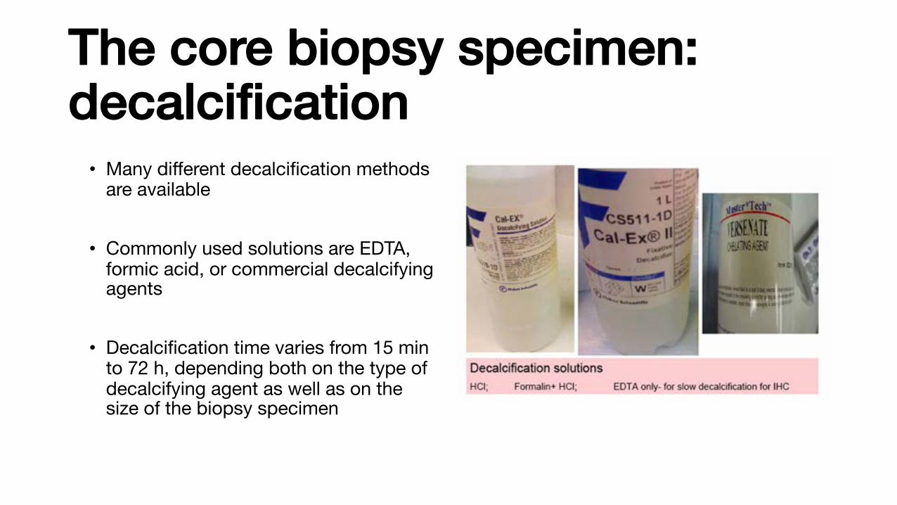
The core biopsy specimen:decalcification• Many different decalcification methods
are available
• Commonly used solutions are EDTA, formic acid, or commercial decalcifying agents
• Decalcification time varies from 15 min to 72 h, depending both on the type of decalcifying agent as well as on the size of the biopsy specimen Decalcification reagents

Q: What are at least two decalcification reagents used in bone marrow processing?
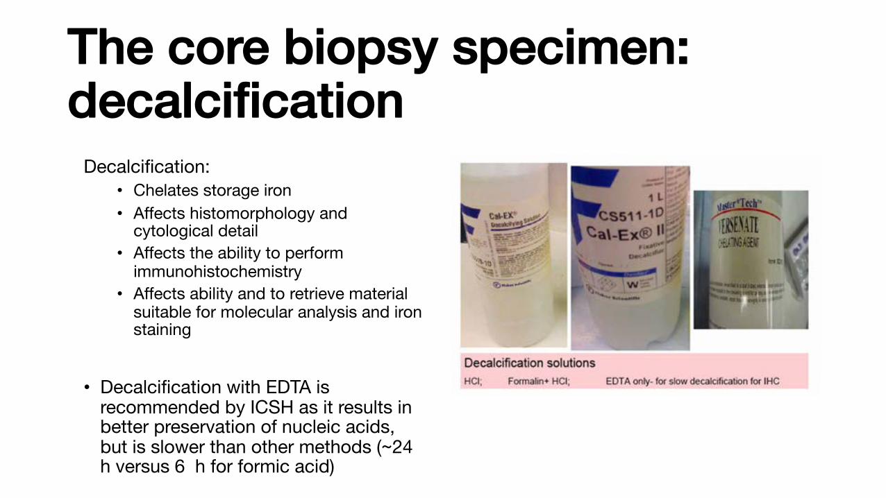
The core biopsy specimen:decalcification
Decalcification:• Chelates storage iron• Affects histomorphology and
cytological detail• Affects the ability to perform
immunohistochemistry • Affects ability and to retrieve material
suitable for molecular analysis and iron staining
• Decalcification with EDTA is recommended by ICSH as it results in better preservation of nucleic acids, but is slower than other methods (~24 h versus 6 h for formic acid)
Decalcification reagents
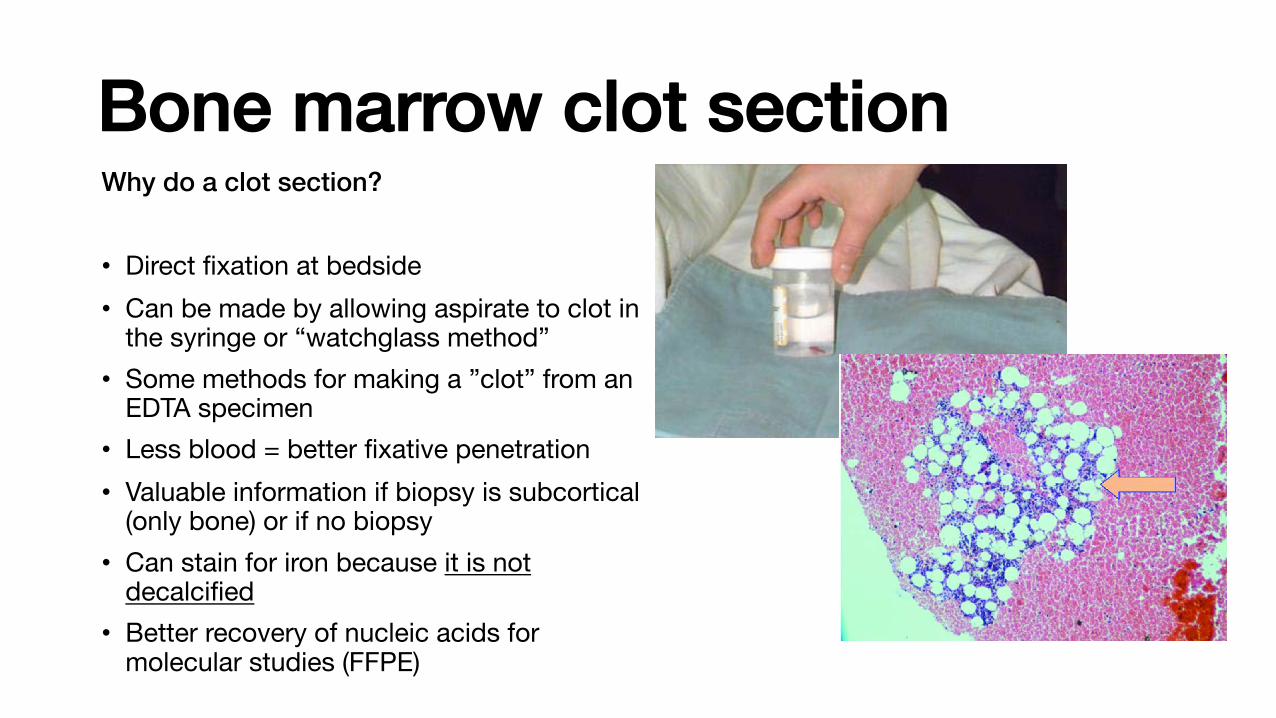
Bone marrow clot sectionWhy do a clot section?
• Direct fixation at bedside• Can be made by allowing aspirate to clot in
the syringe or “watchglass method” • Some methods for making a ”clot” from an
EDTA specimen• Less blood = better fixative penetration • Valuable information if biopsy is subcortical
(only bone) or if no biopsy • Can stain for iron because it is not
decalcified• Better recovery of nucleic acids for
molecular studies (FFPE) Clot section

Bone marrow core biopsy: process flowchartFixation
• NBF• IBF• AZF• 1 -24 h
Decalcification
• Formic acid• EDTA• 15 min – 72
h
Paraffin embed
EtOH / xylene dehydrate
Cut sections
• 2-3 uM
• 3 levels (25%, 50%, 75%)
Staining
• H&E• Special
stains• IHC
Clot section: omit decalcification step.
Time can vary depending on the reagents / methods used by your lab.
Go to your institutions AP histology lab and follow a marrow core from start to finish.

Process flow for bone marrow specimen
Aspirate Accessioning Make smears Fixation / staining NDC
Ancillary studies (flow,
molecular)
Core biopsy AccessioningFix, decal,
paraffin embed
Staining
Specimen collected
Analysis
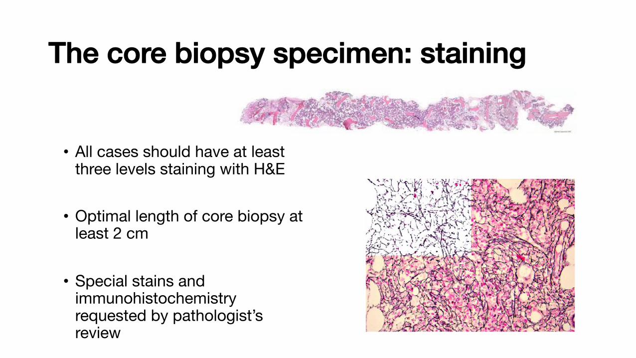
The core biopsy specimen: staining
• All cases should have at least three levels staining with H&E
• Optimal length of core biopsy at least 2 cm
• Special stains and immunohistochemistry requested by pathologist’s review
Core biopsy H&E
Reticulin stain

The core biopsy specimen:immunohistochemistry
In hematopathology immunohistochemistry (IHC) is mainly important for:• Detection of disease (lymphoma)• Lineage assignment (leukemia and
lymphoma) +/- flow cytometry • Maturational stage• Clonality (plasma cells) • Not used for prognostication (BM)
• Immunohistochemistry is not usually standardized across labs
• ICSH guidelines for standardization (2015)
ICSH guidelines for the standardization of bone marrowimmunohistochemistryE. E. TORLAKOVIC*, R. K. BRYNES†, E. HYJEK‡, S.-H. LEE§, H. KREIPE¶, M. KREMER**, R. MCKENNA††,Y. SADAHIRA‡‡, A. TZANKOV§§, M. REIS¶¶, A. PORWIT*,***, FOR THE INTERNATIONAL COUNCIL FORSTANDARDIZATION IN HAEMATOLOGY
*Department of LaboratoryHematology, University HealthNetwork, University of Toronto,Toronto, ON, Canada†Department of Pathology, KeckSchool of Medicine, Universityof Southern California, LosAngeles, CA, USA‡Department of Pathology,University of Chicago, Chicago,IL, USA§Department of Haematology, StGeorge Hospital, SEALS Central,Sydney, NSW, Australia¶Department of Pathology,Hannover Medical School,Hannover, Germany**Munich Municipal Hospital,Institute of Pathology, Munich,Germany††Special Hematology,Department of LaboratoryMedicine and Pathology,University of Minnesota,Minneapolis, MN, USA‡‡Department of Pathology,Kawasaki Medical School,Kurashiki, Japan§§Institute of Pathology,University Hospital Basel, Basel,Switzerland¶¶Department of ClinicalPathology, Sunnybrook HealthSciences Centre, Toronto, ON,Canada***Department of Pathology,Karolinska Institute, Stockholm,Sweden
SUMMARY
Bone marrow (BM) tissue biopsy evaluation, including trephine
biopsy and clot section, is an integral part of BM investigation and
is often followed by ancillary studies, in particular immunohisto-
chemistry (IHC). IHC provides in situ coupling of morphological
assessment and immunophenotype. The number of different IHC
tests that can be applied to BM trephine biopsies and the number
of indications for IHC testing is increasing concurrently with the
development of flow cytometry and molecular diagnostic methods.
An international Working Party for the Standardization of Bone
Marrow IHC was formed by the International Council for Standard-
ization in Hematology (ICSH) to prepare a set of guidelines for the
standardization of BM IHC based on currently available published
evidence and modern understanding of quality assurance principles
as applied to IHC in general. The guidelines were discussed at the
ICSH General Assemblies and reviewed by an international panel
of experts to achieve further consensus and represent further
development of the previously published ICSH guidelines for the
standardization of BM specimens handling and reports.
© 2015 John Wiley & Sons Ltd, Int. Jnl. Lab. Hem. 2015, 37, 431–449 431
REVIEW INTERNATIONAL JOURNAL OF LABORATORY HEMATOLOGY
International Journal of Laboratory HematologyThe Official journal of the International Society for Laboratory Hematology

IHC test classificationCAP (Canadian Association of Pathologists) guidelines (2015):
Class I IHC tests:• IHC test results are both interpreted and used by pathologists• Qualitative with descriptive performance characteristics (ex., positive or negative)
Class II IHC tests:• IHC test results are interpreted by pathologists; however, these tests have either
prognostic or predictive nature and their results are used by treating physician for clinical decision making (used by clinicians).
• Very few, if any in everyday hematopathology practice

ImmunohistochemistryWhy standardize IHC?• IHC testing may provide critical information important for
diagnosis, prognosis and therapy
• Required for building EQA/PT programs (currently not widespread in bone marrow IHC)

Pre-analytical
•?
Analytical
•?
Post-analytical
•?
Think process flow in terms of variables at each analytical step…
Bone marrow IHC standardization

Q: Define the pre-analytical phase for bone marrow IHC. What are the components of this phase?

Pre-analytical
• ischemic time
• fixative and fixation time
• type of decalcifying reagent and time of decalcification
• clot sample preparation
• embedding media
Bone marrow IHC standardization
Define the pre-analytical phase for bone marrow IHC?
• The pre-analytical phase starts at the time of procurement of the BM trephine and aspirate samples and ends by cutting the paraffin-embedded (or plastic-embedded) tissue onto glass slides

Bone marrow IHC standardizationAnalytical phase IHC recommendations (ICSH):
1) Stain validation
2) Monitor performance characteristics using appropriate controls for each IHC test
3) Separate SOP for each IHC test
Analytical
• antibody clone
• antigen retrieval
• colour visualization
• counterstain
• validation
• controls
• SOPs
• QC and EQA

Analytical
• antibody clone
• antigen retrieval
• colour visualization
• counterstain
• validation
• controls
• SOPs
• QC and EQA
Bone marrow IHC standardizationtweaking various parameters amenable to modification
(temperature, time, pH, etc.). As the pre-analytical
phase cannot be absolutely controlled and therefore
cannot be entirely standardized (acceptable ischemic
time is defined as maximum time with a range of
T0–Tmax, and similarly, there is also an acceptable time
range for fixation and decalcification), and this com-
bined with multiple options for selecting other reagents
and equipment as well as changes in equipment perfor-
mance or primary antibody deterioration may lead to
changes in IHC protocols to maintain desired sensitivity
and specificity of the results. See Section 4 for special
considerations relevant to the analytical phase.
Recommendation 1. Immunohistochemistry protocols
need to be validated, that is, designed as such to
reflect optimal calibration of sensitivity and specific-
ity of the IHC test for particular use depending on the
biology of the tested IHC marker and its ultimate use
[4]. This assumes that performance characteristics of
each IHC test, irrespective of its class, are defined
before introduction of the protocol to clinical use.
Recommendation 2. Monitoring of protocol perfor-
mance characteristics using appropriate calibrated
controls is recommended.
Recommendation 3. Separate standard operating pro-
cedures (SOPs) for each IHC test that is performed
in the laboratory need to be developed, as every
IHC test is a different test. All essential components
of IHC protocols for each IHC test need to be
included in the SOPs (Table 2). This information
must be readily available for review for all IHC lab-
oratory staff. The protocols must be regularly
updated. Updating of SOPs is required at the time
any changes in the protocol are introduced. In addi-
tion, periodic review of SOPs is recommended as
per laboratory accreditation requirements.
Recommendation 4. To facilitate methodology trans-
fer, when BM IHC results are published (research,
case reports, etc.), all information included in SOPs
(as shown in Table 2) is recommended to be
included in the ‘Methods’ section.
2.4. Postanalytical standards
Postanalytical standards pertain to the interpretation
of the IHC results by the (hemato)pathologist. See
Section 5 for special considerations relevant to the
postanalytical phase.
Recommendation 1. All BM aspirate results should be
available to (hemato)pathologists directly, that is,
they need to evaluate them and sign them out, or
indirectly, that is, results of cytological evaluation
of BM smears, molecular studies, and cytogenetic
studies are submitted to (hemato)pathologists who
are evaluating BM tissue biopsy optimally before
they are made available to other physicians.
Recommendation 2. Interpretation of IHC results
should be performed in consideration of the sample
adequacy. Proper sampling of the BM cannot be
overemphasized. Although IHC is a powerful
technique and may help detect pathological cells
even when they are present in small numbers and
not apparent morphologically, standardization and
excellence in BM IHC cannot replace proper BM
sampling [26]. If the BM sample does not contain
the lesion, no special studies could compensate
for the deficiency of proper sampling [27–30]. There-
fore, general guidelines for BM biopsy should be fol-
lowed in the first place [19, 23, 24, 31–33].
Recommendation 3. It is recommended that the inter-
pretation starts with the evaluation of the external
control(s), by which it is determined that the
proper antibody was applied and the test is prop-
erly calibrated.
Recommendation 4. Evaluation of the patient sample
starts with detection and evaluation of an internal
positive control if such is present. Detection of non-
specific staining should be observed if present.
Table 2. SOPs components required to be specifiedfor each bone marrow immunohistochemistry tests
SOP component Descriptors
Primary antibody(Ab) type
Monoclonal vs. polyclonal, clone orlot name/number, source,concentrated vs. prediluted, anddilution (if concentrated Ab),incubation time, temperature
Antigen retrievalmethod
Type, pH, concentration(for enzyme-based methods),temperature, time, and source
Detection system Type, name, temperature, time,source
Amplification Type, temperature, time, sourceChromogen Type, timeEnhancement Type, timeAutomated stainer
platformName, source
© 2015 John Wiley & Sons Ltd, Int. Jnl. Lab. Hem. 2015, 37, 431–449
E. E. TORLAKOVIC ET AL. | STANDARDIZATION OF BONE MARROW IMMUNOHISTOCHEMISTRY 437

Thinking questions…Q: What are the components of a method or procedure validation?
Q: What are performance characteristics?
Q: How would you validate a new IHC stain for bone marrow?
Q: What is a common cause of “background” or non-specific binding in bone marrow IHC, and what can be done to mitigate this effect?

Bone marrow IHC standardizationPostanalytical standards pertain to the interpretation and reporting of the IHC results by the hematopathologist.
1) Integrated aspirate and biopsy reported by same person
2) Sample must be adequate3) Proper internal and external controls
(positive and negative)4) Synoptic reporting
Post-analytical
• Ancillarystudies
• Integratedcore and aspirate report
• Synoptic reporting
Controls in bone marrow IHC• Controls should be treated the same way as your specimen…
• External controls should ideally have two levels of staining intensity (weak and strong)
• External controls should ideally be mounted on the same slide as the specimen
• Negative controls if there are internal pigments, necrosis, fibrosis etc. that could interfere with interpretation or when only one one IHC test has been ordered

Bone marrow reporting• Integrated report for bone marrow aspirate and biopsy review
is recommended by ICSH
• It is recommended that the aspirate and core biopsy be read by the same individual
• A final integrated consensus conclusion that includes all test results (e.g. flow cytometry, cytogenetics and molecular genetics) if possible

Bone marrow reportingTable 3. The bone marrow aspirate report
Name of institution
Unique specimen identifier (laboratory accession number)
Details of patient: surname, first name(s), identification number, age or date of birth, gender, contact details(e.g. address, hospital location)
Name of responsible physician
Name of requesting doctor
Date of procedure
Significant clinical history including physical findings, recent chemo/radiotherapy, cytokine therapy and pertinent labresults.
Indication for bone marrow examination
Procedure performed (aspirate/trephine biopsy)
Anatomic site of aspirate/biopsy
Ease/difficulty of aspiration
Blood count: Haemoglobin concentration, total and differential white cell count (neutrophils, eosinophils, basophils,monocytes, lymphocytes) and platelet count
Blood smear description and diagnostic conclusion
Cellularity of particles and cell trails
Nucleated differential cell count
Total number of cells counted
Myeloid:erythroid ratio
Erythropoiesis
Myelopoiesis
Megakaryocytes
Lymphocytes
Plasma cells
Other haemopoietic cells
Abnormal cells (e.g. blast cells, metastatic infiltrates)
Iron stain
Cytochemistry
Other investigations (e.g. cytogenetics, PCR, FISH, microbiology)
Summary of flow cytometry findings, if available
Conclusion
WHO classification (if relevant)
Disease code
Signature and date of report
! 2008 The Authors
Journal compilation ! 2008 Blackwell Publishing Ltd, Int. Jnl. Lab. Hem. 2008, 30, 349–364
358 S.-H. LEE ET AL. ICSH: BM SPECIMENS AND REPORTS
consistent approach. Because not all data may be
available, any pending data should be correlated when
it becomes available. Digital images that demonstrate
the major abnormal findings in specimens may be
appended to the electronic report, if this function is
supported by the laboratory information system.
5. VERBAL REPORTS
When a verbal report is given to a clinician, a com-
ment should be added to indicate the name of the
pathologist providing the information, to whom the
report was given, and the time and date.
6. TURNAROUND TIMES
6.1 Aspirates
The processing TAT for the BM aspirate, or the time
from collection of the aspirate to the time when slides
are available for microscopy should be about two to
six working hours. However, this may not be achiev-
able with remote laboratories. If results are required
urgently by the requesting clinician, the Reporting
TAT, or the time from when the slides are available
for microscopy to the time when a verbal or written
report is issued, should be about three working hours
Table 4. The bone marrow trephine report
Name of institution
Unique specimen identifier (laboratory accession number)
Details of patient: surname, first name(s), identification number, age or date of birth, gender, contact details(e.g. address, hospital location)
Name of responsible physician
Name of requesting doctor
Date of procedure
Significant clinical history including physical findings, recent chemo/radiotherapy, cytokine therapy and pertinentlab results
Indication for bone marrow examination
Procedure performed (aspirate/trephine biopsy)
Anatomic site of aspirate/trephine biopsy
Aggregate length of biopsy core
Adequacy and macroscopic appearance of core
Percentage and pattern of cellularity
Bone architecture
Location, number, morphology and pattern of differentiation for erythroid, myeloid, megakaryocytic lineages,lymphoid cells, plasma cells and macrophages
Abnormal cells and/or infiltrates
Reticulin stain
Immunohistochemistry
Histochemistry
Other investigations (e.g. FISH, PCR)
Conclusion
Disease code
Signature and date of report
! 2008 The Authors
Journal compilation ! 2008 Blackwell Publishing Ltd, Int. Jnl. Lab. Hem. 2008, 30, 349–364
362 S.-H. LEE ET AL. ICSH: BM SPECIMENS AND REPORTS
Bone marrow report components (ICSH)

Synoptic reportingAs currently defined by the CAP, synoptic reporting includes the following elements:
• Reporting of scientifically validated data elements that influence clinical outcome and therapeutic decisions
• Display of each data element in a “variable: result’’ format on a single line
• Display of data elements on separate lines
• Format ensures that critical information is transmitted consistently and succinctly in every report
• Succinct data presentation is free of clutter and irrelevant information
• Supports data mining (ex., machine learning) Synoptic report

Pre-analytical
• ischemic time
• fixative and fixation time
• type of decalcifying reagent and time of decalcification
• clot sample preparation
• embedding media
Analytical
• antibody clone
• antigen retrieval
• colour visualization
• counterstain
• validation
• controls
• SOPs
• QC and EQA
Post-analytical
• Ancillarystudies
• Integrated core and aspirate report
• Synoptic reporting
Bone marrow standardization

Q: What is external quality assurance (EQA)?

Quality assurance in BM IHC• EQA / PT involves comparison of your results to the results of
other laboratories
• Is a means of verifying your lab's performance against an external metric, standard or benchmark (part of accreditation)
• It is recommended that laboratories participate in PT for BM IHC only if the EQA program provides samples with identical or nearly identical BM tissue processing

Inside the lab: bone marrow reportingTurnaround times (TAT)• Quality metric that is measured
and required for lab accreditation• TAT defined as the time between
when at test is ordered or a specimen is submitted to the lab and the time when the results are reported • It is one of the most clinically
apparent/ noticeable signs of laboratory service

Inside the lab: bone marrow reportingTurnaround times (TAT) ICSH recommendations:• Reporting TAT for combined
aspirate and core biopsy report should be < 5 working days• < 7 working days if IHC or special
stains required• Varies based on each institution• Other TAT are time collection,
accessioning, processing etc.

Inside the lab: bone marrow reportingSpecimen storage and retention:• BM slides should be stored at least 20 years
according to most accreditation bodies• Digital images should be stored indefinitely• Unstained slides should be stored at - 80°C
Accreditation and EQA:• Lab should be accredited by recognized
body and subscribe to EQA as part of the accreditation

Summary• In hematopathology, a diagnosis almost always
requires integration of ancillary data and clinical information, which are often not immediately available
• Involves a process of continuous assessment and review as information becomes available
• Standardization of methods is essential for a robust and universally applicable interpretation, especially given the large amount of information that needs to be integrated for a final report
Peripheralsmear
Aspirate
Core biopsy
Flow cytometry
Cytogenetics
Moleculartesting
Pathologistreview
Continuousassessment/
TRIAGE
For the exam (my advice)• Spend time with managers and technologists
• Understand every step of the bone marrow process, from bedside to reporting
• Need fine details AND big picture
• Start thinking of yourself as a laboratory director: how would you design, validate and troubleshoot a bone marrow program (or any other laboratory process)
• Ask why again…and again…and again
• Try to relax, it is not as bad as you think

References1) LEE, S. H. et al. ICSH guidelines for the standardization of bone
marrow specimens and reports. International journal of laboratory hematology 30, 349–364 (2008).
2) Torlakovic, E. E. et al. ICSH guidelines for the standardization of bone marrow immunohistochemistry. Int J Lab Hematol 37, 431–449 (2015).
3) Protocol for the Examination of Specimens From Patients With Hematopoietic Neoplasms Involving the Bone Marrow (CAP, 2013)