the senses
DESCRIPTION
The Senses. Chapter Five. The Senses. Vision Hearing Taste Smell Touch Skin senses Vestibular Kinesthetic. Each sense organ receives some sort of external stimulus such as light, sound waves, or pressure - PowerPoint PPT PresentationTRANSCRIPT

The Senses
Chapter Five

The Senses
• Vision• Hearing• Taste• Smell • Touch• Skin senses
– Vestibular– Kinesthetic
• Each sense organ receives some sort of external stimulus such as light, sound waves, or pressure
• Then, it changes the sensation into a chemical-electrical message transmitted by the nervous system
II. Vision

The Stroop Effect

A. The Eye
1. Most studied of all senses
2. Pupil- the opening in the iris that regulates the amount of light entering the eye a. Where the light enters the eye
3. Lens- a flexible, elastic, transparent structure in the eye that changes its shape to focus light on the retina

A. The Eye (cont.)
4. Retina- the innermost coating of the back of the eye, containing the light-sensitive receptor cellsa. Rods & cones are responsible for changing
light energy into neuronal impulses
5. Optic nerve- the nerve that carries impulses from the retina to the occipital lobe of the brain


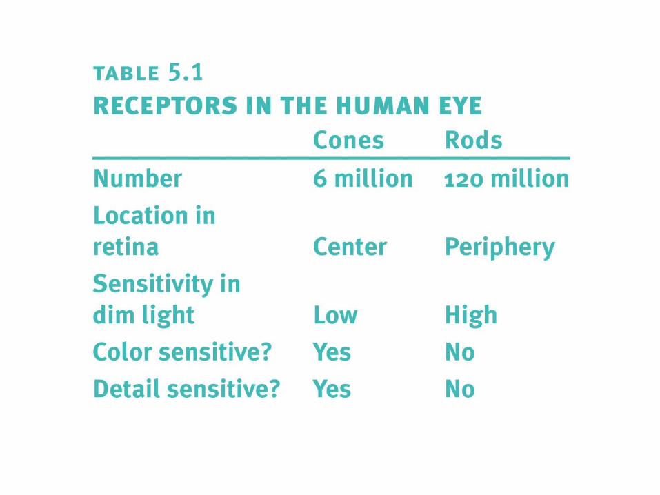
B. Cones and Rods
1. Cones require more light than rods before they begin to responda. Work best in daylight
b. Similar to color film
2. Rods can work in much lower light a. Basis for night vision
b. Similar to sensitive black and white film


C. Light1. Is a form of electromagnetic radiation
2. Other forms of electromagnetic radiation include:
a. Radio wavesb. Microwavesc. Infrared radiationd. Ultraviolet rayse. X-raysf. Gamma rays

The Electromagnetic Spectrum

C. Light (cont.)
3. Visible light only makes up a small portion of the electromagnetic spectrum
4. Passing sunlight through a spectrum breaks the light into a rainbow of colors, each color having a different wavelength
a. Object’s color depends on the light that reaches our eyes
b. Example- A pea looks green because it reflects green light and absorbs other colors

D. Process of Vision
1. Light energy stimulates the eye
– Light rays enter the eye through the cornea
– Then enter the pupil
– Light then passes through the pupil to the lens, which changes its shape to focus or reflect the light rays onto the retina
2. As light rays hit the receptors, the process of conversion (energy is converted into a neural impulse) begins
D. Process of Vision
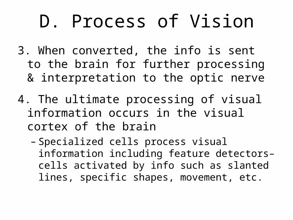
3. When converted, the info is sent to the brain for further processing & interpretation to the optic nerve
4. The ultimate processing of visual information occurs in the visual cortex of the brain– Specialized cells process visual information
including feature detectors– cells activated by info such as slanted lines, specific shapes, movement, etc.

E. Color Vision1. Young-Helmholtz (Trichromatic) theory
(functioning of cones)
– 3 types of cones exist in the eye– yellow/red, blue/violet, & green
– Explains colorblindness
2. Opponent-process theory (functioning of cells that process info from receptors)
– 3 types of cones – red/green, blue/yellow, black/white– Explains afterimages (individual looks at a stimulus for
a period of time, & then shifts gaze away from stimulus, only to see an afterimage of that stimulus in a different color)

Color Deficiency: True or False?
1. If a person’s cones don’t function correctly, the person is color-deficient
2. Fewer people have trouble between red and green than yellow and blue
3. A few can only see the world in blacks, whites, and shades of gray
4. 8% of American men and 1% of American women are color blind/color deficient

Color Blindness Test
• This figure tests for green colorblindness.– They might see
part of the number.– They might not
see the number at all.
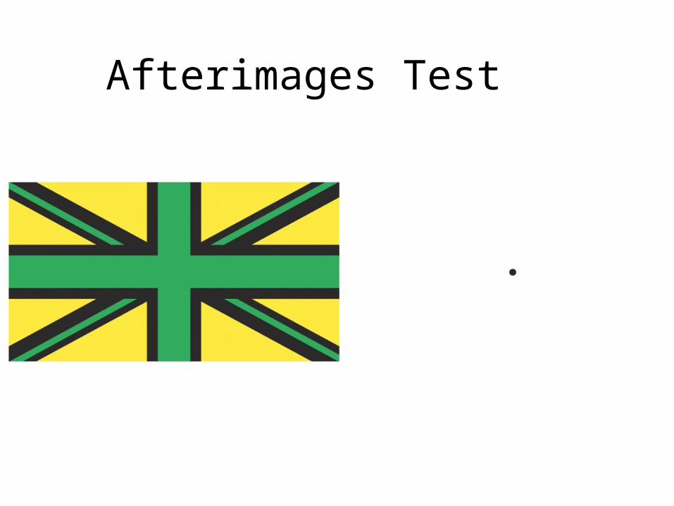
Afterimages Test

F. Binocular Fusion
1. Our visual system receives two images because we have two eyes
2. Instead of seeing double, we see a combination of the two images
a. Large retinal disparity means object is nearby
b. Small retinal disparity means object is distant

Retinal Disparity
1. Each eye projects a slightly different image on the retina
2. Example- Bring an object such as an eraser close to your eyes. Without moving it, look at the eraser first with one eye and then with the other. You will see a difference in the two images because of the different viewpoint each eye has.

Vision in the Brain

G. Nearsightedness and Farsightedness
1. Perfectly shaped eyeball means you have nearly perfect vision
2. Long eyeball usually means you are nearsighteda. You can see objects close to you, but objects in the
distance are blurry
3. Short eyeball usually means you are farsighteda. You can see distant objects clear, but up-close
objects are blurry

Problems with Vision
• Here the eyeball is shaped like an egg on it’s side. • The image is projected
behind the retina.• If you pull the image
farther away from you, you can see it clearly.
• Here the eyeball is shaped like a football.• The image is projected
in front of the retina. • If you pull the object
closer to you, you can see it clearly.

Important Figures in Vision Research
In one experiment, done in 1959, Hubel & Wiesel
inserted a microelectrode into the primary visual
cortex of an anesthetized cat. They then projected
patterns of light and dark on a screen in front of
the cat. They found that some neurons fired rapidly
when presented with lines at one angle, while
others responded best to another angle. These
studies showed how the visual system constructs
complex representations of visual information from
simple stimulus features.

Lasik Surgery
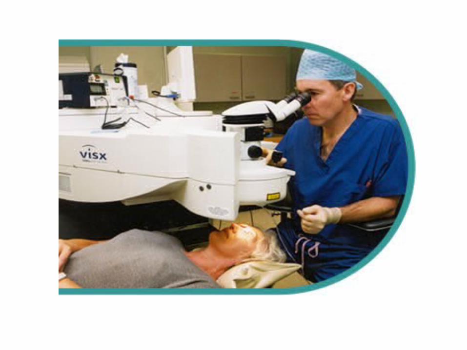


• More than one million patients undergo the LASICprocedure in the United States each year. The U.S. Foodand Drug Administration (FDA) reports that LASIK eyesurgery complications occur in 1 to 5 percent of cases.
According to the FDA’s LASIC surgery statistics:• Glare and sensitivity to light affect 1.7 percent of LASIC
patients.• Visual fluctuations occur in 2.6 percent of LASIK
patients.• Halos around light sources are experienced by 3.5
percent of patients.• About 3 percent of patients report vision worse than
before LASIC.