the senses overview of the senses vision hearing taste smell touch visceral senses
TRANSCRIPT

The Senses
Overview of the Senses
VisionHearingTasteSmellTouchVisceral Senses
receptors receives information about receptors receives information about the environmentthe environment
receptors generate nerve impulses receptors generate nerve impulses and send information to the CNSand send information to the CNS
a coordinated response can be a coordinated response can be directed to maintain homeostasis.directed to maintain homeostasis.

Types of receptors:Types of receptors:
1. Photoreceptors1. Photoreceptors
Vision – rods and cones found in the Vision – rods and cones found in the retina of the eyeretina of the eye
2. Chemoreceptors2. Chemoreceptors
Taste – taste-buds found in the Taste – taste-buds found in the tonguetongue
Smell – olfactory cells found in the Smell – olfactory cells found in the olfactory cavityolfactory cavity
Internal senses – osmoreceptors Internal senses – osmoreceptors that regulate blood pressure, COthat regulate blood pressure, CO22 balance, etc.balance, etc.

3.3. MechanoreceptorsMechanoreceptorsTouch / pressure / pain – receptors in Touch / pressure / pain – receptors in
the skinthe skin
Hearing – hair cells in the inner ear Hearing – hair cells in the inner ear detect sound wavesdetect sound waves
Balance – hair cells in the ear detect Balance – hair cells in the ear detect motionmotion
Body position – proprioceptors and Body position – proprioceptors and stretch receptors in the musclesstretch receptors in the muscles
4. Thermoreceptors4. Thermoreceptors
Temperature – receptors in the skin Temperature – receptors in the skin detect changes in radiant energydetect changes in radiant energy

Sensation Sensation – receiving and processing – receiving and processing by the brain of neural impulses from by the brain of neural impulses from the sensory receptorsthe sensory receptors
Perception-Perception- interpretation of sensory interpretation of sensory information by the cerebral cortex. information by the cerebral cortex.
Sensory Sensory adaptation/fatigadaptation/fatigueue -neuron -neuron becomes becomes accustomed to a accustomed to a stimulus and stops stimulus and stops firing. firing.
-the adaptation -the adaptation indicates that the indicates that the environment is not environment is not dangerous.dangerous.

The Eye- a The Eye- a photoreceptorphotoreceptor (I spy with my little eye)(I spy with my little eye)
regulate and focus lightregulate and focus light translate the wavelengths of light translate the wavelengths of light
into nerve impulses. into nerve impulses. detects visible light spectrumdetects visible light spectrum

The External EyeThe External Eye EyelidEyelid –protects the eye. (5) –protects the eye. (5)
EyelashEyelash – keeps dust and small – keeps dust and small particles from getting into the eye.particles from getting into the eye.
ConjunctivaConjunctiva – a thin protective layer – a thin protective layer on the cornea.on the cornea. (4) (4)
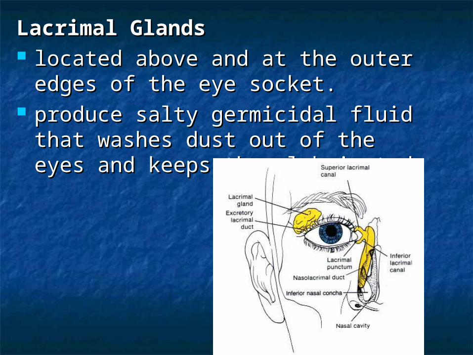
LacrimalLacrimal GlandsGlands located above and at the outer edges located above and at the outer edges
of the eye socket. of the eye socket. produce salty germicidal fluid that produce salty germicidal fluid that
washes dust out of the eyes and washes dust out of the eyes and keeps them lubricated. keeps them lubricated.

This fluid evaporates or drains into This fluid evaporates or drains into the nasal chamber so it must be the nasal chamber so it must be replaced.replaced.
NasalNasal CavityCavity – drains fluid from the – drains fluid from the lacrimal glands into the nose. This is lacrimal glands into the nose. This is why a sharp smell or pinch on the why a sharp smell or pinch on the nose makes your eyes water.nose makes your eyes water.

Internal Structures of the EyeInternal Structures of the Eye
The Eye
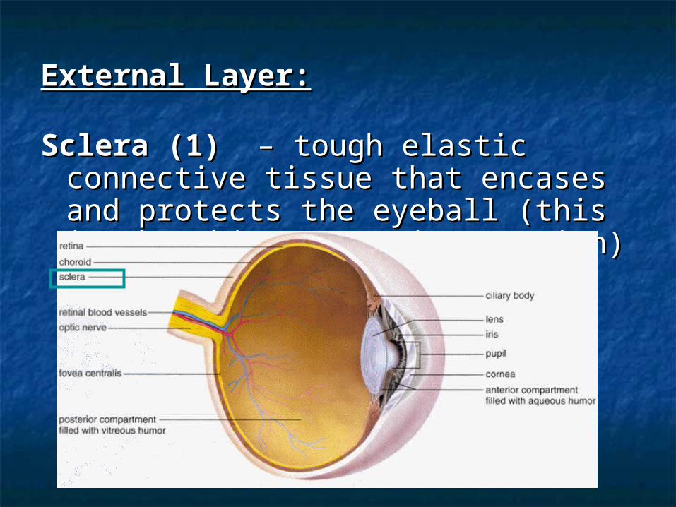
External Layer:External Layer:
Sclera (1) Sclera (1) – tough elastic connective – tough elastic connective tissue that encases and protects the tissue that encases and protects the eyeball (this is the white posterior eyeball (this is the white posterior portion)portion)
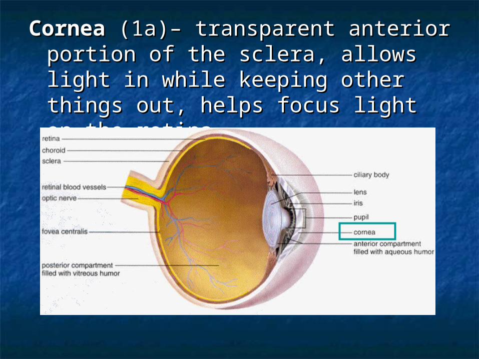
CorneaCornea (1a)– transparent anterior (1a)– transparent anterior portion of the sclera, allows light in portion of the sclera, allows light in while keeping other things out, helps while keeping other things out, helps focus light on the retinafocus light on the retina

Intermediate Layer:Intermediate Layer:
Choroid (2)Choroid (2) – darkly pigmented tissue – darkly pigmented tissue through which blood vessels nourish through which blood vessels nourish the back of the eye, pigment absorbs the back of the eye, pigment absorbs light and prevents reflectionslight and prevents reflections

Ciliary Body (2a)Ciliary Body (2a) – thickened – thickened portion of the choroid attached to the portion of the choroid attached to the suspensory ligamentssuspensory ligaments smooth muscle contracts / relaxes to smooth muscle contracts / relaxes to
change the shape of the lens, also change the shape of the lens, also secretes aqueous humour.secretes aqueous humour.

Suspensory LigamentsSuspensory Ligaments (7a) – attach (7a) – attach to the lens and the ciliary musclesto the lens and the ciliary muscles
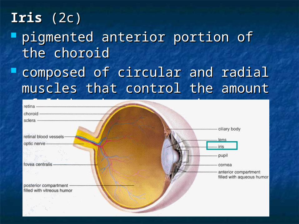
IrisIris (2c) (2c) pigmented anterior portion of the pigmented anterior portion of the
choroidchoroid composed of circular and radial composed of circular and radial
muscles that control the amount of muscles that control the amount of light that enters the eyelight that enters the eye
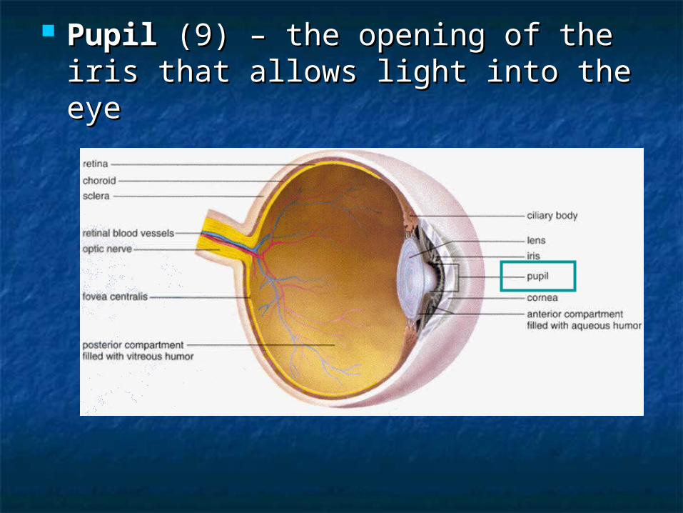
PupilPupil (9) – the opening of the iris (9) – the opening of the iris that allows light into the eyethat allows light into the eye
Internal Layer:Internal Layer:
Retina (3)Retina (3) two types of photoreceptor cells, two types of photoreceptor cells,
rods and conesrods and cones two layers of cells (bipolar cells and two layers of cells (bipolar cells and
ganglionic cells) that organize ganglionic cells) that organize visual information before impulses visual information before impulses are sent to the brainare sent to the brain


FoveaFovea Centralis (3a) –Centralis (3a) – slight slight depression in the back of the eye, depression in the back of the eye, area of highly concentrated cones, area of highly concentrated cones, this is where light is focused for this is where light is focused for clear, precise color visionclear, precise color vision

RodsRods – – night visionnight vision, abundant in the , abundant in the eye, mainly in the peripheral portion eye, mainly in the peripheral portion of the retina, sensitive to slight of the retina, sensitive to slight movements in dim lightmovements in dim light
RhodopsinRhodopsin Pigment in the rods (low light Pigment in the rods (low light
levels)levels) Is broken down (bleached) in high Is broken down (bleached) in high
light and must be replacedlight and must be replaced Vitamin A is used to make retinalVitamin A is used to make retinal Retinal + opsin = rhodopsinRetinal + opsin = rhodopsin

ConesCones – – color vision,color vision, located at the located at the fovea centralis, sensitive to color and fovea centralis, sensitive to color and bright lightbright light
Other:Other:
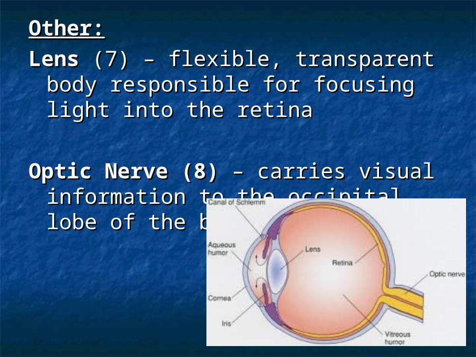
LensLens (7) – flexible, transparent body (7) – flexible, transparent body responsible for focusing light into the responsible for focusing light into the retinaretina
Optic Nerve (8)Optic Nerve (8) – carries visual – carries visual information to the occipital lobe of the information to the occipital lobe of the brain for processingbrain for processing
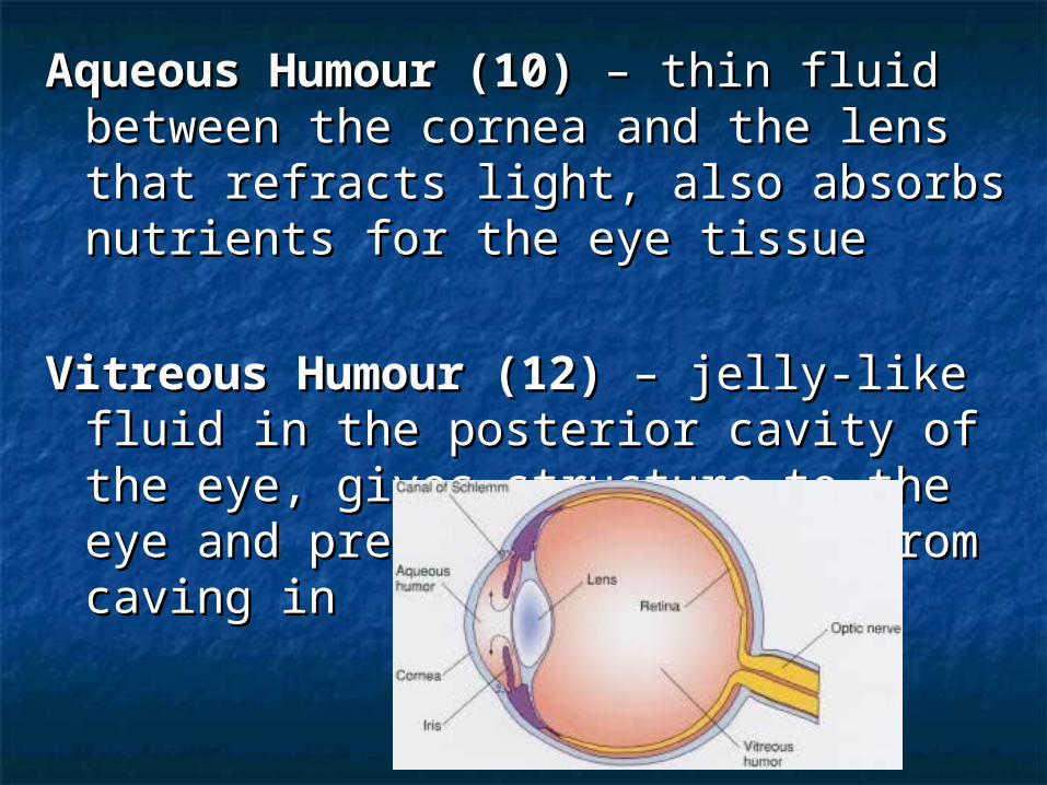
Aqueous Humour (10)Aqueous Humour (10) – thin fluid – thin fluid between the cornea and the lens that between the cornea and the lens that refracts light, also absorbs nutrients for refracts light, also absorbs nutrients for the eye tissuethe eye tissue
Vitreous Humour (12)Vitreous Humour (12) – jelly-like fluid in – jelly-like fluid in the posterior cavity of the eye, gives the posterior cavity of the eye, gives structure to the eye and prevents the structure to the eye and prevents the retina from caving inretina from caving in

Focusing and AccommodationFocusing and Accommodation
Extrinsic eye musclesExtrinsic eye muscles – control – control movements of the eyeball within the movements of the eyeball within the socket (side to side or up and down). socket (side to side or up and down). There are three pairs of muscles.There are three pairs of muscles.
Ciliary musclesCiliary muscles – found within the ciliary – found within the ciliary body, control the shape of the lens body, control the shape of the lens (contracted muscles make the lens round, (contracted muscles make the lens round, relaxed muscles flatten the lens)relaxed muscles flatten the lens)

AccommodationAccommodation – is the flattening or – is the flattening or rounding of the lens in order to focus rounding of the lens in order to focus objects on the retina.objects on the retina.

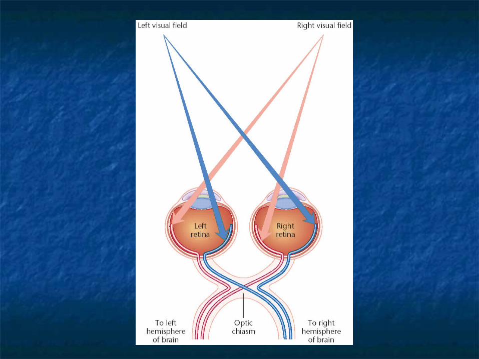
a focused image is inverted (upside down)
The brain rights the image
Neurons join at the optic chiasma and then split to the right and left visual cortex
this allows for stereoscopic vision and depth perception.


Visual DisordersVisual Disorders
Myopia (nearsightedness)Myopia (nearsightedness) – – elongated eyeball, light focuses in elongated eyeball, light focuses in front of the retinafront of the retina
Hyperopia (farsightedness)Hyperopia (farsightedness) – – shortened eyeball, light focuses shortened eyeball, light focuses behind the retinabehind the retina
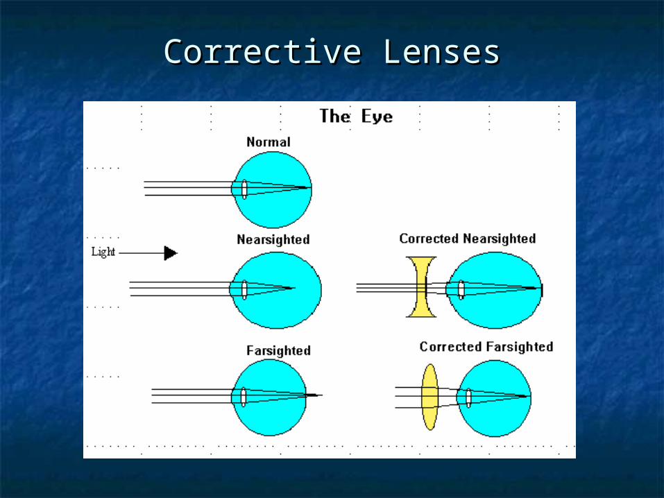
Corrective LensesCorrective Lenses
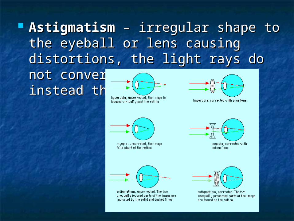
AstigmatismAstigmatism – irregular shape to the – irregular shape to the eyeball or lens causing distortions, the eyeball or lens causing distortions, the light rays do not converge at one light rays do not converge at one point, instead they scatterpoint, instead they scatter

Cataracts Cataracts – proteins in the lens – proteins in the lens change (usually with age) because of change (usually with age) because of a lack of enzymes, causing the lens a lack of enzymes, causing the lens to become opaque instead of to become opaque instead of transparenttransparent
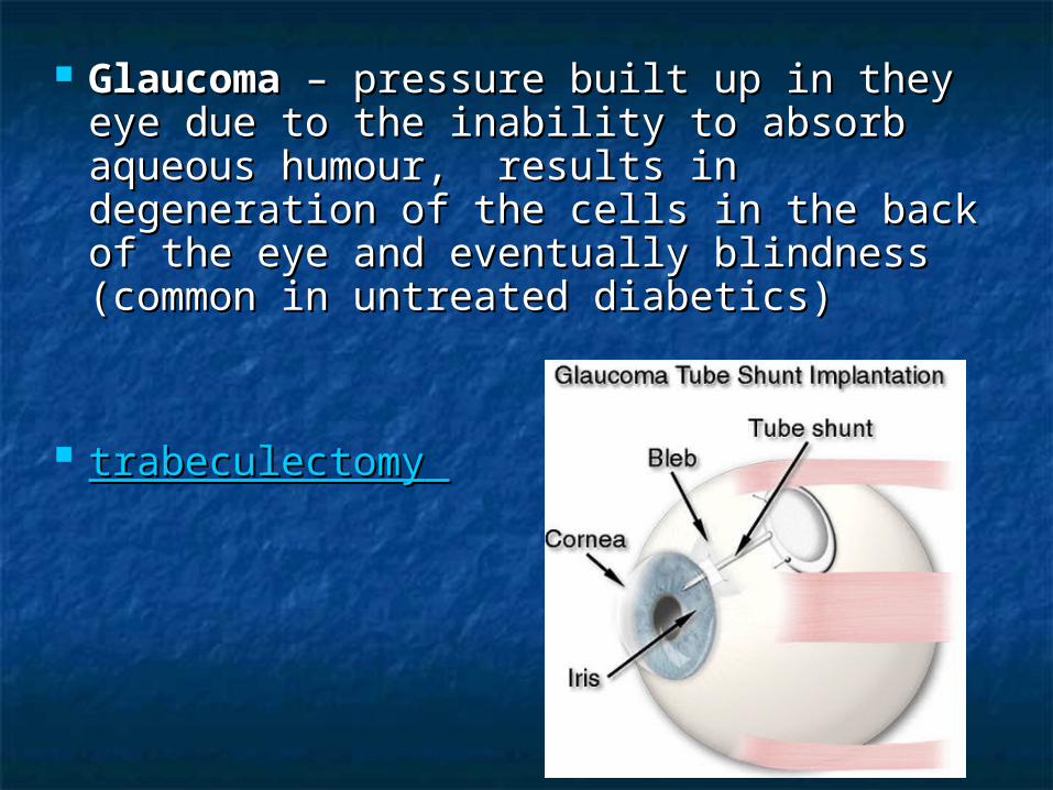
GlaucomaGlaucoma – pressure built up in they – pressure built up in they eye due to the inability to absorb eye due to the inability to absorb aqueous humour, results in aqueous humour, results in degeneration of the cells in the back degeneration of the cells in the back of the eye and eventually blindness of the eye and eventually blindness (common in untreated diabetics)(common in untreated diabetics)
trabeculectomytrabeculectomy
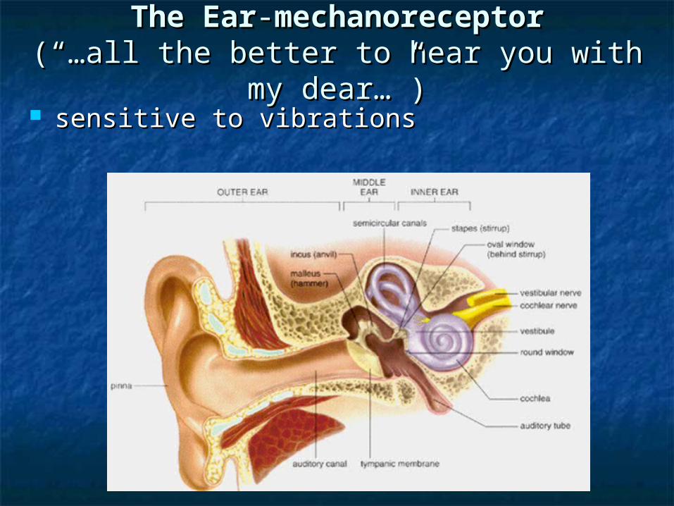
The EarThe Ear--mechanoreceptormechanoreceptor(“…all the better to hear you with my (“…all the better to hear you with my
dear…”)dear…”) sensitive to vibrations sensitive to vibrations

The Outer Ear (1)The Outer Ear (1) Pinna (1a) Pinna (1a) – folds of the visible ear. – folds of the visible ear.
Helps to funnel sound waves into the Helps to funnel sound waves into the ear.ear.
Auditory Canal (1b)Auditory Canal (1b) – carries sound – carries sound waves to the middle ear (eardrum). waves to the middle ear (eardrum). lined with hairs, sweat and glands that lined with hairs, sweat and glands that
produce earwax to prevent foreign bodies produce earwax to prevent foreign bodies from getting into the ear.from getting into the ear.
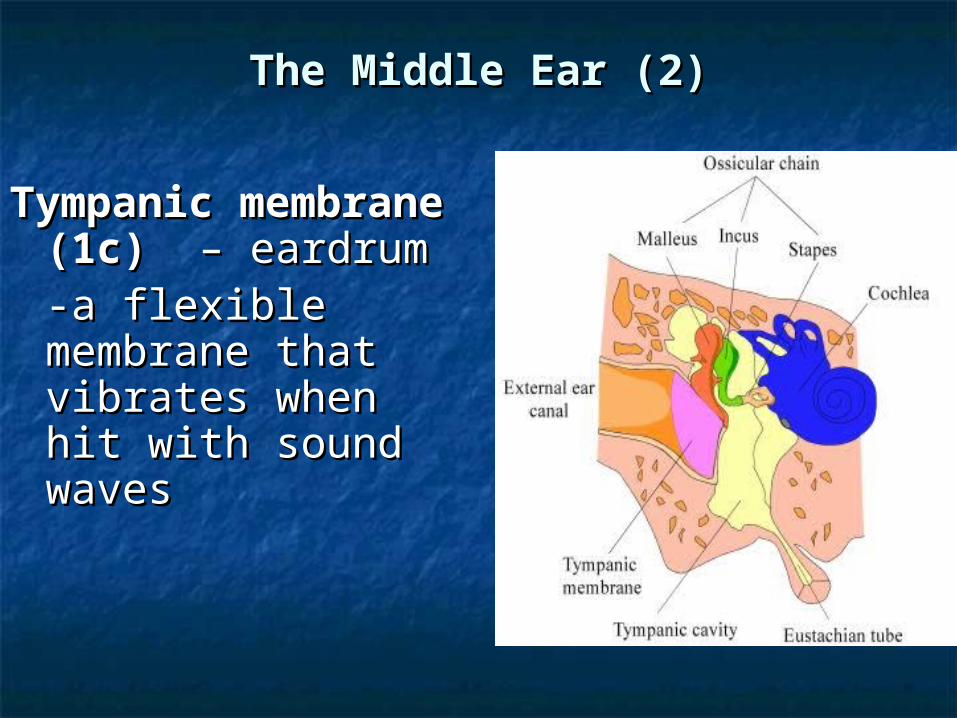
The Middle Ear (2)The Middle Ear (2)
Tympanic Tympanic membrane (1c) membrane (1c) – – eardrumeardrum-a flexible -a flexible membrane that membrane that vibrates when hit vibrates when hit with sound waveswith sound waves
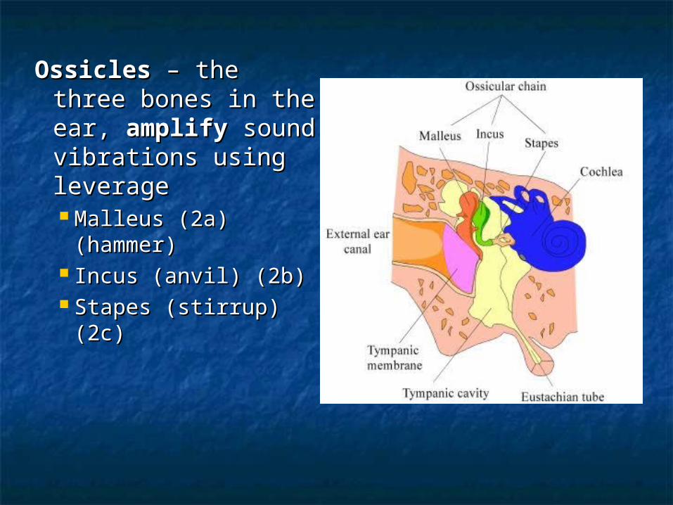
OssiclesOssicles – the three – the three bones in the ear, bones in the ear, amplifyamplify sound sound vibrations using vibrations using leverageleverage Malleus (2a) Malleus (2a)
(hammer)(hammer) Incus (anvil) (2b)Incus (anvil) (2b) Stapes (stirrup) (2c)Stapes (stirrup) (2c)

Eustachian tubeEustachian tube (2d)– extends (2d)– extends from the middle from the middle ear to the naso-ear to the naso-pharynxpharynx
allows for the allows for the equalization of equalization of pressure in the pressure in the ear when you ear when you swallowswallow
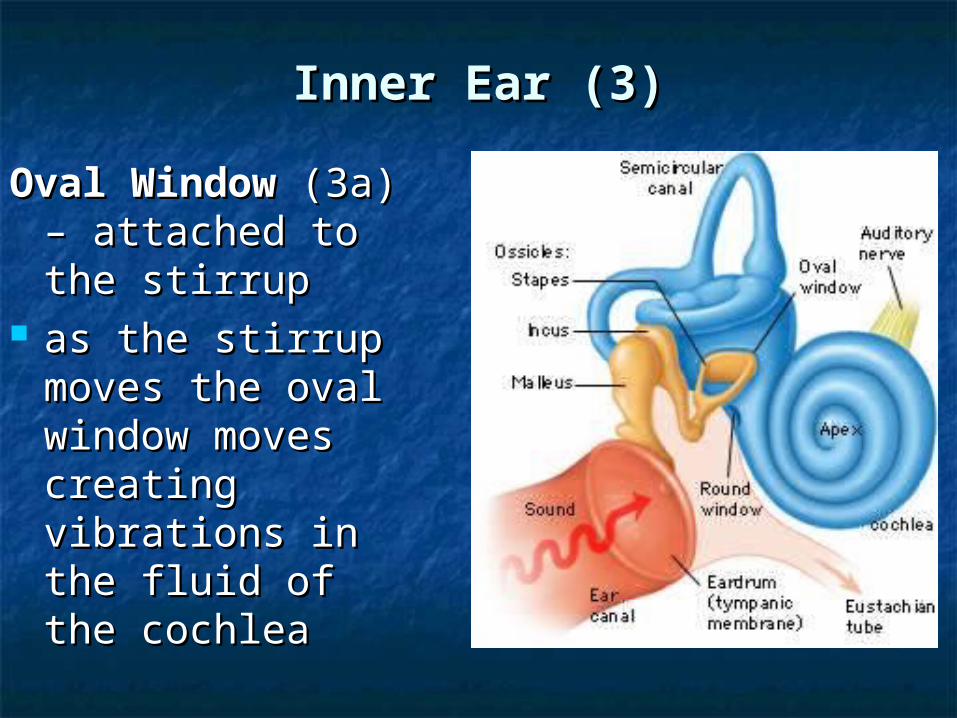
Inner Ear (3)Inner Ear (3)
Oval WindowOval Window (3a) – attached (3a) – attached to the stirrupto the stirrup
as the stirrup as the stirrup moves the oval moves the oval window moves window moves creating creating vibrations in the vibrations in the fluid of the fluid of the cochleacochlea
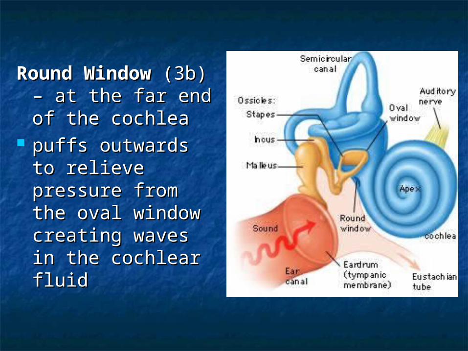
Round WindowRound Window (3b) – at the far (3b) – at the far end of the end of the cochleacochlea
puffs outwards to puffs outwards to relieve pressure relieve pressure from the oval from the oval window creating window creating waves in the waves in the cochlear fluidcochlear fluid
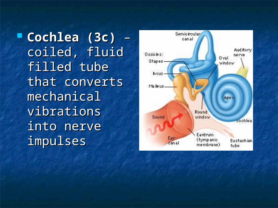
Cochlea (3c) Cochlea (3c) – – coiled, fluid filled coiled, fluid filled tube that tube that converts converts mechanical mechanical vibrations into vibrations into nerve impulsesnerve impulses
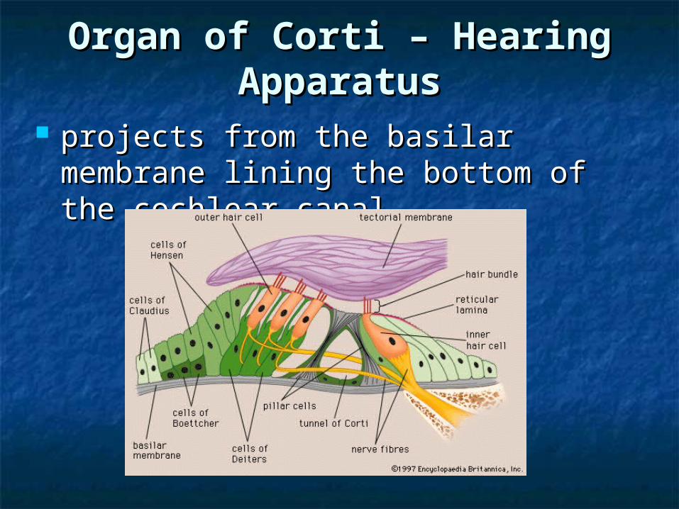
Organ of Corti – Hearing Organ of Corti – Hearing ApparatusApparatus
projects from the basilar membrane projects from the basilar membrane lining the bottom of the cochlear lining the bottom of the cochlear canalcanal

Basilar membraneBasilar membrane lines the bottom of the cochlear canallines the bottom of the cochlear canal
about 25,000 small nerve fibers about 25,000 small nerve fibers ending in hair cells are arranged in ending in hair cells are arranged in order of length along the organ of order of length along the organ of corticorti
each cell vibrates with a different each cell vibrates with a different wavelength of sound giving the wavelength of sound giving the sensation of sensation of pitchpitch

Ex) basil membrane is stiff and narrow Ex) basil membrane is stiff and narrow near the oval membrane: activated near the oval membrane: activated by high frequency wave = high pitchby high frequency wave = high pitch
Basilar Membrane OscillationsBasilar Membrane Oscillations
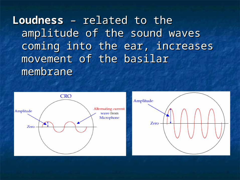
LoudnessLoudness – related to the amplitude – related to the amplitude of the sound waves coming into the of the sound waves coming into the ear, increases movement of the ear, increases movement of the basilar membranebasilar membrane
Tectorial membraneTectorial membrane sits on top of the hair cells in the sits on top of the hair cells in the
basilar membranebasilar membrane
moves up and down with the sound moves up and down with the sound
vibrationsvibrations
causes brushing and bending of the causes brushing and bending of the hairs in the basilar membrane which hairs in the basilar membrane which initiates a nerve impulseinitiates a nerve impulse
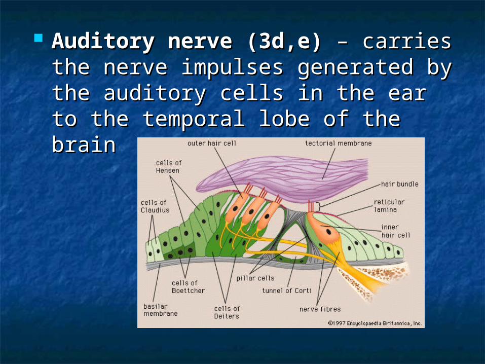
Auditory nerve (3d,e)Auditory nerve (3d,e) – carries the – carries the nerve impulses generated by the nerve impulses generated by the auditory cells in the ear to the auditory cells in the ear to the temporal lobe of the braintemporal lobe of the brain

HearingHearing
BalanceBalance Static equilibrium: movement in one
planeEx) head movement
Dynamic Equilibrium: movement
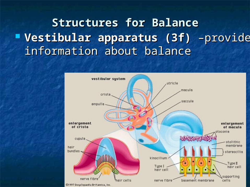
Structures for BalanceStructures for Balance Vestibular apparatus (3f)Vestibular apparatus (3f) –provide –provide
information about balanceinformation about balance
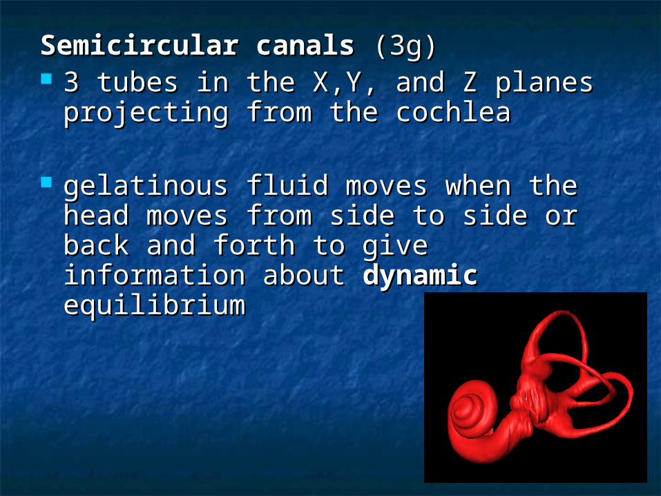
Semicircular canalsSemicircular canals (3g) (3g) 3 tubes in the X,Y, and Z planes 3 tubes in the X,Y, and Z planes
projecting from the cochleaprojecting from the cochlea
gelatinous fluid moves when the gelatinous fluid moves when the head moves from side to side or back head moves from side to side or back and forth to give information about and forth to give information about dynamicdynamic equilibrium equilibrium
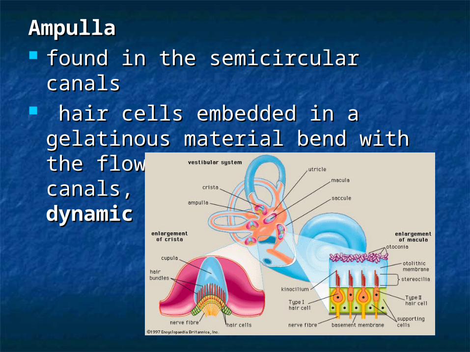
Ampulla Ampulla found in the semicircular canalsfound in the semicircular canals hair cells embedded in a gelatinous hair cells embedded in a gelatinous
material bend with the flow of fluid in material bend with the flow of fluid in the canals, creating a sense of the canals, creating a sense of dynamic dynamic balancebalance

Saccule and UtricleSaccule and Utricle sacs with hair cells covered in small sacs with hair cells covered in small
carbonate crystals called otolithscarbonate crystals called otoliths
as the head is tilted, or the body as the head is tilted, or the body inverted, the crystals shift creating a inverted, the crystals shift creating a sense of sense of staticstatic balance balance

Hearing ProblemsHearing Problems
Conduction DeafnessConduction Deafness: : congenital defect or infection congenital defect or infection ossicles fuseossicles fuse restricts the ability to magnify sound restricts the ability to magnify sound
waves. (hearing aids help)waves. (hearing aids help)
Nerve DeafnessNerve Deafness: : exposure to loud noises and age exposure to loud noises and age breakage or destruction of hairs. breakage or destruction of hairs. neurons don’t fire. (hearing aids are neurons don’t fire. (hearing aids are
ineffective)ineffective)

TinnitusTinnitus: ringing in the ears caused : ringing in the ears caused by damaged hair cells in the cochlea by damaged hair cells in the cochlea or by random “static” within the or by random “static” within the brainbrain
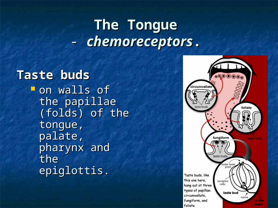
The TongueThe Tongue- - chemoreceptorschemoreceptors..
Taste budsTaste buds on walls of the on walls of the
papillae (folds) of papillae (folds) of the tongue, the tongue, palate, pharynx palate, pharynx and the and the epiglottis.epiglottis.
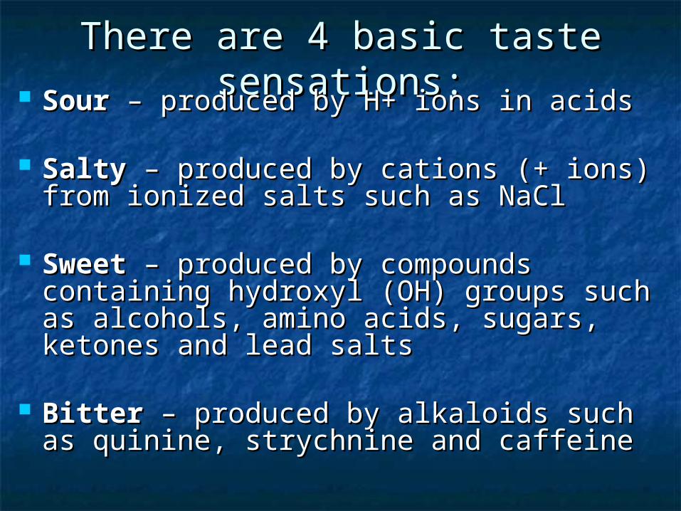
There are 4 basic taste There are 4 basic taste sensations:sensations: SourSour – produced by H+ ions in acids – produced by H+ ions in acids
SaltySalty – produced by cations (+ ions) – produced by cations (+ ions) from ionized salts such as NaClfrom ionized salts such as NaCl
SweetSweet – produced by compounds – produced by compounds containing hydroxyl (OH) groups such containing hydroxyl (OH) groups such as alcohols, amino acids, sugars, as alcohols, amino acids, sugars, ketones and lead saltsketones and lead salts
BitterBitter – produced by alkaloids such as – produced by alkaloids such as quinine, strychnine and caffeinequinine, strychnine and caffeine

Old thinking:Old thinking:

Taste is often a combination of all Taste is often a combination of all four types of receptors giving the four types of receptors giving the perception of different flavors.perception of different flavors.
Taste is enhanced by the sense of Taste is enhanced by the sense of smell. smell.
Taste may also be genetic. Taste may also be genetic.
http://www.bbc.co.uk/science/humanbody/body/fachttp://www.bbc.co.uk/science/humanbody/body/factfiles/taste/taste_ani_f5.swftfiles/taste/taste_ani_f5.swf

The NoseThe Nose(The nose knows. Phew!)(The nose knows. Phew!)
3000 times more receptors for smell 3000 times more receptors for smell than for taste. than for taste.
receptors are located on tissue called receptors are located on tissue called the olfactory epithelium. the olfactory epithelium.
The neurons have modified cilia on the The neurons have modified cilia on the ends of their dendrites that trap and ends of their dendrites that trap and receive airborne (gas or vapor) receive airborne (gas or vapor) molecules.molecules.
http://www.bbc.co.uk/science/humanbohttp://www.bbc.co.uk/science/humanbody/body/factfiles/smell/smell_ani_f5.swfdy/body/factfiles/smell/smell_ani_f5.swf

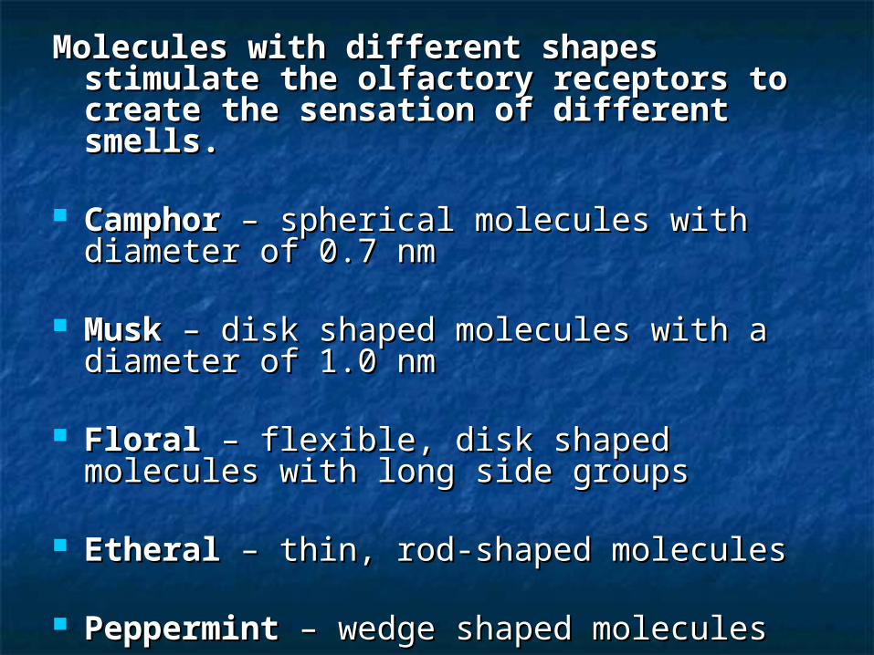
Molecules with different shapes Molecules with different shapes stimulate the olfactory receptors to stimulate the olfactory receptors to create the sensation of different create the sensation of different smells. smells.
CamphorCamphor – spherical molecules with – spherical molecules with diameter of 0.7 nmdiameter of 0.7 nm
MuskMusk – disk shaped molecules with a – disk shaped molecules with a diameter of 1.0 nmdiameter of 1.0 nm
FloralFloral – flexible, disk shaped molecules – flexible, disk shaped molecules with long side groupswith long side groups
EtheralEtheral – thin, rod-shaped molecules – thin, rod-shaped molecules
PeppermintPeppermint – wedge shaped molecules – wedge shaped molecules
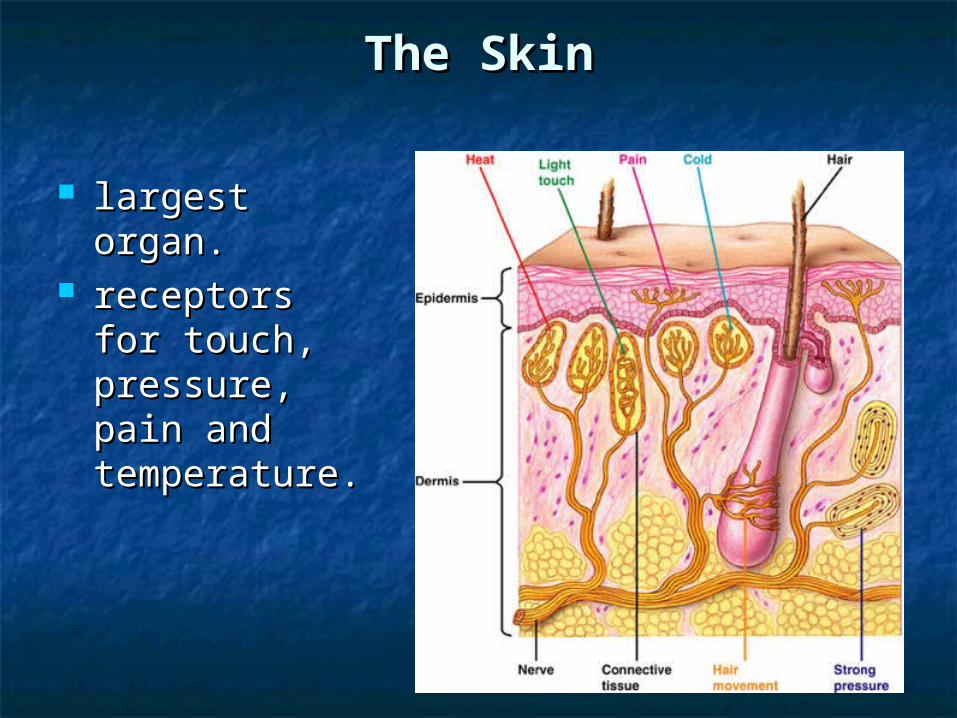
The SkinThe Skin
largest organ. largest organ. receptors for receptors for
touch, touch, pressure, pain pressure, pain and and temperature.temperature.

Proprioceptors – Proprioceptors – provide provide information about the position of information about the position of body partsbody parts
Stretch receptors – Stretch receptors – found in found in muscles and around the lungs muscles and around the lungs

Visceral SensesVisceral Senses
Receptors in internal organs Receptors in internal organs
mediated by the autonomic nervous system mediated by the autonomic nervous system to to
promote internal homeostasis. promote internal homeostasis.
maintain blood pH, blood pressure, oxygen maintain blood pH, blood pressure, oxygen levels, blood glucose levels, water re-levels, blood glucose levels, water re-absorption.absorption.
give sensations of thirst, hunger and nausea.give sensations of thirst, hunger and nausea.