the second most common neurological disease diagnosed in horses
TRANSCRIPT

Volume 29 No 2, July 2011 A publication of the Center for Equine Health • School of Veterinary Medicine • University of California, Davis
— Continued on page 3
The Second Most Common Neurological DiseaseDiagnosed in Horses
INSIDE THIS ISSUE…
The Second Most Common Neurological Disease Diagnosed in Horses ............. 1
Director’s Message ................. 2
Commentary on NAD/EDM by Researchers Involved in Neurological Evaluations ..... 5
Anatomy of an Epidemic: The 2011 EHV-1 Outbreak..... 6
Everyday Biosecurity Consid- erations for Your Horse ......... 9
Biosecurity Measures for Different Situations ........ 10
Neuroaxonal Dystrophy/Equine Degenerative Myeloencephalopathy
by Carrie J. Finno, DVM, DACVIM
Neuroaxonal dystrophy (NAD) is a neurological disease in which both the cell body of
the neurons and the axons throughout the brain and spinal cord undergo degeneration. Axons are the parts of the nerve cells along which impulses travel, so this condition results in abnormal conduction of nervous impulses and associated clinical signs defined by a lack of coordination. The cause of this degeneration is not understood at this time.
Neurological abnormalities associated with neuroaxonal dystrophy or equine degenerative myeloencephalopathy can be subtle and may be missed for years unless the horse is specifically examined for neurological disease. Mild cases may present with performance-related issues, where the horse is just not performing up to the standard expected for its breeding and training.
NAD affects horses, humans, dogs, cats and sheep. A genetic basis for the disease has been identified in humans and is suspected in sheep, cats and dogs. In horses, NAD appears to be inherited, as suggested by pedigree analysis and supported by breeding studies in Morgan horses.
Equine NAD is considered the underlying basis of equine degenerative myeloencephalopathy (EDM), a degenerative and irreversible disease of young horses characterized by hypermetria of the limbs. (Hypermetria
in horses is typified by an overshoot of intended position with the leg. It is sometimes described as an inability to judge distance or scale.)
Horses suffering from NAD or EDM typically display a symmetric (left to right) incoordination (ataxia) that may be more severe in the hind limbs than in the
Volume 29, Number 2 - July 2011
UC Davis Center for Equine Health
2 - The Horse Report
Director’s Message
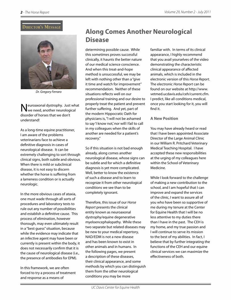
Dr. Gregory Ferraro
Along Comes Another Neurological Disease
Neuroaxonal dystrophy. Just what we need, another neurological
disorder of horses that we don’t understand! As a long-time equine practitioner, I am aware of the problems veterinarians face to achieve a definitive diagnosis in cases of neurological disease. It can be extremely challenging to sort through clinical signs, both subtle and obvious. When there is mild or subclinical disease, it is not easy to discern whether the horse is suffering from a lameness condition or is actually neurologic.
In the more obvious cases of ataxia, one must wade through all sorts of procedures and laboratory tests to rule out any number of possibilities and establish a definitive cause. This process of elimination, however thorough, may even ultimately result in a “best guess” situation, because while the evidence may indicate that an infective agent may have been or currently is present within the body, it does not necessarily confirm that it is the cause of neurological disease (i.e., the presence of antibodies for EPM). In this framework, we are often forced to try a process of treatment and response as a means of
determining possible cause. While this sometimes proves successful clinically, it haunts the better nature of our medical science conscience. And when this treat-and-hope method is unsuccessful, we may be left with nothing other than a “give it time and watch for improvement” recommendation. Neither of these situations reflects well on our professional training and our desire to properly treat the patient and prevent further suffering. And yet, part of the modern Hippocratic Oath for physicians is, “I will not be ashamed to say ‘I know not,’ nor will I fail to call in my colleagues when the skills of another are needed for a patient’s recovery.”
So if this situation is not bad enough already, along comes another neurological disease, whose signs can be subtle and for which a definitive diagnosis is yet more complicated. Well, better to know the existence of such a disease and to learn to recognize it from other neurological conditions we see than to be completely ignorant.
Therefore, this issue of our Horse Report presents the clinical entity known as neuroaxonal dystrophy/equine degenerative myeloencephalopathy. While these two separate but related diseases may be new to your medical repertory, NAD/EDM is not a new disease and has been known to exist in other animals and in humans. In the following pages, we present a description of these diseases, their clinical appearance, and some methods by which you can distinguish them from the other neurological conditions you may be more
familiar with. In terms of its clinical appearance, I highly recommend that you avail yourselves of the video demonstrating the characteristic clinical appearance of affected animals, which is included in the electronic version of this Horse Report. The electronic Horse Report can be found on our website at http://www.vetmed.ucdavis.edu/ceh/current.cfm. I predict, like all conditions medical, once you start looking for it, you will find it.
A New Position
You may have already heard or read that I have been appointed Associate Director of the Large Animal Clinic in our William R. Pritchard Veterinary Medical Teaching Hospital. I have accepted these new responsibilities at the urging of my colleagues here within the School of Veterinary Medicine.
While I look forward to the challenge of making a new contribution to the school, and I am hopeful that I can improve and expand the services of the clinic, I want to assure all of you who have been so supportive of me during my tenure at the Center for Equine Health that I will be no less attentive to my duties there than I have in the past. The CEH is my home, and my true passion and I will continue to serve its mission to the best of my abilities. In fact, I believe that by further integrating the functions of the CEH and our equine clinical services we can maximize the effectiveness of both.

The Horse Report - 3Volume 29, Number 2 - July 2011
UC Davis Center for Equine Health
forelimbs. Affected horses will often stand abnormally at rest (see photos), either too wide or too narrow, and will display proprioceptive deficits, which means that the horses do not know where their limbs are as they walk.
These clinical signs may be subtle and horses should be evaluated walking in slow, tight circles and stopping frequently, backing, walking with their head elevated, and walking up and down hills and curbs. Ataxia (incoordination) is graded on a 5-point scale:
Quarter horses affected with NAD/EDM may also be abnormally quiet and dull. This often makes these horses particularly easy to handle.
Although the neurological status of horses affected with NAD/EDM remains stable throughout the course of their lifetime, their loss of proprioception may make them a risk to handle on the ground. At this time, we have only definitively diagnosed NAD/EDM in horses that were graded as greater than or equal to a grade 2 out of 5 from the above scale. Any horse with a neurological score of 2 or greater should not be ridden and caution should be used when handling these horses on the ground.
A video showing some of the clinical signs of NAD/EDM may be viewed in the electronic (Zmag) version of this Horse Report available athttp://www.vetmed.ucdavis.edu/ceh/current.cfm.
Prevalence
The ataxia observed with NAD/EDM is very similar to that seen with Wobblers Syndrome, or cervical vertebral malformation/malarticulation (CVM), where neurological signs are caused by compression of the spinal cord in the neck, typically due to developmental abnormalities or osteoarthritis. In a study performed at Cornell University, EDM was the second most common neurological disease diagnosed in horses.
Neurological Disease— Continued from page 1
A 5-year-old Quarter horse mare with the abnormal base-wide stance of the forelimbs characteristic of NAD/EDM.
This 2-year-old Quarter horse gelding shows the dull mentation (mental activity) characteristic of NAD/EDM.
Disease Number (n=96) Percentage
EPM 32 33
NAD/EDM 23 24
CVM 11 12
Cervical injury 9 8
Other inflammatory conditions 5 5
Toxicity 1 1
Unknown 15 16
Grading System for Ataxia 0 = Normal; no neurological abnormalities identified
1 = Mild and inconsistent abnormalities
2 = Mild consistent abnormalities
3 = Moderate consistent abnormalities
4 = Severe consistent abnormalities
5 = Recumbent
— Continued on page 4
Cause of Neurological Disease in Horses

Volume 29, Number 2 - July 2011
UC Davis Center for Equine Health
4 - The Horse Report
Age of Onset and Breeds Affected
Typically, horses are affected with NAD or EDM early in life (6-12 months of age); however, these neurological abnormalities can be subtle and may be missed for years unless the horse is specifically examined for neurological disease. Mild cases may present with performance-related issues, where the horse is just not performing up to the standard expected for its breeding and training.
Males and females are equally affected and the disease has been reported in the following breeds:
Quarter horses/Paints/Appaloosas Haflingers Standardbreds Thoroughbreds Pony of the Americas Lusitano/Andalusians Morgans Paso Finos Arabians Tennessee Walking Horse Norwegian Fjord Various Mixed Breeds
Role of Vitamin E in NAD/EDM
A Vitamin E (alpha-tocopherol) deficiency has been reported in some cases of NAD/EDM. Previous research has concluded that Vitamin E is a factor in the development of EDM in the first year of life in genetically predisposed foals. Oxidative stress is known to be a component in human neurodegenerative diseases, including Alzheimer’s, Parkinson’s, Amytrophic lateral sclerosis, multiple sclerosis, and Huntington’s disease. The central nervous system is particularly vulnerable to oxidative insult due to the high rate of oxygen utilization,
relatively poor concentrations of classical antioxidants and high content of polyunsaturated lipids, which are most susceptible to oxidation.
In humans, it is difficult to determine whether the oxidative stress is primary or secondary in many neurodegenerative diseases. It was previously determined that foals with EDM do not demonstrate significant differences in oral Vitamin E absorption as compared to normal foals. Overall, there is very strong evidence that NAD/EDM is an inherited disorder and it may be that Vitamin E acts as a modifier to determine the overall severity of the disease in horses affected with NAD/ EDM. This theory has been supported by the NAD/EDM research to date. Diagnosis
Unfortunately, a definitive diagnosis of NAD/EDM requires examination of the spinal cord on post-mortem examination. Without a necropsy (post-mortem examination), we cannot definitively diagnose the disease. We suspect cases of NAD/EDM in horses that meet the following criteria:
Age of onset of symmetric neurologic signs at 6-36 months of age that either stabilize or are very slowly progressive.
History of other related horses that appear to be affected with similar signs.
History of low vitamin E or poor nutrition during the first year of life.
NEGATIVE EPM titer (blood and/or spinal fluid).
NORMAL neck x-rays +/- NORMAL myelogram (this is performed to
evaluate the horse for CVM, as these cases are indistinguishable based on clinical signs alone).
NEGATIVE for West Nile Virus.
Treatment
At this time, there is no effective treatment for NAD/EDM. Supplementation of affected cases with Vitamin E does NOT appear to improve clinical cases. In genetically predisposed animals, supplementing broodmares and foals with at least 2,000 IU/day of natural alpha-tocopherol does appear to result in less severe neurological signs but does not entirely eliminate the disease.
It appears that the disease may be inherited in an autosomal dominant manner, meaning that only one parent needs to be affected to produce an affected foal. Therefore, it is difficult to make breeding recommendations until a genetic test is available. Although horses do not appear to recover from this disorder, the disease is often either non-progressive or very slowly progressive and horses do not become substantially more debilitated during their lifetime. The neurological deficits present by 3 years of age generally remain unchanged throughout the life of the horse.
Research at UC Davis
A group of investigators at UC Davis has been studying different aspects of NAD/EDM to determine the genetic mutation responsible for the disease. The investigators include Dr. Carrie Finno, Dr. Monica Aleman and Dr. John Madigan (neurological evaluations); Dr. Robert Higgins (pathology
Neurological Disease— Continued from page 3
— Continued on page 8

The Horse Report - 5Volume 29, Number 2 - July 2011
UC Davis Center for Equine Health
Commentary onNeuroaxonal Dystrophy/Equine Degenerative Myelopathy
by Researchers Involved in Neurological Evaluations
John Madigan, DVM, MS, DACVIM, and Monica Aleman, MVZ, PhD, DACVIM
This Horse Report highlights two degenerative dis-orders—neuroaxonal dystrophy (NAD) and equine
degerative myelopathy (EDM)-- that have been large-ly overlooked, as evidenced by a Cornell University study on the causes of neurological disease in horses. In that study, NAD/EDM was identified at postmortem examination in 24% of horses with neurological dis-ease, second only to equine protozoal myeloenceph-alitis (EPM) and more common than cervical vertebral myelopathy (CVM). Since that study was conducted in 1978, more current studies are needed to investigate the prevalence of disease today. Lack of recognition of the disorder may play a role in the sporadic reports found in the literature.
Cervical vertebral myelopathy with all its clinical variants (cervical vertebral malformation/malarticula-tion, cervical vertebral stenotic myelopathy, cervical vertebral compressive myelopathy, cervical vertebral osteoarthritis), also known as “wobblers” syndrome, is a commonly diagnosed disorder that usually presents with symmetrical ataxia (incoordination) of all limbs, tetraparesis (dragging all feet), and proprioceptive deficits (abnormal placement of feet in the ground). These gait and placement deficits could be of various grades of severity, from mild to severe.
NAD/EDM can easily be misdiagnosed as CVM because the clinical signs may be undistinguishable from those seen in horses with CVM. Also, the onset of neurological deficits or age when first noted for both NAD/EDM and CVM is usually in the young popula-tion (not exclusive for all types of CVM). NAD/EDM may present with other signs such as mild obtunda-tion (decreased “alertness”) which is not observed in CVM. Although there is a breed predisposition in horses with CVM, both disorders can affect a variety of breeds. A genetic basis for NAD/EDM is suspected in affected breeds of horses.
A thorough neurological examination is required to note subtle abnormalities that may set NAD/EDM and CVM apart. It is also essential to rule out other causes besides CVM for the observed deficits (e.g., EPM, equine herpesvirus-1 myeloencephalopathy, trauma, among others) with appropriate diagnostic tests. Cau-tion must be taken in the interpretation of radiographic abnormalities of the cervical vertebral column because alterations may be present without causing clinical signs. Therefore, radiographic alterations at this level do not always equal CVM.
The diagnosis of NAD/EDM presents a challenge in the living horse because at present the definitive diagno-sis is possible only through a thorough postmortem examination by an experienced neuropathologist since lesions could be missed.
Keynotes:
Age of onset: Often in young horses but signs may not be noted until the horse is started on training or on more challenging physical activity. Therefore, the first signs may be noted even in adult horses.
There is strong evidence of a genetic basis thataffects several breeds of horses. However, do not dis-miss the possibility of disease because it has not been reported in a certain breed.
Low vitamin E is NOT the cause but is considered an environmental factor associated with onset and sever-ity of signs. Supplement pregnant mares and young-sters if the disease is suspected in breeding farms.
NAD/EDM may look like a “wobbler”; therefore, do not exclude the disease and rule out other disorders.
Definitive diagnosis is reached only through a care-ful postmortem evaluation.

Volume 29, Number 2 - July 2011
UC Davis Center for Equine Health
6 - The Horse Report
Anatomy of an Epidemic: The 2011 EHV-1 Outbreak
Other people may be there to help us, teach us, guide us along our path, but the
lesson to be learned is always ours.
The recent equine herpesvirus-1 (EHV-1) outbreak that kept horse owners across the country in a state of apprehension for two months is a perfect example of how an infectious disease is transmitted and spreads like wildfire.
It started at a horse show where a horse was infected with the virus. It is unclear whether this horse had any clinical signs or whether it was subclinically infected (showing no outward signs of sickness). Before anyone knew, the show was over and exposed horses returned home, unwittingly carrying the virus back with them. Over the next 7 weeks we watched the numbers rise until the virus had finally sickened 90 horses in 10 states; 13 of those horses died or were euthanized. It could have been worse.
The neurological form of EHV-1, known as equine herpesvirus myeloencephalopathy (EHM), has probably existed for centuries, but a strain of the virus has evolved (mutated) in recent decades with an increased virulence over that seen in the past. Its ability to create spontaneous outbreaks of neurological disease is concerning to horse owners and veterinarians alike.
At greatest risk for this re-emerging disease are congregations of horses
assembled from diverse origins and housed under high-density, stress-ridden, and heavily co-mingling conditions for competition, showing or performance. Risk factors are stress (invariably part of traveling to a horse show or event), prolonged transport, intermingling of horses from diverse locations without prior isolation, and high-density stabling in large facilities that share a common air space. Management practices also add to stress, including shipping, abrupt changes in dietary composition, poor infection control, and disruption of established social hierarchies.
Since currently marketed vaccines are not entirely adequate, and reports of neurological outbreaks in fully vaccinated horses are common (AAEP, November 6, 2007), the highest priority and the greatest gap in research is in the development of a new vaccine against the neurological strain of EHV-1.
Currently available treatment includes antiviral drugs such as valacyclovir and ganciclovir along with supportive care, which were effective in treating horses that were brought to the UC Davis Veterinary Medical Teaching Hospital during this year’s EHM outbreak. While most of those horses survived with this treatment, and most affected horses make a full recovery, a small percentage of horses can be left with residual signs such as incoordination or bladder incontinence.
Until a new vaccine is developed, however, the best protection against the neurological form of EHV-1 lies in biosecurity strategies: entry requirements, isolation protocols, movement restriction, and infection control procedures. A year after the 2006-2007 EHV-1 outbreak, the USDA issued a report in which they summarized collective mitigation experiences and lessons learned and recommended future needs with regard to controlling EHM in the future. The report was based on interviews with a number of scientists, researchers and others involved in past outbreaks of EHM. One researcher stated succinctly, “It is important to have a plan for how we will manage the next EHM case or outbreak, as there will be another one.”
In looking back at our very recent experience this year, a number of defensive measures were undertaken quickly and in an organized manner, resulting in efficient handling of the disease outbreak to contain it. It’s worth looking at those measures with the idea that individual horse owners can learn from this experience and formulate a plan of their own for when the next EHM outbreak occurs.
Health officials at the USDA and CDFA became involved immediately, resulting in swift, organized action to contain sick horses and monitor confirmed cases of EHM.

The Horse Report - 7Volume 29, Number 2 - July 2011
UC Davis Center for Equine Health
In California, EHM became a Reportable Disease just this year (January 2011) so that it is now required to be reported to state officials within 48 hours of diagnosis. In the past, EHM was considered a Monitored Condition as EHV-1 and EHV-4 and reportable to the CDFA within 30 days of diagnosis. This change was made based on an increasing incidence of EHM in the United States over the last decade.
The change in reporting status for EHM in California was a good move considering that California had the largest number of primary-exposed horses (59) at the Ogden event as well as the largest number of secondary- and tertiary-exposed horses (628).
Although EHM is not a Reportable Disease with the USDA, federal officials became involved in a monitoring role to provide updates on the outbreak nationwide.
Along with the reporting requirement, horses with fever and other clinical signs that were confirmed positive for EHV-1 were immediately isolated. Other horses that may have been exposed to the sick horse were quarantined for a prescribed period of time.
A significant improvement noted in this outbreak over that in 2007 is that barrier and isolation protocols were developed based on experience and were able to be implemented immediately. This helped in preventing the spread of infection. Biosecurity information also was well publicized on a number of different websites and in print publications. State and federal officials issued frequent (daily in the case of CDFA)
and clear communications to the public via their websites to keep people informed about the status of the disease, the number of infected horses, and geographical locations involved around the country.
The CDFA provided open and timely communication on the Web dedicated to daily EHV-1 status updates for California for the duration of the outbreak. This was extremely helpful in minimizing rumors and giving people confidence that the situation was being handled effectively. The USDA provided status updates on the disease on a weekly basis, summarizing information reported on all states that were affected.
Research into developing improved diagnostic testing for equine herpesvirus paid off.
New diagnostic testing methods that have become available through technological advances helped in providing more rapid confirmation of infection. The real-time polymerase chain reaction assay (RT-PCR) with quantitative assessment allows better characterization of disease stage, better assessment of risk of exposure of in-contact horses, and better monitoring of response to therapy.
RT-PCR has recently been used to document differences in EHV-1 viral loads in blood and nasal secretions between horses in the febrile (fever) and neurological stages of disease and between clinical and subclinical cases. These studies found high viral loads in the nasal secretions of animals with neurological signs, confirming their importance as a potential source of contagion for other horses and highlighting the
need to impose strict biosecurity when horses are identified with suspected or confirmed EHM.
Awareness and understanding of EHM by horse owners and communities resulted in good cooperation in restricting the movement of horses and in canceling events.
There is much more the veterinary profession does not know about the herpes virus than what it does as it is one of the more complicated viruses to understand. Researchers at UC Davis and elsewhere are working to increase our understanding of this organism and its disease-producing ability and from there to begin to develop much needed vaccines and improved treatment.
In the meantime, responsible horse owners can familiarize themselves with the information provided in this Horse Report (pages 9–11) and elsewhere and learn what to do when the next outbreak does occur. Understand that the best defense at the present time is biosecurity and learn what that entails. ❄

Volume 29, Number 2 - July 2011
UC Davis Center for Equine Health
8 - The Horse Report
evaluations); and Dr. Danika Bannasch (genetic studies with Dr. Finno).
Our research group recently published a description of NAD/EDM in the American Quarter horse. Our ultimate goal is to develop a genetic test to identify NAD/EDM, which would enable veterinarians to diagnose the disease without requiring euthanasia and assist breeders in selecting against the mutation. Research into the genetics of NAD/EDM will not only provide a diagnostic/preventive way to test for the disease but will also give us insight into the mechanism underlying the degenerative process and we can begin to consider how horses affected with NAD/EDM can be treated effectively.
For further information on NAD/EDM, please contact Dr. Carrie Finno at [email protected]. ❄
Neurological Disease— Continued from page 4
One of the ways that horses can catch an infectious disease such as EHV-1 is by direct contact with another horse or with equipment used for an infected horse. It’s best to use separate water buckets, feed troughs, tack and grooming equipment for each of your horses. If equip-ment must be shared, it should be dipped in a disinfecting solution (see page 9), washed and dried before use on another horse.

The Horse Report - 9Volume 29, Number 2 - July 2011
UC Davis Center for Equine Health
Recent outbreaks of equine herpesvirus-1 have brought awareness of the need to handle our
horses in a manner that prevents transmission of infection. The term for this is biosecurity, which means taking measures that prevent transmission of an infection to your horse.
Two ways that horses and humans may catch many infectious diseases are by direct contact and/or by aerosol spread via the air.
Direct contact means physical contact with a horse that has an ongoing active infection or is in the early incubation period of infection and ready to break with a fever. It is possible, however, to catch a virus or bacteria from a horse without that horse having a fever or other signs of infection. Direct-contact transmission can also occur when a person touches a horse and has the virus on their hands, tack, bridle, saddle pad, bucket or shoe and then spreads the infectious agent via use of that contaminated equipment on another horse. Herpes viruses are largely spread by direct contact.
Aerosol spread means that the infectious agent travels through the air and causes infections by breathing the agent in. Most respiratory viruses of the influenza group spread this way. Indeed these viruses can travel significant distances, depending on air flow, temperature and other environmental conditions. Strangles horses may cough and spread that bacteria throughout a barn or stable. However, aerosol spread has not been a primary means of transmission of EHV-1.
Biosecurity Measures
Keep your horse in a place where direct contact with other horses is minimal. If there is contact with other horses, it should be preferably with horses that do not leave the premises and return frequently.
Everyday Biosecurity Considerationsfor Your Horse
Have a policy for your stable for horses that leave and attend horse events. Horses returning from distant events should have minimal contact with other horses. Upon returning to the stable, travel horses should ideally be isolated from the home stable population for at least 2 weeks.
While traveling, if it is known that at the event there were sick horses, take your horse’s temperature twice daily and report a fever (101.5°F or greater) to the stable manager and your veterinarian.
Avoid petting and touching other horses to minimize the chance of infecting your horse.
Use separate water buckets, feed troughs, tack and grooming equipment for each of your horses. If equipment must be shared, it should be dipped in a solution of 1 part chlorine bleach to 10 parts water, washed and dried before use on another horse. Commercial preparations for disinfection also are available through your veterinarian.
Other biosecurity measures have been delineated by the U.S. Department of Agriculture’s Animal and Plant Health Inspection Service and are listed on pages 10-11.
The equine herpesvirus as viewed by transmission electron microscopy at a magnification of 167,000 X.

Volume 29, Number 2 - July 2011
UC Davis Center for Equine Health
10 - The Horse Report
The following recommendations from the U.S. Department of Agriculture Plant and Animal Health Inspection Service are based on the belief that you are the best protection your horses have. These guidelines are intended to reduce the chances of an infectious disease being carried onto your farm by people, animals, equipment or vehicles, either accidentally or on purpose.
Showing Your Horse
Use your own trailer whenever possible. Don’t ship your horses with horses from unknown farms.
Ship only in a trailer or van that has been cleaned and disinfected. If you can “smell horse” in the empty trailer, it has not been cleaned and disinfected properly.
Don’t let your horse touch unknown horses, especially nose to nose.
Wash your hands, especially after helping other people with their horses.
Don’t let strangers pet your horse, especially those with horses at home.
Before leaving the show grounds, clean and disinfect tack, boots, equipment and grooming supplies. Brush off dirt or manure; then disinfect (spray or wipes are easy to take with you).
When you get home, shower, blow your nose and put on clean clothes and shoes before going near other horses.
Visiting Other Farms, Horse Shows or Auctions
Have a pair of shoes or boots that you save for visiting and don’t wear around your own horse or wear plastic shoe covers.
Biosecurity Measures for Different Situations
If you are going to be working with horses on another farm, wear coveralls or plan to change clothes before returning home to your horses.
If there are farms you visit all the time and you can’t change clothes and shoes, be sure their vaccination program and biosecurity practices are as good as your own.
For Visitors to Your Farm or Horse
It is best to have only one public access to your farm. Mark this as the main entrance.
Park away from the horses. Doing that will help keep disease-carrying organisms from being tracked from car floors or tires to your horses.
Ask all visitors to wear clean clothes and shoes. Give visitors plastic shoe covers or brush dirt off their shoes and spray them with disinfectant.
If you have many visitors such as at a farm tour or open house, make a footbath for them to walk through (see inset, page 11).
Bringing Horses Back from a Show
If one or more horses travel to horse shows, all horses on the premises should be vaccinated. Horses that show can bring home infectious agents. Discuss with your veterinarian what vaccinations the horses need and how often.
If possible, keep horses that were off the farm isolated for at least 2 weeks. At the very least, make sure there is no nose-to-nose contact between horses in the stable.

The Horse Report - 11Volume 29, Number 2 - July 2011
UC Davis Center for Equine Health
Bringing in New Horses
This is the most likely way for infectious diseases to come in, especially if horses are coming from other states or from foreign countries.
Keep every new horse isolated for 30 days. Don’t use pitchforks, grooming tools, or feed and water buckets on any horse but the new one. Mark these with red tape or use red brushes, etc., only for the isolation area.
Work with the isolated horse last each day. Alternatively, wear boots and coveralls when working with the isolated horse and remove them before working or going near other horses. You can keep these in a plastic-covered tub near the horse. Exercise the isolated horses, alone, at a separate time from others in the stable.
Always wash your hands and blow your nose after working with the new horse. You could carry germs to your other horses in your nose.
Supplies Needed
A low plastic pan or bin, wide enough to fit an adult’s foot, shallow enough to step into easily.
A plastic doormat (the “fake grass” mats work well).
A disinfectant that works when manure or dirt is present is best, such as Tek-trol or One Stroke Environ. A 1:10 Clorox solution is also an effective viralcidal if the solution is replenished often and kept out of direct sunlight.
Water.
Assembling the Footbath
1. Mix the disinfectant with water following label instructions.
2. Put the doormat in the plastic pan.
3. Add disinfectant so that the bottom of the “grass” is wet.
4. Ask visitors to walk through the footbath, wiping their feet on the mat. The “grass” scrubs their shoes a bit as they wipe them, and applies the disinfectant.
5. When the liquid starts to get dirty, empty it and put in new disinfectant.
Making an Easy Footbath

©The Regents of the University of California July 2011
Center for Equine Health(530) 752-6433www.vetmed.ucdavis.edu/ceh
Director:Dr. Gregory L. FerraroE-mail: [email protected]
Senior Editor:Barbara MeierhenryE-mail: [email protected]
Management Services Officer:Katie GlideE-mail: [email protected]
Dean, School of Veterinary Medicine:Dr. Bennie I. Osburn
HORSEREPORTCEH
The Center for Equine Health is supported with funds provided by the State of California Pari-Mutuel Fund and contributions by private donors.
The University of California does not discriminate in any of its policies, procedures or practices.
The University is an affirmative action/equal opportunity employer. The information you provide will be used for University business and will not be released unless required by law. To review your record, contact Advancement Services, 1480 Drew Avenue, Ste. 130, Davis, CA 95616. A portion of all gifts is used to defray the costs of administering the funds. All gifts are tax-deductible as prescribed by law.
View the video in our online Horse Report!
www.vetmed.ucdavis.edu/ceh
The Horse Report is now brought to you in an online format. If you would like to be notified when a new issue is online, send your e-mail address to [email protected].
HORSEREPORTMail ID#1415Center for Equine HealthSchool of Veterinary MedicineUniversity of CaliforniaOne Shields AvenueDavis, CA 95616-8589
RETURN SERVICE REQUESTED
CEH