the roles of cell size and cell number in determining ovariole
TRANSCRIPT

�������� ����� ��
The roles of cell size and cell number in determining ovariole number inDrosophila
Didem Pelin Sarikaya, Abel A. Belay, Abha Ahuja, Aisha Dorta, Del-bert Andre Green II, Cassandra G. Extavour
PII: S0012-1606(11)01437-0DOI: doi: 10.1016/j.ydbio.2011.12.017Reference: YDBIO 5557
To appear in: Developmental Biology
Received date: 17 August 2011Revised date: 9 December 2011Accepted date: 10 December 2011
Please cite this article as: Sarikaya, Didem Pelin, Belay, Abel A., Ahuja, Abha, Dorta,Aisha, Green II, Delbert Andre, Extavour, Cassandra G., The roles of cell size and cellnumber in determining ovariole number in Drosophila, Developmental Biology (2011), doi:10.1016/j.ydbio.2011.12.017
This is a PDF file of an unedited manuscript that has been accepted for publication.As a service to our customers we are providing this early version of the manuscript.The manuscript will undergo copyediting, typesetting, and review of the resulting proofbefore it is published in its final form. Please note that during the production processerrors may be discovered which could affect the content, and all legal disclaimers thatapply to the journal pertain.

ACC
EPTE
D M
ANU
SCR
IPT
ACCEPTED MANUSCRIPTThe cellular basis of ovariole number
Sarikaya et al. Page 1 of 50
The roles of cell size and cell number in determining ovariole number in Drosophila
Didem Pelin Sarikayaa, Abel A. Belay
a, Abha Ahuja
a, Aisha Dorta
b, Delbert André Green
IIc, and Cassandra G. Extavour
a*
a. Department of Organismic and Evolutionary Biology, Harvard University, 16
Divinity Avenue, Cambridge, MA 02138, USA.
b. Brooklyn College, City University New York, 2900 Bedford Avenue, Brooklyn,
NY 11210, USA.
c. Department of Molecular and Cellular Biology, Harvard University, 16 Divinity
Avenue, Cambridge, MA 02138, USA.
* Author for correspondence: email [email protected]
Tel. 617-496-1935
Fax. 617-496-9507

ACC
EPTE
D M
ANU
SCR
IPT
ACCEPTED MANUSCRIPTThe cellular basis of ovariole number
Sarikaya et al. Page 2 of 50
Abstract
All insect ovaries are composed of functional units called ovarioles, which contain
sequentially developing egg chambers. The number of ovarioles varies between and
within species. Ovariole number is an important determinant of fecundity and thus affects
individual fitness. Although Drosophila oogenesis has been intensively studied, the
genetic and cellular basis for determination of ovariole number remains unknown.
Ovariole formation begins during larval development with the morphogenesis of terminal
filament cells (TFCs) into stacks called terminal filaments (TFs). We induced changes in
ovariole number in Drosophila melanogaster by genetically altering cell size and cell
number in the TFC population, and analyzed TF morphogenesis in these ovaries to
understand the cellular basis for the changes in ovariole number. Increasing TFC size
contributed to higher ovariole number by increasing TF number. Similarly, increasing
total TFC number led to higher ovariole number via an increase in TF number. By
analyzing ovarian morphogenesis in another Drosophila species we showed that TFC
number regulation is a target of evolutionary change that affects ovariole number. In
contrast, temperature-dependent plasticity in ovariole number was due to changes in cell-
cell sorting during TF morphogenesis, rather than changes in cell size or cell number. We
have thus identified two distinct developmental processes that regulate ovariole number:
establishment of total TFC number, and TFC sorting during TF morphogenesis. Our data
suggest that the genetic changes underlying species-specific ovariole number may alter
the total number of TFCs available to contribute to TF formation. This work provides for
the first time specific and quantitative developmental tools to investigate the evolution of
a highly conserved reproductive structure.

ACC
EPTE
D M
ANU
SCR
IPT
ACCEPTED MANUSCRIPTThe cellular basis of ovariole number
Sarikaya et al. Page 3 of 50
Keywords: Drosophila melanogaster; ovariole number; reproductive fitness; fecundity;
cell number; cell size

ACC
EPTE
D M
ANU
SCR
IPT
ACCEPTED MANUSCRIPTThe cellular basis of ovariole number
Sarikaya et al. Page 4 of 50
Introduction
All insect ovaries are composed of highly conserved functional units called ovarioles
(Büning, 1994). Ovariole number varies within and between species (Büning, 1998;
Markow and O'Grady, 2007; Telonis-Scott et al., 2005). Because each ovariole produces
eggs autonomously (Extavour and García-Bellido, 2001; R' kha et al., 1997), the number
of ovarioles is an important determinant of fecundity (Cohet and David, 1978; David,
1970; R' kha et al., 1997), thereby influencing evolutionary fitness (Orr, 2009). It is
therefore important to understand the developmental mechanisms that regulate ovariole
number. This will inform our understanding of how evolutionary changes in these
mechainsms might lead to ovariole number differences, and thus fitness differences,
within and between species.
Ovariole development and function are best understood in Drosophila
melanogaster. Each ovariole consists of an anterior germarium and maturing egg
chambers, or follicles. The germarium houses germ line stem cells that divide to produce
oocytes (Wieschaus and Szabad, 1979). As follicles leave the germarium, they move
posteriorly and continue to develop to form mature oocytes. D. melanogaster ovaries
consist of approximately 16 to 23 ovarioles (depending on the strain). Ovariole number is
determined during larval development through the morphogenesis of somatic structures
called terminal filaments (TFs), each of which is composed of a stack of seven to ten
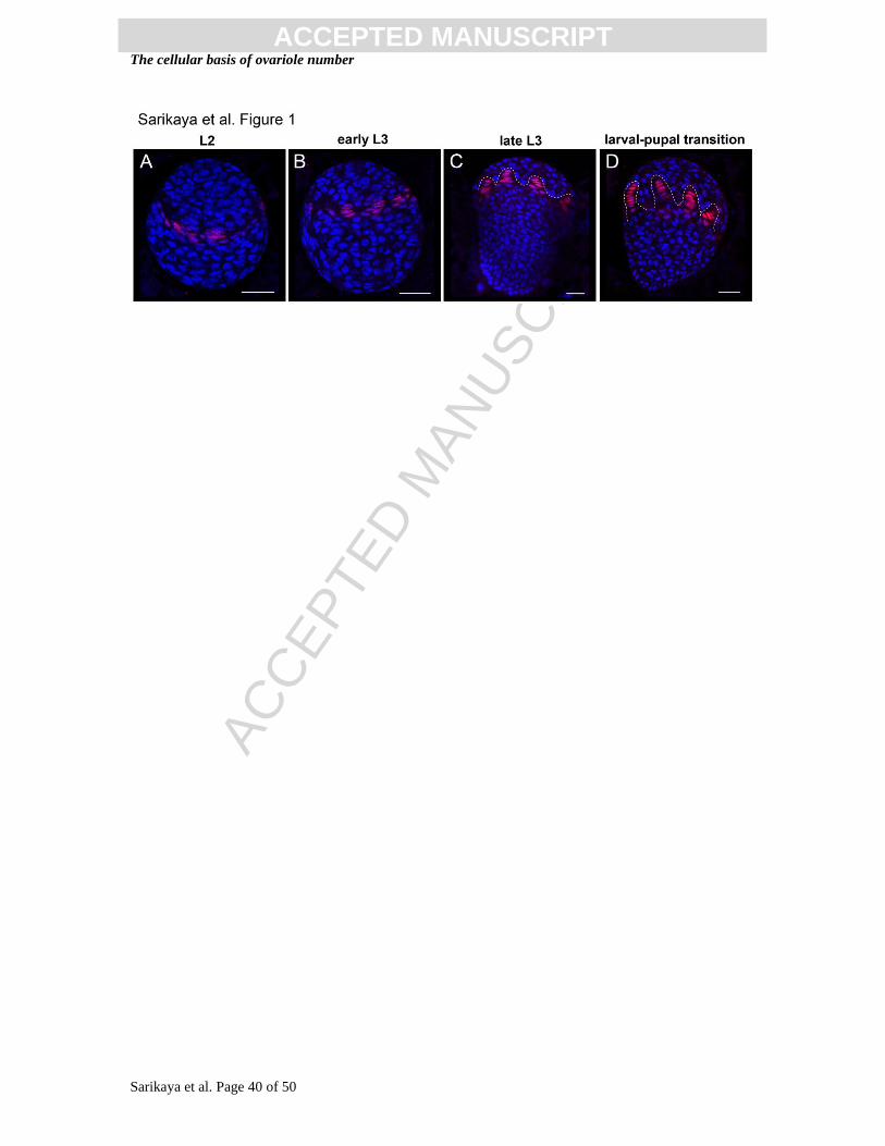
terminal filament cells (TFCs) (Godt and Laski, 1995; King et al., 1968). TFC
specification begins at the second larval instar (L2; Fig. 1A), and proceeds until the onset
of the pupal stage (LP; Fig. 1D) (Godt and Laski, 1995; Sahut-Barnola et al., 1995). TFs
begin to form in the late third larval instar (L3; Fig. 1B) by intercalation of TFCs in a

ACC
EPTE
D M
ANU
SCR
IPT
ACCEPTED MANUSCRIPTThe cellular basis of ovariole number
Sarikaya et al. Page 5 of 50
medial to lateral progression across the ovary (Godt and Laski, 1995). As TF formation is
completed, apical somatic cells migrate posteriorly between the TFs, secreting a
basement membrane that separates TFs from each other. The progressive posterior
migration of these apical cells encapsulates two to three germ line stem cells, and several
early oogonia, into each forming ovariole. Finally, a stack of basal stalk cells is
incorporated into the posterior end of each ovariole. These stalk cells ultimately connect
ovarioles to the oviduct, providing an outlet for the oocytes formed in each ovariole
(King, 1970; King et al., 1968). Because TFs serve as beginning points for ovariole
formation, elucidating how TF number is established is critical to understanding the
developmental and evolutionary basis of ovariole number.
Because TFs are neither created nor destroyed during normal pupal development
(King, 1970), TF number at the larval-pupal transition determines adult ovariole number
(Hodin and Riddiford, 2000). Ovarioles can form in the absence of germ cells (Aboïm,
1945; Engstrom et al., 1982), and changes in germ cell number do not induce changes in
TF number (Barnes et al., 2006; Gilboa and Lehmann, 2006). The germ cell population
thus does not have a major influence on ovariole number. This suggests that
developmental processes that form and sort the somatic cells that create TFs, the TFCs,
determine changes in ovariole number.
Although D. melanogaster oogenesis has been intensively studied, the formation
of ovarioles during ovarian morphogenesis is still not well understood. Specifically, the
genetic and cellular basis for determination of ovariole number remains unknown.
Correct regulation of size and number in other organs, including wings in flies and
somites in frogs (Cooke, 1975; Resino and Garcia-Bellido, 2004), relies on the

ACC
EPTE
D M
ANU
SCR
IPT
ACCEPTED MANUSCRIPTThe cellular basis of ovariole number
Sarikaya et al. Page 6 of 50
coordination of cell number (proliferation), cell size (growth), and cell sorting behavior.
Moreover, evolutionary change in body size is thought to be the result of changes in the
numbers and sizes of cells (French et al., 1998; James et al., 1995; Partridge et al., 1999).
We therefore hypothesized that the developmental parameters influencing ovariole
number might include the numbers, sizes, and cell sorting behaviors of TFCs. In this
context, we analyzed TFC number, size and morphogenesis in ovaries with genetically-
or environmentally-induced differences in ovariole number. To assess the role of TFC
size in determining ovariole number, we changed the activity of S6 kinase (S6K), which
is a downstream regulator of Insulin/TOR signaling (reviewed by Fenton and Gout,
2011b). Altering S6K activity changes cell size without affecting cell number in
ectodermal tissues (Montagne et al., 1999). We also assessed the role of TFC number in
regulating ovariole number, by manipulating the activity of the Hippo pathway. This
recently described pathway plays a conserved role in controlling cell number in fruit flies
and mammals, but does not alter cell size (Dong et al., 2007; Harvey et al., 2003; Wu et
al., 2003). Based on the data from these manipulations, we propose a model for the major
developmental processes that regulate changes in ovariole number.
We used this model to investigate the developmental basis of evolutionary change
in this trait. Ovariole number is species-specific and largely genetically determined. Intra
and inter-species genetic studies on ovariole number indicate that genetic variation in the
trait is additive and polygenic (Coyne et al., 1991; Orgogozo et al., 2006; Telonis-Scott et
al., 2005; Wayne et al., 2001; Wayne and McIntyre, 2002). To determine the roles of
TFC size, number, and sorting behavior in evolutionary change in ovariole number, we
compared TF morphogenesis in two Drosophila species with different ovariole numbers.

ACC
EPTE
D M
ANU
SCR
IPT
ACCEPTED MANUSCRIPTThe cellular basis of ovariole number
Sarikaya et al. Page 7 of 50
Finally, we addressed the role of these cell biological parameters in phenotypic plasticity
in ovariole number. Environmental inputs such as temperature and nutrition can also
influence adult ovariole number (Bergland et al., 2008; Hodin and Riddiford, 2000). To
assess the reasons for ovariole number changes induced by rearing environment, we
compared (1) flies reared at two different temperatures, and (2) flies reared on standard or
reduced nutrition, and analyzed TFC behavior. Our data suggest that genetic and
environmental variation can affect ovariole number through different developmental
processes.
Materials & Methods
Fly strains
TRiP (Harvard Medical School) RNAi lines used to knock down Hippo pathway
members were y1v
1; P{TRiP
hpo}attP2 (Bloomington Drosophila stock center 33614;
abbreviated to UAS:RNAihpo
) and y1v
1; P{TRiP
wts}attP2 (Bloomington Drosophila stock
center 27662; abbreviated to UAS:RNAiwts
). These lines were selected as they have been
reported to increase cell proliferation in the gut epithelium of flies (Karpowicz et al.,
2010). Mutant S6K allele lines used were w; P{w+mC
=UAS-S6k.TE}2 (Bloomington
Drosophila Stock Center 6912) and w; P{w+mC
=UAS-S6k.STDE}2 / CyO actinGFP,
(derived from Bloomington Drosophila Stock Center 6913 and 4533; abbreviated to
UAS:S6KX. These lines were selected as they have been reported to increase cell size (but
not cell proliferation) in the wing (Barcelo and Stewart, 2002). The GAL4 driver lines
used were w; P{GawB}bab1Pgal4-2
/TM6, Tb1 (Bloomington Drosophila Stock Center

ACC
EPTE
D M
ANU
SCR
IPT
ACCEPTED MANUSCRIPTThe cellular basis of ovariole number
Sarikaya et al. Page 8 of 50
6803) (Cabrera et al., 2002) and nubbin:GAL4 (gift of Tassos Pavlopoulos), abbreviated
to bab:GAL4 and nub:GAL4, respectively. The bab:GAL4 driver is expressed in somatic
cells of the larval ovary, most strongly in the somatic cells anterior to the germ cells,
which are largely destined to become TF cells (Cabrera et al., 2002). Additional somatic
cell populations expressing this driver at lower levels are the intermingled cells in direct
contact with germ cells, and at late L3 and prepupal stages, the somatic cells posterior to
the germ cells; neither of these latter cell populations contributes to terminal filaments.
The bab:GAL4 driver is not expressed in germ cells. GAL4 line virgins were crossed to
UAS:RNAihpo
, UAS:RNAiwts
, UAS:dS6KTE
and UAS:dS6KSTDE
males. D. yakuba (UC San
Diego Drosophila Stock Center 1402-0261.01 via Daniel Hartl’s lab) was maintained at
25°C for all experiments.
Rearing conditions: variation of temperature and nutritional regimes
Temperature sensitive experiments were conducted with OregonR-C flies (Bloomington
Drosophila Stock Center 5 via Daniel Hartl’s lab). Flies were reared at 25ºC or 18ºC at
60% humidity on standard fly medium (0.8% agar, 2.75% yeast, 5.2% corn meal, 11%
dextrose) for at least two generations before experiments were conducted (Fig. S1A).
Because reduced nutritional intake of larvae resulting from crowded tubes can reduce
adult ovariole number, adults were permitted to lay eggs in vials for two to six hours and
then removed from the vial to prevent overcrowding of larvae. Only tubes containing
fewer than 100 pupae were used for analysis of larval-pupal ovaries, and for counts of
ovariole number in adults. Adults hatched from these tubes were used to create new
parent cultures at the same temperature. For starvation experiments, flies were reared at

ACC
EPTE
D M
ANU
SCR
IPT
ACCEPTED MANUSCRIPTThe cellular basis of ovariole number
Sarikaya et al. Page 9 of 50
25ºC on ¼ standard fly medium (“quarter food”) made by mixing one part standard fly
medium with three parts 3% agar (VWR); overcrowding of larvae was prevented as
described above.
Adult analysis: ovariole number
As described above, only tubes containing fewer than 100 larvae were used for all
experiments. Adult female flies from non-crowded tubes were placed in 70% ethanol
until sedated, and ovaries were dissected in 1X PBS/0.01% Triton X-100. Ovariole
number was counted in 1X PBS under a dissecting microscope using tungsten needles. At
least 20 ovaries were analyzed for each strain. For temperature comparisons, a two-tailed
t-test was conducted using Microsoft Excel. Ovariole number comparison for Hippo
pathway and S6K experimental adults were conducted by one-way analysis of variance
(ANOVA), followed by Tukey’s Honestly Significant Difference (HSD) using JMP (SAS
Institute Inc., Cary, NC). ANOVA is a standard statistical method based on Fisher’s
methodology (Fisher, 1918) for determining whether significant differences exist
between means from multiple groups; the ANOVA F statistic is the ratio of the variance
of the means of different groups, to the variance between samples comprising a group,
and is reported in the relevant figure legends for all data. The HSD test is a method based
on pairwise comparisons of means in order to determine which means are significantly
different from each other (Bremer and Doerge, 2010).
Adult analysis: wing cell size and number

ACC
EPTE
D M
ANU
SCR
IPT
ACCEPTED MANUSCRIPTThe cellular basis of ovariole number
Sarikaya et al. Page 10 of 50
Rearing temperature was reported to affect wing cell size but not number (Azevedo et al.,
2002). We confirmed these results in our experimental conditions by analyzing wing cell
size and density in flies used in our experiments. Wings were removed from dissected
adults and placed in 100% ethanol overnight. Wings were then washed in 70% ethanol,
1:1 ethanol : glycerol, and 50% glycerol in distilled water for 10 minutes, and mounted in
50% glycerol. Mounted wings were imaged using a Zeiss AxioImager Z1 and a Zeiss
MRm AxioCam driven by AxioVision v4.6, and total surface area of the wing, cell
number per area of interest, and cell size were measured using AxioVision v4.6 or Adobe
Photoshop CS3. Total surface area was measured for the entire wing. Cell number per
unit area was measured in the ventral region of compartment C of the wing (Baena-Lopez
et al., 2005); the number of bristles was counted for the same surface area region of
interest in different wings. Cell size was measured by selecting a single bristle, and
connecting the surrounding six bristles to obtain the surface area. Alternatively, the
distance between trichomes was measured and taken as the diameter of the cell to
calculate cell area. Comparable cell size values were obtained with both methods. At least
ten cells were measured per wing to obtain the individual’s average wing cell size.
Immunohistochemistry
For larval analysis, the transition stage between the larval and pupal stages was used
(referred to as “larval-pupal stage” throughout). Pupae with hardened, white pupal cases
(Ashburner et al., 2005) were collected from vials containing less than 100 pupae. This
stage was chosen for analysis because TF formation, which is gradual throughout the
third larval instar, ends at the larval-pupal stage, and so the TF number of these ovaries is

ACC
EPTE
D M
ANU
SCR
IPT
ACCEPTED MANUSCRIPTThe cellular basis of ovariole number
Sarikaya et al. Page 11 of 50
the final TF number for that individual. Ovaries with incomplete TFs (still in the process
of intercalating) were discarded from the dataset, and only ovaries where all TFs were
separated by migrating anterior cells were used. Samples were dissected in 1X PBS, fixed
in 4% paraformaldehyde/1X PBS for 25 minutes at room temperature, and blocked in
0.5% goat serum (Jackson ImmunoLabs) in 1X PBS/0.01% Triton-X for 30 minutes at
room temperature. Primary antibody incubation in mouse anti-Engrailed (Developmental
Studies Hybridoma Bank 4D9, 1:40) and/or anti-Traffic jam (gift of D. Godt, 1:4000) in
blocking solution was conducted overnight at 4ºC. Engrailed labels the TF population
(Forbes et al., 1996), and Traffic jam (Tj) labels intermingled cells and cap cells (Li et al.,
2003). Samples were washed in 1X PBS/0.01% Triton-X twice for 15 minutes at room
temperature, and incubated with FITC-Phalloidin or A555-Phalloidin (Invitrogen, 1:120
of 200 U/ml stock solution), Hoechst 33342 (Sigma, 1:500 of 10 mg/ml stock solution),
and goat anti-Mouse Alexa 568 (Invitrogen, 1:500) and/or goat anti-Guinea Pig Cy5
(Jackson ImmunoLabs, 1:500) overnight at 4ºC. Samples were mounted in Vectashield
mounting medium (Vector labs), and imaged using a Zeiss LSM 710 confocal
microscope.
Larval analysis: TFC number per TF
Z-stack confocal images of stained ovaries were taken with a 40X objective and 1.2-1.6x
zoom to capture the entire ovary at 1 m intervals (Fig. S1B). Total TF number was
counted and comparisons between samples were conducted using a two-tailed t-test.
TFCs were identified by morphology and Engrailed expression. Engrailed-expressing
cuboidal cells at the posterior of the TF were excluded from the TFC number count, as

ACC
EPTE
D M
ANU
SCR
IPT
ACCEPTED MANUSCRIPTThe cellular basis of ovariole number
Sarikaya et al. Page 12 of 50
they were adjacent to germ cells and had characteristics of cap cells. Ten ovaries were
analyzed per temperature for the environmental manipulations, and five individual
ovaries for each genetic condition were analyzed for TFC number per TF. The dataset for
each manipulation (temperature/genetic) contained measurements of several cells from
each of for five or ten individuals, which were randomly selected. To account for
potential individual variation affecting the dataset, we conducted a nested mixed model
ANOVA (JMP, SAS Institute Inc., Cary, NC) with a fixed manipulation term (genetic or
environmental) and a random-effects individual term nested within manipulation. Sample
sizes reported reflect the individual ovary number, rather than the number of
measurements made per individual, unless indicated otherwise.
Larval analysis: TFC size
Larval TFC size (= volume in m3) was obtained specifically in the third and fourth TFC
of each TF, counting from the anterior tip of the TF in order to ensure the cells were
comparable in size (Fig. 2A). Cell outlines were visualized by Phalloidin staining. For
each individual ovary, four to ten cells (average 7.8 cells per sample) were analyzed by
measuring the surface area of the cell through serial confocal image stacks for all stacks
where the selected cell was visible (Fig. 2A). The sum of the surface areas was multiplied
by the thickness of each individual stack to obtain the cell size. In the case of GAL4/UAS
experiments, the maternal strain (w; babGAL4p4.2
/TM6b, Tb1) and F1 siblings (w; UAS-
S6KX/+; TM6b/+) carry the TM6b balancer that contains a mutant allele of the gene
Tubby. Flies carrying this Tubby allele are visibly shorter and stouter than wild type as
adults and larvae (Linsdsley and Zimm, 1992). The mechanistic causes for the phenotype

ACC
EPTE
D M
ANU
SCR
IPT
ACCEPTED MANUSCRIPTThe cellular basis of ovariole number
Sarikaya et al. Page 13 of 50
are unknown, but they may affect cell size. Because cell size is one of the parameters
under analysis, and the TM6b chromosome is not present in any experimental animals of
interest, we excluded these genotypes from the analysis. Similar to TFC number per TF, a
mixed model nested ANOVA (see above) was used to analyze the data by setting
temperature/genetic treatment as a fixed effect, and the individual nested within
temperature/genetic treatment as random effect using JMP (SAS Institute Inc., Cary, NC).
Larval analysis: total TFC number
Obtaining images of larval ovaries where all TFCs can be resolved at the late L3 and
larval-pupal stage is inefficient. As an alternative, we estimated total TFC number by
calculating an average TFC number per TF (by averaging measurements from more than
eight TFs; Fig. 2B)), and multiplying by the TF number of that ovary (Fig. 2C). We
tested the validity of this method by randomly choosing eight TFC number per TF
measurements from ovaries where total TFC number had also been counted manually,
and comparing the calculated and counted measurements. For all eight ovaries analyzed,
our calculation method gave total TFC numbers that matched the counted number with an
accuracy of ±4 (average TFC number was 134.5 for manual counts and 135.8 for proxy
calculation; p=0.79, two-tailed t-test; Fig. S1D). Calculating total TFC number in this
manner thus provides an accurate proxy for total TFC number.
Results
Constitutively active S6K in TFCs results in increased adult ovariole number

ACC
EPTE
D M
ANU
SCR
IPT
ACCEPTED MANUSCRIPTThe cellular basis of ovariole number
Sarikaya et al. Page 14 of 50
We hypothesized that TF number would be affected by cellular behaviors during TF
morphogenesis. Specifically, we examined TFC size, TFC number per TF, and total TFC
number to gain insight into how the dynamics of TF morphogenesis could affect TF
number, and therefore ovariole number. To test whether changes in TFC size could
influence TF number, we used mutations in S6 Kinase (S6K) as a tool to change cell size.
S6K phosphorylates the ribosomal subunit S6 and as a result, regulates translation
downstream of the Insulin and TOR signaling pathways (Jefferies et al., 1997).
Expression of constitutively active S6K alleles in the wing increases cell size, but does
not alter cell number (Montagne et al., 1999) (Fig. S2). We took advantage of the
GAL4/UAS system (Brand and Perrimon, 1993) to increase S6K activity in the TFC
population (Fig. S3) with a bab:GAL4 driver line (Cabrera et al., 2002) (Fig. 2A, B; see
Methods).
Expression of two different constitutively active alleles of S6K (S6KTE
and
S6KSTDE
; see Methods) in TFCs resulted in an increase in ovariole number of females
from the experimental cross (Fig. 3A, B). Ovariole number in the experimental F1 flies
(w; UAS-S6KX/+; bab:GAL4
p4.2 /+) was compared to ovariole numbers in GAL4-only or
UAS-only parental and sibling controls. We compared samples using a one-way ANOVA
followed by Tukey’s HSD, and found that in both cases, F1 adult females had
significantly more ovarioles compared to parents and siblings (S6KTE
: p<0.001; S6KSTDE
:
p<0.001). This increase was also reflected in larval TF number (S6KTE
p<0.01; S6KSTDE
p<0.05) (Fig. 3C).
Constitutively active S6K increases both size and number of TFCs

ACC
EPTE
D M
ANU
SCR
IPT
ACCEPTED MANUSCRIPTThe cellular basis of ovariole number
Sarikaya et al. Page 15 of 50
In Drosophila ectodermal tissues (wing, eye) and in mouse ectodermal tissues (adrenal
gland) and embryonic fibroblasts, S6K activity is linked to control of cell size, but not of
cell number (Lawlor et al., 2002; Montagne et al., 1999). However, the function of S6K
in mesodermal tissues in Drosophila has not yet been investigated. We therefore asked
whether the effect on adult ovariole number caused by constitutive S6K activity was due
to a size change in TFCs. Cell size measurements were taken manually at the larval-pupal
transition stage (referred to as “larval-pupal stage” throughout; see Methods) using
confocal z-stacks of TFs (Fig. 2A) from four to ten cells per sample (average 7.8; see
Methods). F1s (w; UAS-S6KX/+; babGAL4
p4.2/+) and UAS-only controls were compared
using a mixed-model nested ANOVA (see Methods). Average TFC size increased in both
S6K alleles as compared to controls, although the increase was not statistically significant
in the case of the STDE allele (S6KTE
: p<0.05; S6KSTDE
: p=0.52; Fig. 4A, B).
A model that could explain how larger TFCs would result in more terminal
filaments, is one where developmental regulation controls total overall TF size. This
model predicts that TFs made of larger cells would contain fewer TFCs per TF, in order
to maintain constant TF size. To test this model we measured TFC number per TF, and
found that it was significantly lower (p<0.01) for the S6KTE
allele and slightly lower
(p=0.075) in the S6KSTDE
allele (Fig. 4C, D). However, TFC size was not correlated with
the number of cells per TF (Fig. S4). This suggests that S6K may have a role in sorting
TFCs that is independent of cell size.
The reductions in TFC number per TF were not steep enough to account for the
generation of all supernumerary TFs induced by S6K constitutive expression. We
therefore analyzed the effect of constitutive S6K activity on total TFC number.
ACC
EPTE
D M
ANU
SCR
IPT
ACCEPTED MANUSCRIPTThe cellular basis of ovariole number
Sarikaya et al. Page 16 of 50
Surprisingly, expression of both S6KTE
and S6KSTDE
resulted in a significant increase in
total TFC number in the experimental cross (S6KTE
: p<0.01; S6KSTDE
: p<0.05) (Fig. 4E,
F), indicating that constitutively active S6K alleles alter cell number in the developing
ovary. This contrasts with what has been observed in ectodermal tissues, where S6K only
affects cell size (Lawlor et al., 2002; Montagne et al., 1999) (Fig. S2). In summary, the
increase in ovariole number induced by overexpression of constitutively active S6K
results from an increase both in TFC size and in cell number. This shows that S6K
activity can have cell-type specific effects in D. melanogaster.
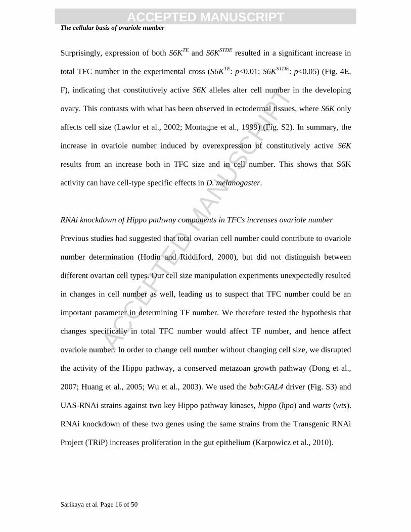
RNAi knockdown of Hippo pathway components in TFCs increases ovariole number
Previous studies had suggested that total ovarian cell number could contribute to ovariole
number determination (Hodin and Riddiford, 2000), but did not distinguish between
different ovarian cell types. Our cell size manipulation experiments unexpectedly resulted
in changes in cell number as well, leading us to suspect that TFC number could be an
important parameter in determining TF number. We therefore tested the hypothesis that
changes specifically in total TFC number would affect TF number, and hence affect
ovariole number. In order to change cell number without changing cell size, we disrupted
the activity of the Hippo pathway, a conserved metazoan growth pathway (Dong et al.,
2007; Huang et al., 2005; Wu et al., 2003). We used the bab:GAL4 driver (Fig. S3) and
UAS-RNAi strains against two key Hippo pathway kinases, hippo (hpo) and warts (wts).
RNAi knockdown of these two genes using the same strains from the Transgenic RNAi
Project (TRiP) increases proliferation in the gut epithelium (Karpowicz et al., 2010).

ACC
EPTE
D M
ANU
SCR
IPT
ACCEPTED MANUSCRIPTThe cellular basis of ovariole number
Sarikaya et al. Page 17 of 50
RNAi knockdown of hippo and warts in TFCs (Fig. 2C, D) increased ovariole
number of females from the experimental cross (w; UAS:RNAi /+ ; bab:GAL4p4.2
/+)
compared with GAL4-only and UAS-only parental and sibling controls (Fig. 5A, B).
One-way ANOVA revealed a significant difference in ovariole number between these
genotypes (hpo-RNAi: p<0.0001 ; wts-RNAi: p<0.0001 ), and comparisons using
Tukey’s HSD revealed that in both cases, F1 adult females had significantly more
ovarioles compared to parents and siblings (p<0.05). This increase was reflected in larval
TF number (hpo-RNAi: p=0.028; wts-RNAi: p=0.037; Fig. 5C), suggesting that the
cellular behaviors underlying the increase in ovariole number take place during larval
stages.
Reduced Hippo pathway activity increases total TFC number
To investigate the developmental causes underlying the increase in ovariole number, we
then analyzed larval-pupal TFCs with reduced hpo and wts activity. As expected, TFC
cell size was unchanged from controls (hpo-RNAi: p=0.93; wts-RNAi: p=0.23; Fig. 6A,
B), and we did not observe a difference in TFC number per TF (hpo-RNAi: p=0.58; wts-
RNAi: p=0.72; Fig 6C, D). However, there was a significant increase in total TFC
number (hpo-RNAi: p<0.01; wts-RNAi: p=0.028; Fig. 6E, F). This shows that TF number
can be modified by direct changes in total TFC number, without affecting the stacking
mechanism that creates TFs. In summary, downregulating the Hippo pathway in TFCs
increased total TFC number, thereby increasing the number of TFs created and resulting
in higher ovariole number.

ACC
EPTE
D M
ANU
SCR
IPT
ACCEPTED MANUSCRIPTThe cellular basis of ovariole number
Sarikaya et al. Page 18 of 50
Ovariole number differences between D. melanogaster and D. yakuba result from
differences in TFC number
Because we found that TFC number was a key regulator of TF number and thus ovariole
number in D. melanogaster, we hypothesized that evolutionary changes in TFC number
could be responsible for ovariole number differences in different Drosophila species. To
test this hypothesis, we examined TFC number in D. yakuba (Fig. 7A, B). This species
diverged from the lineage containing D. melanogaster 4-6 million years ago (Li et al.,
1999), and has an average of 14 ovarioles per ovary (Markow and O'Grady, 2007). We
first confirmed that this difference in adult ovariole number correlated with a difference
in TF number in larval-pupal stage ovaries (Fig. 7C, E, p<0.05). Consistent with our
hypothesis, this reduced TF number was the result of a smaller total number of TFCs
(Fig. 7D, p<0.01), which were organized into TFs that contained the same number of
TFCs per TF as D. melanogaster (Fig. 7E, p=0.72). This shows that the developmental
basis of evolutionary change in ovariole number between these two species is a change in
proliferation of a specific cell population within the ovary, the TFCs.
Adult ovariole number and larval TF number decrease in response to lower rearing
temperature or decreased nutrition
Finally, we asked if temperature- and nutrition-dependent phenotypic plasticity in
ovariole number could proceed through the same developmental mechanisms as genetic
variation. Previous studies reported an effect of temperature on ovariole number in D.
melanogaster, in both wild and laboratory populations (Chakir et al., 2007; Delpuech et
al., 1995; Hodin and Riddiford, 2000; Moreteau et al., 1997). Similarly, nutrient intake

ACC
EPTE
D M
ANU
SCR
IPT
ACCEPTED MANUSCRIPTThe cellular basis of ovariole number
Sarikaya et al. Page 19 of 50
can also affect ovariole number in D. melanogaster: increasing yeast content in the
medium increases ovariole number (Bergland et al., 2008), and relatively reduced
nutrient levels results in reduced ovariole number (Hodin and Riddiford, 2000;
Robertson, 1957). To understand the developmental causes for temperature-induced
differences in ovariole number, we analyzed OregonR flies reared at 18ºC and 25ºC on
standard fly medium (Fig. S1A). To investigate the developmental basis for nutrition-
dependent reduction in ovariole number, we raised OregonR flies at 25ºC on a diet with
one quarter the nutrient level of control flies (“quarter food”). In both of these conditions
we counted adult ovariole number per ovary and observed, as expected, a significant
decrease in ovariole number at 18°C compared to 25°C, and on full medium compared to
quarter food (p<0.001 for both comparisons) (Fig. 8A). Similarly, larval-pupal stage TF
number corresponded with adult ovariole number in all conditions (Fig. 8B). The
difference was statistically significant in both cases (p<0.05 for temperature comparisons
and p<0.01 for nutrition comparison). This confirms that the decrease in adult ovariole
number caused by lower rearing temperature or reducing nutritional intake is a result of
reduced larval TF number.
Nutrition affects ovariole number by altering TFC number
We asked whether a second environmental variable, nutrition, also affected ovariole
number via the same developmental processes as those altered in our temperature
experiments. We found that in fact, variation of different developmental parameters was
involved. Flies raised on quarter food had significantly smaller and fewer TFCs than
controls (Fig. 8D, E; p<0001 in both cases). This is consistent with previous observations

ACC
EPTE
D M
ANU
SCR
IPT
ACCEPTED MANUSCRIPTThe cellular basis of ovariole number
Sarikaya et al. Page 20 of 50
that limiting nutrition reduces both cell size and cell number in epithelial tissues (Neel,
1940; Robertson, 1959) (Fig. 8C, S5). However, in contrast to the temperature
experiments, the number of TFCs per TF was not significantly different between quarter
food-raised flies and full food-raised controls (Fig. 8F; p=0.96). This indicates that,
similar to what we observed when altering cell size with the S6KTE
alleles (Fig. 4A, S4A),
altering cell size via nutrition does have a significant impact on TF morphogenesis . The
largest contributor to reduced ovariole number in flies raised on quarter-food is therefore
the reduction in total TF number (Fig. 8E), which results in fewer TFs being formed.
Rearing temperature does not affect ovariole number by altering TFC number
We next examined TFC size, TFC number per TF, and total TFC number in ovaries of
larvae reared at different temperatures. Because temperature correlates negatively with
cell size in somatic epithelial tissues (Azevedo et al., 2002) (Fig. 8C), we expected that
TFCs would also be enlarged by a colder rearing temperature. Surprisingly however, we
found no significant difference in TFC size between the two rearing temperatures
(p=0.58) (Fig. 8D). As temperature also affects cell cycle and therefore might be
expected to change total cell number, we analyzed total TFC number per ovary at 18ºC
and 25ºC. In wing cell populations, cell number is not affected by temperature (Azevedo
et al., 2002) (Fig. S2). Similarly, no differences were observed in total TFC number
between larvae reared at 18ºC and 25ºC (Figure 8E; p=0.45). This demonstrates that,
unlike the species-specific differences in ovariole number, the temperature effect on
ovariole number is not achieved by changing the number or size of TFCs. Furthermore,

ACC
EPTE
D M
ANU
SCR
IPT
ACCEPTED MANUSCRIPTThe cellular basis of ovariole number
Sarikaya et al. Page 21 of 50
the contrast with the temperature effects observed on wing cell size (Fig. S5) indicates
that temperature-induced changes in development can be tissue-specific.
Rearing temperature affects ovariole number by altering TFC number per TF
Even though there was a significant decrease in TF number in larval ovaries reared at
18ºC compared with 25ºC, there was no corresponding significant decrease in total TFC
number. This suggests that temperature-induced changes in TF morphogenesis might
account for differences in total TF number. Accordingly, we found a significant increase
in TFC number per TF in ovaries from larvae reared at 18ºC (p<0.01) (Fig. 8F). This
suggests that during early ovarian morphogenesis, the size and starting number of TFCs
is similar regardless of the temperature. However, as morphogenesis proceeds and TFs
form, lower temperatures result in changes to the mechanism that organizes cells, such
that a larger number of TFCs are incorporated into each TF. As a result, fewer TFs are
formed at lower temperatures.
Discussion
Here we have shown that two distinct developmental mechanisms can alter ovariole
number: the establishment of total TFC number (Fig. 9A), and the local cell-cell sorting
process during TF formation (Fig. 9B). These two processes appear to be differently
employed to alter ovariole number. Specifically, by genetically altering the activity of
developmental growth pathways, we observed that change in ovariole number was
achieved by changes in total TFC number, rather than by changing TFC number per TF.
Similarly, changes in TFC number appeared responsible for ovariole number differences

ACC
EPTE
D M
ANU
SCR
IPT
ACCEPTED MANUSCRIPTThe cellular basis of ovariole number
Sarikaya et al. Page 22 of 50
between two Drosophila species, and for starvation-induced reduction in ovariole
number. In contrast, temperature-induced differences in ovariole number were caused by
changes in TFC number per TF, rather than changes in total TFC number or TFC size.
We postulate that at least some of the genetic changes underlying species-specific
ovariole number may alter total TFC number, while temperature-dependent variation may
result from differences in TFC sorting during TF formation.
In this work we have examined specifically the TFC population of the somatic
ovary. Previous work has analyzed total ovarian cell number in relation to ovariole
number, and did not always find a direct correlation (Hodin and Riddiford, 2000). We
therefore suggest that total ovarian cell number is unlikely to be the parameter targeted
for evolutionary variation in this trait. Consistent with the hypothesis that total ovarian
cell number is not necessarily a useful predictor of ovariole number, D. mauritiana has
fewer ovarioles than D. simulans, but more total ovarian cells than D. simulans (Hodin
and Riddiford, 2000). Thorax length (a proxy index? for body size) can correlate
positively with ovariole number, but this correlation is strong only under poor nutritional
conditions (Bergland et al., 2008): when grown on food with high yeast concentrations,
genetic correlations between thorax length and ovariole number are not significant
(Telonis-Scott et al., 2005; Wayne et al., 1997). Our dissection of the response of cellular
populations and processes to ovariole number variation suggests that the specific ovarian
cell population likely to be the target of evolutionary change is the TFC population.
Further studies will be needed to determine if modification of the TFC population is a
conserved mechanism of evolutionary change in insect species that differ in ovariole
number.

ACC
EPTE
D M
ANU
SCR
IPT
ACCEPTED MANUSCRIPTThe cellular basis of ovariole number
Sarikaya et al. Page 23 of 50
Tissue-specific response to temperature and constitutively active S6K
While investigating the mechanisms underlying ovariole number change, we identified
tissue-specific responses to both temperature and overexpression of constitutively active
S6K. Larval rearing temperature affects overall body size of D. melanogaster by causing
a change in cell size of the epidermal cells (Azevedo et al., 2002) (Fig. 8C). When the
same strain of flies are reared at colder temperatures, the flies are larger, and the cells that
compose the epidermal tissues are larger, but there is no difference in cell number. In
contrast, we did not observe cell size differences in the ovarian TFC cells, but rather
observed a change in the cell-cell sorting behavior during TF formation.
S6K activity in the Drosophila wing influences cell size without affecting cell
number (Montagne et al., 1999). Our analysis showed that constitutively active S6K
activity could also increase TFC size, but this increase was only statistically significant
with the S6KTE
allele. In contrast to the wing, however, constitutive S6K activity in TFCs
significantly increased cell number. The mammalian S6K orthologues S6K1 and S6K2
have been implicated in proliferation in some tissues (reviewed by Fenton and Gout,
2011a), but to our knowledge S6K has not previously been reported to influence cell
proliferation in Drosophila.
Interestingly, while constitutively active S6K significantly increased TFC number,
it also decreased TFC number per TF. This was true even for the S6KSTDE
allele, where
cell size was not significantly increased compared to controls. Since a clear correlation
between TFC size and number of cells per TF was not observed (Fig. S5), TF size may
not contribute significantly to regulating TF number (Figure 9C). Instead, it is possible

ACC
EPTE
D M
ANU
SCR
IPT
ACCEPTED MANUSCRIPTThe cellular basis of ovariole number
Sarikaya et al. Page 24 of 50
that Insulin or TOR signaling, which both act via S6K (reviewed by Fenton and Gout,
2011a), may also be involved in the process of cell-cell sorting of TFCs.
The developmental mechanisms influencing evolutionary change in ovariole number
The ovaries of all insects are composed of ovarioles, and ovariole number changes
frequently in insect evolution (Büning, 1994). One of the best-studied examples of
ovariole number change is in honeybees, where females develop into queens with
hundreds of ovarioles, or workers with only five to ten ovarioles, depending on larval
nutrition (Haydak, 1970). The developmental process that ultimately results in ovariole
number difference between queens and workers is increased apoptosis in worker ovaries
during late larval instars, which actively reduces ovarian structures, and higher ovarian
cell proliferation in queens (Reginato and Cruz-Landim, 2001; Reginato and Cruz-
Landim, 2003; Reginato and da Cruz-Landim, 2002). However, it is unclear which
specific cell population is the dominant contributor to either apoptosis or proliferation in
shaping honeybee ovariole number (Capella and Hartfelder, 1998; Hartfelder and
Steinbruck, 1997; Reginato and da Cruz-Landim, 2002). In contrast to honeybees,
apoptosis is not a regulator of ovariole number in Drosophila species (Hodin and
Riddiford, 2000), but our now data demonstrate that higher TFC proliferation can also
increase ovariole number in D. melanogaster. This suggests that proliferation control of
the TFC population may be a developmental process that is a target of evolutionary
change in ovariole number in Drosophila, and perhaps in other insects.
Ovariole number in Drosophilid flies can change relatively rapidly within a clade.
For example, the melanogaster subgroup contains D. simulans, D. mauritiana, and D.

ACC
EPTE
D M
ANU
SCR
IPT
ACCEPTED MANUSCRIPTThe cellular basis of ovariole number
Sarikaya et al. Page 25 of 50
sechellia, three species that diverged from a common ancestor less than one million years
ago., Their species-specific ovariole numbers are approximately 35, 28, and 17
respectively, and are proportional to fecundity: D. simulans is the most fecund of these
three species, and D. sechellia the least, under standard laboratory rearing conditions (R'
kha et al., 1997). These Drosophilids do not display a difference in apoptosis in the
developing ovary, but rather have different numbers of total ovarian cells in late larval
stages (Hodin and Riddiford, 2000), consistent with the differences in total TFC number
that we observed here for D. melanogaster and D. yakuba. This further supports our
hypothesis that TFC proliferation, rather than differential apoptosis, is a developmental
process subject to evolutionary change in Drosophilid ovariole number. Because
developmental studies on this group have thus far been limited, future work could take
advantage of this clade as an opportunity to study the developmental basis for ovariole
number variation across shorter evolutionary time scales.
Cell types and evolutionary change
Evolutionary change in Drosophila wing size occurs through changes in both cell number
and cell size, where selective pressures are proposed to act on the size of the entire wing,
rather on specific mechanisms of cell proliferation or growth (Zwaan et al., 2000).
Dipteran wing development comprises a continuous, interlocked set of processes, in
which proliferation, growth and patterning of all wing disc cells show a high degree of
coupling throughout development (Baena-Lopez and Garcia-Bellido, 2006; Garcia-
Bellido and Garcia-Bellido, 1998; Rafel and Milan, 2008; Resino and Garcia-Bellido,
2004). By contrast, in ovariole development discrete steps of proliferation, patterning,

ACC
EPTE
D M
ANU
SCR
IPT
ACCEPTED MANUSCRIPTThe cellular basis of ovariole number
Sarikaya et al. Page 26 of 50
movement and sorting by one of many distinct ovarian cell types are required to produce
TFs. Each step of TF formation is relatively autonomous with respect to the behaviors of
other ovarian cell types during morphogenesis, and to global body-wide processes of
growth and patterning (Green & Extavour, unpublished observations; Boyle and
DiNardo, 1995; Gilboa and Lehmann, 2006; Kerkis, 1931; King, 1970; Li et al., 2003;
Riechmann et al., 1998). TFC behavior may thus be able to change in response to a
particular evolutionary pressure, without large effects on the other aspects of ovarian or
general somatic development. In this context, Drosophila ovaries provide an interesting
model for addressing the role of different cell types in organ size? evolution.
In summary, we have taken a developmental approach to a long-standing question
regarding the evolution of a quantitative fitness trait, and shed new light on the specific
cell population likely to be the target of evolutionary change in ovariole number. We
hypothesize that the most promising candidate pathways for future investigation of
species-specific genetic changes affecting ovariole number are pathways that control
growth and cell proliferation in TFCs. These may include cell cycle genes, long-range
signaling molecules, and organ-level proliferation and growth control pathways.
Consistent with this hypothesis, several such genes, including the insulin receptor, are
contained in the Drosophila QTL that have been identified as linked to inter- and
intraspecies variation in this trait (Orgogozo et al., 2006; Wayne et al., 2001; Wayne and
McIntyre, 2002), and insulin pathway genes are present in some honeybee QTL linked to
ovariole number differences (Hunt et al., 2007). Intriguingly, differential activity of the
insulin pathway can alter ovariole number in both D. melanogaster (Green & Extavour,
unpublished observations; Richard et al., 2005; Tu and Tatar, 2003) and in honeybees

ACC
EPTE
D M
ANU
SCR
IPT
ACCEPTED MANUSCRIPTThe cellular basis of ovariole number
Sarikaya et al. Page 27 of 50
(Mutti et al., 2011; Patel et al., 2007; Wolschin et al., 2011). Our work provides novel
developmental and cell biological tools to test the hypotheses that these and other genes
have been the direct targets of evolutionary change leading to ovariole number variation.
Author contributions
CE conceived the idea for the research; AAB (initial S6K analyses), AA (hpo:RNAi
analyses of TF number and TFC/TF number), DAG (analysis of the bab:GAL4
expression pattern), AD (D. yakuba analysis) and DS (all other experiments) performed
experiments and collected data; CE, AAB and DS analyzed data; CE and DS wrote the
paper with input from AA and DAG; CE obtained funding for the research.
Acknowledgements
Thanks to Dorothea Godt, the Hartl Lab, Jessica Cande, and Tassos Pavlopoulos for
kindly sharing reagents, Adam Bahrami for advice on statistics, Michelle Ang for
performing preliminary experiments that contributed to Figure S4, Tripti Gupta and
members of the Extavour lab for discussion of the data and manuscript. This work was
partially supported by funds from Harvard University. DPS is supported by a
Postgraduate Scholarship from the Natural Sciences and Engineering Research Council
of Canada (NSERC). DAG is supported by an NSF predoctoral fellowship. AD was
supported by a research fellowship from the SROH/MCO Summer Research Program at
Harvard University as part of the Leadership Alliance Consortium.

ACC
EPTE
D M
ANU
SCR
IPT
ACCEPTED MANUSCRIPTThe cellular basis of ovariole number
Sarikaya et al. Page 28 of 50
Figure Legends
Figure 1. Adult ovarioles, terminal filament cell (TFC) specification, and TF
morphogenesis during larval development in D. melanogaster. Progressive
specification and intercalation of TFCs (red) begins in the second larval instar L2 (A) and
progresses throughout the third larval instar L3 (B, C), where mature terminal filaments
(TFs) are found at the larval-pupal stage (D). Dotted line in (C, D) outlines the forming
TFs. Red: Engrailed; blue: Hoechst. Anterior is up;. Scale bar = 20 m.
Figure 2. Methodology for measuring TFC parameters in late larval ovaries. (A)
Examples of optical sections used to measure TFC size. Measurements of TFC sizes were
performed on the third and fourth cell from the anterior of the TF. For a given TF cell
(identifiable by Engrailed-positive signal and flattened morphology), the surface area
(visible with phalloidin-labeled cell outlines) is measured in every optical section where
the cell is present, and cell size is obtained by multiplying the sum of the surface area
measurements by the thickness of the optical sections. Examples of three Z-plane optical
sections through a TF are shown. Optical section thickness is uniform for all images of a
given ovary. (B) Example of an optical section used to count TFC number per TF. TFCs
are identified (white dots) as engrailed-positive cells (red) with flattened nuclei in stacks.
Cuboidal cells at the posterior of stacks with lower levels of Engrailed signal (arrowhead)
are not included in TFC number counts, as they are cap cell precursors. (C) Examples of
optical sections used to count total TF number. Sections are taken through a larval-pupal
stage ovary, and reconstructed in three dimensions in order to visualize TFs where all

ACC
EPTE
D M
ANU
SCR
IPT
ACCEPTED MANUSCRIPTThe cellular basis of ovariole number
Sarikaya et al. Page 29 of 50
TFCs are visible; the average number of TFCs per TF is then multiplied by the total TF
number for that ovary (see Methods and Figure S1B). To count TF number manually
(Fig. S1B), all optical sections of an ovary were examined. Red: Engrailed; blue:
Hoechst; green: phalloidin. Anterior is up and scale bar = 6 m in A and B; in C anterior
is to the top right and scale bar = 10 m..
Figure 3. Expression of constitutively active S6K alleles in TFCs increases ovariole
number. (A) Ovariole number in ovaries expressing S6KTE
with the bab:GAL4 driver
(Fig. S3) (F1) is significantly higher than that of siblings (F1sib) and both parental strains
(P♂, P♀) (F(3,75)=26.14, p<0.0001), indicated by **. (B) Ovariole number in larval
ovaries expressing S6KSTDE
with the bab:GAL4 driver (Fig. S3) (F1) is significantly
higher (p<0.01) than that of siblings (F1sib) and parental strains (P♂, P♀) (F(3,76)=14.12,
p<0.0001). indicated by **. (C) TF number in larval/pupal stage ovaries expressing the
different S6K alleles (F1) with the bab:GAL4 driver (Fig. S3) compared with the parental
strains (P♂). n = 20 per genotype for adult ovariole number analysis, and n = 5 per
genotype for larval analysis. In (C) * p<0.05, ** p<0.01. Error bars indicate 95%
confidence interval.
Figure 4. Expression of constitutively active S6K alleles in TFCs increases TFC cell
size and cell number. TFC size in larval-pupal stage ovaries expressing S6KTE
(A) and
S6KSTDE
(B) with the bab:GAL4 driver (Fig. S3) compared with the parental strains (P♂).

ACC
EPTE
D M
ANU
SCR
IPT
ACCEPTED MANUSCRIPTThe cellular basis of ovariole number
Sarikaya et al. Page 30 of 50
Total TFC number in larval-pupal stage ovaries expressing S6KTE
(C) and S6KSTDE
(D)
compared with the parental strain (P♂). n = 5 per genotype for analysis. * p<0.05, **
p<0.01. Error bars indicate 95% confidence interval.
Figure 5. Decreasing Hippo pathway activity in TFCs increases ovariole number.
(A-B) Ovariole number in ovaries expressing hpo-RNAi (A) or wts-RNAi (B) with the
bab:GAL4 driver (Fig. S3) during development (F1) is significantly higher than F1
siblings carrying only a balancer (F1sib) and than both parental strains (P♂, P♀). hpo:
F(1,80)=18.16, p<0.0001; wts: F(1,80)=16.29, p<0.0001. (C) TF number in ovaries at larval-
pupal stage ovaries expressing the different Hippo pathway RNAi lines (F1) compared
with the parental strain (P♂). n = 20 per genotype for adult ovariole number analysis, and
n = 5 per genotype for larval analysis. * p<0.05, ** p<0.01. Error bars indicate 95%
confidence interval.
Figure 6. Decreasing Hippo pathway activity in TFCs increases total TFC number,
without affecting cell size or sorting. (A-B) TFC volume of larval-pupal stage ovaries
expressing hpo-RNAi (A) or wts-RNAi (B) with the bab:GAL4 driver (Fig. S3) compared
to parental strain y1v
1; UAS-RNAi. hpo: F(1,76)=0.0066, p=0.93 ; wts: F(1,80)=1.6, p=0.23.
(C-D) TFC number per TF of larval-pupal stage ovaries expressing hpo-RNAi (C) or wts-
RNAi (D) compared to parental strains. hpo: F(1,112)=0.32, p=0.58 ; wts: F(1,102)=0.13,
p=0.72. (E-F) Total TFC number of larval-pupal stage ovaries expressing (E) hpo-RNAi
ACC
EPTE
D M
ANU
SCR
IPT
ACCEPTED MANUSCRIPTThe cellular basis of ovariole number
Sarikaya et al. Page 31 of 50
and (F) wts-RNAi compared to parental strain. n = 5 per genotype for analysis. * p<0.05.
Error bars indicate 95% confidence interval.
Figure 7. TF number, TFC number and TF morphogenesis in D. yakuba. Larval-
pupal stage ovaries in D. melanogaster (A) and D. yakuba (B). (C) TF number at larval-
pupal stage of D. yakuba and D. melanogaster . (D) TFC number at larval-pupal stage in
D. yakuba and D. melanogaster. (E) TFC number per TF at larval-pupal stage in D.
yakuba and D. melanogaster (F(1, 148) = 0.1323, p=0.72). Animals were reared at 25ºC for
all experiments. In (A, B) anterior is up and scale bar = 20 m. * p<0.05, ** p<0.01.
Error bars indicate 95% confidence interval. Apparent morphological differences
between (A) and (B) are an artifact of flattened preparation in (B).
Figure 8. The effects of temperature and nutrition on ovariole number, TF number
and TFC morphogenesis. (A) Ovariole number decreases at colder temperatures and in
flies raised on quarter food. Mean ovariole number of OregonR flies reared at 18ºC
(blue), 25ºC (red) and on quarter food (green). (B) TF number at larval-pupal stage of
animals reared at 18ºC (blue), 25ºC (red) and on quarter food (green). (C) Wing size
increases at colder temperatures and decreases with reduced nutrition. Outlines of total
adult wing surface area of flies reared at 18ºC, 25ºC, and on quarter food; n=5 for all
conditions. Scale bar = 100 m. Anterior is to the left. (D) TFC size in larval-pupal stage
OregonR ovaries reared at 18ºC, 25ºC, and on quarter food (green) (between
temperatures F(1,57)=0.3288, p=0.58; between nutritional regimes F(1,177)=25.69, p<0.001).
(E) Total TFC number of larval-pupal stage OregonR ovaries reared at 18ºC, 25ºC and on

ACC
EPTE
D M
ANU
SCR
IPT
ACCEPTED MANUSCRIPTThe cellular basis of ovariole number
Sarikaya et al. Page 32 of 50
quarter food (between temperatures p=0.45; between nutritional regimes p<0.001)). (F)
TFC number per TF of larval-pupal stage OregonR ovaries reared at 18ºC (blue), 25ºC
(red) and on quarter food (green) (between temperatures F(1,182)=12.22, p<0.01; between
nutritional regimes F(1,67)=0.0019, p=0.96). n = 40 adults per temperature for adult
ovariole counts, and n = 10 larvae per temperature for TF number counts. Error bars
indicate 95% confidence interval. * p<0.05, ** p<0.01, *** p<0.001
Figure 9. Models for developmental parameters that determine ovariole number.
(A) Model I: TFC number determines TF number. This model predicts that ovariole
number variation is achieved through changes in total TF cell number, but not by altering
TF cell number per TF. (B) Model II: TFC number per TF determines TF number. Under
this model, variation in ovariole number occurs by changes in TF Cell number per TF,
regardless of total TF cell number. (C) Model III: TF morphogenesis controls for TF size.
This model predicts that the TF morphogenesis program detects and controls overall TF
size; changes in ovariole number would therefore come about through differences in TFC
size. This would be predicted to have a secondary effect on TF cell number per TF, such
that TFs with larger cells would have fewer cells per TF; however, our data do not
support this corollary of Model III (Figure S4). Our data suggest that growth pathway
activity can affect ovariole number via Models I and III, and that both nutritional intake
and genetic variation between species may change ovariole number via these
developmental mechanisms. In contrast, temperature effects on ovariole number in D.
melanogaster proceed via Model II.
ACC
EPTE
D M
ANU
SCR
IPT
ACCEPTED MANUSCRIPTThe cellular basis of ovariole number
Sarikaya et al. Page 33 of 50
References
Aboïm, A.N., 1945. Développement embryonnainre et post-embryonnaire des gonades
normales et agamétiques de Drosophila melanogaster. Revue Suisse de Zoologie 3, 53-
154.
Ashburner, M., Golic, K.G., Hawley, R.S., 2005. Drosophila: A Laboratory Handbook,
2nd ed. Cold Spring Harbor Laboratory Press, Cold Spring Harbor, NY.
Azevedo, R.B.R., French, V., Partridge, L., 2002. Temperature modulates epidermal cell
size in Drosophila melanogaster. J Insect Physiol 48, 231-237.
Baena-Lopez, L.A., Baonza, A., Garcia-Bellido, A., 2005. The orientation of cell
divisions determines the shape of Drosophila organs. Curr Biol 15, 1640-1644.
Baena-Lopez, L.A., Garcia-Bellido, A., 2006. Control of growth and positional
information by the graded vestigial expression pattern in the wing of Drosophila
melanogaster. Proc. Natl. Acad. Sci. USA 103, 13734-13739.
Barcelo, H., Stewart, M.J., 2002. Altering Drosophila S6 kinase activity is consistent
with a role for S6 kinase in growth. Genesis 34, 83-85.
Barnes, A.I., Boone, J.M., Jacobson, J., Partridge, L., Chapman, T., 2006. No extension
of lifespan by ablation of germ line in Drosophila. Proc. R. Soc. Lond. B. Biol. Sci. 273,
939-947.
Bergland, A.O., Genissel, A., Nuzhdin, S.V., Tatar, M., 2008. Quantitative trait loci
affecting phenotypic plasticity and the allometric relationship of ovariole number and
thorax length in Drosophila melanogaster. Genetics 180, 567-582.
Boyle, M., DiNardo, S., 1995. Specification, migration and assembly of the somatic cells
of the Drosophila gonad. Development 121, 1815-1825.
Brand, A.H., Perrimon, N., 1993. Targeted gene expression as a means of altering cell
fates and generating dominant phenotypes. Development 118, 401-415.
Bremer, M., Doerge, R.W., 2010. Statistics at the Bench: A Step-by-Step Handbook for
Biologists. Cold Spring Harbor Laboratory Press, Cold Spring Harbor.
Büning, J., 1994. The Insect Ovary: ultrastructure, previtellogenic growth and evolution.
Chapman and Hall, London.

ACC
EPTE
D M
ANU
SCR
IPT
ACCEPTED MANUSCRIPTThe cellular basis of ovariole number
Sarikaya et al. Page 34 of 50
Büning, J., 1998. The Ovariole: Structure, Type, and Phylogeny, in: Harrison, F.W. (Ed.),
Microscopic Anatomy of Invertebrates. Wiley-Liss, Inc., pp. 897-932.
Cabrera, G.R., Godt, D., Fang, P.Y., Couderc, J.L., Laski, F.A., 2002. Expression pattern
of Gal4 enhancer trap insertions into the bric a brac locus generated by P element
replacement. Genesis 34, 62-65.
Capella, I.C.S., Hartfelder, K., 1998. Juvenile hormone effect on DNA synthesis and
apoptosis in caste-specific differentiation of the larval honey bee (Apis mellifera L.)
ovary. J. Insect Physiol. 44, 385-391.
Chakir, M., Moreteau, B., Capy, P., David, J.R., 2007. Phenotypic variability of wild
living and laboratory grown Drosophila: Consequences of nutritional and thermal
heterogeneity in growth conditions. Journal of Thermal Biology 32, 1-11.
Cohet, Y., David, J.R., 1978. Control of the Adult reproductive Potential by Preimaginal
thermal Conditions. Oecologia 36.
Cooke, J., 1975. Control of somite number during morphogenesis of a vertebrate,
Xenopus laevis. Nature 254, 196-199.
Coyne, J.A., Rux, J., David, J.R., 1991. Genetics of morphological differences and hybrid
sterility between Drosophila sechellia and its relatives. Genetics Research 57, 113-122.
David, J.R., 1970. Le nombre d'ovarioles chez Drosophila melanogaster: relation avec la
fécondité et valeur adaptive. Arch. Zool. Exp. Gen. 111, 357-370.
Delpuech, J.-M., Noreteau, B., Chiche, J., Pla, E., Vouidibio, J., David, J.R., 1995.
Phenotypic plasticity and reaction norms in temperate and tropical populations of
Drosophila melanogaster: Ovarian size and developmental temperature. Evolution 4,
670-675.
Dong, J., Feldmann, G., Huang, J., Wu, S., Zhang, N., Comerford, S.A., Gayyed, M.F.,
Anders, R.A., Maitra, A., Pan, D., 2007. Elucidation of a universal size-control
mechanism in Drosophila and mammals. Cell 130, 1120-1133.
Engstrom, L., Caulton, J.H., Underwood, E.M., Mahowald, A.P., 1982. Developmental
Lesions in the Agametic Mutant of Drosophila melanogaster. Dev. Biol. 91, 163-170.
Extavour, C.G., García-Bellido, A., 2001. Germ cell selection in genetic mosaics in
Drosophila melanogaster. Proc. Natl. Acad. Sci. USA 98, 11341-11346.

ACC
EPTE
D M
ANU
SCR
IPT
ACCEPTED MANUSCRIPTThe cellular basis of ovariole number
Sarikaya et al. Page 35 of 50
Fenton, T.R., Gout, I.T., 2011a. Functions and regulation of the 70kDa ribosomal S6
kinases. Int. J. Biochem. Cell Biol. 43, 47-59.
Fenton, T.R., Gout, I.T., 2011b. Functions and regulation of the 70kDa ribosomal S6
kinases, Int J Biochem Cell Biol, pp. 47-59.
Fisher, R.A., 1918. The correlation between relatives on the supposition of Mendelian
inheritance. Transactions of the Royal Society of Edinburgh 52, 399-3433.
Forbes, A.J., Spradling, A.C., Ingham, P.W., Lin, H., 1996. The role of segment polarity
genes during early oogenesis in Drosophila. Development 122, 3283-3294.
French, V., Feast, M., Partridge, L., 1998. Body size and cell size in Drosophila: the
developmental response to temperature. J Insect Physiol 44, 1081-1089.
Garcia-Bellido, A.C., Garcia-Bellido, A., 1998. Cell proliferation in the attainment of
constant sizes and shapes: the Entelechia model. Int. J. Dev. Biol. 42, 353-362.
Gilboa, L., Lehmann, R., 2006. Soma-germline interactions coordinate homeostasis and
growth in the Drosophila gonad. Nature 443, 97-100.
Godt, D., Laski, F.A., 1995. Mechanisms of cell rearrangement and cell recruitment in
Drosophila ovary morphogenesis and the requirement of bric à brac. Development 121,
173-187.
Hartfelder, K., Steinbruck, G., 1997. Germ cell cluster formation and cell death are
alternatives in caste-specific differentiation of the larval honey bee ovary. Invertebr.
Reprod. Dev. 31, 237-250.
Harvey, K.F., Pfleger, C.M., Hariharan, I.K., 2003. The Drosophila Mst ortholog, hippo,
restricts growth and cell proliferation and promotes apoptosis. Cell 114, 457-467.
Haydak, M.H., 1970. Honey bee nutrition, Annual Rev. Entomology, pp. 143-156.
Hodin, J., Riddiford, L.M., 2000. Different mechanisms underlie phenotypic plasticity
and interspeficic variation for a reproductive character in Drosophilds (Insecta: Diptera).
Evolution 5, 1638-1653.
Huang, J., Wu, S., Barrera, J., Matthews, K., Pan, D., 2005. The Hippo signaling pathway
coordinately regulates cell proliferation and apoptosis by inactivating Yorkie, the
Drosophila Homolog of YAP, Cell, pp. 421-434.

ACC
EPTE
D M
ANU
SCR
IPT
ACCEPTED MANUSCRIPTThe cellular basis of ovariole number
Sarikaya et al. Page 36 of 50
Hunt, G.J., Amdam, G.V., Schlipalius, D., Emore, C., Sardesai, N., Williams, C.E.,
Rueppell, O., Guzmán-Novoa, E., Arechavaleta-Velasco, M., Chandra, S., Fondrk, M.K.,
Beye, M., Page, R.E., 2007. Behavioral genomics of honeybee foraging and nest defense.
Die Naturwissenschaften 94, 247-267.
James, A.C., Azevedo, R.B., Partridge, L., 1995. Cellular basis and developmental timing
in a size cline of Drosophila melanogaster, Genetics, pp. 659-666.
Jefferies, H.B., Fumagalli, S., Dennis, P.B., Reinhard, C., Pearson, R.B., Thomas, G.,
1997. Rapamycin suppresses 5'TOP mRNA translation through inhibition of p70s6k.
EMBO J. 16, 3693-3704.
Karpowicz, P., Perez, J., Perrimon, N., 2010. The Hippo tumor suppressor pathway
regulates intestinal stem cell regeneration, Development, pp. 4135-4145.
Kerkis, J., 1931. The growth of the gonads in Drosophila melanogaster. Genetics 16,
212-224.
King, R.C., 1970. Ovarian Development in Drosophila melanogaster. Academic Press,
New York.
King, R.C., Aggarwal, S.K., Aggarwal, U., 1968. The Development of the Female
Drosophila Reproductive System. J. Morphol., 143-166.
Lawlor, M.A., Mora, A., Ashby, P.R., Williams, M.R., Murray-Tait, V., Malone, L.,
Prescott, A.R., Lucocq, J.M., Alessi, D.R., 2002. Essential role of PDK1 in regulating
cell size and development in mice. EMBO J. 21, 3728-3738.
Li, M.A., Alls, J.D., Avancini, R.M., Koo, K., Godt, D., 2003. The large Maf factor
Traffic Jam controls gonad morphogenesis in Drosophila. Nature cell biology 5, 994-
1000.
Li, Y.J., Satta, Y., Takahata, N., 1999. Paleo-demography of the Drosophila melanogaster
subgroup: application of the maximum likelihood method, Genes Genet Syst, pp. 117-
127.
Linsdsley, D.L., Zimm, G.G., 1992. The Genome of Drosophila melanogaster. Academic
Press.
Markow, T.A., O'Grady, P.M., 2007. Drosophila biology in the genomic age, Genetics,
pp. 1269-1276.

ACC
EPTE
D M
ANU
SCR
IPT
ACCEPTED MANUSCRIPTThe cellular basis of ovariole number
Sarikaya et al. Page 37 of 50
Montagne, J., Stewart, M.J., Stocker, H., Hafen, E., Kozma, S.C., Thomas, G., 1999.
Drosophila S6 kinase: a regulator of cell size. Science 285, 2126-2129.
Moreteau, B., Morin, J.P., Gibert, P., Pétavy, G., Pla, E., David, J.R., 1997. Evolutionary
changes of nonlinear reaction norms according to thermal adaptation: a comparison of
two Drosophila species. C. R. Acad. Sci. III 320, 833-841.
Mutti, N.S., Dolezal, A.G., Wolschin, F., Mutti, J.S., Gill, K.S., Amdam, G.V., 2011. IRS
and TOR nutrient-signaling pathways act via juvenile hormone to influence honey bee
caste fate. J. Exp. Biol. 214, 3977-3984.
Neel, J.V., 1940. The Interrelations of Temperature, Body Size, and Character Expression
in Drosophila Melanogaster. Genetics 25, 225-250.
Orgogozo, V., Broman, K.W., Stern, D.L., 2006. High-resolution quantitative trait locus
mapping reveals sign epistasis controlling ovariole number between two Drosophila
species. Genetics 173, 197-205.
Orr, H.A., 2009. Fitness and its role in evolutionary genetics, Nat Rev Genet, pp. 531-
539.
Partridge, L., Langelan, R., Fowler, K., Zwaan, B., French, V., 1999. Correlated
responses to selection on body size in Drosophila melanogaster, Genet Res, pp. 43-54.
Patel, A., Fondrk, M.K., Kaftanoglu, O., Emore, C., Hunt, G., Frederick, K., Amdam,
G.V., 2007. The making of a queen: TOR pathway is a key player in diphenic caste
development. PLoS ONE 2, e509.
R' kha, S., Moreteau, B., Coyne, J.A., David, J.R., 1997. Evolution of a lesser fitness
trait: egg production in the specialist Drosophila sechellia, Genet Res, pp. 17-23.
Rafel, N., Milan, M., 2008. Notch signalling coordinates tissue growth and wing fate
specification in Drosophila. Development 135, 3995-4001.
Reginato, R., Cruz-Landim, C., 2001. Differentiation of the worker's ovary in Apis
mellifera L. (Hymenoptera, Apidae) during life of the larvae. Invert. Repr. Dev. 39, 127-
134.
Reginato, R.D., Cruz-Landim, C., 2003. Ovarian growth during larval development of
queen and worker of Apis mellifera (Hymenoptera: Apidae): a morphometric and
histological study, Braz J Biol, pp. 121-127.

ACC
EPTE
D M
ANU
SCR
IPT
ACCEPTED MANUSCRIPTThe cellular basis of ovariole number
Sarikaya et al. Page 38 of 50
Reginato, R.D., da Cruz-Landim, C., 2002. Morphological characterization of cell death
during the ovary differentiation in worker honey bee. Cell Biol. Int. 26, 243-251.
Resino, J., Garcia-Bellido, A., 2004. Drosophila genetic variants that change cell size and
rate of proliferation affect cell communication and hence patterning. Mech Dev 121, 351-
364.
Richard, D.S., Rybczynski, R., Wilson, T.G., Wang, Y., Wayne, M.L., Zhou, Y.,
Partridge, L., Harshman, L.G., 2005. Insulin signaling is necessary for vitellogenesis in
Drosophila melanogaster independent of the roles of juvenile hormone and ecdysteroids:
female sterility of the chico1 insulin signaling mutation is autonomous to the ovary.
Journal of Insect Physiology 51, 455-464.
Riechmann, V., Rehorn, K.P., Reuter, R., Leptin, M., 1998. The genetic control of the
distinction between fat body and gonadal mesoderm in Drosophila. Development 125,
713-723.
Robertson, F., 1959. Studies in quantitative inheritance. XII. Cell size and number in
relation to genetic and environmental variation of body size in Drosophila. Genetics 44,
869-896.
Robertson, F.W., 1957. Studies in Quantitative Inheritance X. Genetic variation of ovary
size in Drosophila. J. Genet., 410-427.
Sahut-Barnola, I., Godt, D., Laski, F.A., Couderc, J.-L., 1995. Drosophila Ovary
Morphogenesis: Analysis of Terminal Filament Formation and Identification of a Gene
Required for This Process. Dev. Biol. 170, 127-135.
Telonis-Scott, M., McIntyre, L.M., Wayne, M.L., 2005. Genetic architecture of two
fitness-related traits in Drosophila melanogaster: ovariole number and thorax length.
Genetica 125, 211-222.
Tu, M.P., Tatar, M., 2003. Juvenile diet restriction and the aging and reproduction of
adult Drosophila melanogaster. Aging Cell 2, 327-333.
Wayne, M.L., Hackett, J.B., Dilda, C.L., Nuzhdin, S.V., Pasyukova, E.G., Mackay, T.F.,
2001. Quantitative trait locus mapping of fitness-related traits in Drosophila
melanogaster. Genetical research 77, 107-116.
Wayne, M.L., Hackett, J.B., Mackay, T.F.C., 1997. Quantitative Genetics of Ovariole
Number in Drosophila melanogaster. I. Segregating Variation and Fitness. Evolution 4,
1156-1163.

ACC
EPTE
D M
ANU
SCR
IPT
ACCEPTED MANUSCRIPTThe cellular basis of ovariole number
Sarikaya et al. Page 39 of 50
Wayne, M.L., McIntyre, L.M., 2002. Combining mapping and arraying: An approach to
candidate gene identification. Proc. Natl. Acad. Sci. USA 99, 14903-14906.
Wieschaus, E., Szabad, J., 1979. The Development and Function of the Female Germ
Line in Drosophila melanogaster: A Cell Lineage Study. Dev. Biol. 68, 29-46.
Wolschin, F., Mutti, N.S., Amdam, G.V., 2011. Insulin receptor substrate influences
female caste development in honeybees. Biology letters 7, 112-115.
Wu, S., Huang, J., Dong, J., Pan, D., 2003. hippo encodes a Ste-20 family protein kinase
that restricts cell proliferation and promotes apoptosis in conjunction with salvador and
warts. Cell 114, 445-456.
Zwaan, B.J., Azevedo, R.B., James, A.C., Van 't Land, J., Partridge, L., 2000. Cellular
basis of wing size variation in Drosophila melanogaster: a comparison of latitudinal
clines on two continents. Heredity 84, 338-347.
ACC
EPTE
D M
ANU
SCR
IPT
ACCEPTED MANUSCRIPTThe cellular basis of ovariole number
Sarikaya et al. Page 40 of 50

ACC
EPTE
D M
ANU
SCR
IPT
ACCEPTED MANUSCRIPTThe cellular basis of ovariole number
Sarikaya et al. Page 41 of 50

ACC
EPTE
D M
ANU
SCR
IPT
ACCEPTED MANUSCRIPTThe cellular basis of ovariole number
Sarikaya et al. Page 42 of 50

ACC
EPTE
D M
ANU
SCR
IPT
ACCEPTED MANUSCRIPTThe cellular basis of ovariole number
Sarikaya et al. Page 43 of 50

ACC
EPTE
D M
ANU
SCR
IPT
ACCEPTED MANUSCRIPTThe cellular basis of ovariole number
Sarikaya et al. Page 44 of 50

ACC
EPTE
D M
ANU
SCR
IPT
ACCEPTED MANUSCRIPTThe cellular basis of ovariole number
Sarikaya et al. Page 45 of 50

ACC
EPTE
D M
ANU
SCR
IPT
ACCEPTED MANUSCRIPTThe cellular basis of ovariole number
Sarikaya et al. Page 46 of 50

ACC
EPTE
D M
ANU
SCR
IPT
ACCEPTED MANUSCRIPTThe cellular basis of ovariole number
Sarikaya et al. Page 47 of 50

ACC
EPTE
D M
ANU
SCR
IPT
ACCEPTED MANUSCRIPTThe cellular basis of ovariole number
Sarikaya et al. Page 48 of 50

ACC
EPTE
D M
ANU
SCR
IPT
ACCEPTED MANUSCRIPTThe cellular basis of ovariole number
Sarikaya et al. Page 49 of 50

ACC
EPTE
D M
ANU
SCR
IPT
ACCEPTED MANUSCRIPTThe cellular basis of ovariole number
Sarikaya et al. Page 50 of 50
Highlights
Terminal filament cell (TFC) size and number regulate ovariole number (ON)
Changes in TFC number between species underlies species-specific ON
Temperature changes ON by altering TF morphogenesis
Nutrition changes ON by altering TFC number
Genetic and environmental variation change ON via different developmental
processes