the role played by bile in the absorption of role played by bile in the absorption of vitamin d in...
TRANSCRIPT
THE ROLE PLAYED BY BILE IN THE ABSORPTION OF VITAMIN D IN THE RAT
BY JOSEPH D. GREAVES AND CARL L. A. SCHMIDT
(Prom the Division of Biochemistry, University of California Medical School, Berkeley)
(Received for publication, May 22, 1933)
The work of Pavlov (l), Looser (2), Wisner and Whipple (3), Diittmann (4), Dietrich (5), Gilbert (6), Bushbinder and Kern (7), Seidel (8), and Rigano (9) points to the fact that exclusion of bile from the intestinal tract of man and of certain other animals leads to osteoporosis. Two explanations to account for this phenomenon have been advanced. Klinke (10) postulated that it is due to faulty calcium absorption resulting from the formation of calcium soaps. Seifert (11) has suggested that in the absence of bile vitamin D is not absorbed which in turn leads to a negative calcium and phosphorus balance. Support to the latter hypoth- esis was furnished by Tammann (12) who showed that parenteral administration of vitamin D alleviated the condition.
There is a good deal of evidence which points to the r61e played by bile in the absorption of the lipids. Little or no fat is absorbed when bile is totally excluded from the intestinal tract (13). The absorption of cholesterol is markedly decreased in icteric patients (14). The work of Schijnheimer and others (15) indicates that cholesterol absorption is facilitated by desoxycholic acid. Curi- ously, however, plant sterols are not absorbed in appreciable amounts from the alimentary tracts of some animals (16). When these compounds are fed to the rabbit together with cholesterol, only the latter substance is found in the chyle from the thoracic duct. Non-irradiated ergosterol is apparently not absorbed or at least not stored in the rat, mouse, and rabbit (17). It is absorbed in small amounts by the hen (18). On the other hand, irradiated ergosterol or vitamin D must be absorbed since it is administered
101
by guest on July 3, 2018http://w
ww
.jbc.org/D
ownloaded from
102 Absorption of Vitamin D
therapeutically by mouth.’ From the close relationship between the fat-soluble vitamins and the sterols it might be expected that the absorption of the former would also depend upon the carrier action of bile.
Several papers dealing with this subject have been published from this laboratory. In the case of vitamin A it was shown that under the conditions of the experiments in which bile was withheld from the intestinal tract by ligating and sectioning the common bile duct, sufficient vitamin A was absorbed to restore the vaginal smear picture of rats which had been depleted of this factor to normal (19). These experiments are perhaps subject to criticism from the standpoint that traces of bile may have been present in the intestinal tracts of the animals for several days following the operation. Experiments which were carried out on two chole- cystonephrostomized dogs showed that the negative calcium and phosphorus balances could be changed to positive ones by the subcutaneous administration of viosterol, indicating that in the absence of bile from the intestinal tract, little or no absorption of this factor takes place (20). These experiments have now been extended to rats.
A search of the literature failed to reveal any reference to bile fistula rats. The rat does not possess a gallbladder; thus a chole- cystonephrostomy is not possible. External fistulas are trouble- some inasmuch as constant care must be taken to avoid infection and to prevent access of the animal to the bile. In our experience, such animals usually die within 3 to 4 days following the operation. We, therefore, turned our attention to the development of an
internal fistula. The absorption which takes place from the lower colon is small
and consists primarily of water and a small amount of sugars and possibly also amino acids. There is no evidence of fat absorption in the lower colon. Thus, if the bile duct could be connected to the lower colon, the greater part of the intestinal tract could be freed from bile. After some practise, it was found possible to do this in the following manner.
1 In this connection, see Koehne, M., and Mendel, L. B., J. Nutrition, 1, 399 (1928-29).
by guest on July 3, 2018http://w
ww
.jbc.org/D
ownloaded from

J. D. Greaves and C. L. A. Schmidt 103
EXPERIMENTAL
Adult rats were fasted for 24 hours without food or water. Ether was used as an anesthetic. A medial incision was made, and the common bile duct was cut from the duodenum in such a manner as to take a small piece of the duodenum with it. The bile duct was passed under the duodenum and sewed into a small hole made in the descending colon. The opening in the duodenum, as well as the incision, was then surgically closed. Food and water were withheld for another 24 hours. Great care was necessary in order to avoid biliary obstruction. Only animals free from jaundice, as shown by absence of bile pigments in the urine and skin 48 hours after the operation, were used in this work. Of 63 animals which were operated upon, only twenty-one of them met the above conditions. The remaining forty-two animals showed extended periods of jaundice and were discarded. Fifteen of the twenty-one successfully operated animals lived for a sufficient length of time to obtain balances of four or more periods. For the sake of space economy, data obtained on three animals only are presented in this paper. They are illustrative of the general trend of the experimental results. All animals had been on the following diet for periods ranging from 4 to 9 weeks previous to the beginning of these experiments: wheat (ground fine), 30 pounds; fish-meal, 5 pounds; milk powder, 5 pounds; alfalfa meal, 1 pound; sodium chloride, 100 gm.; calcium carbonate, 100 gm.
This diet was selected because experience had shown that the bile fistula rat can tolerate it well. Its fat content is low by necessity. All dietary essentials are included. The diet was mixed and remixed until repeated analysis showed it to have a constant calcium and phosphorus content of 0.993 and 0.623 per cent respectively. It has a calcium to phosphorus ratio of 1.6.
All animals were kept in standard individual metabolism cages in an air bath maintained at 26”. An electric fan was used to keep the air in motion. All air was drawn into the air bath through a cotton filter and forced out of doors through a window.
All mineral balances, except four, were carried out over a period of 1 week. The food consumption was determined daily. At the end of each period, the wire screens were removed from the cages and boiled with water to remove the last traces of excreta. The washings were evaporated to dryness and added to the week’s
by guest on July 3, 2018http://w
ww
.jbc.org/D
ownloaded from

104 Absorption of Vitamin D
sample. They were ashed at a dull red heat, taken up in dilute HCl, and calcium and phosphorus determinations were made on aliquot portions by standard methods (21, 22).
Two 1 week balance periods were carried out on a number of animals prior to the operation to test the adequacy of the diet. The results show that all of the animals were in slightly positive calcium and phosphorus balance during this period. The diet therefore seemed adequate and these normal periods were omitted in the later experiments.
The animals were then operated upon and the balances continued for periods of 1 to 4 weeks, after which the animals received Squibb’s viosterol 250 D in oil, orally or subcutaneously, with or without desoxycholic acid. The viosterol solution contained 3333 rat units per gm. of solution.
Autopsies were performed on all animals at the conclusion of the experiments to verify the operation.
Results
It is evident from the data given in Tables I and II that, al- though all of the rats were in slight calcium and phosphorus bal- ances during the preoperative period, as a result of the bile fistula operation the calcium and phosphorus balances on two of the animals (Rats 708 and 709) became negative, while the third (Rat 706) showed a negative phosphorus balance but a slightly positive calcium balance. The results are essentially in accord with the negative calcium and phosphorus balances obtained with bile fistula dogs (20). The quantitative variations in the calcium and phosphorus balances obtained on different animals are prob- ably due to the amounts of vitamin D stored by the animals at, the time the experiments were begun.
Oral administration of 333 rat units of viosterol to Rat 708 (see Table I) did not lead to an appreciable shift in the negative calcium and phosphorus balance values. In fact, for two periods the animal was in negative calcium and phosphorus balance despite the oral administration of vitamin D. Results essentially in accord with the above were obtained on five other animals, the data for which are not included here. The conclusion is drawn that the absorption of irradiated ergosterol is markedly reduced in the bile fistula rat. On subcutaneous administration of vit,amin D to Rat
by guest on July 3, 2018http://w
ww
.jbc.org/D
ownloaded from

J. D. Greaves and C. L. A. Schmidt 105
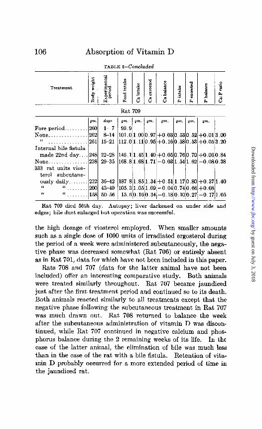
708, the negative calcium and phosphorus balances became posi- tive. This is likewise true of Rat 709 (see Table I) and Rat 706 (see Table II). However, on continued administration of 333 rat units subcutaneously, the calcium and phosphorus balances on Rats 708 and 709 became either negative or decreased in amounts over the previous period. This is apparently due to
TABLE 1
Calcium and Phosphorus Balances on Rats 708 and 709
Treatment
Rat 708
g7n.l days pm. gm. gm. gm. gm. gm. gm.
Fore period.. 290 l- 7 89.1 None. . . . 289 8- 14 90.10.890.88+0.010.470.45+0.020.50
“ . . . . . . . . . . . . . . . 294 15- 21 95.60.990.93 +0.060.500.47 j-0.031.66 Internal bile fistula
made 22nd day 281 22- 28 73.30.730.73 0.000.380.38 0.00 None . . . 257 29 35 99.90.991.03 -0.040.620.64 -0.022.09 333 rat unit5 vios-
terol orally daily. 247 36- 42130.31.291.25+0.040.820.77+0.050.80 “ “ . . . 239 43- 49107.11.061.12 -0.060.670.70 -0.032.00 “ “ . . . . 240 50- 56 90.10.900.93 -0.030.560.61-0.050.60
333 rat units vios- terol subcutane- ously 3 times per wk................259 57- 63143.41.421.04+0.380.890.63+0.261.50 “ “ . . 248 64- 70133.51.331.75 -0.420.831.09 -0.261.60
None . . . . . . . . . . . . . . . 248 71- 77113.41.131.14-0.010.710.71 0.06 “ . . . . . . . . . . . . . 249 78 84117.11.161.16 0.000.730.74 -0.01
1000 rat units vios- terol subcutane- ously 85th day.... 259 85- 91149.11.481.38+0.100.930.83+0.101.00
None. . . . . 245 92- 98 160.5 1.59 1.58 +O.Ol 1.000.97 +0.030.30 “ . . . . . . . . . . . . . . 241 99-105126.31.251.30 -0.050.790.84 -0.051.00
95.5 mg. desoxy- cholic acid + 3180 rat units vioster- olinfood.........24010&112 95.50.950.87+0.080.600.55+0.051.60
None ,,.......,...__ 236113-119113.61.131.19-0.060.710.80-0.090.66 ‘I ..,...,........ 261120-126141.21.401.45 -0.050.880.92 -0.041.25
by guest on July 3, 2018http://w
ww
.jbc.org/D
ownloaded from
106 Absorption of Vitamin D
TABLE I-Concluded
Rat 709
l7m. days
Fore period.. 260 l- 7 None.. . 262 8-14
“ . . . . . . . . . . . 261 15-21 Internal bile fistula
made 22nd day.. .248 22-28 None...............238 29-35 333 rat units vios-
terol subcutane- ously daily.. 232 36-42 ‘I “ 200 43-49 ‘I “ . 158 50-56
pm. I gm. gm. gm. gm. gm. gm.
99.9 101.01.000.97+0.030.530.52+0.013.09 112.01.110.95+0.160.580.53+0.053.20
146.11.451.40+0.050.760.70+0.060.84 168.81.681.71 -0.031.541.62 -0.080.38
187.81.851.34+0.511.170.30+0.371.40 105.31.051.09 -0.040.740.66+0.08
15.60.160.34-0.180.100.27-0.171.05
Rat 709 died 56th day. Autopsy; liver darkened on under side and edges; bile duct enlarged but operation was successful.
the high dosage of viosterol employed. When smaller amounts such as a single dose of 1000 units of irradiated ergosterol during the period of a week were administered subcutaneously, the nega- tive phase was decreased somewhat (Rat 706) or entirely absent as in Rat 701, data for which have not been included in this paper.
Rats 708 and 707 (data for the latter animal have not been included) offer an interesting comparative study. Both animals were treated similarly throughout. Rat 707 became jaundiced just after the first treatment period and continued so to its death. Both animals reacted similarly to all treatments except that the negative phase following the subcutaneous treatment in Rat 707 was much drawn out. Rat 708 returned to balance the week after the subcutaneous administration of vitamin D was discon- tinued, while Rat 707 continued in negative calcium and phos- phorus balance during the 2 remaining weeks of its life. In the case of the latter animal, the elimination of bile was much less than in the case of the rat with a bile fistula. Retention of vita- min D probably occurred for a more extended period of time in the jaundiced rat.
by guest on July 3, 2018http://w
ww
.jbc.org/D
ownloaded from
J. D. Greaves and C. L. A. Schmidt 107
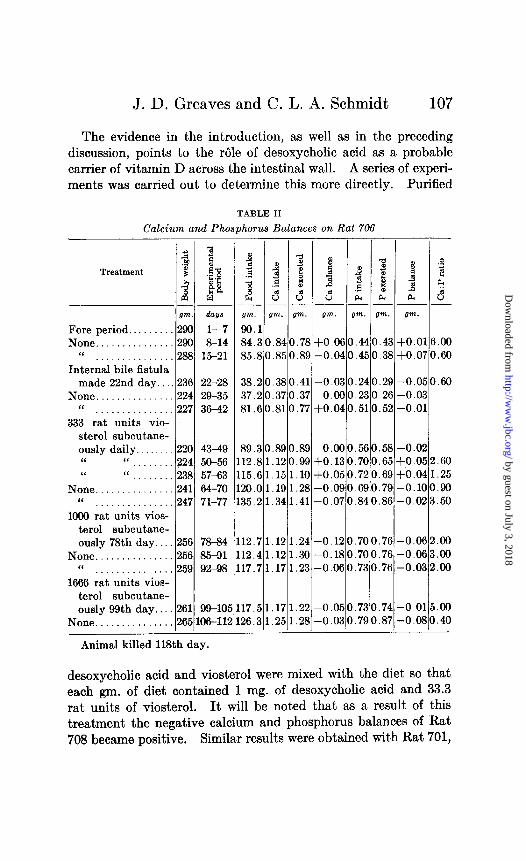
The evidence in the introduction, as well as in the preceding discussion, points to the role of desoxycholic acid as a probable carrier of vitamin D across the intestinal wall. A series of experi- ments was carried out to determine this more directly. Purified
TABLE II
Calcium and Phosphorus Balances on Rat 706
gm. his
Fore period.. . 290 l- 7 None. . 290 8-14
“ 288 15-21 Internal bile fistula
made 22nd day. ,236 22-28 None. 224 29-35
“ . . . . . . . . . . . . . 227 36-42 333 rat units vio-
sterol subcutane- ously daily.. 220 4349 “ “ 224 50-56 “ “ 238 57-63
None. : 1. 241 64-70 1‘ . . . . . . . . . 247 71-77
1000 rat units vios- terol subcutane- ously 78th day.. 256 78-84
None. . 256 85-91 “ . . . . . . . . . . . . . . 259 92-98
1666 rat units vios- terol subcutane- ously 99th day.. 261 99-lo!
None...............265106-11:
Animal killed 118th day.
gm. 1 gm. I gm. I bvn. I gm. I g*. I gm. l
gfd I I I I I I 84.30.840.78+0.060.440.43+0.016.00 85.80.850.89 -0.040.450.38+0.070.60
38.20.380.41 -0.030.240.29 -0.050.60 37.20.370.37 0.000.230.26 -0.03 81.60.810.77+0.040.510.52-0.01
89.30.890.89 0.000.560.58-0.02 12.81.120.99+0.130.700.65+0.052.60
.15.61.151.10+0.050.720.69+0.041.25 20.01.191.28 -0.090.690.79 -0.100.90 35.21.341.41 -0.070.840.86-0.023.50
12.71.121.24 -0.120.700.76 -0.062.00 12.41.121.30 -0.180.700.76 -0.063.00 17.71.171.23 -0.060.730.76 -0.032.00
17.51.171.22 -0.050.730.74 -0.015.00 26.31.251.28 -0.030.790.87 -0.080.40
desoxycholic acid and viosterol were mixed with the diet so that each gm. of diet contained 1 mg. of desoxycholic acid and 33.3 rat units of viosterol. It will be noted that as a result of this treatment the negative calcium and phosphorus balances of Rat 708 became positive. Similar results were obtained with Rat 701,
by guest on July 3, 2018http://w
ww
.jbc.org/D
ownloaded from
108 Absorption of Vitamin D
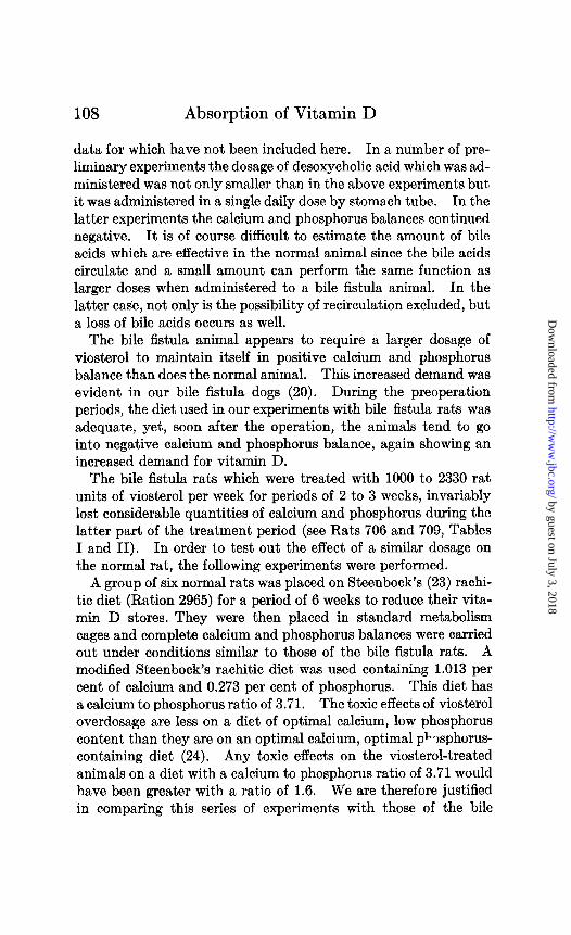
data for which have not been included here. In a number of pre- liminary experiments the dosage of desoxycholic acid which was ad- ministered was not only smaller than in the above experiments but it was administered in a single daily dose by stomach tube. In the latter experiments the calcium and phosphorus balances continued negative. It is of course difficult to estimate the amount of bile acids which are effective in the normal animal since the bile acids circulate and a small amount can perform the same function as larger doses when administered to a bile fistula animal. In the latter case, not only is the possibility of recirculation excluded, but a loss of bile acids occurs as well.
The bile fistula animal appears to require a larger dosage of viosterol to maintain itself in positive calcium and phosphorus balance than does the normal animal. This increased demand was evident in our bile fistula dogs (20). During the preoperation periods, the diet used in our experiments with bile fistula rats was adequate, yet, soon after the operation, the animals tend to go into negative calcium and phosphorus balance, again showing an increased demand for vitamin D.
The bile fistula rats which were treated with 1000 to 2330 rat units of viosterol per week for periods of 2 to 3 weeks, invariably lost considerable quantities of calcium and phosphorus during the latter part of the treatment period (see Rats 706 and 709, Tables I and II). In order to test out the effect of a similar dosage on the normal rat, the following experiments were performed.
A group of six normal rats was placed on Steenbock’s (23) rachi- tic diet (Ration 2965) for a period of 6 weeks to reduce their vita- min D stores. They were then placed in standard metabolism cages and complete calcium and phosphorus balances were carried out under conditions similar to those of the bile fistula rats. A modified Steenbock’s rachitic diet was used containing 1.013 per cent of calcium and 0.273 per cent of phosphorus. This diet has a calcium to phosphorus ratio of 3.71. The toxic effects of viosterol overdosage are less on a diet of optimal calcium, low phosphorus content than they are on an optimal calcium, optimal p%phorus- containing diet (24). Any toxic effects on the viosterol-treated animals on a diet with a calcium to phosphorus ratio of 3.71 would have been greater with a ratio of 1.6. We are therefore justified in comparing this series of experiments with those of the bile
by guest on July 3, 2018http://w
ww
.jbc.org/D
ownloaded from
J. D. Greaves and C. L. A. Schmidt 109
fistula animals even though the calcium to phosphorus ratios of the diet are different. The actual differences in viosterol tolerance are probably greater than this comparison indicates.
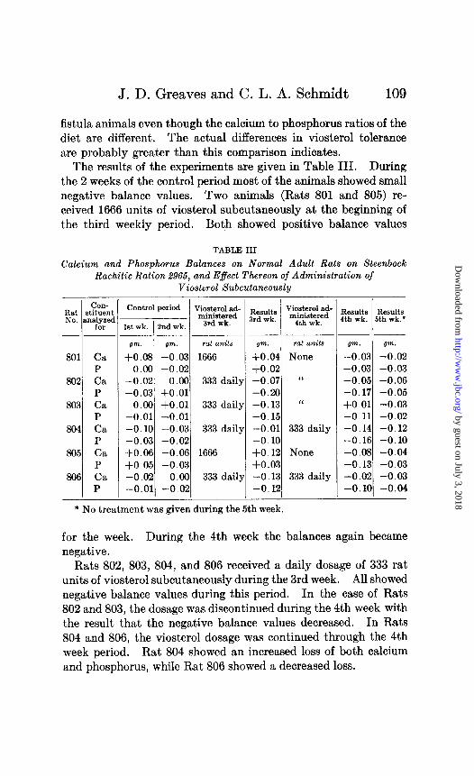
The results of the experiments are given in Table 111. During the 2 weeks of the control period most of the animals showed small negative balance values. Two animals (Rats 801 and 805) re- ceived 1666 units of viosterol subcutaneously at the beginning of the third weekly period. Both showed positive balance values
TABLE III
Calcium and Phosphorus Balances on Normal Adult Rats on Steenbock Rachitic Ration .%%5, and Effect Thereon of Administration of
Viosterol Subcutaneously
COII- Rat stituent No. nna$yd
-___
801 ca P
802 Ca P
803 ca P
804 Ca P
805 Ca P
806 Ca P
__- ~-
gm. !w. rat units gm. rat units gm. gm.
+o.os -0.03 1666 +0.04 None -0.03 -0.02 0.00 -0.02 f0.02 -0.03 -0.03
-0.02 0.00 333 daily -0.07 “ -0.05 -0.06 -0.03 +0.01 -0.20 -0.17 -0.05
0.00 +O.Ol 333 daily -0.13 “ +0.01 -0.03 -0.01 -0.01 -0.15 -0.11 -0.02 -0.10 -0.03 333 daily -0.01 333 daily -0.14 -0.12 -0.03 -0.02 -0.10 -0.16 -0.10 +0.06 -0.06 1666 +O. 12 None -0.08 -0.04 $0.05 -0.03 +0.03 -0.13 -0.03 -0.02 0.00 333 daily -0.13 333 daily -0.02 -0.03 -0.01 -0.02 -0.12 -0.10 -0.04
-
Results Results Ith wk. 5th wk.*
* No treatment was given during the 5th week.
for the week. During the 4th week the balances again became negative.
Rats 802, 803, 804, and 806 received a daily dosage of 333 rat units of viosterol subcutaneously during the 3rd week. All showed negative balance values during this period. In the case of Rats 802 and 803, the dosage was discontinued during the 4th week with the result that the negative balance values decreased. In Rats 804 and 806, the viosterol dosage was continued through the 4th week period. Rat 804 showed an increased loss of both calcium and phosphorus, while Rat 806 showed a decreased loss.
by guest on July 3, 2018http://w
ww
.jbc.org/D
ownloaded from
110 Absorption of Vitamin D
It is thus seen that positive shifts in the balance values were obtained only when a single dose of 1666 rat units of viosterol during the week was administered. When the larger dosages (2332 units) were administered, a marked loss of both calcium and phosphorus invariably followed.
In the case of the bile fistula rats, positive balances values were obtained when 333 rat units of viosterol were administered sub- cutaneously three times per week for the 1st week to Rat 708, and when 333 rat units were administered daily to Rat 709 during the similar period. These data, while not directly comparable, give additional weight to the hypothesis that the bile fistula animal requires more vitamin D than does the normal animal. The de- cided negative phase of icteric Rat 707 during the 2nd week when 333 units of viosterol per week were administered subcutaneously (data not given here) points strongly to the fact that the smaller response to a like dose of viosterol is due to increased excretion of the vitamin by the bile fistula rat. The most probable channel is through the bile.
It is recognized that in experiments of the type recorded in this paper not only considerable errors in the estimation of small amounts of calcium and phosphorus enter but individual variations in the response of different animals to the same dose of viosterol as well as possible differences in the flow of bile must be considered. The conclusions are based on general trends rather than on precise quantities. It appears, however, that the data which were ob- tained in these experiments point to the fact that the bile acts as a carrier of vitamin D across the intestinal wall of the rat and that in the absence of bile from the small intestines as in the case of the bile fistula rat little or no vitamin D is absorbed. Absence of bile from the intestinal tract is followed by a loss of calcium and of phosphorus.
The complete data, of which only a part has been included in this paper, are on file in the University of California library.
SUMMARY
The results of a study of the absorption of vitamin D in chole- dochocolostomized rats when calcium and phosphorus balances are used as the criterion for absorption are reported. The follow- ing are indicated:
by guest on July 3, 2018http://w
ww
.jbc.org/D
ownloaded from
J. D. Greaves and C. L. A. Schmidt
1. Bile fistula rats tend to be in negative calcium and phosphorus balance soon after the operation.
2. Little or no irradiated ergosterol is absorbed from the in- testinal tract of the bile fistula rat.
3. Desoxycholic acid when administered by mouth can serve as a carrier of irradiated ergosterol across the intestinal wall of the bile fistula rat.
4. In the presence of bile, the absorption of irradiated ergosterol which takes place from the descending colon of the rat, as shown in these experiments on rats with bile fistulas, is small or absent.
5. The hypothesis is advanced that the increased need for anti- rachitic factor by the bile fistula animal is due to increased excre- tion of vitamin D which probably proceeds by way of the bile.
BIBLIOGRAPHY
1. Pavlov, I. P., VW. Ges. russ. Aerzt., ‘73, 314 (1904). 2. Looser, E., Verhandl. deutsch. path. Ges., 11, 291 (1907). 3. Wisner, F. P., and Whipple, G. H., Am. J. PhysioZ., 60, 119 (1922). 4. Dtittmann, G., Be&. klin. Chir., 139, 720 (1927). 5. Dietrich, H., Be&. klin. Chir., 134, 530 (1925). 6. Gilbert, E., Z. ges. esp. Med., 43, 539 (1924). 7. Bushbinder, W. C., and Kern, R., Arch. Znt. Med., 40, 900 (1927); Am.
J. Physiol., 80, 273 (1927). 8. Seidel, H., Miinch. med. Woch., 67, 2034 (1910). 9. Rigano, I., Cultura med. mod., 10, 43 (1931).
10. Klinke, K., KZin. Woch., 1, 385 (1928); Ergebn. PhysioZ., 26, 279 (1928). 11. Seifert, E., Beitr. klin. Chir., 136, 496 (1926). 12. Tammann, H., Beitr. klin. Chir., 142, 83 (1928). 13. Literature cited by Schmidt, C. L. A., Physiol. Rev., 7, 129 (1927).
Verzar, F., Nutrition Abst. and Rev., 2, 441 (1933). 14. Burger, M., and Winterseel, W., 2. ges. ezp. Med., 66, 463 (1929). Ep-
stein, %. Z., Arch. Int. Med., 47,821 (1931); 60,203 (1932). Gardner, J. A., and Gainsborough, H., Quart. J. Med., 23,465 (1930). Muller, G. L., Medicine, 9, 119 (1930). Ssokoloff, N. A., Virchows Arch. path. Anat., 246, 203 (1923).
15. Schonheimer, R., Biochem. Z., 147,258 (1924). Wieland, H., and Sorge, H., 2. physiol. Chem., 97, 1 (1916).
16. Schonheimer, R., 2. physiol. Chem., 160, 16, 24, 36 (1929). SchBn- heimer, R., and von Behring, H., 2. physiol. Chem., 192, 97 (1930). Schonheimer, R., von Behring, H., and Hummel, H., 2. physiol. Chem., 192, 117 (1930).
17. Page, I. H., and Menschick, W., Biochem. Z., 221, 6 (1930). Schijn- heimer, R., 2. physiol. Chem., 186, 119 (1929); J. BioZ. Chem., 92, v (1931). Schonheimer, R., and von Behring, H., KZin. Woch., 9, 1308 (1930).
by guest on July 3, 2018http://w
ww
.jbc.org/D
ownloaded from

112 Absorption of Vitamin D
18. Menschick, W., and Page, I. H., 2. physiol. Chem,, 211, 246 (1932). Schiinheimer, R., and Dam, H., Z. physiol. Chem., 211, 241 (1932). Schiinheimer, R., and Hrdine, L., 2. physiol. Chem., 212, 161 (1932).
19. Schmidt, W., and Schmidt, C. L. A., Univ. California Pub, Physiol., 7, 211 (1930).
20. Greaves, J. D., and Schmidt, C. L. A., Proc. Sot. Exp. Biol. and Med., 29, 373 (1932).
21. McCrudden, F. H., J. Biol. Chem., 7, 83 (1909-10); 10, 187 (1911-12). 22. Pemberton, D., J. Am. Chem. Sot., 16, 278 (1894). Hibbard, P. L., J.
Ind. and Eng. Chem., 6, 998 (1913). 23. Steenbock, H., and Black, A., J. Biol. Chem., 64,274 (1925). 24. Shelling, D. H., and Asher, D. E., Bull. Johns Hopkins Hosp., 60, 318
(1932).
by guest on July 3, 2018http://w
ww
.jbc.org/D
ownloaded from
Joseph D. Greaves and Carl L. A. SchmidtRAT
ABSORPTION OF VITAMIN D IN THE THE RÔLE PLAYED BY BILE IN THE
1933, 102:101-112.J. Biol. Chem.
http://www.jbc.org/content/102/1/101.citation
Access the most updated version of this article at
Alerts:
When a correction for this article is posted•
When this article is cited•
alerts to choose from all of JBC's e-mailClick here
tml#ref-list-1
http://www.jbc.org/content/102/1/101.citation.full.haccessed free atThis article cites 0 references, 0 of which can be
by guest on July 3, 2018http://w
ww
.jbc.org/D
ownloaded from