the role of pontine reticular formation gabaa receptors - deep blue
TRANSCRIPT

GABAA RECEPTORS IN THE PONTINE RETICULAR FORMATION OF C57BL/6J MOUSE MODULATE NEUROCHEMICAL, ELECTROGRAPHIC,
AND BEHAVIORAL PHENOTYPES OF WAKEFULNESS
by
RaShonda R. Flint
A dissertation submitted in partial fulfillment of the requirements for the degree of
Doctor of Philosophy (Pharmacology)
in The University of Michigan 2010
Doctoral Committee: Professor Helen A. Baghdoyan, Chair Professor Stephen K. Fisher Professor Margaret E. Gnegy Professor Robert T. Kennedy Professor Ralph Lydic

DEDICATION
To my family
ii

ACKNOWLEDGEMENTS
As I come to the completion of one chapter of my life and prepare to begin the
next, I reflect upon a quote I have carried with me throughout my graduate studies from
the African-American educator and author Booker T. Washington - “Success is to be
measured not so much by the position that one has reached in life as by the obstacles by
which s(he) has overcome while trying to succeed”. As I prepare to complete my PhD
and leave The University of Michigan after six years, I reflect on the journey that brought
me here, the experiences that shaped my time as a PhD student, and most importantly the
people who were there along the way. I have been fortunate to have many people
guiding and supporting me, and I am FOREVER indebted to them all.
The first two people I want to thank are my advisors, Dr. Helen A. Baghdoyan
and Dr. Ralph Lydic. Their role in my graduate education as both mentors and teachers
was invaluable and I am grateful to them for their dedication to me throughout this
journey.
I would also like to thank the other members of my doctoral committee, Dr.
Steve Fisher, Dr. Peggy Gnegy, and Dr. Bob Kennedy, whose guidance and feedback
during my graduate years was much appreciated. I cannot forget to thank the faculty,
staff, and students of both the Pharmacology and Anesthesiology Departments for all of
their support and help along the way, many of you will remain lifelong friends.
iii

Last, but certainly not least, I would like to thank ALL of my family and friends
who have been with me throughout this journey, especially my parents and my sister.
Your love, encouragement, and support made so much of this dream a reality. Words
cannot express what you all mean to me, but know that my success is your success.
iv

TABLE OF CONTENTS
DEDICATION .................................................................................................................... ii
ACKNOWLEDGEMENTS ............................................................................................... iii
LIST OF FIGURES .......................................................................................................... vii
LIST OF TABLES ............................................................................................................. ix
LIST OF ABBREVIATIONS ..............................................................................................x
CHAPTER
1. INTRODUCTION AND STATEMENT OF RESEARCH QUESTION .............1
THE ROLE OF THE PONTINE RETICULAR FORMATION IN
AROUSAL STATE CONTROL ...................................................................4
NEUROTRANSMITTER REGULATION OF SLEEP AND
WAKEFULNESS ..........................................................................................6
TECHNICAL APPROACHES AND CONSIDERATIONS.......................11
OVERVIEW OF RESEARCH OBJECTIVES ............................................15
REFERENCES ............................................................................................21
2. GABAA RECEPTORS IN THE PONTINE RETICULAR FORMATION OF C57BL/6J MOUSE MODULATE NEUROCHEMICAL, ELECTROGRAPHIC, AND BEHAVIORAL PHENOTYPES OF WAKEFULNESS ..............................................................................................32
SUMMARY .................................................................................................32
INTRODUCTION .......................................................................................33
v

METHODS ..................................................................................................36
RESULTS ....................................................................................................42
DISCUSSION ..............................................................................................67
REFERENCES ............................................................................................74
3. SUMMARY AND CONCLUSIONS .................................................................81
GABA ..........................................................................................................85
ACETYLCHOLINE ....................................................................................87
GLUTAMATE .............................................................................................89
REFERENCES ............................................................................................96
vi

LIST OF FIGURES
CHAPTER 2
2.1. Microinjection of muscimol into the PnO increased EEG power during wakefulness and REM sleep ....................................................................... 45
2.2. PnO microinjection of muscimol disrupted sleep architecture by increasing
wakefulness ................................................................................................. 46 2.3. Muscimol caused a concentration dependent increase in wakefulness and
decrease in sleep ......................................................................................... 49 2.4. Bicuculline blocked the sleep inhibition caused by muscimol ..................... 51 2.5. PnO microinjection of bicuculline blocked the increase in wakefulness, decrease in NREM sleep, and decrease in REM sleep caused by muscimol ..................................................................................................... 53 2.6. EEG power was not altered by PnO microinjection of bicuculline .............. 55 2.7. PnO microinjection of bicuculline altered sleep architecture by increasing sleep ............................................................................................................ 57
2.8. Microinjection of bicuculline into the PnO caused a concentration dependent decrease in wakefulness and increase in NREM sleep and REM sleep ..............................................................................................................59 2.9. ACh release in mouse PnO ............................................................................62
2.10. Blocking GABAA receptors in the PnO caused a concentration dependent increase in ACh release in the PnO .............................................................63 2.11. Bicuculline caused a concentration dependent decrease in breathing rate...............................................................................................................65 2.12. Bicuculline administered to the PnO caused a concentration dependent increase in anesthesia recovery time ...........................................................66
vii

CHAPTER 3 3.1. Synaptic model illustrating neurons in the pontine reticular formation (PRF) of B6 mouse through which GABA may promote wakefulness ................................................................................................93
viii

LIST OF TABLES
CHAPTER 3
3.1. The PRF contains GABA inter-neurons and GABAA receptors. GABA in the PRF promotes wakefulness and respiratory rate ................................... 86
3.2. ACh neurons in the LDT/PPT project to the PRF, which contains muscarinic
receptors. ACh in the PRF modulates REM sleep ..................................... 88 3.3. Glutamate neurons in the PRF project to the LDT/PPT. Glutamate receptors
in the PPT induce REM sleep ..................................................................... 90
ix

LIST OF ABBREVIATIONS
˚C degrees Celsius
% percent
ACh acetylcholine
ANOVA analysis of variance
ARAS ascending reticular activating system
B6 C57BL/6J
BDZ benzodiazepine
BRA benzodiazepine receptor agonist
df degrees of freedom
EEG electroencephalogram
EMG electromyogram
FFT Fast Fourier transform
GABA γ-aminobutyric acid
h hour(s)
HPLC/ED high performance liquid chromatography with electrochemical
detection
Hz hertz
i.p. intraperitoneal
kDa kilo Dalton
x

xi
KO knockout
LDT laterodorsal tegmental
min minute(s)
µm micromolar
mM millimolar
ng nanogram
nl nanoliter
NREM non-rapid eye movement
p probability
pmol picomoles
PnO pontine reticular nucleus, oral part
PPT pedunculopontine tegmental
PRF pontine reticular formation
REM rapid eye movement
s second(s)
SEM standard error of the mean

CHAPTER 1
INTRODUCTION AND STATEMENT OF RESEARCH QUESTION
States of sleep and anesthesia are defined by specific traits. Sleep is a naturally
occurring behavioral state characterized by a decrease in voluntary body movement, an
unawareness of surroundings, and an increased threshold to sensory stimulation.
Anesthesia on the other hand is a pharmacologically induced reversible state with
characteristics of amnesia, analgesia, unconsciousness, immobility, and blunted
autonomic responses. One commonality between sleep and anesthesia is that some of the
drugs that are used to produce sleep, sedation, or general anesthesia act to enhance
transmission at GABAA receptors. Understanding the cellular and molecular mechanisms
through which both sleep and anesthesia occur can provide insights into each of their
specific traits (Lydic, 2001; Lydic and Baghdoyan, 2006; Watson et al., 2009). Teasing
out these mechanisms can provide relief for the burdens that arise from the disorders
associated with sleep or the complications that arise from anesthesia through the rational
design of therapeutically selective drugs.
Insomnia is a sleep disorder that affects 10-15% of the United States population
(Drake et al., 2003) and is characterized by difficulty initiating and/or maintaining sleep
despite ample time spent in bed. Insomnia can have severe consequences including
fatigue, social impairment, and daytime accidents. These consequences result in large
1

direct and indirect costs occurring from such things as medical care and absenteeism
from work. The economic burden of insomnia in the US based on direct costs alone in
the 1990s was over 13.9 billion dollars, and indirect costs added in during this time
period led to totals exceeding 35 billion dollars, with the cost continuing to rise sharply in
the last decade (Walsh and Engelhardt, 1999; Ozminkowski et al., 2007). Current
pharmacological treatments for insomnia include benzodiazepine receptor agonists
(BRAs), which enhance transmission at GABAA receptors. Although these drugs
improve sleep by increasing overall sleep time, some BRAs produce undesirable side
effects such as reduced slow wave sleep and decreased rapid eye movement (REM) sleep
(Mendelson, 2005), making the need for rational drug design increasingly important.
Anesthesia is administered to millions of patients each year in the United States,
despite little being known about the mechanisms by which these drugs produce their
effects or side effects. Postoperative nausea and vomiting is the most common anesthetic
complication, and it occurs in 30-50% of all surgical patients (Gundzik, 2008; Conway,
2009). In the elderly population, postoperative delirium following anesthesia is a
common occurrence, observed in anywhere from 5-15% of patients (Silverstein et al.,
2007; Sieber, 2009). It is also estimated that each year between 0.007% and 0.13% of
patients administered general anesthesia experience some type of awareness (Sebel et al.,
2004; Pollard et al., 2007), and 1 in 250,00 die as a direct result of anesthesia (Lydic,
2001). Further understanding of the mechanisms through which both sleep and
anesthesia produce their effects can contribute to the safer use of drugs to treat sleep
disorders and produce anesthesia (Lydic, 2001).
2

Both sleep and anesthesia are modulated by a number of neurotransmitters in
different brain regions. Even though the two states have their own distinctive traits,
anesthetics act through some of the same neural circuitry and brain regions that generate
sleep (Lydic and Baghdoyan, 2006; Ishizawa, 2007; Franks, 2008; Watson et al., 2009).
Studies have demonstrated the brain region selective effects of γ-aminobutyric acid
(GABA), and increase of GABAergic transmission in the pontine reticular formation
(PRF) has been shown to promote wakefulness and suppress sleep (Xi et al., 1999;
Vanini et al., 2008; Watson et al., 2008). In an attempt to characterize how multiple
brain regions and neurotransmitters work together to regulate states of arousal, it is
necessary to examine the individual contributions of these neurotransmitters and brain
regions in the regulation of arousal states. This dissertation research focused on the PRF
and two of the neurotransmitters in the PRF associated with arousal state control, GABA
and acetylcholine (ACh). The introduction begins with a discussion of the PRF and its
role in both sleep and anesthesia. There is consideration of neurotransmitter regulation of
state, with a focus on GABA and ACh. After that is a brief examination of the technical
approaches and limitations of the dissertation studies in the context of the results. The
chapter closes with an overview of the dissertation objectives and results.
3

THE ROLE OF THE PONTINE RETICULAR FORMATION IN AROUSAL
STATE CONTROL
The pontine reticular formation contributes to the generation of arousal states
The functional concept of the PRF became apparent with the pioneering work of
Moruzzi and Magoun in 1949, who proposed the existence of a neuronal network
responsible for the control of arousal states that they named the ascending reticular
activating system (ARAS) (Moruzzi and Magoun, 1949). This system of neurons and
neural fibers, including the reticular formation, contributes to the regulation of states of
arousal such as sleep, wakefulness, and anesthesia. The PRF, one of the phylogenetically
oldest portions of the brain, is made up of cells of varying sizes and shapes and is
surrounded by networks of fibers that run in all directions (Rossi and Brodal, 1956).
Within the PRF there are two main divisions, the pontine reticular nucleus, oral part
(PnO) and the pontine reticular nucleus, caudal part (PnC). The PRF contains GABAA
receptors, GABA inter-neurons, and glutamatergic neurons, and receives cholinergic
input from the laterodorsal tegmental (LDT) and pedunculopontine tegmental (PPT)
nuclei (Kosaka et al., 1988; Mitani et al., 1988; Shiromani et al., 1988; Jones, 1990; Ford
et al., 1995; Pirker et al., 2000; de la Roza and Reinoso-Suarez, 2006; Rodrigo-Angulo et
al., 2008). Work by Jouvet and others highlighted the pons as a brain region that
contributes to arousal state control and is key for REM sleep generation (Jouvet, 1962;
Lavie et al., 1984; Webster and Jones, 1988; Reinoso-Suarez et al., 2001).
4

Studies of the pontine reticular formation enhance the overall knowledge of sleep
and anesthesia
The fields of sleep and anesthesia have benefited tremendously from research
aimed at understanding the different brain regions involved in the regulation of arousal
states. Early studies focused at the level of the PRF used brain transections to establish
the role of this region in the generation of non-REM (NREM) sleep and REM sleep
(Jouvet, 1962; Carli and Zanchetti, 1965; Jouvet, 1965). In addition, the Reciprocal
Interaction Model conceptualized the neural mechanisms generating states of
wakefulness and sleep using both structural and mathematical models (McCarley and
Hobson, 1975). This model hypothesized a shift between NREM sleep and REM sleep
based on the reciprocal connections of pontine structures (Hobson et al., 1975). The
mathematical model is unique in that it has predictive value. Pontine lesion studies in
both humans (Lavie et al., 1984; Webster and Jones, 1988; Kushida et al., 1991; Gironell
et al., 1995; Kimura et al., 2000; Reinoso-Suarez et al., 2001) and animals (Webster and
Jones, 1988) demonstrate a disruption in REM sleep, validating Jouvet’s findings. These
studies prompt further research examining the role of pontine neurotransmitters in the
generation of arousal states, particularly REM sleep.
5

NEUROTRANSMITTER REGULATION OF SLEEP AND WAKEFULNESS
GABAergic modulation of sleep and wakefulness
The main inhibitory neurotransmitter in the mammalian central nervous system
is GABA, which plays a vital role in regulating neuronal excitability throughout the
entire nervous system. GABA was discovered in large amounts in the brain in 1950 by
three different groups of researchers (Awapara et al., 1950; Roberts and Frankel, 1950;
Udenfriend, 1950). Studies for the next 20 years studied the inhibitory actions of GABA
as it related to physiological processes in the brain, until the 1970s and 1980s when the
focus shifted to identifying and characterizing the receptors upon which GABA acts
(Krnjevic, 2004; Bowery and Smart, 2006). There are two types of transmembrane
receptors with GABA as their endogenous ligand, GABAA and GABAB, with GABAA
receptors also having a subclass of receptors known as GABAA-ρ receptors (formerly
GABAC receptors) (Olsen and Sieghart, 2008).
The focus for these dissertation studies was on GABAA receptors because of their
direct link to both sleep and anesthesia. GABAA receptors were discovered toward the
end of the 1980s and classified as ionotropic receptors based on their ability to open their
channel upon binding of the ligand GABA. These receptors consist of five protein
subunits that surround a central pore preferentially permeable to negative chloride ions,
and there are multiple subunit combinations and binding sites for the receptor. Generally
speaking, drugs acting as agonists or allosteric modulators of GABAA receptors typically
6

have anticonvulsant, anxiolytic, amnesic, sedative, or hypnotic effects. These properties
are useful for both anesthesia and sleep.
GABA and its inhibitory neurotransmitter actions have been defined and
characterized extensively over the last 60 years, and more recently studies have begun to
investigate the role GABA plays in specific physiological processes such as arousal state.
Administration of GABAA receptor agonists to brain regions such as the posterior
hypothalamus, locus coeruleus, medial preoptic area, and the dorsal raphe nucleus
increases sleep (Mallick et al., 2001; Tung et al., 2001; Baghdoyan and Lydic, 2002),
whereas in the PRF increasing GABAergic transmission increases wakefulness
(Camacho-Arroyo et al., 1991; Xi et al., 1999; Sanford et al., 2003). Increasing GABA
levels in the PRF with the GABA reuptake inhibitor nipecotic acid increases wakefulness
(Watson et al., 2008). These studies provide support for analyzing neurotransmitter
effects on arousal states in a brain region specific manner. Doing so will allow for a
greater understanding of the mechanisms involved in regulating both sleep and anesthesia
and may lead to rational therapeutic drug design.
GABAA receptors as binding sites for drugs that produce sleep and anesthesia
Some anesthetics and pharmacological treatments for insomnia are known to act
on GABAA receptors to produce their effects, as mentioned above. Anesthetics are used
to produce a reversible state characterized by five traits: analgesia, amnesia, hypnosis,
paralysis, and obtundation of reflexes. Anesthetics act on a multitude of receptors, most
importantly for these dissertation studies are those working at GABAA receptors (Bonin
and Orser, 2008). Most of these anesthetics act as allosteric modulators which potentiate
7

the effects of GABA at GABAA receptors and include barbiturates, etomidate, propofol,
and volatile anesthetics. Benzodiazepines (BDZs), which will be discussed in further
detail below, are also routinely used in the practice of anesthesia. In clinical settings
anesthetics act in numerous ways to induce anesthesia, maintain anesthesia, or produce
conscious sedation (Grasshoff et al., 2006). Many studies have been done analyzing how
these drugs work when administered systemically and distribute throughout the brain.
However, it is also necessary to examine the brain region specific effects of these
anesthetics in order to elucidate their mechanisms of action. Injection of propofol or
pentobarbital into the medial preoptic area of the rat elicited significant decreases in sleep
latency and increases in NREM sleep (Mendelson, 1996; Tung et al., 2001), suggesting
potential sites of action for these drugs. More pre-clinical studies investigating the role
of anesthetics that modulate GABAergic transmission are needed to further understand
the brain regions and mechanisms that regulate anesthetic effects.
To produce the anxiolysis and hypnosis important for anesthesia, BDZs are
often employed. These drugs are also widely used in the treatment of conditions ranging
from seizures to muscle spasms, and most importantly for these dissertation studies, as a
pharmacological treatment for anxiety and insomnia. Similar to the anesthetics described
above, BDZs act as allosteric modulators of the GABAA receptor. Non-BDZs, which are
structurally different from the classic BDZs, also act as allosteric modulators at the same
binding site as BDZs, referred to as BRAs, and are commonly used to treat insomnia
(Gottesmann, 2002). There are numerous BDZs used today in the practice of anesthesia,
midazolam, diazepam, and lorazepam being the most common (White and Eng, 2009).
For the treatment of insomnia, many drugs are prescribed “off-label”, but currently
8

approved drugs for the treatment of the disorder include the BDZs estazolam, flurazepam,
quazepam, temazepam, and triazolam, and the BRAs zaleplon, zolpidem, and eszopiclone
(2005).
In the treatment of insomnia BRAs work to decrease the latency to sleep onset,
decrease the time spent awake after sleep onset, and increase total sleep time. As with
anesthetics, studies have been performed investigating the effects of systemic BRA
administration. Intraperitoneal (i.p.) injections of lorazepam in mice significantly
increase NREM sleep while reducing motor activity (Tang et al., 2009). Eszopiclone and
zolpidem administered i.p. to guinea pigs also produce significant increases in NREM
sleep (Xi and Chase, 2008). Pre-clinical studies have begun studying the role of BRAs in
various brain regions. Interestingly, microinjection of triazolam into the ventrolateral
preoptic area of rat produced no significant effect on sleep (Mendelson, 1999), in contrast
to the medial optic area where triazolam produced a significant increase in NREM sleep
(Mendelson et al., 1989; Mendelson and Martin, 1992) and the dorsal raphe nucleus
where triazolam increased sleep latency and decreased NREM sleep (Mendelson et al.,
1987). This suggests a possible role for the medial optic area in the generation of NREM
sleep following BDZ administration and further supports the need for brain region
specific studies in efforts to understand where anesthetic and arousal state effects are
mediated.
9

Acetylcholine modulates arousal states
Another neurotransmitter important in the PRF and for this dissertation work is
ACh. ACh is found in both the peripheral and central nervous systems. This
neurotransmitter holds great significance as the first to be identified (1914) and
confirmed (1921) by Henry Dale and Otto Loewi. This discovery led them to share the
1936 Nobel Prize in Physiology or Medicine. After over 60 years of research aimed at
understanding this neurotransmitter and chemical transmission, focus in the 1980s turned
to the identification and characterization of the two types of acetylcholine receptors,
nicotinic and muscarinic (Brown, 2006). Nicotinic receptors are ionotropic receptors that
are stimulated by both ACh and nicotine, whereas muscarinic receptors are metabotropic
receptors stimulated by both ACh and muscarine. There are five molecularly distinct
types of muscarinic acetylcholine receptors which are G protein-coupled, M1-M5 (Fukuda
et al., 1987; Brown, 2006). M1, M3, and M5 receptors are Gq/11-coupled and M2 and M4
are Gi/o-coupled (Caulfield and Birdsall, 1998). ACh and its receptors play a key role in
regulating states of arousal (Lydic and Baghdoyan, 2005, 2008).
Within the brain there are two main populations of cholinergic projection
neurons, those in the basal forebrain and those in the LDT/PPT (Kimura et al., 1981;
Woolf and Butcher, 1986), the latter and its contribution to arousal being important for
this dissertation work. The LDT/PPT provides cholinergic input to the PRF (Jones and
Beaudet, 1987). Administration of a muscarinic antagonist to normal humans increases
REM sleep latency (Sagales et al., 1969). Pre-clinical and clinical studies using
cholinergic agonists and acetylcholinesterase inhibitors decrease the latency to REM
sleep, increase the duration of REM sleep, and increase total REM sleep time (Domino et
10

al., 1968; Sitaram et al., 1976; Sitaram et al., 1978), supporting the role of ACh in REM
sleep.
Brain region specific studies have helped to identify areas playing a role in
cholinergic regulation of REM sleep. Microinjecting cholinomimetics into the PRF
produced a REM sleep-like state (Baghdoyan and Lydic, 2002; Lydic et al., 2002;
Coleman et al., 2004b; Douglas et al., 2005), and microdialysis studies demonstrated
significant increases in ACh release during REM sleep (Leonard and Lydic, 1997) and
the REM sleep-like state (Lydic et al., 1991). Studies using specific agonists and
antagonists for muscarinic receptors have supported the role of ACh in REM sleep. M1,
M2, and M3 type muscarinic receptors have been localized to the PRF (Baghdoyan et al.,
1994a; Baghdoyan et al., 1994b; Mallios et al., 1995; Baghdoyan, 1997), and animal
studies have implicated M2 and possibly M3 type receptors in the PRF contributing to
REM sleep (Imeri et al., 1994; Sakai and Onoe, 1997; Baghdoyan and Lydic, 1999;
Coleman et al., 2004b, a). All of these data are consistent with the interpretation that
cholinergic transmission regulates REM sleep.
TECHNICAL APPROACHES AND CONSIDERATIONS
The mouse model
It is known that 99% of mouse genes have homologues in humans (Waterston et
al., 2002), making the mouse an excellent resource for identifying neuropharmacological
mechanisms and molecular targets for rational drug design (Watters and McLeod, 2002).
Understanding the relationship between genotype and sleep phenotype (Valatx et al.,
11
1972; Toth, 2001; Tafti and Franken, 2002) is essential to the future understanding and
treatment of sleep disorders. This dissertation work used the C57BL/6J (B6) mouse as a
tool to elucidate brain region specific effects of GABAergic transmission on arousal
states (Lydic et al., 2002; Coleman et al., 2004b; Douglas et al., 2005; Coleman et al.,
2006; Van Dort et al., 2009). The major advantage of the B6 mouse is that it is the most
widely used inbred mouse strain, and it is the first to have its genome sequenced
(Waterston et al., 2002). Research studies using this mouse strain have studied
everything from behavior and learning to drug addiction and cancer (Bursch et al., 2004;
Singh et al., 2007).
The present dissertation work provides background for future sleep studies
utilizing genetically altered mouse strains in an attempt to identify specific receptors and
neurotransmitters involved in the physiological processes underlying sleep. Future
studies could involve conditional knockout (KO) mice, which have a specific gene,
neuron, or channel inactivated, and would allow for functional studies of targets involved
in regulating arousal states based on comparisons of observed differences from this
dissertation work. Conditional KO mice have provided novel insights into cancer,
cardiovascular disease, anxiety, and obesity, and have provided advancement in both the
biomedical and pharmaceutical arenas (Rudmann and Durham, 1999).
The field of sleep has also been furthered by the use of conditional KO mice.
Conditional KO mice are a powerful tool for understanding sleep as well as its disorders,
providing insight into the function of specific neurons and neuronal systems.
Orexin/ataxin-3 transgenic mice (Chou et al., 2001; Hara et al., 2001) have been used to
study orexinergic involvement in the circadian control of sleep and wakefulness (Kantor
12

et al., 2009) as well as narcoleptic sleep/wake fragmentation in relation to metabolism
(Zhang et al., 2007). The role of calcium channels in the blockade of arousal signal
transmission was studied in thalamic-Cav3.1 KO mice (Anderson et al., 2005). The
present dissertation work used intracranial drug administration to examine the
neurochemical mechanisms modulating states of arousal. These studies set the stage for
future work using these same methods in different transgenic mouse models.
Measures of arousal
Electroencephalogram: The electroencephalogram (EEG) is established as the
primary tool used in evaluating sleep in both humans and animals. An EEG is a
recording of the electrical activity of the brain resulting from neuronal firing, which can
be analyzed to determine sleep stages based on classically characterized traits. Neurons
fire relatively fast during the active states of wakefulness and REM sleep. This firing
produces desynchronized waves that are low in voltage and not consistent in pattern.
During NREM sleep, also known as slow wave sleep, neurons fire at slower rates. Brain
activity during NREM sleep is slow in frequency with large amplitude waves which are
synchronized and more consistent in pattern (McCormick and Bal, 1997; Steriade, 2006;
Miller, 2007).
In addition to the EEG signal, states of arousal can also be characterized by
analyzing a breakdown of the EEG signal into its many waveform components using a
Fast Fourier transform (FFT). An FFT is a mathematical algorithm designed to extract
information from signals, in this case EEG signals, and transform the signals from the
time domain to the frequency domain (Mager and Abernethy, 2007). The application of
13

the FFT to sleep studies allows for the EEG signals to be broken down into different
waveforms with varying frequencies (Dumermuth and Fluhler, 1967). These waveform
components are correlated with various states of arousal when quantified as power for a
given frequency (Muthuswamy and Thakor, 1998; Campbell, 2009). Behavioral EEG
characteristics are the same in both animals and humans, making it possible to correlate
behavioral arousal in animals to arousal in humans. The present dissertation studies used
EEG signals recorded from mice to objectively characterize states of wakefulness,
NREM sleep, and REM sleep following drug administration. The FFT was analyzed
along with the EEGs to help in characterizing arousal states and to study the effects of
drug on quality of sleep. There were two main waveforms analyzed in this dissertation,
delta waves and theta waves. Delta waves occur during NREM sleep (restorative sleep)
and are between the frequencies of 0.5 and 4 Hz. REM sleep has dominant theta waves
with frequencies between 4 and 9 Hz.
Recovery of Righting Response: The recovery of righting response is widely
used as an established measure of wakefulness following administration of commonly
used drugs such as anesthetics, opioids, and alcohol in humans (Straw and Mitchell,
1967; Kissin et al., 1993; York and Chan, 1993; Ajadi et al., 2009) as well as rodents
(Bignall, 1974; Tung et al., 2002; Demarco et al., 2004; Kelz et al., 2008; Van Dort et al.,
2009). For these dissertation studies mice were placed in dorsal recumbency under a
heated lamp and the time (in minutes) required for a mouse to resume a normal upright
posture was recorded. This recovery of righting response was used as a measure of
wakefulness following microdialysis delivery of the GABAA receptor antagonist
bicuculline to the PnO.
14

OVERVIEW OF RESEARCH OBJECTIVES
The unifying goal of this dissertation research was to elucidate the neurochemical
mechanisms by which GABAA receptors in the PRF of B6 mouse modulate wakefulness
and sleep. The PRF is part of the ARAS and plays an important role in generating REM
sleep (Steriade and McCarley, 2005). Administration of GABAergic drugs to the PRF
produces alterations in sleep and wakefulness (Lydic and Baghdoyan, 2005).
Administration of the GABAA receptor agonist muscimol into cat PRF or the oral portion
of rat PRF (PnO) increases wakefulness and decreases sleep (Camacho-Arroyo et al.,
1991; Xi et al., 1999; Sanford et al., 2003). Microinjection of the GABAA receptor
antagonist bicuculline into the same brain region of cat or rat causes an increase in REM
sleep (Xi et al., 1999; Sanford et al., 2003). Microinjection of the GABA uptake inhibitor
nipecotic acid (NPA) or the GABA synthesis inhibitor 3-mercaptopropionic acid (3-
MPA) into rat PnO, respectively, increases or decreases wakefulness (Watson et al.,
2008). GABA levels in cat PRF decrease during the isoflurane-induced loss of
wakefulness, and microinjection of NPA or 3-MPA into rat PnO increases or decreases,
respectively, isoflurane induction time (Vanini et al., 2008). Collectively, these data
support the interpretation that GABAergic transmission in the PRF promotes
wakefulness. Results from the mouse can be compared to results from other species in
order to determine how the mouse is similar to or different from previous work. The
studies done in mouse can help to provide further insight into the mechanisms regulating
arousal.
Within the PRF, GABAergic and cholinergic neurons interact to modulate sleep
and wakefulness. The PRF contains GABAergic neurons inter-neurons (Ford et al.,
15

1995) and GABAA receptors (Heldt and Ressler, 2007). The PRF is also cholinoceptive
(Baghdoyan, 1997), receiving ACh from the more rostrally and dorsally located LDT and
PPT nuclei (Steriade and McCarley, 2005). Glutamatergic neurons that project to the
LDT/PPT originate in the PRF (Steininger et al., 1992). Electrical stimulation of
LDT/PPT neurons increases both REM sleep (Thakkar et al., 1996) and ACh release in
the PRF (Lydic and Baghdoyan, 1993). The discharge rate of LDT/PPT neurons
increases prior to and during REM sleep (Kayama et al., 1992). In the opposite manner,
neurotoxic lesions of the LDT/PPT decrease REM sleep (Webster and Jones, 1988).
Cholinomimetics administered to the PRF increase REM sleep (Baghdoyan and Lydic,
1999) by activating M2 muscarinic receptors (Coleman et al., 2004b) putatively localized
to GABAergic neurons in the PRF (Brown et al., 2008). Muscarinic autoreceptors also
modulate ACh release presynaptically in the PRF (Baghdoyan et al., 1998; Coleman et
al., 2004a). Bicuculline administered to the PRF increases ACh release within the PRF
(Vazquez and Baghdoyan, 2004) and ACh in the PRF increases during REM sleep
(Leonard and Lydic, 1997). The mechanisms by which enhancing transmission at GABA
receptors in the PRF increases wakefulness are largely unknown but may involve
cholinergic transmission from the LDT/PPT and GABAergic inhibition in the PRF. The
following dissertation research aims were designed to provide insight into the
mechanisms by which GABAergic and cholinergic neurotransmission in the PRF
modulate sleep and wakefulness.
16

Specific Aims
Specific Aim 1 tested the hypotheses that microinjection of the GABAA
receptor agonist muscimol into the PnO concentration dependently increases
wakefulness and decreases sleep, and that co-administration of the GABAA receptor
antagonist bicuculline blocks the increase in wakefulness and decrease in sleep
caused by muscimol alone. Aim 1 also tested the hypothesis that bicuculline
microinjected into the PnO causes a concentration dependent decrease in
wakefulness and increase in sleep. The results of Aim 1 are presented in Chapter 2.
Aim 1 used mice implanted with microinjection guide tubes stereotaxically aimed for the
PnO as well as EEG and electromyogram (EMG) recording electrodes. Muscimol,
bicuculline, or muscimol and bicuculline were microinjected into the PnO of the freely
moving mouse. EEG and EMG signals were recorded for 4 h post-injection prior to
analysis of dependent measures of sleep and wakefulness. In addition, EEG power was
analyzed during states of wakefulness, NREM sleep, and REM sleep. Four
concentrations (0.5, 5, 50, and 500 pmol/50 nl) of muscimol were studied along with the
vehicle control saline. These studies were done in order to determine if GABAA
receptors in the PnO modulate states of wakefulness and sleep.
Microinjection of muscimol into the PnO of B6 mouse caused a significant,
concentration dependent increase in the amount of wakefulness and decrease in the
amount of NREM sleep and REM sleep compared to the vehicle control saline.
Administration of muscimol also caused a significant, concentration dependent increase
in the latency to the onset of both NREM sleep and REM sleep and decrease in the
17

number of episodes of wakefulness, NREM sleep, and REM sleep. Muscimol caused a
significant, concentration dependent decrease in the number of transitions between states
of wakefulness, NREM sleep, and REM sleep, and an increase in the average duration of
wakefulness episodes. Muscimol increased EEG delta power during wakefulness and
EEG theta power during REM sleep, with no effect on EEG power during NREM sleep.
Co-administration of muscimol (50 pmol/nl) and bicuculline (5 pmol/50 nl) was
performed to determine whether the muscimol-induced effects on sleep could be
antagonized. Co-administration of bicuculline with muscimol blocked the muscimol-
induced increase in the amount of wakefulness, the decrease in the amount of NREM
sleep, and the decrease in the amount of REM sleep. Muscimol co-administered with
bicuculline blocked the increase in NREM sleep latency and partially blocked the
increase in REM sleep latency. Co-administration of bicuculline and muscimol blocked
the muscimol-induced decrease in the number of episodes of wakefulness, NREM sleep,
and REM sleep, as well as the decrease in the number of transitions and the increase in
the average duration of wakefulness. The results support the interpretation that GABAA
receptors in the PRF promote wakefulness.
In order to determine whether endogenous GABA within the PnO modulates
sleep, EEG and EMG recordings were obtained after microinjecting three concentrations
(0.5, 5, and 50 pmol/50 nl) of bicuculline. Bicuculline caused a significant, concentration
dependent decrease in wakefulness and increase in both NREM sleep and REM sleep
when compared with the vehicle control saline. There were no alterations in EEG power
during any state following PnO administration of bicuculline. The results are consistent
with the interpretation that GABAA receptors in the PnO promote wakefulness. Taken
18

together, these Aim 1 results demonstrate that PnO GABAA receptors promote
wakefulness.
Specific Aim 2 tested the hypotheses that GABAA receptors in the PnO of B6
mouse modulate ACh release in the PnO, respiratory rate, and time to recovery of
righting following isoflurane anesthesia. The results of Aim 2 are reported in Chapter
2. The behavioral effects of GABA are known to be brain region specific, and selectively
increasing endogenous GABA levels in the PRF promotes wakefulness and suppresses
sleep (Vanini et al., 2008; Watson et al., 2008). In both cat and rat, blockade of GABAA
receptors in the PRF causes an increase in REM sleep (Xi et al., 1999; Sanford et al.,
2003; Marks et al., 2008). In cat, blockade of GABAA receptors in the PRF also causes
an increase in PRF ACh release (Vazquez and Baghdoyan, 2004). ACh in the PRF may
trigger REM sleep by inhibiting GABAergic wakefulness promoting neurons via M2
muscarinic receptor activation (Baghdoyan and Lydic, 1999; Coleman et al., 2004b;
Brischoux et al., 2008; Brown et al., 2008). Cholinergic transmission in the PRF
promotes REM sleep, and the increase in REM sleep caused by antagonizing GABAA
receptors can be blocked by antagonizing muscarinic cholinergic receptors (Marks et al.,
2008). Taken together, these data suggest that GABA in the PRF inhibits REM sleep by
inhibiting ACh release in the PRF (Vazquez and Baghdoyan, 2004).
The Aim 1 results reviewed above support the interpretation that GABAergic
transmission in the PnO promotes wakefulness. In order to analyze the mechanisms by
which GABAA receptors in mouse PnO modulate states of arousal, Aim 2 examined ACh
release in the PnO. Aim 2 studies were performed using microdialysis delivery of
19

bicuculline to the PnO of the isoflurane-anesthetized B6 mouse while simultaneously
measuring ACh release in the PnO. Dependent measures were ACh release in the PnO,
respiratory rate during anesthesia, and time to recovery of righting following isoflurane
anesthesia. A concentration response curve was generated for the effects of bicuculline
on ACh release, respiratory rate, and recovery time using the vehicle control Ringer’s and
five concentrations of bicuculline (0.1, 0.3, 1, 3, and 10 mM).
Bicuculline caused a significant, concentration dependent increase in ACh release
in the PnO and increase in anesthesia recovery time compared to Ringer’s. Respiratory
rate was concentration dependently decreased during dialysis with bicuculline. Taken
together with the results from Aim 1, the present data supports the interpretation that
GABAA receptors in the PnO of B6 mouse modulate sleep and wakefulness, ACh release
in the PnO, behavioral arousal, and breathing rate. The finding that ACh release in the
PnO is modulated by GABAA receptors in the PnO is consistent with the interpretation
that the blockade of GABAergic transmission in the PnO leads to decreases in
wakefulness and increases in sleep, in part, by increasing ACh release.
20

REFERENCES
(2005) NIH State-of-the-Science Conference Statement on manifestations and
management of chronic insomnia in adults. NIH Consens Sci Statements 22:1-30. Ajadi AR, Olusa TA, Smith OF, Ajibola ES, Adeleye OE, Adenubi OT, Makinde FA
(2009) Tramadol improved the efficacy of ketamine-xylazine anaesthesia in young pigs. Vet Anaesth Analg 36:562-566.
Anderson MP, Mochizuki T, Xie J, Fischler W, Manger JP, Talley EM, Scammell TE,
Tonegawa S (2005) Thalamic Cav3.1 T-type Ca2+ channel plays a crucial role in stabilizing sleep. Proc Natl Acad Sci U S A 102:1743-1748.
Awapara J, Landua AJ, Fuerst R, Seale B (1950) Free gamma-aminobutyric acid in brain.
J Biol Chem 187:35-39. Baghdoyan HA (1997) Location and quantification of muscarinic receptor subtypes in rat
pons: implications for REM sleep generation. Am J Physiol 273:R896-904. Baghdoyan HA, Lydic R (1999) M2 muscarinic receptor subtype in the feline medial
pontine reticular formation modulates the amount of rapid eye movement sleep. Sleep 22:835-847.
Baghdoyan HA, Lydic R (2002) Neurotransmitters and neuromodulators regulating sleep.
In: Sleep and Epilepsy: The Clinical Spectrum (Bazil C, Malow B, Sammaritano M, eds), pp 17-44. New York: Elsevier Science.
Baghdoyan HA, Carlson BX, Roth MT (1994a) Pharmacological characterization of
muscarinic cholinergic receptors in cat pons and cortex. Preliminary study. Pharmacology 48:77-85.
Baghdoyan HA, Lydic R, Fleegal MA (1998) M2 muscarinic autoreceptors modulate
acetylcholine release in the medial pontine reticular formation. J Pharmacol Exp Ther 286:1446-1452.
Baghdoyan HA, Mallios VJ, Duckrow RB, Mash DC (1994b) Localization of muscarinic
receptor subtypes in brain stem areas regulating sleep. Neuroreport 5:1631-1634. Bignall KE (1974) Ontogeny of levels of neural organization: the righting reflex as a
model. Exp Neurol 42:566-573. Bonin RP, Orser BA (2008) GABAA receptor subtypes underlying general anesthesia.
Pharmacol Biochem Behav 90:105-112.
21
Bowery NG, Smart TG (2006) GABA and glycine as neurotransmitters: a brief history. Br J Pharmacol 147 Suppl 1:S109-119.
Brischoux F, Mainville L, Jones BE (2008) Muscarinic-2 and orexin-2 receptors on
GABAergic and other neurons in the rat mesopontine tegmentum and their potential role in sleep-wake state control. J Comp Neurol 510:607-630.
Brown DA (2006) Acetylcholine. Br J Pharmacol 147 Suppl 1:S120-126. Brown RE, McKenna JT, Winston S, Basheer R, Yanagawa Y, Thakkar MM, McCarley
RW (2008) Characterization of GABAergic neurons in rapid-eye-movement sleep controlling regions of the brainstem reticular formation in GAD67-green fluorescent protein knock-in mice. Eur J Neurosci 27:352-363.
Bursch W, Grasl-Kraupp B, Wastl U, Hufnagl K, Chabicovsky M, Taper H, Schulte-
Hermann R (2004) Role of apoptosis for mouse liver growth regulation and tumor promotion: comparative analysis of mice with high (C3H/He) and low (C57Bl/6J) cancer susceptibility. Toxicol Lett 149:25-35.
Camacho-Arroyo I, Alvarado R, Manjarrez J, Tapia R (1991) Microinjections of
muscimol and bicuculline into the pontine reticular formation modify the sleep-waking cycle in the rat. Neurosci Lett 129:95-97.
Campbell IG (2009) EEG recording and analysis for sleep research. Curr Protoc Neurosci
Chapter 10.2:1-19. Carli G, Zanchetti A (1965) A study of pontine lesions suppressing deep sleep in the cat.
Arch Ital Biol 103:751-788. Caulfield MP, Birdsall NJ (1998) International Union of Pharmacology. XVII.
Classification of muscarinic acetylcholine receptors. Pharmacol Rev 50:279-290. Chou TC, Lee CE, Lu J, Elmquist JK, Hara J, Willie JT, Beuckmann CT, Chemelli RM,
Sakurai T, Yanagisawa M, Saper CB, Scammell TE (2001) Orexin (hypocretin) neurons contain dynorphin. J Neurosci 21:RC168.
Coleman CG, Lydic R, Baghdoyan HA (2004a) M2 muscarinic receptors in pontine
reticular formation of C57BL/6J mouse contribute to rapid eye movement sleep generation. Neuroscience 126:821-830.
Coleman CG, Lydic R, Baghdoyan HA (2004b) Acetylcholine release in the pontine
reticular formation of C57BL/6J mouse is modulated by non-M1 muscarinic receptors. Neuroscience 126:831-838.
22

Coleman CG, Baghdoyan HA, Lydic R (2006) Dialysis delivery of an adenosine A2A agonist into the pontine reticular formation of C57BL/6J mouse increases pontine acetylcholine release and sleep. J Neurochem 96:1750-1759.
Conway B (2009) Prevention and management of postoperative nausea and vomiting in
adults. AORN J 90:391-413. de la Roza C, Reinoso-Suarez F (2006) GABAergic structures in the ventral part of the
oral pontine reticular nucleus: An ultrastructural immunogold analysis. Neuroscience 142:1183-1193.
Demarco GJ, Baghdoyan HA, Lydic R (2004) Carbachol in the pontine reticular
formation of C57BL/6J mouse decreases acetylcholine release in prefrontal cortex. Neuroscience 123:17-29.
Domino EF, Yamamoto K, Dren AT (1968) Role of cholinergic mechanisms in states of
wakefulness and sleep. Prog Brain Res 28:113-133. Douglas CL, Bowman GN, Baghdoyan HA, Lydic R (2005) C57BL/6J and B6.V-LEPOB
mice differ in the cholinergic modulation of sleep and breathing. J Appl Physiol 98:918-929.
Drake CL, Roehrs T, Roth T (2003) Insomnia causes, consequences, and therapeutics: an
overview. Depress Anxiety 18:163-176. Dumermuth G, Fluhler H (1967) Some modern aspects in numerical spectrum analysis of
multichannel electroencephalographic data. Med Biol Eng 5:319-331. Ford B, Holmes CJ, Mainville L, Jones BE (1995) GABAergic neurons in the rat
pontomesencephalic tegmentum: codistribution with cholinergic and other tegmental neurons projecting to the posterior lateral hypothalamus. J Comp Neurol 363:177-196.
Franks NP (2008) General anaesthesia: from molecular targets to neuronal pathways of
sleep and arousal. Nat Rev Neurosci 9:370-386. Fukuda K, Kubo T, Akiba I, Maeda A, Mishina M, Numa S (1987) Molecular distinction
between muscarinic acetylcholine receptor subtypes. Nature 327:623-625. Gironell A, de la Calzada MD, Sagales T, Barraquer-Bordas L (1995) Absence of REM
sleep and altered non-REM sleep caused by a haematoma in the pontine tegmentum. J Neurol Neurosurg Psychiatry 59:195-196.
Gottesmann C (2002) GABA mechanisms and sleep. Neuroscience 111:231-239.
23

Grasshoff C, Drexler B, Rudolph U, Antkowiak B (2006) Anaesthetic drugs: linking molecular actions to clinical effects. Curr Pharm Des 12:3665-3679.
Gundzik K (2008) Nausea and vomiting in the ambulatory surgical setting. Orthop Nurs
27:182-188. Hara J, Beuckmann CT, Nambu T, Willie JT, Chemelli RM, Sinton CM, Sugiyama F,
Yagami K, Goto K, Yanagisawa M, Sakurai T (2001) Genetic ablation of orexin neurons in mice results in narcolepsy, hypophagia, and obesity. Neuron 30:345-354.
Heldt SA, Ressler KJ (2007) Forebrain and midbrain distribution of major
benzodiazepine-sensitive GABAA receptor subunits in the adult C57 mouse as assessed with in situ hybridization. Neuroscience 150:370-385.
Hobson JA, McCarley RW, Wyzinski PW (1975) Sleep cycle oscillation: reciprocal
discharge by two brainstem neuronal groups. Science 189:55-58. Imeri L, Bianchi S, Angeli P, Mancia M (1994) Selective blockade of different brain
stem muscarinic receptor subtypes: effects on the sleep-wake cycle. Brain Res 636:68-72.
Ishizawa Y (2007) Mechanisms of anesthetic actions and the brain. J Anesth 21:187-199. Jones BE (1990) Immunohistochemical study of choline acetyltransferase-
immunoreactive processes and cells innervating the pontomedullary reticular formation in the rat. J Comp Neurol 295:485-514.
Jones BE, Beaudet A (1987) Distribution of acetylcholine and catecholamine neurons in
the cat brainstem: a choline acetyltransferase and tyrosine hydroxylase immunohistochemical study. J Comp Neurol 261:15-32.
Jouvet M (1962) Research on the neural structures and responsible mechanisms in
different phases of physiological sleep. Arch Ital Biol 100:125-206. Jouvet M (1965) Paradoxical Sleep--a Study of Its Nature and Mechanisms. Prog Brain
Res 18:20-62. Kantor S, Mochizuki T, Janisiewicz AM, Clark E, Nishino S, Scammell TE (2009)
Orexin neurons are necessary for the circadian control of REM sleep. Sleep 32:1127-1134.
Kayama Y, Ohta M, Jodo E (1992) Firing of 'possibly' cholinergic neurons in the rat
laterodorsal tegmental nucleus during sleep and wakefulness. Brain Res 569:210-220.
24

Kelz MB, Sun Y, Chen J, Cheng Meng Q, Moore JT, Veasey SC, Dixon S, Thornton M, Funato H, Yanagisawa M (2008) An essential role for orexins in emergence from general anesthesia. Proc Natl Acad Sci U S A 105:1309-1314.
Kimura H, McGeer PL, Peng JH, McGeer EG (1981) The central cholinergic system
studied by choline acetyltransferase immunohistochemistry in the cat. J Comp Neurol 200:151-201.
Kimura K, Tachibana N, Kohyama J, Otsuka Y, Fukazawa S, Waki R (2000) A discrete
pontine ischemic lesion could cause REM sleep behavior disorder. Neurology 55:894-895.
Kissin I, Stanski DR, Brown PT, Bradley EL, Jr. (1993) Pentobarbital-morphine
anesthetic interactions in terms of intensity of noxious stimulation required for arousal. Anesthesiology 78:744-749.
Kosaka T, Tauchi M, Dahl JL (1988) Cholinergic neurons containing GABA-like and/or
glutamic acid decarboxylase-like immunoreactivities in various brain regions of the rat. Exp Brain Res 70:605-617.
Krnjevic K (2004) How does a little acronym become a big transmitter? Biochem
Pharmacol 68:1549-1555. Kushida CA, Rye DB, Nummy D, Milton JG, Spire JP, Rechtschaffen A (1991) Cortical
asymmetry of REM sleep EEG following unilateral pontine hemorrhage. Neurology 41:598-601.
Lavie P, Pratt H, Scharf B, Peled R, Brown J (1984) Localized pontine lesion: nearly
total absence of REM sleep. Neurology 34:118-120. Leonard TO, Lydic R (1997) Pontine nitric oxide modulates acetylcholine release, rapid
eye movement sleep generation, and respiratory rate. J Neurosci 17:774-785. Lydic R (2001) Pain: a bridge linking anesthesiology and sleep research. Sleep 24:10-12. Lydic R, Baghdoyan HA (1993) Pedunculopontine stimulation alters respiration and
increases ACh release in the pontine reticular formation. Am J Physiol 264:R544-554.
Lydic R, Baghdoyan HA (2005) Sleep, anesthesiology, and the neurobiology of arousal
state control. Anesthesiology 103:1268-1295. Lydic R, Baghdoyan HA (2006) Sleep and anesthesia. In: Foundations of Anesthesia :
Basic Sciences for Clinical Practice, 2nd Edition (Hemmings HC, Hopkins PM, eds), pp 361-371. Philadelphia: Mosby Elsevier.
25

Lydic R, Baghdoyan HA (2008) Acetylcholine modulates sleep and wakefulness: a synaptic perspective. In: Neurochemistry of Sleep and Wakefulness (Monti JM, Pandi-Perumal SR, Sinton CW, eds), pp 109-143. Cambridge: Cambridge University Press.
Lydic R, Baghdoyan HA, Lorinc Z (1991) Microdialysis of cat pons reveals enhanced
acetylcholine release during state-dependent respiratory depression. Am J Physiol 261:R766-770.
Lydic R, Douglas CL, Baghdoyan HA (2002) Microinjection of neostigmine into the
pontine reticular formation of C57BL/6J mouse enhances rapid eye movement sleep and depresses breathing. Sleep 25:835-841.
Mager DE, Abernethy DR (2007) Use of wavelet and fast Fourier transforms in
pharmacodynamics. J Pharmacol Exp Ther 321:423-430. Mallick BN, Kaur S, Saxena RN (2001) Interactions between cholinergic and
GABAergic neurotransmitters in and around the locus coeruleus for the induction and maintenance of rapid eye movement sleep in rats. Neuroscience 104:467-485.
Mallios VJ, Lydic R, Baghdoyan HA (1995) Muscarinic receptor subtypes are
differentially distributed across brain stem respiratory nuclei. Am J Physiol 268:L941-949.
Marks GA, Sachs OW, Birabil CG (2008) Blockade of GABA, type A, receptors in the
rat pontine reticular formation induces rapid eye movement sleep that is dependent upon the cholinergic system. Neuroscience 156:1-10.
McCarley RW, Hobson JA (1975) Neuronal excitability modulation over the sleep cycle:
a structural and mathematical model. Science 189:58-60. McCormick DA, Bal T (1997) Sleep and arousal: thalamocortical mechanisms. Annu
Rev Neurosci 20:185-215. Mendelson WB (1996) Sleep induction by microinjection of pentobarbital into the medial
preoptic area in rats. Life Sci 59:1821-1828. Mendelson WB (1999) Effects of microinjections of triazolam into the ventrolateral
preoptic area on sleep in the rat. Life Sci 65:PL301-307. Mendelson WB (2005) Hypnotic medications: Mechanisms of action and pharmalogic
effects. In: Principles and Practice of Sleep Medicine (Kryger MH, Roth T, Dement WC, eds), pp 444-451. Philadephia: Elsevier Saunders.
Mendelson WB, Martin JV (1992) Characterization of the hypnotic effects of triazolam
microinjections into the medial preoptic area. Life Sci 50:1117-1128.
26

Mendelson WB, Martin JV, Perlis M, Wagner R (1987) Arousal induced by injection of
triazolam into the dorsal raphe nucleus of rats. Neuropsychopharmacology 1:85-88.
Mendelson WB, Martin JV, Perlis M, Wagner R (1989) Enhancement of sleep by
microinjection of triazolam into the medial preoptic area. Neuropsychopharmacology 2:61-66.
Miller R (2007) Theory of the normal waking EEG: from single neurones to waveforms
in the alpha, beta and gamma frequency ranges. Int J Psychophysiol 64:18-23. Mitani A, Ito K, Hallanger AE, Wainer BH, Kataoka K, McCarley RW (1988)
Cholinergic projections from the laterodorsal and pedunculopontine tegmental nuclei to the pontine gigantocellular tegmental field in the cat. Brain Res 451:397-402.
Moruzzi G, Magoun HW (1949) Brain stem reticular formation and activation of the
EEG. Electroencephalogr Clin Neurophysiol 1:455-473. Muthuswamy J, Thakor NV (1998) Spectral analysis methods for neurological signals. J
Neurosci Methods 83:1-14. Olsen RW, Sieghart W (2008) International Union of Pharmacology. LXX. Subtypes of
gamma-aminobutyric acid(A) receptors: classification on the basis of subunit composition, pharmacology, and function. Update. Pharmacol Rev 60:243-260.
Ozminkowski RJ, Wang S, Walsh JK (2007) The direct and indirect costs of untreated
insomnia in adults in the United States. Sleep 30:263-273. Pirker S, Schwarzer C, Wieselthaler A, Sieghart W, Sperk G (2000) GABAA receptors:
immunocytochemical distribution of 13 subunits in the adult rat brain. Neuroscience 101:815-850.
Pollard RJ, Coyle JP, Gilbert RL, Beck JE (2007) Intraoperative awareness in a regional
medical system: a review of 3 years' data. Anesthesiology 106:269-274. Reinoso-Suarez F, de Andres I, Rodrigo-Angulo ML, Garzon M (2001) Brain structures
and mechanisms involved in the generation of REM sleep. Sleep Med Rev 5:63-77.
Roberts E, Frankel S (1950) gamma-Aminobutyric acid in brain: its formation from
glutamic acid. J Biol Chem 187:55-63.
27

Rodrigo-Angulo ML, Heredero S, Rodriguez-Veiga E, Reinoso-Suarez F (2008) GABAergic and non-GABAergic thalamic, hypothalamic and basal forebrain projections to the ventral oral pontine reticular nucleus: their implication in REM sleep modulation. Brain Res 1210:116-125.
Rossi GF, Brodal A (1956) Corticofugal fibres to the brain-stem reticular formation; an
experimental study in the cat. J Anat 90:42-62. Rudmann DG, Durham SK (1999) Utilization of genetically altered animals in the
pharmaceutical industry. Toxicol Pathol 27:111-114. Sagales T, Erill S, Domino EF (1969) Differential effects of scopolamine and
chlorpromazine on REM and NREM sleep in normal male subjects. Clin Pharmacol Ther 10:522-529.
Sakai K, Onoe H (1997) Critical role for M3 muscarinic receptors in paradoxical sleep
generation in the cat. Eur J Neurosci 9:415-423. Sanford LD, Tang X, Xiao J, Ross RJ, Morrison AR (2003) GABAergic regulation of
REM sleep in reticularis pontis oralis and caudalis in rats. J Neurophysiol 90:938-945.
Sebel PS, Bowdle TA, Ghoneim MM, Rampil IJ, Padilla RE, Gan TJ, Domino KB (2004)
The incidence of awareness during anesthesia: a multicenter United States study. Anesth Analg 99:833-839.
Shiromani PJ, Armstrong DM, Gillin JC (1988) Cholinergic neurons from the
dorsolateral pons project to the medial pons: a WGA-HRP and choline acetyltransferase immunohistochemical study. Neurosci Lett 95:19-23.
Sieber FE (2009) Postoperative delirium in the elderly surgical patient. Anesthesiol Clin
27:451-464. Silverstein JH, Timberger M, Reich DL, Uysal S (2007) Central nervous system
dysfunction after noncardiac surgery and anesthesia in the elderly. Anesthesiology 106:622-628.
Singh SM, Treadwell J, Kleiber ML, Harrison M, Uddin RK (2007) Analysis of behavior
using genetical genomics in mice as a model: from alcohol preferences to gene expression differences. Genome 50:877-897.
Sitaram N, Moore AM, Gillin JC (1978) Induction and resetting of REM sleep rhythm in
normal man by arecholine: blockade by scopolamine. Sleep 1:83-90. Sitaram N, Wyatt RJ, Dawson S, Gillin JC (1976) REM sleep induction by
physostigmine infusion during sleep. Science 191:1281-1283.
28

Steininger TL, Rye DB, Wainer BH (1992) Afferent projections to the cholinergic
pedunculopontine tegmental nucleus and adjacent midbrain extrapyramidal area in the albino rat. I. Retrograde tracing studies. J Comp Neurol 321:515-543.
Steriade M (2006) Grouping of brain rhythms in corticothalamic systems. Neuroscience
137:1087-1106. Steriade M, McCarley RW (2005) Brain Control of Wakefulness and Sleep, 2nd Edition.
New York: Kluwer Academic/Plenum Publishers. Straw RN, Mitchell CL (1967) A comparison of the effects of phenobarbital and
pentobarbital on motor cortical threshold and righting reflex response in the cat. J Pharmacol Exp Ther 156:598-601.
Tafti M, Franken P (2002) Invited review: genetic dissection of sleep. J Appl Physiol
92:1339-1347. Tang X, Yang L, Fishback NF, Sanford LD (2009) Differential effects of lorazepam on
sleep and activity in C57BL/6J and BALB/cJ strain mice. J Sleep Res 18:365-373. Thakkar M, Portas C, McCarley RW (1996) Chronic low-amplitude electrical stimulation
of the laterodorsal tegmental nucleus of freely moving cats increases REM sleep. Brain Res 723:223-227.
Toth LA (2001) Identifying genetic influences on sleep: an approach to discovering the
mechanisms of sleep regulation. Behav Genet 31:39-46. Tung A, Bluhm B, Mendelson WB (2001) The hypnotic effect of propofol in the medial
preoptic area of the rat. Life Sci 69:855-862. Tung A, Szafran MJ, Bluhm B, Mendelson WB (2002) Sleep deprivation potentiates the
onset and duration of loss of righting reflex induced by propofol and isoflurane. Anesthesiology 97:906-911.
Udenfriend S (1950) Identification of gamma-aminobutyric acid in brain by the isotope
derivative method. J Biol Chem 187:65-69. Valatx JL, Bugat R, Jouvet M (1972) Genetic studies of sleep in mice. Nature 238:226-
227. Van Dort CJ, Baghdoyan HA, Lydic R (2009) Adenosine A1 and A2A receptors in mouse
prefrontal cortex modulate acetylcholine release and behavioral arousal. J Neurosci 29:871-881.
29
Vanini G, Watson CJ, Lydic R, Baghdoyan HA (2008) Gamma-aminobutyric acid-mediated neurotransmission in the pontine reticular formation modulates hypnosis, immobility, and breathing during isoflurane anesthesia. Anesthesiology 109:978-988.
Vazquez J, Baghdoyan HA (2004) GABAA receptors inhibit acetylcholine release in cat
pontine reticular formation: implications for REM sleep regulation. J Neurophysiol 92:2198-2206.
Walsh JK, Engelhardt CL (1999) The direct economic costs of insomnia in the United
States for 1995. Sleep 22 Suppl 2:S386-393. Waterston RH et al. (2002) Initial sequencing and comparative analysis of the mouse
genome. Nature 420:520-562. Watson CJ, Baghdoyan HA, Lydic R (2009) A neurochemical perspective on states of
consciousness. In: Suppressing the Mind: Anesthetic Modulation of Memory and Consciousness (Hudetz A, Pearce R, eds), pp 33-80. New York: Springer/Humana Press.
Watson CJ, Soto-Calderon H, Lydic R, Baghdoyan HA (2008) Pontine reticular
formation (PnO) administration of hypocretin-1 increases PnO GABA levels and wakefulness. Sleep 31:453-464.
Watters JW, McLeod HL (2002) Murine pharmacogenomics: using the mouse to
understand the genetics of drug therapy. Pharmacogenomics 3:781-790. Webster HH, Jones BE (1988) Neurotoxic lesions of the dorsolateral pontomesencephalic
tegmentum-cholinergic cell area in the cat. II. Effects upon sleep-waking states. Brain Res 458:285-302.
White PF, Eng MR (2009) Intravenous anesthetics. In: Clinical Anesthesia, 6th Edition
(Barash PG, Cullen BF, Stoelting RK, Cahalan M, Stock MC, eds). Philadelphia: Wolters Kluwer/Lippincott Williams & Wilkins.
Woolf NJ, Butcher LL (1986) Cholinergic systems in the rat brain: III. Projections from
the pontomesencephalic tegmentum to the thalamus, tectum, basal ganglia, and basal forebrain. Brain Res Bull 16:603-637.
Xi M, Chase MH (2008) Effects of eszopiclone and zolpidem on sleep and waking states
in the adult guinea pig. Sleep 31:1043-1051. Xi MC, Morales FR, Chase MH (1999) Evidence that wakefulness and REM sleep are
controlled by a GABAergic pontine mechanism. J Neurophysiol 82:2015-2019.
30

York JL, Chan AW (1993) Age-related differences in sensitivity to alcohol in the rat. Alcohol Clin Exp Res 17:864-869.
Zhang S, Zeitzer JM, Sakurai T, Nishino S, Mignot E (2007) Sleep/wake fragmentation
disrupts metabolism in a mouse model of narcolepsy. J Physiol 581:649-663.
31

CHAPTER 2
GABAA RECEPTORS IN THE PONTINE RETICULAR FORMATION OF
C57BL/6J MOUSE MODULATE NEUROCHEMICAL, ELECTROGRAPHIC, AND BEHAVIORAL PHENOTYPES OF
WAKEFULNESS
SUMMARY
Drugs that potentiate transmission at gamma-aminobutyric acidA (GABAA)
receptors are widely used to produce sleep and general anesthesia. The mechanisms
underlying these effects are unknown. This study tested the hypothesis that GABAA
receptors in mouse pontine reticular nucleus, oral part (PnO) modulate five phenotypes of
arousal: sleep and wakefulness, cortical electroencephalogram (EEG) activity,
acetylcholine (ACh) release in the PnO, breathing rate, and recovery time from general
anesthesia. PnO microinjections of saline (vehicle control), muscimol, muscimol with
bicuculline, and bicuculline alone were performed in C57BL/6J (B6) mice (n = 33)
implanted with EEG recording electrodes. Muscimol caused a significant increase in
wakefulness and decrease in rapid eye movement (REM) sleep and non-REM (NREM)
sleep. These effects were reversed by co-administration of bicuculline. Bicuculline
alone caused a significant decrease in wakefulness and increase in NREM sleep and
REM sleep. Muscimol significantly increased EEG power in the delta range (0.5-4 Hz)
during wakefulness and in the theta range (4-9 Hz) during REM sleep. Dialysis delivery
of bicuculline to the PnO of isoflurane-anesthetized B6 mice (n = 18) caused a significant
increase in ACh release in the PnO, breathing rate, and anesthesia recovery time. All
32

drug effects were concentration dependent. These data support the conclusion that one
function of GABAA receptors in the PnO of B6 mouse is to promote wakefulness.
Increasing GABAergic transmission in the PnO may be one mechanism by which some
hypnotics produce state dissociations.
INTRODUCTION
Sleep is a fundamental biological process and disorders of sleep and
wakefulness are now recognized to have a major negative impact on human health and
productivity (Walsh and Engelhardt, 1999; Leger et al., 2002; Ting and Malhotra, 2005;
Wilson, 2005; Colten et al., 2006). Because sleep is characterized by similar
physiological traits in most mammals, animal models are of key importance in
understanding the neurochemical mechanisms underlying normal as well as disordered
human sleep. Sleep is a heritable phenotype in human (Katzenberg et al., 1998; Toh et
al., 2001) and mouse (Franken et al., 1999; Franken et al., 2001), and all human genes
have mouse homologues (O'Brien et al., 1999; Bradley, 2002). The mouse facilitates
identifying genetic mechanisms underlying mammalian physiology and pathophysiology,
and permits direct manipulation of the genome to create disease phenotypes (Paigen and
Eppig, 2000).
Sleep and wakefulness are generated by complex interactions between many
brain regions, neurotransmitters, and neuromodulators (Hobson and Pace-Schott, 2002;
Lydic and Baghdoyan, 2005; Datta and Maclean, 2007; Stenberg, 2007). In humans,
drugs that enhance the actions of the inhibitory neurotransmitter gamma-aminobutyric
acid (GABA) cause sleep (Winsky-Sommerer, 2009), sedation, or general anesthesia
(Franks, 2008). Preclinical studies using intracranial drug administration demonstrate
33
that GABAergic drugs promote either sleep or wakefulness, depending upon site of drug
administration within the brain (Lin et al., 1989; Sallanon et al., 1989; Sastre et al., 1996;
Kaur et al., 1997; Nitz and Siegel, 1997; Ali et al., 1999; Xi et al., 1999; Manfridi et al.,
2001; Boissard et al., 2002; Pollock and Mistlberger, 2003; Sanford et al., 2003; Vanini
et al., 2007; Watson et al., 2008; Pal and Mallick, 2009; Sapin et al., 2009).
The pontine reticular formation is a component of the ascending reticular
activating system and contributes to the generation of cortical electroencephalogram
(EEG) activation and rapid eye movement (REM) sleep (Lydic and Baghdoyan, 2005;
Steriade and McCarley, 2005). Pharmacological enhancement of GABAergic
transmission in the pontine reticular formation of cat and rat increases wakefulness and
decreases sleep (Camacho-Arroyo et al., 1991; Xi et al., 1999; Sanford et al., 2003;
Marks et al., 2008; Watson et al., 2008), whereas blocking pontine reticular formation
GABAA receptors decreases time spent in wakefulness and increases time spent in REM
sleep (Xi et al., 1999; Sanford et al., 2003; Marks et al., 2008). Increasing GABAergic
transmission in the PnO also lengthens anesthesia induction time, and endogenous GABA
levels in the PnO are greater during wakefulness than during anesthesia (Vanini et al.,
2008)] or sleep (Vanini et al., 2009). Acetylcholine (ACh) in the pontine reticular
formation promotes REM sleep (Lydic and Baghdoyan, 2008), and direct administration
of bicuculline to cat pontine reticular formation increases ACh release and triggers the
onset of REM sleep (Vazquez and Baghdoyan, 2004). Taken together, these
pharmacological data support the interpretation that GABAergic transmission within the
pontine reticular formation promotes wakefulness, inhibits ACh release, and inhibits
REM sleep.
34

No previous studies have determined whether GABAergic transmission in
mouse PnO modulates states of behavioral arousal or traits that characterize these states.
The present study used in vivo microinjection and microdialysis to test the hypothesis
that GABAA receptors in the PnO of C57BL/6J (B6) mouse modulate sleep and
wakefulness, cortical EEG activity, ACh release in the PnO, rate of breathing, and
recovery time from general anesthesia. Microinjection experiments conducted in awake
mice were designed to determine whether PnO microinjection of muscimol increases
wakefulness and decreases sleep, and whether bicuculline increases REM sleep and
decreases wakefulness. These experiments also investigated whether administering
muscimol and bicuculline directly into the PnO alters EEG power. Microdialysis
experiments performed with isoflurane anesthetized mice aimed to determine whether
PnO delivery of bicuculline increases ACh release in the PnO and alters breathing and
anesthesia recovery time. All responses were predicted to be concentration dependent.
The B6 mouse was selected for these studies because its genome has been sequenced
(Waterston et al., 2002), it is one of the strains that has been recommended for
phenotyping by the Phenome Committee of the Jackson Laboratory
(www.jax.org/phenome) (Paigen and Eppig, 2000), and it serves as a background strain
for many genetically modified mice (Silver, 1995). Preliminary reports of these data
have been published (Flint et al., 2007; Flint et al., 2007; Flint et al., 2008; Flint et al.,
2009; Flint et al., 2009).
35

METHODS
Animals. Experiments were approved by the University of Michigan Committee on Use
and Care of Animals and conducted in accordance with the U.S. Department of
Agriculture Animal Welfare Act and the Public Health Service Policy on Humane Care
and Use of Laboratory Animals (National Institutes of Health Publication 80-23, National
Academy of Sciences Press, Washington DC, 1996). Adult male B6 mice (25-30 g, n =
51) were purchased from the Jackson Laboratory (Bar Harbor, ME, USA) and housed in
a humidity controlled facility under constant light. Mice had ad libitum access to food
and water, and were kept for a minimum of one week before being used for experiments.
Surgical procedures for implantation of microinjection guide tubes and recording
electrodes. Mice were anesthetized with 2-3% isoflurane (Abbott Laboratories, North
Chicago, IL, USA) delivered in 100% oxygen at a flow rate of 1 l/min. Once
unconscious, mice were placed in a stereotaxic frame (Model 962, David Kopf, Tujunga,
CA, USA) fitted with a mouse adaptor (Model 921) and mouse anesthesia mask (Model
907). Delivered isoflurane concentration was reduced to 1.5% and a flow rate of 0.5
l/min. The concentration of isoflurane delivered to the anesthesia mask was measured
continuously by spectrophotometry (Cardiocap/5, Datex-Ohmeda, Louisville, CO, USA).
Core body temperature was maintained at 36-37°C using a heating pad filled with
continuously circulating hot water (TP400 T/Pump Heat Therapy System, Gaymar,
Orchard Park, NY, USA). One 26 gauge stainless steel guide tube (Cannula Guide #
C315GS-4-SPC, Plastics One, Roanoke, VA, USA) occluded by a stylet (Dummy
Cannula # C315DCS-4-SPC, Plastics One) was implanted 3 mm dorsal to the PnO at
36

stereotaxic coordinates 4.24 mm caudal to bregma, 0.8 mm lateral to the midline, and 1.5
mm ventral to bregma (Paxinos and Franklin, 2001). Three electrodes (H-Formvar Wire,
0.005" diameter, California Fine Wire Company, Grover City, CA, USA) for recording
the cortical EEG were placed directly under the skull approximately 0.5 mm caudal and
1.5 mm lateral to bregma, 2.0 mm caudal and 1.5 mm lateral to bregma, and 1.0 mm
rostral and 1.5 mm lateral to bregma. One pair of electrodes for recording the
electromyogram (EMG) (Biomed Bare Braided Wire, 0.12" overall diameter, Cooner
Wire, Chatsworth, CA, USA) were inserted into the neck muscles. Recording electrodes
terminated in gold pins (# E363-0, Plastics One) that were gathered together and placed
into a plastic connector (6-pin Collector Pedestal # MS363, Plastics One). Two stainless
steel anchor screws (# 39052, Plastics One) were inserted into the skull and the plastic
connector, screws, and guide tube were secured to the skull with dental acrylic (Jet
Acrylic Self Curing Resin and Liquid, Lang Dental Manufacturing Co., Inc., Wheeling,
IL, USA). Isoflurane delivery was discontinued and mice were kept warm and under
continuous observation until ambulatory. Mice were housed individually and allowed
one week to recover from surgery.
Intracranial microinjection procedure. Mice were conditioned to being housed in a
Raturn recording chamber (Bioanalytical Systems Inc., West Lafayette, IN, USA) where
they had ad libitum access to food and water. The conditioning period included handling,
removing the stylet from the guide tube, reinserting the stylet to simulate a
microinjection, and tethering the mice to a recording cable (Connector Cable System #
363-441/6-150cm-6TC, Plastics One). After one week of conditioning, mice entered the
37

microinjection protocol. Before each microinjection and recording session, mice spent
18 h in the recording environment to ensure a normal sleep cycle (Tang et al., 2005).
Solutions of muscimol (Sigma Chemical Co., St. Louis, MO, USA) and
bicuculline methiodide (Sigma Chemical Co.) were prepared immediately prior to use.
Unilateral microinjections (50 nl) were made using a 1 μl Hamilton syringe (Thomas
Scientific, Swedesboro, NJ, USA) mounted in a manual microdrive and connected to a 33
gauge microinjector (Internal Cannula # C315IS-4-SPC, Plastics One) via PE-20 tubing
(Fisher Scientific, Pittsburgh, PA, USA). Microinjections occurred between 8:00 and
10:00 a.m. and were followed immediately by 4 h of continuous recording. Each mouse
received one microinjection of saline (0.9%, vehicle control) and either four
concentrations of muscimol (0.5, 5, 50, and 500 pmol/50 nl, corresponding to 0.057,
0.571, 5.71, and 57.1 ng) or three concentrations of bicuculline (0.5, 5, and 50 pmol/50
nl, corresponding to 0.25, 2.5, and 25 ng) for a total of five or four microinjections,
respectively, per mouse. A second group of mice each received one microinjection of
saline, muscimol (50 pmol), and muscimol (50 pmol) co-administered with bicuculline (5
pmol), for a total of three microinjections per mouse. Microinjection duration was one
min. The order of the microinjections was randomized and microinjections in the same
mouse were separated by one week.
Recording and objectively identifying states of sleep and wakefulness. EEG and EMG
signals were amplified, filtered, digitized at 128 Hz, and scored as previously described
(Watson et al., 2007) using Icelus Data Acquisition and Analysis software (Opp, 1998).
States of sleep and wakefulness for each 4 h recording were scored in 10 s epochs
38

according to the following criteria. Wakefulness was characterized by a mixed frequency
EEG (0.5-25 Hz) and a relatively high amplitude EMG containing periods of movement
artifact. Non-REM (NREM) sleep was scored according to the presence of slow waves
in the EEG (0.5-4 Hz), lower EMG amplitude than wakefulness, and lack of movement
artifact. REM sleep was identified by a dominant EEG theta rhythm (6-8 Hz) and EMG
hypotonia. Sixty five percent of all sleep records were scored by two investigators, one
of which was blinded to the treatment condition. Inter-rater reliability between the two
scorers was 93%. To construct plots of average EEG power (VRMS), five 1 min intervals
of recording time were analyzed for each of the three behavioral states from selected
recordings. Fast Fourier Transform analysis identified dominant EEG frequencies in 2 s
bins. Frequencies ranging from 0.5 to 25 Hz were analyzed in increments of 0.5 Hz, and
five consecutive 2 s bins were averaged for each 10 s epoch.
In vivo microdialysis experiments and quantification of ACh. Microdialysis experiments
were performed using isoflurane anesthetized mice (one per experiment) and procedures
that have been described in detail (Van Dort et al., 2009). Briefly, mice were placed in a
Kopf stereotaxic frame and a CMA/7 microdialysis probe (CMA Microdialysis, Solna,
Sweden) was aimed unilaterally for the PnO at coordinates 4.7 mm posterior, 0.8 mm
lateral, and 5.4 mm ventral to bregma (Paxinos and Franklin, 2001). The probe was
perfused continuously (2 µl/min) with Ringer’s solution (147 mM NaCl, 2.4 mM CaCl2,
4 mM KCl, 10 µM neostigmine) followed by Ringer’s containing bicuculline methiodide
(0, 0.1, 0.3, 1, 3, or 10 mM). Dialysis samples (25 µl) were collected sequentially every
12.5 min. Core body temperature and breathing rate were recorded each time a dialysis
39

sample was collected. For each experiment, five samples were obtained during dialysis
with Ringer’s (control) and five samples were acquired during dialysis administration of
bicuculline. Only one concentration of bicuculline was tested per experiment, and each
concentration of bicuculline was tested in three mice. After collecting the last sample,
the dialysis probe was removed from the brain, the scalp incision was closed, isoflurane
delivery was discontinued, and mice were placed on their backs under a heating lamp.
The time (min) from cessation of isoflurane delivery until the mice righted themselves
was recorded. Recovery of righting response is a well recognized measure of time to
wakefulness following general anesthesia (Van Dort et al., 2009). Total anesthesia time
(min) was defined as the time from onset of induction to cessation of isoflurane delivery
and was recorded for each experiment.
Each 25 µl dialysis sample was injected into a high performance liquid
chromatography system (Bioanalytical Systems Inc.) immediately after collection. ACh
was separated and quantified as previously described (Van Dort et al., 2009).
Chromatograms were digitized using ChromGraph software (Bioanalytical Systems) and
the amount of ACh in each dialysis sample was calculated using a standard curve based
on seven known amounts of ACh ranging from 0.05 to 1.0 pmol.
Recovery of ACh by all dialysis probes was determined in vitro before and after
each experiment by placing the probe in a known concentration of ACh and collecting
five dialysis samples. There was no significant difference in pre- and post-experimental
probe recoveries. This information is important for two reasons. First, changes in ACh
measured in vivo can be ascribed to the effects of bicuculline and did not result from
intra-experimental changes in the dialysis membrane. Second, the percent probe
40

recovery can be used to estimate the amount of drug delivered to the brain (Watson et al.,
2006). The average ACh recovery by the dialysis membranes used for this study was
5.5%, indicating that the concentrations of bicuculline delivered to the PnO ranged from
approximately 5.5 to 550 μM.
Histological analysis of microinjection and microdialysis sites. Following completion of
the last microinjection and 4 h recording or following each microdialysis experiment,
mice were deeply anesthetized and decapitated. Brains were rapidly removed, frozen,
and sectioned coronally at 40 μm in thickness. Slide mounted tissue sections were dried,
fixed in paraformaldehyde vapor (80°C), and stained with cresyl violet. Tissue sections
were digitized and compared with a mouse brain atlas (Paxinos and Franklin, 2001) to
assign stereotaxic coordinates to each microinjection or microdialysis site. Only data
obtained from sites localized to the PnO were included in the statistical analyses.
Quantitative analysis of drug effects. All dependent measures were tested and found to
be normally distributed. Data were analyzed using descriptive and inferential statistics.
Dependent measures of sleep and wakefulness included the percent of total recording
time spent in wakefulness, NREM sleep, and REM sleep, the average duration and
number of episodes of each state, the number of transitions between states, and the
latency to onset of NREM sleep and REM sleep. Drug effects on dependent measures of
sleep and wakefulness were analyzed by repeated measures one-way analysis of variance
(ANOVA) or ANOVA using a linear mixed model adjusted for incomplete block design.
Each ANOVA was followed by a Dunnett’s multiple comparisons test. ACh release
41

during dialysis administration of bicuculline is reported as the percent of ACh measured
during dialysis with Ringer’s (control). The effect of bicuculline on ACh release in the
PnO was analyzed using ANOVA and a post hoc Dunnett’s test. Drug effects on EEG
power were assessed using repeated measures two-way ANOVA and post hoc
comparison of treatment at each frequency. The effects of bicuculline on recovery of
righting response, core body temperature, and rate of breathing were analyzed by
ANOVA and Dunnett’s test. Statistical programs used included GBStat v.6.5.6 for
Macintosh, GraphPad Prism 5.0a for Windows, and SAS 9.1.2 for Windows. A
probability (p) value of < 0.05 was considered significant. Data are reported as mean ±
standard error of the mean (SEM).
RESULTS
Muscimol microinjected into the PnO increased EEG power and caused a
concentration dependent increase in wakefulness and decrease in sleep
The effects of muscimol on sleep and wakefulness are summarized by Figures 2.1, 2.2,
and 2.3. The Figure 2.1 power spectral analyses revealed that PnO microinjection (Fig.
2.1D) of muscimol significantly increased EEG power during wakefulness (Fig. 2.1A; F
= 20.54; df = 2, 6; p = 0.0021). During REM sleep there was a significant treatment by
EEG frequency interaction (Fig. 2.1C; F = 4.72; df = 12, 12; p = 0.0059). Muscimol did
not alter EEG power during NREM sleep (Fig. 2.1B).
Figure 2.2 plots the temporal distribution of sleep and wakefulness for two mice
following microinjection of saline and muscimol. These figures, which are representative
of the group data, illustrate that muscimol increased the time spent in wakefulness,
decreased the time spent in NREM sleep, and decreased (Figs. 2.2B and 2.2E) or
42

43
abolished (Figs. 2.2C and 2.2F) REM sleep compared to control (Figs. 2.2A and 2.2D).
Normal aspects of the sleep cycle following microinjection of muscimol included the
findings that NREM sleep was always preceded by wakefulness, and REM sleep was
always preceded by NREM sleep.

Figure 2.1. Microinjection of muscimol into the PnO increased EEG power during wakefulness and REM sleep. Data are from four mice. Black bars directly above the abscissa in parts A and C indicate the frequencies at which muscimol caused a significant increase in EEG power relative to saline. A, during wakefulness muscimol (500 pmol) significantly increased cortical EEG power between 0.5-1.5 Hz and at 3 Hz. B, muscimol did not alter EEG power in NREM sleep. C, during REM sleep muscimol (50 pmol) caused a significant frequency-by-treatment interaction. Post hoc analyses comparing the means at each frequency revealed that muscimol increased EEG power between 7.5-9.5 Hz. The insets in parts A - C show representative pairs of 10 s EEG recordings obtained from the same mouse following microinjection of saline and muscimol. D, this digitized image of a cresyl violet-stained coronal section shows a representative microinjection site (arrow) in the PnO, located approximately 4.2 mm posterior to bregma.
44

45

Figure 2.2. PnO microinjection of muscimol disrupted sleep architecture by increasing wakefulness. Black bars plot the time course of sleep and wakefulness recorded from two mice. The height of the bars corresponds to arousal state, with the lowest bars indicating wakefulness, intermediate bars representing NREM sleep, and highest bars showing the occurrence of REM sleep.
46

47
Figure 2.3 summarizes the group data for the quantitative effects of muscimol on
sleep and wakefulness in 10 mice. Because the highest concentration of muscimol (500
pmol/50 nl) eliminated REM sleep, each ANOVA for dependent measures of REM sleep
was run using four rather than five concentrations of muscimol. Muscimol increased the
percent of time spent in wakefulness (Fig. 2.3A; F = 27.4; df = 4, 36; p < 0.0001) and
decreased the time spent in both NREM sleep (Fig. 2.3B; F = 18.9; df = 4, 36; p <
0.0001) and REM sleep (Fig. 2.3C; F = 16.2; df = 3, 27; p < 0.0001). Microinjection of
muscimol increased the latency to onset of NREM sleep (Fig. 2.3D; F = 15.5; df = 4, 36;
p < 0.0001) and REM sleep (Fig. 2.3E; F = 39.0; df = 3, 27; p < 0.0001), and decreased
the number of episodes of wakefulness (Fig. 2.3F; F = 6.4; df = 4, 36; p = 0.0005),
NREM sleep (Fig. 2.3G; F = 7.2; df = 4, 36; p = 0.0002), and REM sleep (Fig. 2.3H; F =
14.9; df = 3, 27; p < 0.0001). Muscimol also decreased the number of transitions
between states of wakefulness, NREM sleep, and REM sleep (Fig. 2.3I; F = 8.4; df = 4,
36; p < 0.0001). Muscimol significantly increased the average duration of wakefulness (F
= 7.04; df = 4, 36; p = 0.0003) and had no effect on the average duration of NREM sleep
or REM sleep (data not shown). All microinjection sites were localized to the PnO (Fig.
2.3J). Mean ± SEM stereotaxic coordinates for the PnO microinjection sites were 4.6 ±
0.1 mm posterior to bregma, 0.9 ± 0.1 mm lateral to the midline, and 4.6 ± 0.1 mm below
the skull surface (Paxinos and Franklin, 2001).
Figure 2.3. Muscimol caused a concentration dependent increase in wakefulness and decrease in sleep. A-I, asterisks indicate a significant (p < 0.05) difference from control (0 pmol muscimol). J, a coronal diagram modified from a mouse brain atlas (Paxinos and Franklin, 2001) illustrates the mean ± SEM location of the 10 microinjection sites (black dot and bars in left PnO).
48

49
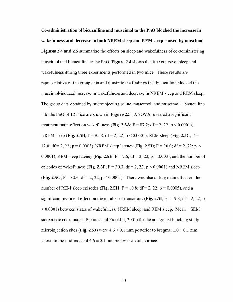
Co-administration of bicuculline and muscimol to the PnO blocked the increase in
wakefulness and decrease in both NREM sleep and REM sleep caused by muscimol
Figures 2.4 and 2.5 summarize the effects on sleep and wakefulness of co-administering
muscimol and bicuculline to the PnO. Figure 2.4 shows the time course of sleep and
wakefulness during three experiments performed in two mice. These results are
representative of the group data and illustrate the findings that bicuculline blocked the
muscimol-induced increase in wakefulness and decrease in NREM sleep and REM sleep.
The group data obtained by microinjecting saline, muscimol, and muscimol + bicuculline
into the PnO of 12 mice are shown in Figure 2.5. ANOVA revealed a significant
treatment main effect on wakefulness (Fig. 2.5A; F = 87.2; df = 2, 22; p < 0.0001),
NREM sleep (Fig. 2.5B; F = 85.8; df = 2, 22; p < 0.0001), REM sleep (Fig. 2.5C; F =
12.0; df = 2, 22; p = 0.0003), NREM sleep latency (Fig. 2.5D; F = 20.0; df = 2, 22; p <
0.0001), REM sleep latency (Fig. 2.5E; F = 7.6; df = 2, 22; p = 0.003), and the number of
episodes of wakefulness (Fig. 2.5F; F = 30.3; df = 2, 22; p < 0.0001) and NREM sleep
(Fig. 2.5G; F = 30.6; df = 2, 22; p < 0.0001). There was also a drug main effect on the
number of REM sleep episodes (Fig. 2.5H; F = 10.8; df = 2, 22; p = 0.0005), and a
significant treatment effect on the number of transitions (Fig. 2.5I; F = 19.8; df = 2, 22; p
< 0.0001) between states of wakefulness, NREM sleep, and REM sleep. Mean ± SEM
stereotaxic coordinates (Paxinos and Franklin, 2001) for the antagonist blocking study
microinjection sites (Fig. 2.5J) were 4.6 ± 0.1 mm posterior to bregma, 1.0 ± 0.1 mm
lateral to the midline, and 4.6 ± 0.1 mm below the skull surface.
50
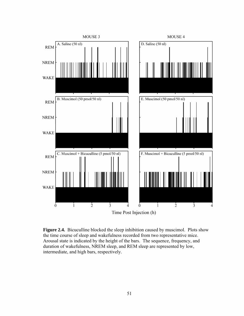
Figure 2.4. Bicuculline blocked the sleep inhibition caused by muscimol. Plots show the time course of sleep and wakefulness recorded from two representative mice. Arousal state is indicated by the height of the bars. The sequence, frequency, and duration of wakefulness, NREM sleep, and REM sleep are represented by low, intermediate, and high bars, respectively.
51

Figure 2.5. PnO microinjection of bicuculline (5 pmol/50 nl) blocked the increase in wakefulness, decrease in NREM sleep, and decrease in REM sleep caused by muscimol (50 pmol/50 nl). A-I, asterisks indicate a significant (p < 0.05) difference from control (saline). J, a coronal diagram at 4.60 mm posterior to bregma was modified from a mouse brain atlas (Paxinos and Franklin, 2001) to indicate the mean ± SEM location of 12 histologically confirmed microinjection (black dot and bars in left PnO).
52
53
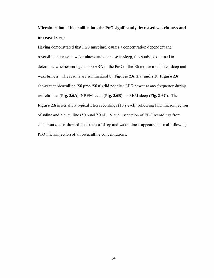
Microinjection of bicuculline into the PnO significantly decreased wakefulness and
increased sleep
Having demonstrated that PnO muscimol causes a concentration dependent and
reversible increase in wakefulness and decrease in sleep, this study next aimed to
determine whether endogenous GABA in the PnO of the B6 mouse modulates sleep and
wakefulness. The results are summarized by Figures 2.6, 2.7, and 2.8. Figure 2.6
shows that bicuculline (50 pmol/50 nl) did not alter EEG power at any frequency during
wakefulness (Fig. 2.6A), NREM sleep (Fig. 2.6B), or REM sleep (Fig. 2.6C). The
Figure 2.6 insets show typical EEG recordings (10 s each) following PnO microinjection
of saline and bicuculline (50 pmol/50 nl). Visual inspection of EEG recordings from
each mouse also showed that states of sleep and wakefulness appeared normal following
PnO microinjection of all bicuculline concentrations.
54

Figure 2.6. EEG power was not altered by PnO microinjection of bicuculline. Graphs plot average (n = 6 mice) EEG power during A, wakefulness, B, NREM sleep, and C, REM sleep following PnO microinjection of saline (gray line) and bicuculline (blue line). Cortical EEG power did not differ following PnO microinjection of bicuculline or saline. The insets show representative pairs of EEG recordings from the same mouse and illustrate the indistinguishable signals following microinjection of bicuculline and saline.
55

Plotting the temporal distribution of sleep and wakefulness from two
representative mice following microinjection of saline (Fig. 2.7A and 2.7C) and
bicuculline (50 pmol/50 nl; Fig. 2.7B and 2.7D) shows that bicuculline decreased the
amount of wakefulness and increased the amount of NREM sleep and REM sleep. The
group data (n = 11 mice) are summarized by Figure 2.8. Bicuculline caused a
significant, concentration dependent decrease in the amount of wakefulness (Fig. 2.8A; F
= 3.0; df = 3, 27; p = 0.05) and increase in the amount of NREM sleep (Fig. 2.8B; F =
2.9; df = 3, 27; p = 0.05) and REM sleep (Fig. 2.8C; F = 7.9; df = 3, 27; p = 0.0006).
Histological analyses revealed that all bicuculline microinjections were made within the
PnO. Mean ± SEM stereotaxic coordinates for the bicuculline microinjection sites (Fig.
2.8D) were 4.6 ± 0.1 mm posterior to bregma, 0.9 ± 0.04 mm lateral to the midline, and
4.3 ± 0.1 mm below the skull surface (Paxinos and Franklin, 2001).
56

Figure 2.7. PnO microinjection of bicuculline altered sleep architecture by increasing sleep. Black bars illustrate the time course of sleep and wakefulness recorded from two mice for 4 h following PnO microinjection of A, saline and B, bicuculline (50 pmol). Bicuculline decreased the time spent in wakefulness and increased the time spent in NREM sleep and REM sleep.
57

Figure 2.8. Microinjection of bicuculline into the PnO caused a concentration dependent decrease in wakefulness and increase in NREM sleep and REM sleep. A-C, asterisks indicate a significant (p < 0.05) difference from control (0 pmol bicuculline). D, a coronal diagram at 4.60 mm posterior to bregma was modified from a mouse brain atlas (Paxinos and Franklin, 2001) with a black dot and bars indicating the mean ± SEM location of the 11 histologically confirmed PnO microinjection sites.
58

59

Microdialysis delivery of bicuculline to the PnO caused a concentration dependent
increase in ACh release in the PnO, decrease in respiratory rate, and increase in
anesthesia recovery time
Figure 2.9 illustrates the use of in vivo microdialysis to determine whether GABAA
receptors in the PnO of the B6 mouse modulate ACh release, rate of breathing, and
anesthesia recovery time. Dialysis probes were aimed for the PnO (Fig. 2.9A) and
placement was confirmed histologically. Mean ± SEM stereotaxic coordinates of the
dialysis sites in the PnO (Fig. 2.9B) were 4.8 ± 0.1 mm posterior to bregma, 1.0 ± 0.1
mm lateral to the midline, and 4.9 ± 0.1 mm below the skull surface (Paxinos and
Franklin, 2001). Dialysis with Ringer’s followed by dialysis with Ringer’s containing 1
mM bicuculline (Figs. 2.9C and 2.9D) revealed that bicuculline caused a 210% increase
in ACh release in the PnO. Control experiments (Fig. 2.9E and 2.9F) measured ACh
during dialysis with Ringer’s and demonstrated that ACh release was stable across the
125 min sampling period. Figure 2.10 summarizes the group data showing that
bicuculline caused a significant, concentration dependent increase in ACh release in the
PnO (F = 16.73; df = 5, 12; p < 0.0001).
60

61
Figure 2.9. ACh release in mouse PnO. A, a coronal diagram modified from a mouse brain atlas (Paxinos and Franklin, 2001) schematizes a microdialysis probe in the PnO. The dialysis membrane is drawn to scale (1 mm length and 0.24 mm diameter). B, vertical lines on the sagittal diagram of the mouse brain (Paxinos and Franklin, 2001) show the anterior-posterior range of the histologically confirmed microdialysis sites in the PnO. C, time course plots of ACh release in the PnO. Each bar represents the average ACh release from 3 mice. Dialysis administration of bicuculline (black bars) increased ACh release within the first 12.5 min of administration. The inset shows a chromatogram with a representative ACh peak obtained during control dialysis of the PnO. D, demonstrates that ACh measures were collapsed across time. For each experiment the first five dialysis samples shown in part C were averaged to provide control levels of ACh release (Ringer’s) and the last five samples were averaged to determine the effect of bicuculline on ACh release. E, control experiments confirmed that ACh release did not change as a function of time. Each bar represents the average ACh release from 3 mice. F, ACh release was stable during isoflurane anesthesia.

62

Figure 2.10. Blocking GABAA receptors in the PnO caused a concentration dependent increase in ACh release in the PnO. Asterisks indicate a significant difference from control (0 mM bicuculline, 100% ACh release). The concentration of bicuculline accounted for 87% of the variance in ACh release. Data are from 3 mice per concentration, for a total of 18 mice.
63
Figure 2.11 shows that bicuculline caused a significant, concentration dependent
decrease in rate of breathing (F = 6.04; df = 5, 12; p = 0.005). There was no significant
effect of bicuculline on core body temperature, which averaged 37.3 ± 0.06 ˚C across all
dialysis experiments. Upon completion of dialysis sample collection, the same mice were
used to determine the effect of PnO bicuculline on time to recovery from isoflurane
anesthesia. Figure 2.12 shows that blocking GABAA receptors in the PnO caused a
significant, concentration dependent increase in anesthesia recovery time (F = 8.67; df =
3, 8; p = 0.007). The two highest concentrations of bicuculline caused seizures followed
by death, thus measures of recovery time following PnO dialysis with 3 mM (n = 3 mice)
or 10 mM (n = 3 mice) bicuculline were not included in the final data sets. Duration of
exposure to anesthetics can influence recovery time, and the duration of isoflurane
administration was held constant at 234 ± 1 min across all experiments.
64

Figure 2.11. Bicuculline caused a concentration dependent decrease in breathing rate. Asterisks indicate a significant difference from control (0 mM bicuculline). The concentration of bicuculline accounted for 76% of the variance in breathing rate. Data are from 3 mice per concentration, for a total of 18 mice.
65

Figure 2.12. Bicuculline administered to the PnO caused a concentration dependent increase in anesthesia recovery time. Asterisks indicate a significant difference from control (0 mM bicuculline). Data are from 3 mice per concentration, for a total of 12 mice. A, the concentration of bicuculline accounted for 76% of the variance in recovery time. B, shows that total anesthesia time was held constant across all experiments.
66

DISCUSSION
Drugs that increase transmission at GABAA receptors are used extensively in clinical
practice to produce states of sleep (Winsky-Sommerer, 2009) or anesthesia (Franks,
2008). Selectively altering GABAergic transmission in specific brain regions, however,
has widely varying effects on arousal states (Vanini et al., 2010; Watson et al., 2010).
Studies using cat and rat have shown that GABAergic transmission in the PRF
contributes to generating states of sleep, wakefulness, and general anesthesia (Lydic and
Baghdoyan, 2005; Steriade and McCarley, 2005). The present study is the first to
characterize the effects of GABAergic transmission in mouse PnO on states of arousal
and traits characterizing those states. The findings demonstrate that GABAA receptors in
PnO of the B6 mouse modulate occurrence of sleep/wake states, activity of the cortical
EEG, release of ACh in the PnO, breathing rate, and time needed to regain wakefulness
following isoflurane anesthesia. The ensuing discussion considers the results in relation
to clinical implications of brain region-specific effects of GABAergic transmission on
arousal states, and evidence that endogenous GABA in the PRF promotes wakefulness.
Technical considerations
Limitations of intracranial microinjection as an approach for elucidating the behavioral
state-related roles of neurotransmitter receptors have been reviewed (Myers, 1966;
Routtenberg, 1972; Nicholson, 1985). The small size of the mouse brain presents
particular challenges for the microinjection approach because distances between
structures are short and microinjected drugs diffuse away from their injection sites into
neighboring brain regions (Edeline et al., 2002). Compensatory measures include use of
67

small injection volumes and selection of relatively large brain nuclei as targets. With
these approaches, the B6 mouse has been used successfully for microinjection studies to
characterize drug effects on behavioral states and their physiological traits (Lydic et al.,
2002; Coleman et al., 2004b; Douglas et al., 2005; Van Dort et al., 2009).
Limitations of microdialysis for drug delivery (Boschi and Scherrmann, 2000)
and recovery of endogenous neurotransmitters (Watson et al., 2006) are well known.
One disadvantage of microdialysis compared to microinjection is the inability to
determine exactly how much drug is administered to the brain. This limitation can be
addressed by demonstrating concentration response relationships for the effects of
receptor agonists and antagonists on transmitter levels, as has been done successfully in
the PnO (Douglas et al., 2001; Coleman et al., 2004a; Coleman et al., 2006) and
prefrontal cortex (Van Dort et al., 2009) of B6 mouse.
GABAergic transmission causes brain region specific effects on sleep and
wakefulness
This study is the first to demonstrate that microinjecting muscimol into the PnO of B6
mouse caused a concentration-dependent increase in wakefulness and decrease in sleep
(Figs. 2.2 & 2.3) that was blocked by co-administration of bicuculline (Figs. 2.4 & 2.5).
The data indicate that these changes in sleep and wakefulness are mediated by GABAA
receptors localized to the PnO. Additionally, by showing that microinjection of
bicuculline caused a concentration dependent decrease in wakefulness and increase in
sleep (Figs. 2.7 & 2.8), this study provides the first in vivo evidence that endogenous
GABA in B6 mouse PnO promotes wakefulness and inhibits sleep.
68

Preclinical studies using intracranial drug administration have revealed that the
effects of GABAA receptor agonists and antagonists on sleep and wakefulness vary
significantly as a function of brain region. Within cat hypothalamus, the GABAA
receptor agonist muscimol induces sleep onset with short latency and increases sleep time
when microinjected into posterior hypothalamus, but increases wakefulness and
decreases sleep time when microinjected into preoptic and anterior hypothalamic areas
(Lin et al., 1989). Opposite effects of muscimol on sleep and wakefulness also can be
evoked from within the pontine portion of the brainstem. REM sleep is increased by
microinjecting muscimol into cat dorsal raphé nucleus (Nitz and Siegel, 1997) and
inhibited by delivering muscimol to the PRF of cat (Xi et al., 1999), rat (Camacho-
Arroyo et al., 1991), and mouse (Figs. 2.2-2.5). Studies using intracranial administration
of drugs that block transmission at GABAA receptors, such as bicuculline (Figs. 2.7 &
2.8) (Camacho-Arroyo et al., 1991; Xi et al., 1999; Sanford et al., 2003), gabazine (Marks
et al., 2008), and picrotoxin (Kaur et al., 1997; Nitz and Siegel, 1997; Ali et al., 1999; Pal
and Mallick, 2009), provide convincing evidence that endogenous GABA differentially
alters sleep and wakefulness depending upon brain site of action. One unifying
explanation for these findings is that in brain regions that function to generate
wakefulness, GABAergic transmission causes sleep, whereas in brain regions that
generate sleep GABAergic transmission increases wakefulness.
The discovery that effects of GABAergic transmission on arousal states are
anatomically site-specific within the brain provides insight into how systemically
administered benzodiazepines produce sleep architecture that differs from that of
spontaneously occurring sleep. Benzodiazepine hypnotics increase total sleep time and
69

shorten sleep latency, but some of these drugs increase REM sleep latency (Borbély and
Achermann, 1991). Drug actions at the level of the PRF, where GABAergic transmission
increases wakefulness, decreases sleep, and inhibits ACh release may account for the
increased REM latency caused by some benzodiazepine hypnotics.
Muscimol but not bicuculline caused state-trait dissociations
States of wakefulness, NREM sleep, and REM sleep can occur in a humbling complexity
of admixtures that combine traits characteristic of one state with traits typical of a
completely different state. These mixed states are referred to as dissociated states and
represent a significant clinical concern (Mahowald and Schenck, 1992, 2001). The
present study found that direct administration of muscimol into the PnO caused a
dissociation between waking behavior and EEG activity. During the increase in
wakefulness caused by muscimol (500 pmol) mice showed a marked increase in running
activity yet significantly greater EEG power in the delta range (0.5-5 Hz) (Fig. 2.1).
Increased delta activity normally occurs during NREM sleep which is characterized by
decreased motor activity and a loss of consciousness. A previous study that administered
muscimol to B6 mice by intraperitoneal injection reported periods of vigorous wheel
running activity during which the frontal EEG was dominated by high amplitude slow
waves characteristic of NREM sleep (Vyazovskiy et al., 2007). The finding that the
slowing of the EEG caused by systemic administration of muscimol also occurs
following muscimol delivery directly into the PnO supports the interpretation that the
increase in delta power caused by intraperitoneal muscimol is mediated, at least in part,
by GABAA receptors in the PnO. This interpretation is consistent with data showing that
70

the benzodiazepine receptor agonist hypnotics zolpidem and diazepam increase EEG
delta power when administered to the PnO of isoflurane anesthetized rat (Hambrecht et
al., 2009).
PnO microinjection of muscimol significantly decreased the amount of time
spent in REM sleep, and the REM sleep that did occur following muscimol (50 pmol)
administration was characterized by increased EEG power in the theta range (4-9 Hz)
(Fig. 2.1). Theta activity normally is prominent during REM sleep and increases during
wakefulness when animals are exploring their environment (Poe et al., 2009). Although
the mechanisms underlying GABA-induced state-trait dissociation are not known, the
phenomenon of paradoxical activation by benzodiazepines has been documented by
multiple case reports (Paton, 2002; Brefel-Courbon et al., 2007; Schiff and Posner, 2007;
Hall et al., 2008). Efforts to synthesize these case reports have led to a network model by
which the benzodiazepine receptor agonist zolpidem might produce EEG and behavioral
activation (Schiff, 2008, 2010). The present results expand this network model by
showing that increasing GABAergic transmission in the PRF may be another mechanism
through which hypnotics produce state dissociations.
GABAA receptors in the PRF function to inhibit local ACh release, stimulate
breathing, and decrease anesthesia recovery time: implications for arousal state
control
Dialysis delivery of bicuculline to the PnO caused a concentration dependent increase in
ACh release (Fig. 2.10). Bicuculline also increases ACh release in cat PRF (Vazquez
and Baghdoyan, 2004), and these findings support the conclusion that one functional role
71

of endogenous GABA in the PnO is to inhibit ACh release. Extensive evidence
demonstrates that cholinergic transmission within the PRF promotes REM sleep (Lydic
and Baghdoyan, 2008), and the increase in REM sleep caused by blocking GABAA
receptors in the PnO can be prevented by pretreating the PnO with the muscarinic
receptor antagonist atropine (Marks et al., 2008). Considered together, these findings
suggest that GABAergic tone in the PRF normally suppresses REM sleep, in part by
inhibiting PRF ACh release.
The finding that human rate of breathing declines significantly during the loss of
wakefulness caused by anesthesia (Fink, 1961) or sleep (Phillipson and Sullivan, 1978)
initiated ongoing efforts to identify cellular and molecular substrates comprising a
wakefulness stimulus for breathing. Although the PRF does not contain neurons with
respiratory related discharge patterns, breathing is significantly altered by PRF neurons
that regulate EEG and behavioral arousal (Lydic and Baghdoyan, 1993). Blockade of
GABAA receptors in the PnO with bicuculline decreased rate of breathing (Fig. 2.11).
This finding implies that endogenous GABA in the PRF stimulates breathing, and
suggests that GABAergic transmission in the PnO contributes to the wakefulness
stimulus for breathing. Consistent with this interpretation is the finding that bicuculline
delivered to the PnO delayed recovery time from isoflurane anesthesia (Fig. 2.12A). In
rat, administering drugs that decrease or increase GABAergic transmission in the PnO
shorten or lengthen, respectively, the time required for isoflurane to induce loss of
consciousness (Vanini et al., 2008). Induction of and emergence from general anesthesia
are mechanistically distinct processes (Kelz et al., 2008), and the present data are the first
in any species to show that inhibition of GABAA receptors in the PnO increases
72

anesthesia recovery time. Taken together, the findings of increased ACh release,
decreased breathing rate, and increased anesthesia recovery time caused by blocking PnO
GABAA receptors provide novel evidence that GABAergic transmission in the PnO
promotes wakefulness.
73

REFERENCES
Ali M, Jha SK, Kaur S, Mallick BN (1999) Role of GABA-A receptor in the preoptic
area in the regulation of sleep-wakefulness and rapid eye movement sleep. Neurosci Res 33:245-250.
Boissard R, Gervasoni D, Schmidt MH, Barbagli B, Fort P, Luppi PH (2002) The rat
ponto-medullary network responsible for paradoxical sleep onset and maintenance: a combined microinjection and functional neuroanatomical study. Eur J Neurosci 16:1959-1973.
Borbély AA, Achermann P (1991) Ultradian dynamics of sleep after a single dose of
benzodiazepine hypnotics. Eur J Pharmacol 195:11-18. Boschi G, Scherrmann J-M (2000) Microdialysis in mice for drug delivery research. Adv
Drug Deliv Rev 45:271-281. Bradley A (2002) Mining the mouse genome. Nature 420:512-514. Brefel-Courbon C, Payoux P, Ory F, Sommet A, Slaoui T, Raboyeau G, Lemesle B, Puel
M, Montastruck J-L, Cardebat D (2007) Clinical and imaging evidence of zolpidem effect in hypoxic encephalopathy. Ann Neurol 62:102-105.
Camacho-Arroyo I, Alvarado R, Manjarrez J, Tapia R (1991) Microinjections of
muscimol and bicuculline into the pontine reticular formation modify the sleep-waking cycle in the rat. Neurosci Lett 129:95-97.
Coleman CG, Lydic R, Baghdoyan HA (2004a) M2 muscarinic receptors in pontine
reticular formation of C57BL/6J mouse contribute to rapid eye movement sleep generation. Neuroscience 126:821-830.
Coleman CG, Lydic R, Baghdoyan HA (2004b) Acetylcholine release in the pontine
reticular formation of C57BL/6J mouse is modulated by non-M1 muscarinic receptors. Neuroscience 126:831-838.
Coleman CG, Baghdoyan HA, Lydic R (2006) Dialysis delivery of an adenosine A2A
agonist into the pontine reticular formation of C57BL/6J mouse increases pontine acetylcholine release and sleep. J Neurochem 96:1750-1759.
Colten HR, Altevogt BM, Institute of Medicine (U.S.). Committee on Sleep Medicine
and Research. (2006) Sleep Disorders and Sleep Deprivation : An Unmet Public Health Problem. Washington, DC: Institute of Medicine : National Academies Press.
74

Datta S, Maclean RR (2007) Neurobiological mechanisms for the regulation of mammalian sleep-wake behavior: reinterpretation of historical evidence and inclusion of contemporary cellular and molecular evidence. Neurosci Biobehav Rev 31:775-824.
Douglas CL, Baghdoyan HA, Lydic R (2001) M2 muscarinic autoreceptors modulate
acetylcholine release in prefrontal cortex of C57BL/6J mouse. J Pharmacol Exp Ther 299:960-966.
Douglas CL, Bowman GN, Baghdoyan HA, Lydic R (2005) C57BL/6J and B6.V-LEPOB
mice differ in the cholinergic modulation of sleep and breathing. J Appl Physiol 98:918-929.
Edeline J-M, Hars B, Hennevin E, Cotillon N (2002) Muscimol diffusion after
intracerebral microinjections: a reevaluation based on electrophysiological and autoradiographic quantifications. Neurobiol Learn Mem 78:100-124.
Fink BR (1961) Influence of cerebral activity in wakefulness on regulation of breathing. J
Appl Physiol 16:15-20. Flint RR, Lydic R, Baghdoyan HA (2007) Microinjection of the GABAA receptor agonist
muscimol into the pontine reticular nucleus, oral part (PnO) of C57BL/6J (B6) mouse increases wakefulness and decreases sleep. Sleep 30:0006.
Flint RR, Lydic R, Baghdoyan HA (2007) Microinjection of the GABAA receptor agonist
muscimol into the pontine reticular nucleus, oral part (PnO) of C57BL/6J (B6) mouse causes a concentration dependent increase in wakefulness and decrease in sleep. Neuroscience Meeting Planner Program No. 631.5.
Flint RR, Lydic R, Baghdoyan HA (2008) In C57BL/6J (B6) mouse the GABAA receptor
antagonist bicuculline blocks the increase in wakefulness and decrease in sleep caused by the GABAA receptor agonist muscimol. Neuroscience Meeting Planner Program No. 285.15.
Flint RR, Lydic R, Baghdoyan HA (2009) Electroencephalogram (EEG) power during
wakefulness and rapid eye movement (REM) sleep is increased by microinjection of the GABAA receptor agonist muscimol into the pontine reticular nucleus, oral part (PnO) of C57BL/6J (B6) mouse. Sleep 32:0014.
Flint RR, Lydic R, Baghdoyan HA (2009) Microdialysis delivery of the GABAA receptor
antagonist bicuculline to the pontine reticular formation (PRF) of C57BL/6J (B6) mouse increases PRF acetylcholine (ACh) release. Neuroscience Meeting Planner Program No. 375.12.
Franken P, Malafosse A, Tafti M (1999) Genetic determinants of sleep regulation in
inbred mice. Sleep 22:155-169.
75

Franken P, Chollet D, Tafti M (2001) The homeostatic regulation of sleep need is under
genetic control. J Neurosci 21:2610-2621. Franks NP (2008) General anaesthesia: from molecular targets to neuronal pathways of
sleep and arousal. Nat Rev Neurosci 9:370-386. Hall SD, Yamawaki N, Fisher AE, Clauss RP, Woodhall GL, Stanford IM (2008)
Desynchronization of pathological low-frequency brain activity by the hypnotic drug zolpidem. Nature Precedings hdl:10101/npre.2008.1966.1.
Hambrecht VS, Baghdoyan HA, Lydic R (2009) Pontine reticular formation (PRF)
administration of diazepam and zolpidem, but not eszopiclone, increases electroencephalogram (EEG) delta power in Sprague-Dawley rat. Sleep 32 (Abstr Suppl):0028.
Hobson JA, Pace-Schott EF (2002) The cognitive neuroscience of sleep: neuronal
systems, consciousness and learning. Nat Rev Neurosci 3:679-693. Katzenberg D, Young T, Finn L, Lin L, King DP, Takahashi JS, Mignot E (1998) A
CLOCK polymorphism associated with human diurnal preference. Sleep 21:569-576.
Kaur S, Saxena RN, Mallick BN (1997) GABA in locus coeruleus regulates spontaneous
rapid eye movement sleep by acting on GABAA receptors in freely moving rats. Neurosci Lett 223:105-108.
Kelz MB, Sun Y, Chen J, Cheng Meng Q, Moore JT, Veasey SC, Dixon S, Thornton M,
Funato H, Yanagisawa M (2008) An essential role for orexins in emergence from general anesthesia. Proc Natl Acad Sci U S A 105:1309-1314.
Leger D, Guilleminault C, Bader G, Levy E, Paillard M (2002) Medical and socio-
professional impact of insomnia. Sleep 25:625-629. Lin JS, Sakai K, Vanni-Mercier G, Jouvet M (1989) A critical role of the posterior
hypothalamus in the mechanisms of wakefulness determined by microinjection of muscimol in freely moving cats. Brain Res 479:225-240.
Lydic R, Baghdoyan HA (1993) Pedunculopontine stimulation alters respiration and
increases ACh release in the pontine reticular formation. Am J Physiol 264:R544-554.
Lydic R, Baghdoyan HA (2005) Sleep, anesthesiology, and the neurobiology of arousal
state control. Anesthesiology 103:1268-1295.
76

Lydic R, Baghdoyan HA (2008) Acetylcholine modulates sleep and wakefulness: a synaptic perspective. In: Neurochemistry of Sleep and Wakefulness (Monti JM, Pandi-Perumal SR, Sinton CW, eds), pp 109-143. Cambridge: Cambridge University Press.
Lydic R, Douglas CL, Baghdoyan HA (2002) Microinjection of neostigmine into the
pontine reticular formation of C57BL/6J mouse enhances rapid eye movement sleep and depresses breathing. Sleep 25:835-841.
Mahowald MW, Schenck CH (1992) Dissociated states of wakefulness and sleep.
Neurology 42:44-51; discussion 52. Mahowald MW, Schenck CH (2001) Evolving concepts of human state dissociation.
Arch Ital Biol 139:269-300. Manfridi A, Brambilla D, Mancia M (2001) Sleep is differentially modulated by basal
forebrain GABAA and GABAB receptors. Am J Physiol Regulatory Integrative Comp Physiol 281:R170-R175.
Marks GA, Sachs OW, Birabil CG (2008) Blockade of GABA, type A, receptors in the
rat pontine reticular formation induces rapid eye movement sleep that is dependent upon the cholinergic system. Neuroscience 156:1-10.
Myers RD (1966) Injection of solutions into cerebral tissue: relation between volume and
diffusion. Physiol Behav 1:171-174. Nicholson C (1985) Diffusion from an injected volume of a substance in brain tissue with
arbitrary volume fraction and tortuosity. Br Res 333:325-329. Nitz D, Siegel J (1997) GABA release in the dorsal raphe nucleus: role in the control of
REM sleep. Am J Physiol 273:R451-455. O'Brien SJ, Menotti-Raymond M, Murphy WJ, Nash WG, Wienberg J, Stanyon R,
Copeland NG, Jenkins NA, Womack JE, Marshall Graves JA (1999) The promise of comparative genomics in mammals. Science 286:458-462, 479-481.
Opp MR (1998) Rat strain differences suggest a role for corticotropin-releasing hormone
in modulating sleep. Physiol Behav 63:67-74. Paigen K, Eppig JT (2000) A mouse phenome project. Mamm Genome 11:715-717. Pal D, Mallick BN (2009) GABA in pedunculopontine tregmentum increases rapid eye
movement sleep in freely moving rats: possible role of GABA-ergic inputs from substania nigra pars reticulata. Neuroscience 164:404-414.
77

Paton C (2002) Benzodiazepines and disinhibition: a review. Psychiatric Bulletin 26:460-462.
Paxinos G, Franklin KBJ (2001) The Mouse Brain in Stereotaxic Coordinates, 2nd
Edition. San Diego: Academic Press. Phillipson EA, Sullivan C (1978) Arousal: The forgotten response to respirator stimuli.
Am Rev Resp Dis 118:807-809. Poe GR, Booth V, Bjorness TE, Riley BT, Watts AC (2009) Sleep is for unfinished
business. Growing evidence that sleep is important for learning and memory. In: Current Advances in Sleep Biology (Frank MG, ed), pp 141-176: Nova Science Publishers, Inc.
Pollock MS, Mistlberger RE (2003) Rapid eye movement sleep induction by
microinjection of the GABA-A antagonist bicuculline into the dorsal subcoeruleus area of the rat. Brain Res 962:68-77.
Routtenberg A (1972) Intracranial chemical injection and behavior: a critical review.
Behav Biol 7:601-641. Sallanon M, Denoyer M, Kitahama K, Aubert C, Gay N, Jouvet M (1989) Long-lasting
insomnia induced by preoptic neuron lesions and its transient reversal by muscimol injection into the posterior hypothalamus in the cat. Neuroscience 32:669-683.
Sanford LD, Tang X, Xiao J, Ross RJ, Morrison AR (2003) GABAergic regulation of
REM sleep in reticularis pontis oralis and caudalis in rats. J Neurophysiol 90:938-945.
Sapin E, Lapray D, Berod A, Goutagny R, Leger L, Ravassard P, Clement O, Hanriot L,
Fort P, Luppi PH (2009) Localization of the brainstem GABAergic neurons controlling paradoxical (REM) sleep. PLoS One 4:e4272.
Sastre JP, Buda C, Kitahama K, Jouvet M (1996) Importance of the ventrolateral region
of the periaqueductal gray and adjacent tegmentum in the control of paradoxical sleep as studied by muscimol microinjections in the cat. Neuroscience 74:415-426.
Schiff ND (2008) Central thalamic contributions to arousal regulation and neurological
disorders of consciousness. Ann NY Acad Sci 1129:105-118. Schiff ND (2010) Recovery of consciousness after brain injury: a mesocircuit hypothesis.
Trends Neurosci 33:1-9. Schiff ND, Posner JB (2007) Another "Awakenings". Ann Neurol 62:5-6.
78

Silver LM (1995) Mouse Genetics : Concepts and Applications. New York: Oxford
University Press. Stenberg D (2007) Neuroanatomy and neurochemistry of sleep. Cell Mol Life Sci
64:1187-1204. Steriade M, McCarley RW (2005) Brain Control of Wakefulness and Sleep, 2nd Edition.
New York: Kluwer Academic/Plenum Publishers. Tang X, Xiao J, Parris BS, Fang J, Sanford LD (2005) Differential effects of two types of
environmental novelty on activity and sleep in BALB/cJ and C57BL/6J mice. Physiol Behav 85:419-429.
Ting L, Malhotra A (2005) Disorders of sleep: an overview. Prim Care 32:305-318. Toh KL, Jones CR, He Y, Eide EJ, Hinz WA, Virshup DM, Ptacek LJ, Fu YH (2001) An
hPer2 phosphorylation site mutation in familial advanced sleep phase syndrome. Science 291:1040-1043.
Van Dort CJ, Baghdoyan HA, Lydic R (2009) Adenosine A1 and A2A receptors in mouse
prefrontal cortex modulate acetylcholine release and behavioral arousal. J Neurosci 29:871-881.
Vanini G, Lydic R, Baghdoyan HA (2010) GABAergic modulation of REM sleep. In:
Rapid Eye Movement Sleep – Regulation and Function (Mallick BN, Pandi-Perumal R, McCarley RW, Morrison AR, eds), p in press: Cambridge University Press.
Vanini G, Watson CJ, Lydic R, Baghdoyan HA (2008) Gamma-aminobutyric acid-
mediated neurotransmission in the pontine reticular formation modulates hypnosis, immobility, and breathing during isoflurane anesthesia. Anesthesiology 109:978-988.
Vanini G, Wathen BL, Lydic R, Baghdoyan HA (2009) GABA levels in cat pontine
reticular formation (PRF) are lower during rapid eye movement (REM) sleep and the neostigmine-induced REM sleep-like state (REM-Neo) than during wakefulness. Sleep 32 (Abstr Suppl):0011.
Vanini G, Torterolo P, McGregor R, Chase MH, Morales FR (2007) GABAergic
processes in the mesencephalic tegmentum modulate the occurrence of active (rapid eye movement) sleep in guinea pigs. Neuroscience 145:1157-1167.
Vazquez J, Baghdoyan HA (2004) GABAA receptors inhibit acetylcholine release in cat
pontine reticular formation: implications for REM sleep regulation. J Neurophysiol 92:2198-2206.
79

Vyazovskiy VV, Tobler I, Winsky-Sommerer R (2007) Alteration of behavior in mice by
muscimol is associated with regional electroencephalogram synchronization. Neuroscience 147:833-841.
Walsh JK, Engelhardt CL (1999) The direct economic costs of insomnia in the United
States for 1995. Sleep 22 Suppl 2:S386-393. Waterston RH et al. (2002) Initial sequencing and comparative analysis of the mouse
genome. Nature 420:520-562. Watson CJ, Venton BJ, Kennedy RT (2006) In vivo measurements of neurotransmitters
by microdialysis sampling. Anal Chem 78:1391-1399. Watson CJ, Lydic R, Baghdoyan HA (2007) Sleep and GABA levels in the oral part of
rat pontine reticular formation are decreased by local and systemic administration of morphine. Neuroscience 144:375-386.
Watson CJ, Baghdoyan HA, Lydic R (2010) A neurochemical perspective on states of
consciousness. In: Suppressing the Mind: Anesthetic Modulation of Memory and Consciousness (Hudetz A, Pierce R, eds), pp 33-80. New York: Springer.
Watson CJ, Soto-Calderon H, Lydic R, Baghdoyan HA (2008) Pontine reticular
formation (PnO) administration of hypocretin-1 increases PnO GABA levels and wakefulness. Sleep 31:453-464.
Wilson JF (2005) Is sleep the new vital sign? Ann Int Med 142:8-880. Winsky-Sommerer R (2009) Role of GABAA receptors in the physiology and
pharmacology of sleep. Eur J Neurosci 29:1779-1794. Xi MC, Morales FR, Chase MH (1999) Evidence that wakefulness and REM sleep are
controlled by a GABAergic pontine mechanism. J Neurophysiol 82:2015-2019.
80
CHAPTER 3
SUMMARY AND CONCLUSIONS
Many drugs that produce both sleep and anesthesia act on GABAA receptors, but
the mechanisms underlying their effects remain largely unknown. The pontine reticular
nucleus, oral part (PnO) is a brain region long known for contributing to arousal state
control (Jouvet, 1962; Lydic and Baghdoyan, 2005). This dissertation research utilized a
pharmacological approach to investigate the role of GABAA receptors in the PnO in the
regulation of five arousal phenotypes: sleep and wakefulness, cortical EEG activity, ACh
release in the PnO, respiratory rate, and recovery from anesthesia. The results of this
research are consistent with the interpretation that GABAA receptors in the PnO of B6
mouse promote wakefulness (Flint et al., 2007; Flint et al., 2007; Flint et al., 2008; Flint
et al., 2009; Flint et al., 2009). Involvement of GABAA receptors in the PnO in
regulating arousal are discussed in the context of these results using a proposed synaptic
model.
The studies in Chapter 2 were designed to study the effects of GABAergic
transmission in the PnO on ACh release in the PnO. The data demonstrated that
administration of the GABAA receptor agonist muscimol to the PnO increased cortical
EEG activity during wakefulness and REM sleep (Fig. 2.1). Administration of muscimol
to the PnO also increased wakefulness and decreased sleep (Figs. 2.2 and 2.3). Co-
administration of the GABAA receptor antagonist bicuculline reversed the effects of
81

muscimol on wakefulness and sleep (Figs. 2.4 and 2.5). Bicuculline administered to the
PnO decreased wakefulness and increased sleep (Figs. 2.7 and 2.8). Microdialysis
delivery of bicuculline to the PnO increased ACh release in the PnO (Fig. 2.10),
decreased respiratory rate (Fig. 2.11), and increased the recovery of righting following
isoflurane anesthesia (Fig. 2.12A). The data in Chapter 2 are novel in that they
characterize for the first time, the role of GABAA receptors in the phenotypes of arousal
in the mouse PnO. These data support the conclusion that GABA in the pontine reticular
formation (PRF) promotes wakefulness, and is consistent with studies done in cat (Xi et
al., 1999; Vazquez and Baghdoyan, 2004) and rat (Camacho-Arroyo et al., 1991; Sanford
et al., 2003; Marks et al., 2008). These consistencies across species suggest that the
wakefulness promoting effects of GABAA receptors in the PRF are somewhat conserved,
making it possible to ultimately make comparisons between species as they relate to
arousal state.
Both anatomical and functional studies have determined that cholinergic
projections to the PRF originate in the LDT/PPT (Mitani et al., 1988; Shiromani et al.,
1988; Jones, 1990; Lydic and Baghdoyan, 1993; Semba, 1993) (Table 3.2). Electrical
stimulation of LDT neurons increases REM sleep (Thakkar et al., 1996), and ACh release
in the PRF increases during REM sleep (Kodama et al., 1990; Lydic et al., 1991; Leonard
and Lydic, 1995, 1997). The PRF also contains GABAergic inter-neurons (Ford et al.,
1995) (Table 3.1). Studies in cat and rat show that increasing GABAergic transmission
in the PRF increases wakefulness and decreases sleep (Camacho-Arroyo et al., 1991; Xi
et al., 1999; Sanford et al., 2003; Watson et al., 2008). One interpretation for the results
in Chapter 2 showing that muscimol administered to the PnO increased wakefulness and
82

decreased sleep is that GABAergic transmission in the PRF inhibits the release of ACh
from LDT/PPT terminals (Fig. 3.1A&B), and therefore produces wakefulness. In
contrast, the blockade of GABAA receptors in the PRF with bicuculline allows for the
release of ACh from LDT/PPT terminals (Fig 3.1A&B) and leads to REM sleep. The
PRF also contains glutamatergic neurons that project to the LDT/PPT (Kaneko et al.,
1989; Steininger et al., 1992; Lai et al., 1993) (Table 3.3). GABAergic transmission in
the PRF could inhibit the ability of LDT/PPT cholinergic neurons to be excited by
glutamate (Fig. 3.1A&C), thereby decreasing ACh release in the PRF and decreasing
REM sleep. Blockade of GABAA receptors in the PRF would allow for glutamatergic
excitation of ACh neurons in the LDT/PPT (Fig. 3.1A&C) to cause ACh release in the
PRF, and REM sleep.
In Chapter 2, PnO dialysis with bicuculline was shown to increase ACh release
in the PnO. Along with data in cat demonstrating that the blockade of GABAA receptors
in the PRF increases REM sleep (Vazquez and Baghdoyan, 2004) (Table3.1), this finding
is consistent with the interpretation that GABA in the PRF suppresses REM sleep, at least
in part, by inhibiting ACh release in the PRF. Two possible mechanisms could be
contributing to the increase in ACh release caused by blocking GABAA receptors in the
PRF. First, bicuculline could block GABA inhibition of cholinergic terminals (Fig. 3.1B)
and thus result in an increase of ACh release in the PRF. Second, the normal inhibition
of glutamate neurons in the PRF by GABA (Fig. 3.1C) could be prevented by
bicuculline, and glutamatergic excitation of LDT/PPT ACh neurons (Fig. 3.1A&C)
would lead to an increase in ACh release in the PRF.
83

Breathing is altered by PRF neurons that regulate arousal despite the fact that
the PRF contains no neurons with respiratory related discharge patterns (Lydic and
Baghdoyan, 1993). Electrical stimulation of the PPT increases ACh release in the PRF
and causes respiratory rate depression similar to PRF microinjection of carbachol (Lydic
and Baghdoyan, 1993) (Table 3.2). Increasing GABA levels in the PnO increases
respiration rate (Vanini et al., 2008) (Table 3.1). The finding in Chapter 2 that
respiratory rate decreased following bicuculline administration to the PRF implies that
GABA in the PRF stimulates breathing. Based on these findings, one interpretation for
the decrease in breathing is that bicuculline prevents the inhibition of GABA on
glutamate release (Fig. 3.1B), allowing glutamate to excite ACh neurons in the LDT/PPT
(Fig. 3.1C) and depress breathing.
Drugs that alter GABA levels by inhibiting GABA uptake (NPA) or GABA
synthesis (3-MPA) have been studied in relation to states of arousal. Microinjection of
NPA into the PnO increases wakefulness and decreases sleep, whereas 3-MPA delivered
to the PnO increases sleep and decreases wakefulness (Watson et al., 2008) (Table 3.1).
Increasing or decreasing GABA levels in the PnO increases or decreases, respectively,
time to loss of righting (Vanini et al., 2008) (Table 3.1). Bicuculline delivered to the PnO
increases time to recovery of righting after anesthesia (Fig. 2.12A). Taken together, these
data also support the interpretation that GABA in the PnO promotes wakefulness. Based
on this interpretation, the bicuculline-mediated increase in recovery time could occur by
blockade of the wakefulness promoting effects of GABA in the PRF (Fig. 3.1B&C).
84

GABA
These dissertation results support the interpretation that GABA in the PRF
promotes wakefulness. Within the PRF are GABA neurons and receptors that play a key
role in arousal state control (Table 3.1). The PRF has been shown to contain local and
distant projecting GABAergic neurons (Ford et al., 1995; de la Roza and Reinoso-Suarez,
2006; Brown et al., 2008; Rodrigo-Angulo et al., 2008; Liang and Marks, 2009) as well
as GABAA receptors (Waldvogel et al.; Fritschy and Mohler, 1995; Pirker et al., 2000;
Heldt and Ressler, 2007) (Fig. 3.1B&C). GABAergic neurons are also present in the
LDT/PPT (Jia et al., 2003; Boucetta and Jones, 2009; Wang and Morales, 2009).
Facilitation of GABAergic transmission in the PRF increases wakefulness and decreases
sleep (Camacho-Arroyo et al., 1991; Xi et al., 1999, 2001; Sanford et al., 2003; Watson et
al., 2008; Fig. 2.3). Furthermore, elevating GABA levels in the PnO increases time to
loss of righting as well as respiratory rate (Vanini et al., 2008). Dialysis delivery of
bicuculline to the PRF increases ACh release (Vazquez and Baghdoyan, 2004; Fig. 2.10)
and REM sleep (Xi et al., 1999, 2001; Sanford et al., 2003; Vazquez and Baghdoyan,
2004), supporting the role of both GABA and ACh in the PRF in REM sleep.
85

Table 3.1. The PRF contains GABA inter-neurons and GABAA receptors. GABA in the PRF promotes wakefulness and respiratory rate.
86

ACETYLCHOLINE
As seen in Table 3.2, cholinergic neurons and receptors provide ACh to the PRF
and play an important role in regulating sleep and wakefulness. The PRF is
cholinoceptive, receiving ACh from LDT/PPT cholinergic neurons (Jones and Beaudet,
1987; Mitani et al., 1988; Shiromani et al., 1988; Lydic and Baghdoyan, 1993; Semba,
1993) (Fig. 3.1A). Stimulation of the PPT increases both ACh release and respiratory
depression (Lydic and Baghdoyan, 1993). ACh release in the PRF is also modulated by
muscarinic receptors. Muscarinic receptors proposed to be localized either
presynaptically on LDT/PPT cholinergic terminals (Fig. 3.1B) or postsynaptically on
glutamatergic neurons (Fig. 3.1C), decrease ACh release (Baghdoyan et al., 1998;
Coleman et al., 2004a). M2 muscarinic receptors shown to be localized postsynaptically
on GABAergic neurons in the PRF (Brischoux et al., 2008; Brown et al., 2008) (Fig.
3.1B) contribute to the generation of REM sleep (Baghdoyan and Lydic, 1999; Coleman
et al., 2004b). Muscarinic receptors also modulate ACh release in the PRF (Baghdoyan
et al., 1998; Coleman et al., 2004). Taken together, the findings are consistent with the
Chapter 2 results that blockade of GABAergic transmission in the PRF increases ACh
release.
87

Table 3.2. ACh neurons in the LDT/PPT project to the PRF, which contains muscarinic receptors. ACh in the PRF modulates REM sleep.
88

GLUTAMATE
As the major excitatory neurotransmitter in the mammalian central nervous
system, glutamate acts on ionotropic NMDA, AMPA, or kainate receptors and
metabotropic glutamate receptors (Lodge, 2009). The involvement of glutamate in sleep
and wakefulness has not been well characterized, although glutamatergic neurons and
receptors have been localized to brain regions implicated in sleep-wake control such as
the PRF and LDT/PPT (Table 3.3). Within the PRF, there are glutamatergic neurons that
project to the LDT/PPT (Kaneko et al., 1989; Steininger et al., 1992; Lai et al., 1993)
(Fig. 3.1C), which also has glutamatergic neurons (Boucetta and Jones, 2009; Wang and
Morales, 2009). In addition, glutamate kainate receptors have been localized in the PPT
(Fig. 3.1A), where local glutamate produces REM sleep (Datta, 2002; Datta et al., 2002).
GABAA receptors on glutamate neurons in the PRF may inhibit glutamate excitation of
LDT/PPT cholinergic neurons and lead to a decrease in ACh release in the PRF and an
increase in wakefulness, consistent with the Chapter 2 results that GABAergic
transmission increases wakefulness and the interpretation that GABA promotes
wakefulness.
89

Table 3.3. Glutamate neurons in the PRF project to the LDT/PPT. Glutamate receptors in the PPT induce REM sleep.
90

Figure 3.1 is a synaptic model of the PRF depicting possible GABAergic,
cholinergic, and glutamatergic interactions regulating states of arousal.
91

92
Figure 3.1. Synaptic model illustrating neurons in the pontine reticular formation (PRF) of B6 mouse through which GABA may promote wakefulness. A, Laterodorsal tegmental (LDT) and pedunculopontine tegmental (PPT) cholinergic neurons (blue) provide ACh to the PRF (Jones and Beaudet, 1987; Mitani et al., 1988; Shiromani et al., 1988; Lydic and Baghdoyan, 1993; Semba, 1993). B, The PRF contains locally projecting GABA (green) neurons (Ford et al., 1995; de la Roza and Reinoso-Suarez, 2006; Brown et al., 2008). C, PRF glutamate neurons (orange) project to the LDT/PPT (Kaneko et al., 1989; Steininger et al., 1992; Lai et al., 1993). The findings in Chapter 2 that muscimol delivered to the PRF increases wakefulness and bicuculline administered to the PRF increases sleep and ACh release are consistent with the effects of muscimol and bicuculline on GABA neurons to inhibit or allow, respectively, ACh release from the LDT/PPT (B) and/or inhibit or allow, respectively, glutamate excitation of LDT/PPT cholinergic neurons (C). Inhibition or excitation of LDT/PPT cholinergic nerve terminals or neurons would result in either a decrease or increase in ACh release in the PRF and lead to wakefulness or REM sleep. The Chapter 2 results reporting an increase in recovery of righting following bicuculline administration in the PRF are consistent with the idea that blocking GABA neurons in the PRF causes ACh release by blocking inhibition of LDT/PPT ACh neurons (B) or exciting LDT/PPT ACh through PRF glutamate neurons (C). Both scenarios could lead to REM sleep and support the interpretation that GABA promotes wakefulness. One interpretation of the Chapter 2 finding that bicuculline delivered to the PRF decreases respiratory rate is that bicuculline is acting to block the inhibitory actions of GABA in the PRF on LDT/PPT ACh neurons (B), and in turn allowing ACh to be released into the PRF. When PPT neurons are stimulated they release ACh in the PRF and can cause respiratory rate depression. Plus signs (+) indicate excitatory effects and minus signs (-) indicate inhibitory effects. Abbreviations in model: ACh, cholinergic neuron; GABA, GABAergic neuron; GABAAR, GABAA receptor; GLUT, glutamate neuron; GlutR, glutamate receptor; LDT/PPT, laterodorsal tegmental and pedunculopontine tegmental; M2, M2 subtype muscarinic receptor; mAChR, muscarinic cholinergic receptor; PRF, pontine reticular formation
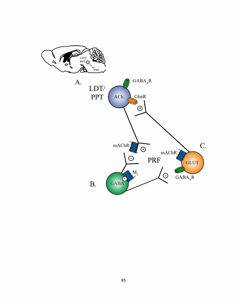
93
93

In conclusion, five novel findings were reported in this dissertation pertaining to
the effects of GABA in the PnO of B6 mouse on states and traits of arousal. These
findings showed that GABAA receptors in the PnO of B6 mouse alter states of sleep and
wakefulness, cortical EEG activity, ACh release in the PnO, respiratory rate, and
recovery of righting from anesthesia. Taken together, these results demonstrate that
GABAA receptors in the PnO of B6 mouse regulate arousal phenotypes, and that GABA
acting at GABAA receptors in the PnO promotes wakefulness.
GABAA receptors remain a major target in the pharmacological treatment of sleep
disorders such as insomnia. Despite the emergence of insomnia treatments that act on
other receptors such as melatonin, the clinical development of drugs acting to enhance
GABAergic transmission remains viable for research programs at pharmaceutical
companies such as Neurocrine Biosciences, Evotec, and Neurogen (Winsky-Sommerer,
2009). All three companies have reached at least Phase II trials with their potential
drugs, and the early clinical results encourage further exploration of their use as therapies
for insomnia in hopes that they may prevent some of the negative side effects patients
experience with current drug treatments (Lemon et al., 2009; Walsh et al., 2009).
Insomnia patients currently exhibit increases in total sleep time following systemic BDZ
administration, but often times at the expense of slow wave sleep and REM sleep
(Feinberg et al., 1979; Borbély and Achermann, 1991). Pharmacologically-induced state
dissociations can also occur (Mendelson, 2005) when the characteristic traits which make
up individual states of wakefulness, NREM sleep, and REM sleep are observed in a
completely different state (Mahowald and Schenck, 1992, 2001). Interestingly, my
research showed that increasing GABAergic transmission in the PnO produced decreases
94

in NREM sleep and REM sleep as well as state dissociations (Chapter 2). Potential
effects of increasing GABAergic neurotransmission in the PnO may be to decrease REM
sleep and produce state dissociations similar to the actions of current treatments for
insomnia. Future investigations into the roles of other brain regions and
neurotransmitters, in addition to genetically modified mouse strains, may lead to the
rational design of drugs to treat the sleep loss associated with insomnia, without affecting
other stages of sleep or producing state dissociations.
The present dissertation results demonstrate the use of pharmacological
manipulations in the mouse to investigate arousal state control. The main focus of this
dissertation research was to investigate the role of GABAA receptors in waking
phenotypes in the PnO, and these data supported the finding that GABA promotes
wakefulness. These dissertation results encourage the use of mouse strains, such as those
that selectively knockout GABA subunits (Tobler et al., 2001; Wisor et al., 2002; Kopp et
al., 2003; Winsky-Sommerer et al., 2007; Jia et al., 2008; Winsky-Sommerer et al., 2008;
Rau et al., 2009), in order to uncover the synaptic mechanisms involved in arousal state
control. Additional microinjection and microdialysis studies examining the role that
other neurotransmitters in the PnO, such as glutamate, play in regulating states of sleep
and wakefulness might be helpful in filling in the gaps of the current synaptic model (Fig.
3.1). Understanding synaptic mechanisms in the PRF as they relate to sleep and
wakefulness would provide insight into the regulation of sleep and lead to a greater
understanding of both sleep disorders and anesthetic action.
95

REFERENCES
Baghdoyan HA, Lydic R (1999) M2 muscarinic receptor subtype in the feline medial
pontine reticular formation modulates the amount of rapid eye movement sleep. Sleep 22:835-847.
Baghdoyan HA, Lydic R, Fleegal MA (1998) M2 muscarinic autoreceptors modulate
acetylcholine release in the medial pontine reticular formation. J Pharmacol Exp Ther 286:1446-1452.
Borbély AA, Achermann P (1991) Ultradian dynamics of sleep after a single dose of
benzodiazepine hypnotics. Eur J Pharmacol 195:11-18. Boucetta S, Jones BE (2009) Activity profiles of cholinergic and intermingled
GABAergic and putative glutamatergic neurons in the pontomesencephalic tegmentum of urethane-anesthetized rats. J Neurosci 29:4664-4674.
Brischoux F, Mainville L, Jones BE (2008) Muscarinic-2 and orexin-2 receptors on
GABAergic and other neurons in the rat mesopontine tegmentum and their potential role in sleep-wake state control. J Comp Neurol 510:607-630.
Brown RE, McKenna JT, Winston S, Basheer R, Yanagawa Y, Thakkar MM, McCarley
RW (2008) Characterization of GABAergic neurons in rapid-eye-movement sleep controlling regions of the brainstem reticular formation in GAD67-green fluorescent protein knock-in mice. Eur J Neurosci 27:352-363.
Camacho-Arroyo I, Alvarado R, Manjarrez J, Tapia R (1991) Microinjections of
muscimol and bicuculline into the pontine reticular formation modify the sleep-waking cycle in the rat. Neurosci Lett 129:95-97.
Coleman CG, Lydic R, Baghdoyan HA (2004a) M2 muscarinic receptors in pontine
reticular formation of C57BL/6J mouse contribute to rapid eye movement sleep generation. Neuroscience 126:821-830.
Coleman CG, Lydic R, Baghdoyan HA (2004b) Acetylcholine release in the pontine
reticular formation of C57BL/6J mouse is modulated by non-M1 muscarinic receptors. Neuroscience 126:831-838.
Datta S (2002) Evidence that REM sleep is controlled by the activation of brain stem
pedunculopontine tegmental kainate receptor. J Neurophysiol 87:1790-1798. Datta S, Spoley EE, Mavanji VK, Patterson EH (2002) A novel role of pedunculopontine
tegmental kainate receptors: a mechanism of rapid eye movement sleep generation in the rat. Neuroscience 114:157-164.
96

de la Roza C, Reinoso-Suarez F (2006) GABAergic structures in the ventral part of the oral pontine reticular nucleus: An ultrastructural immunogold analysis. Neuroscience 142:1183-1193.
Feinberg I, Fein G, Walker JM, Price LJ, Floyd TC, March JD (1979) Flurazepam effects
on sleep EEG. Visual, computer, and cycle analysis. Arch Gen Psychiatry 36:95-102.
Flint RR, Lydic R, Baghdoyan HA (2007) Microinjection of the GABAA receptor agonist
muscimol into the pontine reticular nucleus, oral part (PnO) of C57BL/6J (B6) mouse increases wakefulness and decreases sleep. Sleep 30:0006.
Flint RR, Lydic R, Baghdoyan HA (2007) Microinjection of the GABAA receptor agonist
muscimol into the pontine reticular nucleus, oral part (PnO) of C57BL/6J (B6) mouse causes a concentration dependent increase in wakefulness and decrease in sleep. Neuroscience Meeting Planner Program No. 631.5.
Flint RR, Lydic R, Baghdoyan HA (2008) In C57BL/6J (B6) mouse the GABAA receptor
antagonist bicuculline blocks the increase in wakefulness and decrease in sleep caused by the GABAA receptor agonist muscimol. Neuroscience Meeting Planner Program No. 285.15.
Flint RR, Lydic R, Baghdoyan HA (2009) Electroencephalogram (EEG) power during
wakefulness and rapid eye movement (REM) sleep is increased by microinjection of the GABAA receptor agonist muscimol into the pontine reticular nucleus, oral part (PnO) of C57BL/6J (B6) mouse. Sleep 32:0014.
Flint RR, Lydic R, Baghdoyan HA (2009) Microdialysis delivery of the GABAA receptor
antagonist bicuculline to the pontine reticular formation (PRF) of C57BL/6J (B6) mouse increases PRF acetylcholine (ACh) release. Neuroscience Meeting Planner Program No. 375.12.
Ford B, Holmes CJ, Mainville L, Jones BE (1995) GABAergic neurons in the rat
pontomesencephalic tegmentum: codistribution with cholinergic and other tegmental neurons projecting to the posterior lateral hypothalamus. J Comp Neurol 363:177-196.
Fritschy JM, Mohler H (1995) GABAA-receptor heterogeneity in the adult rat brain:
differential regional and cellular distribution of seven major subunits. J Comp Neurol 359:154-194.
Heldt SA, Ressler KJ (2007) Forebrain and midbrain distribution of major
benzodiazepine-sensitive GABAA receptor subunits in the adult C57 mouse as assessed with in situ hybridization. Neuroscience 150:370-385.
97

Jia F, Yue M, Chandra D, Homanics GE, Goldstein PA, Harrison NL (2008) Isoflurane is a potent modulator of extrasynaptic GABAA receptors in the thalamus. J Pharmacol Exp Ther 324:1127-1135.
Jia HG, Yamuy J, Sampogna S, Morales FR, Chase MH (2003) Colocalization of
gamma-aminobutyric acid and acetylcholine in neurons in the laterodorsal and pedunculopontine tegmental nuclei in the cat: a light and electron microscopic study. Brain Res 992:205-219.
Jones BE (1990) Immunohistochemical study of choline acetyltransferase-
immunoreactive processes and cells innervating the pontomedullary reticular formation in the rat. J Comp Neurol 295:485-514.
Jones BE, Beaudet A (1987) Distribution of acetylcholine and catecholamine neurons in
the cat brainstem: a choline acetyltransferase and tyrosine hydroxylase immunohistochemical study. J Comp Neurol 261:15-32.
Jouvet M (1962) Research on the neural structures and responsible mechanisms in
different phases of physiological sleep. Arch Ital Biol 100:125-206. Kaneko T, Itoh K, Shigemoto R, Mizuno N (1989) Glutaminase-like immunoreactivity in
the lower brainstem and cerebellum of the adult rat. Neuroscience 32:79-98. Kodama T, Takahashi Y, Honda Y (1990) Enhancement of acetylcholine release during
paradoxical sleep in the dorsal tegmental field of the cat brain stem. Neurosci Lett 114:277-282.
Kopp C, Rudolph U, Keist R, Tobler I (2003) Diazepam-induced changes on sleep and
the EEG spectrum in mice: role of the α3-GABAA receptor subtype. Eur J Neurosci 17:2226-2230.
Lai YY, Clements JR, Siegel JM (1993) Glutamatergic and cholinergic projections to the
pontine inhibitory area identified with horseradish peroxidase retrograde transport and immunohistochemistry. J Comp Neurol 336:321-330.
Lemon MD, Strain JD, Hegg AM, Farver DK (2009) Indiplon in the management of
insomnia. Drug Des Devel Ther 3:131-142. Leonard TO, Lydic R (1995) Nitric oxide synthase inhibition decreases pontine
acetylcholine release. Neuroreport 6:1525-1529. Leonard TO, Lydic R (1997) Pontine nitric oxide modulates acetylcholine release, rapid
eye movement sleep generation, and respiratory rate. J Neurosci 17:774-785. Liang CL, Marks GA (2009) A novel GABAergic afferent input to the pontine reticular
formation: the mesopontine GABAergic column. Brain Res 1297:32-40.
98

Lodge D (2009) The history of the pharmacology and cloning of ionotropic glutamate
receptors and the development of idiosyncratic nomenclature. Neuropharmacology 56:6-21.
Lydic R, Baghdoyan HA (1993) Pedunculopontine stimulation alters respiration and
increases ACh release in the pontine reticular formation. Am J Physiol 264:R544-554.
Lydic R, Baghdoyan HA (2005) Sleep, anesthesiology, and the neurobiology of arousal
state control. Anesthesiology 103:1268-1295. Lydic R, Baghdoyan HA, Lorinc Z (1991) Microdialysis of cat pons reveals enhanced
acetylcholine release during state-dependent respiratory depression. Am J Physiol 261:R766-770.
Mahowald MW, Schenck CH (1992) Dissociated states of wakefulness and sleep.
Neurology 42:44-51; discussion 52. Mahowald MW, Schenck CH (2001) Evolving concepts of human state dissociation.
Arch Ital Biol 139:269-300. Marks GA, Sachs OW, Birabil CG (2008) Blockade of GABA, type A, receptors in the
rat pontine reticular formation induces rapid eye movement sleep that is dependent upon the cholinergic system. Neuroscience 156:1-10.
Mendelson WB (2005) Hypnotic medications: Mechanisms of action and pharmalogic
effects. In: Principles and Practice of Sleep Medicine (Kryger MH, Roth T, Dement WC, eds), pp 444-451. Philadephia: Elsevier Saunders.
Mitani A, Ito K, Hallanger AE, Wainer BH, Kataoka K, McCarley RW (1988)
Cholinergic projections from the laterodorsal and pedunculopontine tegmental nuclei to the pontine gigantocellular tegmental field in the cat. Brain Res 451:397-402.
Pirker S, Schwarzer C, Wieselthaler A, Sieghart W, Sperk G (2000) GABAA receptors:
immunocytochemical distribution of 13 subunits in the adult rat brain. Neuroscience 101:815-850.
Rau V, Iyer SV, Oh I, Chandra D, Harrison N, Eger EI, 2nd, Fanselow MS, Homanics
GE, Sonner JM (2009) Gamma-aminobutyric acid type A receptor alpha 4 subunit knockout mice are resistant to the amnestic effect of isoflurane. Anesth Analg 109:1816-1822.
99
Rodrigo-Angulo ML, Heredero S, Rodriguez-Veiga E, Reinoso-Suarez F (2008) GABAergic and non-GABAergic thalamic, hypothalamic and basal forebrain projections to the ventral oral pontine reticular nucleus: their implication in REM sleep modulation. Brain Res 1210:116-125.
Sanford LD, Tang X, Xiao J, Ross RJ, Morrison AR (2003) GABAergic regulation of
REM sleep in reticularis pontis oralis and caudalis in rats. J Neurophysiol 90:938-945.
Semba K (1993) Aminergic and cholinergic afferents to REM sleep induction regions of
the pontine reticular formation in the rat. J Comp Neurol 330:543-556. Shiromani PJ, Armstrong DM, Gillin JC (1988) Cholinergic neurons from the
dorsolateral pons project to the medial pons: a WGA-HRP and choline acetyltransferase immunohistochemical study. Neurosci Lett 95:19-23.
Steininger TL, Rye DB, Wainer BH (1992) Afferent projections to the cholinergic
pedunculopontine tegmental nucleus and adjacent midbrain extrapyramidal area in the albino rat. I. Retrograde tracing studies. J Comp Neurol 321:515-543.
Thakkar M, Portas C, McCarley RW (1996) Chronic low-amplitude electrical stimulation
of the laterodorsal tegmental nucleus of freely moving cats increases REM sleep. Brain Res 723:223-227.
Tobler I, Kopp C, Deboer T, Rudolph U (2001) Diazepam-induced changes in sleep: role
of the α1 GABAA receptor subtype. Proc Natl Acad Sci U S A 98:6464-6469. Vanini G, Watson CJ, Lydic R, Baghdoyan HA (2008) Gamma-aminobutyric acid-
mediated neurotransmission in the pontine reticular formation modulates hypnosis, immobility, and breathing during isoflurane anesthesia. Anesthesiology 109:978-988.
Vazquez J, Baghdoyan HA (2004) GABAA receptors inhibit acetylcholine release in cat
pontine reticular formation: implications for REM sleep regulation. J Neurophysiol 92:2198-2206.
Waldvogel HJ, Baer K, Eady E, Allen KL, Gilbert RT, Mohler H, Rees MI, Nicholson
LF, Faull RL Differential localization of gamma-aminobutyric acid type A and glycine receptor subunits and gephyrin in the human pons, medulla oblongata and uppermost cervical segment of the spinal cord: an immunohistochemical study. J Comp Neurol 518:305-328.
Walsh JK, Thacker S, Knowles LJ, Tasker T, Hunneyball IM (2009) The partial positive
allosteric GABAA receptor modulator EVT 201 is efficacious and safe in the treatment of adult primary insomnia patients. Sleep Med 10:859-864.
100
101
Wang HL, Morales M (2009) Pedunculopontine and laterodorsal tegmental nuclei contain distinct populations of cholinergic, glutamatergic and GABAergic neurons in the rat. Eur J Neurosci 29:340-358.
Watson CJ, Soto-Calderon H, Lydic R, Baghdoyan HA (2008) Pontine reticular
formation (PnO) administration of hypocretin-1 increases PnO GABA levels and wakefulness. Sleep 31:453-464.
Winsky-Sommerer R (2009) Role of GABAA receptors in the physiology and
pharmacology of sleep. Eur J Neurosci 29:1779-1794. Winsky-Sommerer R, Vyazovskiy VV, Homanics GE, Tobler I (2007) The EEG effects
of THIP (Gaboxadol) on sleep and waking are mediated by the GABAA δ-subunit-containing receptors. Eur J Neurosci 25:1893-1899.
Winsky-Sommerer R, Knapman A, Fedele DE, Schofield CM, Vyazovskiy VV, Rudolph
U, Huguenard JR, Fritschy JM, Tobler I (2008) Normal sleep homeostasis and lack of epilepsy phenotype in GABAA receptor α3 subunit-knockout mice. Neuroscience 154:595-605.
Wisor JP, DeLorey TM, Homanics GE, Edgar DM (2002) Sleep states and sleep
electroencephalographic spectral power in mice lacking the β3 subunit of the GABAA receptor. Brain Res 955:221-228.
Xi MC, Morales FR, Chase MH (1999) Evidence that wakefulness and REM sleep are
controlled by a GABAergic pontine mechanism. J Neurophysiol 82:2015-2019. Xi MC, Morales FR, Chase MH (2001) Induction of wakefulness and inhibition of active
(REM) sleep by GABAergic processes in the nucleus pontis oralis. Arch Ital Biol 139:125-145.