the role of biophotonics to address unmet needs in neurology · the role of biophotonics to address...
TRANSCRIPT

The Role of Biophotonics to Address Unmet Needs in Neurology
Yama Akbari, MD, PhD Assistant Professor
Staff Neurointensivist Principal Investigator: Laboratory of Neurocritical Care Research
Departments of Neurology & Neurological Surgery University of California, Irvine
July 25, 2017 Computational Photonics 2017 Short Course

DISCLOSURES
• NONE • No financial interests with any device or
company mentioned

What is the Neuro-Intensive Care Unit (“Neuro-ICU”)?
• Intensive Care Unit (ICU) geared to treat acute brain injuries
• Anyone with a “primary” acute brain injury, despite any and all other organ dysfunction can be admitted to the Neuro-ICU
Neurointensivists take care of patients in the Neuro-Intensive Care Unit
• Training in neurology and critical care

Traumatic Brain Injury
Epidural Hematoma Subarachnoid Hemorrhage
Hydrocephalus
Stroke Intracerebral Hemorrhage
Spinal Cord Injury
Cardiac Arrest (Anoxia/Ischemia)

Neurologic Failure + “Multi-System Organ Failure”

Brain vs other organs
• Brain is the most sensitive and most important organ • Determines our identity, who we are • ~All organs can be transplanted except for our brain • As longevity increases (now and in future decades &
centuries), rate limiting step of oldest old will likely remain brain dysfunction (dementia)
Li et al, 2017

Primary & Secondary Brain Injury
• Primary Injury = Brain Trauma, Stroke, ICH, SAH, Tumor (compression), etc…
• Secondary Injury = High ICP, hypotension
(low BP), hypoxemia (low O2), reperfusion, inflammation, electrolytes, hypo or hyperglycemia (sugar levels), infections (e.g. lactate levels), etc.

TBI Epidural Hematoma Subarachnoid Hemorrhage
Hydrocephalus Stroke
Intracerebral Hemorrhage
Examples of 1° Injury (all can lead to ICP-related 2° Injury)
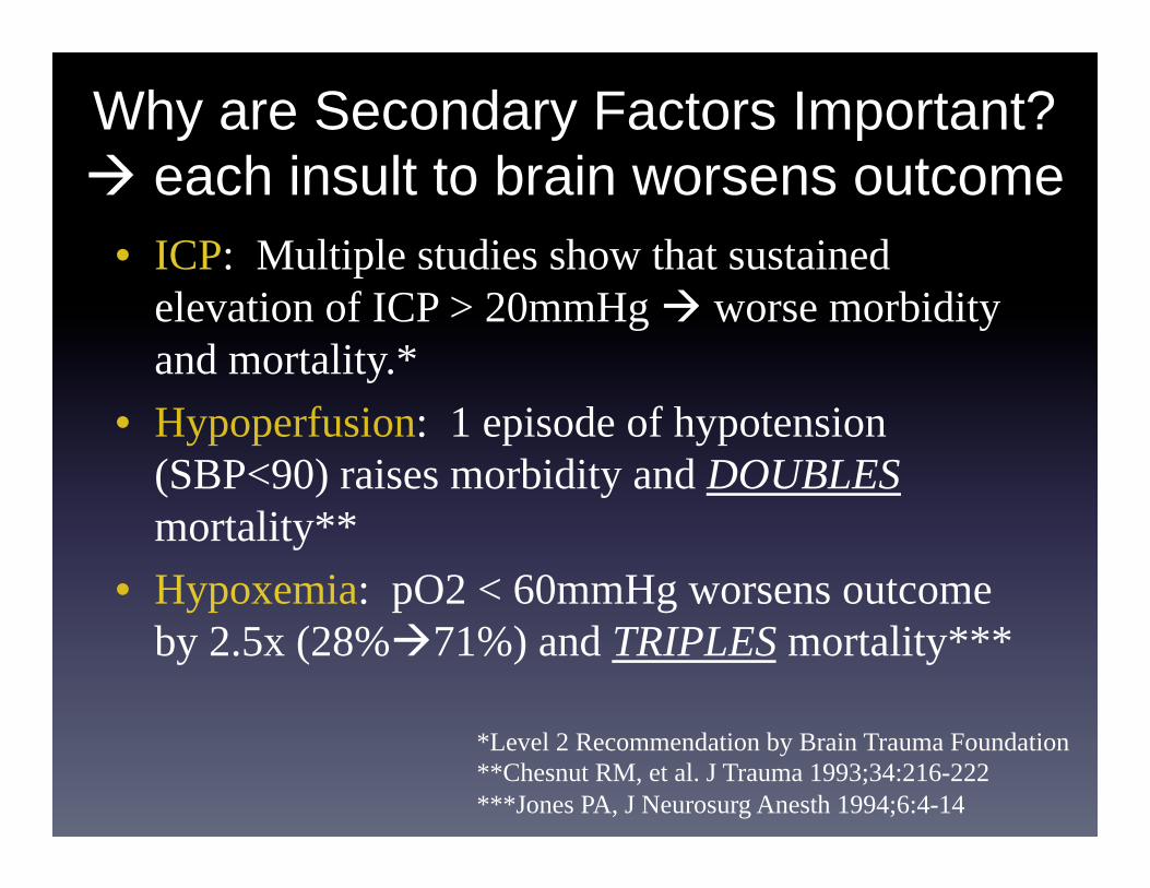
Why are Secondary Factors Important? each insult to brain worsens outcome
• ICP: Multiple studies show that sustained elevation of ICP > 20mmHg worse morbidity and mortality.*
• Hypoperfusion: 1 episode of hypotension (SBP<90) raises morbidity and DOUBLES mortality**
• Hypoxemia: pO2 < 60mmHg worsens outcome by 2.5x (28% 71%) and TRIPLES mortality***
*Level 2 Recommendation by Brain Trauma Foundation **Chesnut RM, et al. J Trauma 1993;34:216-222 ***Jones PA, J Neurosurg Anesth 1994;6:4-14

Time is Brain!
• During each minute of an ongoing “stroke”, millions of neurons are lost!
• Every second counts!

Coma and Neuro-Monitoring
• Coma = a severe disturbance of consciousness, including lack of both arousal (wakefulness) and awareness. • Spectrum of Arousal & Awareness: Coma, Minimally
Conscious State, Vegetative State • Coma = “neurological failure”
• ~Half of Neuro-ICU patients in a coma (25% in MICU) • Independent predictor of death and length of ICU stay • Heterogeneity of causes thus hard to study
• Neuromonitoring is most critical for acutely comatose patients since cause/origin must be identified and brain / other organs must be properly supported, otherwise death or permanent disability may occur.

How do we monitor our ICU patients? Non-invasive Methods
• Pulse Oximeter, Cardiac Telemetry (Continuous) • Blood Pressure (cuff) every 15-60 minutes
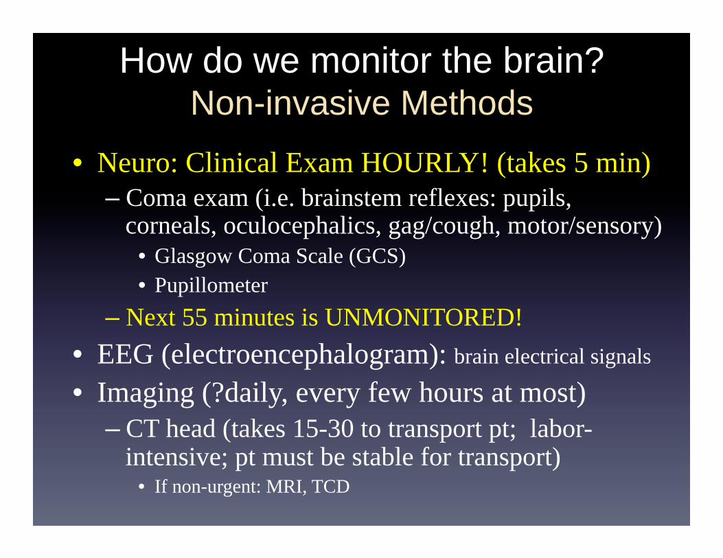
How do we monitor the brain? Non-invasive Methods
• Neuro: Clinical Exam HOURLY! (takes 5 min) – Coma exam (i.e. brainstem reflexes: pupils,
corneals, oculocephalics, gag/cough, motor/sensory) • Glasgow Coma Scale (GCS) • Pupillometer
– Next 55 minutes is UNMONITORED! • EEG (electroencephalogram): brain electrical signals
• Imaging (?daily, every few hours at most) – CT head (takes 15-30 to transport pt; labor-
intensive; pt must be stable for transport) • If non-urgent: MRI, TCD

Sometimes radiographic image do not correlate with
neurological status of patient

JHU – NCCU 12-24-10 JHU – NCCU 12-8-10
Difficult to predict coma level based on our technology
Case #2 57-year-old male
Case #1 40-year-old male
Predict the Neuro exam of these Neuro-ICU patients

GCS=3 Deepest Coma
GCS=15 Completely awake & normal cognition
JHU – NCCU 12-24-10 JHU – NCCU 12-8-10
Predict the Neuro exam of these NCCU patients
Difficult to predict coma level based on our technology

How do we monitor our patients?

Elusive brain parameters or critically ill neurological patients
• Cerebral Blood Flow (“The Holy Grail”)
• Intracranial Pressure
• Brain Metabolism
• Brain Electrical Activity
• Systemic Parameters: electrolytes, glucose,
inflammatory markers, ischemia markers

Cerebral Blood Flow (CBF) • CT Angiography (contrast, Xe, other)
• Transcranial Doppler (Ultrasound)
• Conventional Angiography (invasive)
• PET or SPECT scans
• MRI (inc MR Angiography/Perfusion, fMRI)
• Thermodilution techniques
• qEEG
Stroke

Continuous CBF monitors
Vajkoczy et al 2007(Hemedex device)
Invasive: Thermal Diffusion Flowmetry (Current standard)
Non-invasive: NIRS + Ultrasound (New device by Ornim, now commercially available)
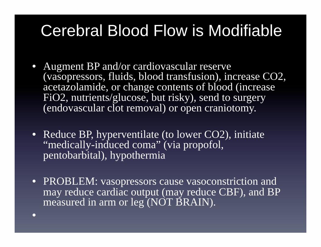
Cerebral Blood Flow is Modifiable
• Augment BP and/or cardiovascular reserve (vasopressors, fluids, blood transfusion), increase CO2, acetazolamide, or change contents of blood (increase FiO2, nutrients/glucose, but risky), send to surgery (endovascular clot removal) or open craniotomy.
• Reduce BP, hyperventilate (to lower CO2), initiate “medically-induced coma” (via propofol, pentobarbital), hypothermia
• PROBLEM: vasopressors cause vasoconstriction and may reduce cardiac output (may reduce CBF), and BP measured in arm or leg (NOT BRAIN).
•

L. Meng, M. Cannesson, B. S. Alexander, Z. Yu,
Z. N. Kain, A. E. Cerussi, B. J. Tromberg,
and W. W. Mantulin, Br. J. Anaesth. 107 (2),
209-217 (2011).
Near-Infrared Optical
Spectroscopy (NIRS) can also non-invasively
probe brain tissue oxygen saturation
(SctO2) in a critical care
setting
(e.g., to quantify the effect of
phenylephrine and ephedrine treatments
on patients under anaesthesia)
Phenylephrine treatment Ephedrine treatment
Start of treatment
Start of treatment


Clinical guidelines are “1 size fits all” • BP goals are standard for everyone:
• But 1 person’s baseline BP may be 110 and another person’s baseline BP may be 160
• “SBP<160” for postoperative patients • “SBP<140” for brain hemorrhage • “SBP 160-200 (or 220) for stroke and SAH vasospasm patients”
• “Optimizing BP” is all relative for different patients!
• Need to modify BP and assess change in CBF, then tailor hemodynamic goals on an individual basis.
• Monitoring brain hemodynamics, CBF, and cerebral metabolic activity while attempting various treatments is the key.
• Must NOT be afraid to question classical dogma!!

What is ICP? – Pressure inside the cranial vault – Indirect measure of cerebral blood flow (CBF)
Why is ICP important? Sustained Elevation in ICP is an emergency! leads to brain herniation (strangulation) • Severe reduction in CBF death or severe neurological disability

1. Cranial Vault (Skull) & Meninges 2. Cerebral blood vessels (arteries & veins) 3. Ventricles and Cerebrospinal Fluid Brain 4. Parenchyma (actual brain matter)
What determines ICP?

ICP Concepts
• Skull volume is fixed – Overcome only by craniectomy
• Cerebral Perfusion Pressure (CPP) CPP = MAP-ICP
– MAP=Mean Arterial Pressure, as measured from peripheral artery in arm or leg
Parenchyma 1200cc
Blood 150
CSF 150
• Normal ICP = 3-15 mmHg (<20cmH20) • High ICP = sustained >20 mmHg sustained > 5min
– Many studies show high ICP morbidity mortality.* • Thus ICP is an indirect surrogate for measuring CBF • High ICP is synonymous to “Reduced CBF”
*Level 2 Recommendation by Brain Trauma Foundation

Clinical Sx’s of High ICP • Cushing’s Triad: bradycardia, hypertension,
abnormal respirations – only found in 1/3-1/2 of patients w/ high ICP, even until
the very final moments of herniation! • Pupillary changes, posturing of limbs
“extensor posturing” “blown pupil”
• If not comatose, then headache, confusion, N/V, hiccups, diplopia, (CN6 > CN3), e.g. brain tumor, large aneurysm, pseudotumor cerebri

Herniation Syndromes (extreme ICP issue)

Body position & external devices are important in ICP dynamics

Invasive (continuous) ICP Monitoring
• Indications (Brain Trauma Foundation): – Traumatic Brain Injury (if comatose) – Hydrocephalus – SAH w/ abnormal CT, cerebral edema, poor exam – ICH with >5mm midline shift and poor exam – Large cerebellar lesion/ICH – ?Malignant ischemic (MCA) stroke?

A. Parenchymal ICP Monitor • Fiberoptic 20-35mm deep • Negligible drift (even w/ time per BTF)
B. Ventriculostomy, aka External Ventricular Drain (EVD) or “intraventricular catheter” (IVC) • Current gold standard
C. Epidural Transducer D. Subarachnoid Bolt
• Prone to occlusion, drift
E. Lumbar Catheter/Drain • Lower risk of hemorrhage & infection than EVD
(Schade et al 2005 J Neurosurg).
Invasive ICP monitoring (if suspecting high ICP)


Video of ICP Monitor Placement

Difference between ventriculostomy & ICP monitor is CSF drainage

ICP Compliance Intracranial Pressure/Volume Curve
From: Suarez, Critical Care Neurology & Neurosurgery 2004

Risks associated with invasive ICP monitors
• Hemorrhage or other non-infectious complications: 5% • Infection: 5-10% (risk rises daily) • May prolong hospital stay and lead to unnecessary treatment • May require blood transfusions to un-thin blood • Requires sedation/analgesia

To monitor invasively or not?
1st ever RCT on invasive ICP monitoring vs non-invasive measures

No statistical difference between invasive & non-invasive group

Treatment of High ICP • Mass (tumor, blood) Surgical Resection
• Tumor edema Steroids
• Infarct Hyperosmolar Therapy, Craniectomy
• Hydrocephalus CSF drainage

Tier 2
Tier 3
Tier 1=Vital Signs
Chang, C. Society of Critical Care Medicine 2013
Treatments for High ICP
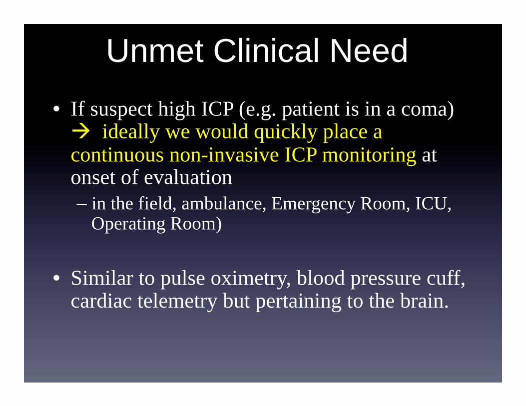
Unmet Clinical Need • If suspect high ICP (e.g. patient is in a coma)
ideally we would quickly place a continuous non-invasive ICP monitoring at onset of evaluation – in the field, ambulance, Emergency Room, ICU,
Operating Room) • Similar to pulse oximetry, blood pressure cuff,
cardiac telemetry but pertaining to the brain.

Non-invasive, continuous ICP monitor: Potential Game Changer
• Can be placed simultaneously with other monitors (pulse oximetry, telemetry, BP cuff) on initial evaluation
• Would not require specialized physicians • Offer continuous measurements • Would allow quick diagnosis, treatment, and triaging of
patients • May avoid unnecessary procedures (less guess work) • May improve neurological outcome
Multiple photonic devices being developed for ICP monitoring
• Diffuse correlation Spectroscopy (DCS)
• Ultrasound (pulsatility of blood vessels)
• NIR devices
• Pupillometry
