the response of human epithelial cells to tnf involves an ... · the response of human epithelial...
TRANSCRIPT

The Response of Human EpithelialCells to TNF Involves anInducible Autocrine CascadeKevin A. Janes,1,2 Suzanne Gaudet,1,3 John G. Albeck,1,3 Ulrik B. Nielsen,4 Douglas A. Lauffenburger,1,2,3
and Peter K. Sorger1,2,3,*1Center for Cell Decision Processes2Department of Biological Engineering3Department of Biology
Massachusetts Institute of Technology, 77 Massachusetts Avenue, Cambridge, MA 02139, USA4Merrimack Pharmaceuticals, Cambridge, MA 02139, USA*Contact: [email protected]
DOI 10.1016/j.cell.2006.01.041
SUMMARY
Tumor necrosis factor (TNF) is a proinflamma-tory cytokine that induces conflicting pro- andantiapoptotic signals whose relative strengthsdetermine the extent of cell death. TNF receptor(TNFR) has been studied in considerable detail,but it is not known how crosstalk amongantagonistic pro- and antiapoptotic signals isachieved. Here we report an experimental andcomputational analysis of crosstalk betweenprodeath TNF and prosurvival growth factorsin human epithelial cells. By applying classi-fier-based regression to a cytokine-signalingcompendiumof�8000 intracellularproteinmea-surements, we demonstrate that cells respondto TNF both directly, via activated TNF receptor,and indirectly, via the sequential release oftransforming growth factor-a (TGF-a), interleu-kin-1a (IL-1a), and IL-1 receptor antagonist(IL-1ra). We refer to the contingent and time-varying series of extracellular signals inducedby TNF as an ‘‘autocrine cascade.’’ Time-dependent crosstalk of synergistic and antago-nistic autocrine circuits may serve to link cellularresponses to the local environment.
INTRODUCTION
Cytokines and their receptors activate complex signaling
networks composed of diverse proteins whose overall
function is to control cell fate and function (Downward,
2001). Understanding these intracellular networks is com-
plicated in part because many receptors generate com-
peting, and even antagonistic, intracellular signals. For ex-
ample, tumor necrosis factor (TNF) promotes cell death by
C
inducing activation of the cysteine proteases caspase-8
and caspase-3 (Nicholson and Thornberry, 1997) but
also promotes cell survival by activating the nuclear fac-
tor-kB (NF-kB) transcription factor (Karin and Ben-Neriah,
2000). In tissues, the connection between signals and cell
fate is even more complex because cells are exposed to
multiple cytokines that act together in synergistic and an-
tagonistic combinations. Conflicting stimuli often arise
when cells are exposed to paracrine cytokines from neigh-
boring cells together with autocrine cytokines secreted by
the cell itself. In colonic epithelia, for example, TNF se-
creted by inflammatory cells is a key mediator of inflam-
matory bowel disease (Rutgeerts et al., 2004) and can
lead to epithelial cell death, whereas locally produced epi-
dermal growth factor (EGF) and insulin-like growth factor
(IGF) are critical for cell division and repair of the mucosa
(Chailler and Menard, 1999; Singh and Rubin, 1993).
Individual cytokines and receptors have been studied
extensively, but relatively little is known about intracellular
processing of antagonistic signals. It is likely that crosstalk
between individual receptors and their downstream sig-
naling pathways is important. We reasoned that crosstalk
could be studied effectively by using a large-scale sys-
tematic approach in which cells were treated with antag-
onistic cytokines and multiple downstream signals were
then measured to create a data compendium. We antici-
pated that most crosstalk would be intracellular and in-
volve the joint regulation of signaling proteins such as
mitogen-activated protein kinases, caspases, etc. We
therefore assayed intracellular signaling proteins for which
reliable quantitative measurements were available (Janes
et al., 2003; Nielsen et al., 2003). By focusing on known
pathways and proteins, the compendium approach does
not seek to identify new signaling proteins but rather to
uncover how the activities of known molecules are coor-
dinated during a physiological stimulus. For TNF, our sys-
tematic analysis revealed the surprising importance of
crosstalk via extracellular autocrine signaling in specifying
cell fate.
ell 124, 1225–1239, March 24, 2006 ª2006 Elsevier Inc. 1225

1226 Cell 124, 1225–1239, March 24, 2006 ª2006 Elsevier Inc.
TNF is the prototypical member of a family of 19 related
proapoptotic and proinflammatory cytokines (Aggarwal,
2003; Chen and Goeddel, 2002). Trimeric TNF binds to
a TNF receptor (TNFR) trimer, leading to intracellular as-
sembly of two multicomponent death-inducing signaling
complexes, DISC-I and DISC-II (Figure 1A; Barnhart and
Peter, 2003; Micheau and Tschopp, 2003). DISC-I is an
early membrane bound complex that signals through the
NF-kB, JNK, and p38 pathways, whereas DISC-II is a later
cytoplasmic complex that activates caspases-8 and -3
(Micheau and Tschopp, 2003). NF-kB signaling from
DISC-I antagonizes caspase signaling from DISC-II by in-
ducing transcription of survival factors, such as c-FLIPL,
which regulate caspase-8 activation (Micheau et al.,
2001, 2002). In many cell types, the proapoptotic func-
tions of TNF are also antagonized by mitogenic cytokines
such as EGF, insulin, and IGFs (Garcia-Lloret et al., 1996;
Qian et al., 2001; Wu et al., 1996). EGF, insulin, and IGFs
bind dimeric receptors that signal via adaptor proteins to
ERK, Akt, and other kinase pathways (Figure 1A; Avruch,
1998; Schlessinger, 2004). Growth-factor receptors re-
spond acutely to changes in exogenous ligands and also
transduce chronic signals arising from constitutive auto-
crine stimulation. Autocrine signaling is critically important
for the survival and the progression of many cancers
(Hambek et al., 2001; Torrance et al., 2000), and small-
molecule- and antibody-based therapies have therefore
been developed to disrupt receptor-ligand binding, recep-
tor dimerization, and signaling (Hofmann and Garcia-
Echeverria, 2005; Mendelsohn and Baselga, 2000). The
possibility that these drugs have secondary effects on
inflammatory signaling has not been investigated exten-
sively, despite evidence linking inflammation and cancer
(Karin and Greten, 2005).
In this paper, we combine conventional and high-
throughput experimental methods with numerical analysis
to uncover mechanisms of crosstalk involving pro- and
antiapoptotic signals induced by TNF. Time-dependent
profiles of 19 intracellular signals were measured in cells
costimulated with TNF and either EGF or insulin (two pro-
totypical survival factors) to create a cytokine-signaling
C
compendium of�8000 measurements. We describe else-
where the experimental methods used to collect, validate,
and systematize the compendium (Gaudet et al., 2005)
and focus here on the surprising finding that a large frac-
tion of TNF-induced crosstalk occurs extracellularly via
regulated shedding of autocrine cytokines. We find that
TNF activates a multistep, time-varying ‘‘autocrine cas-
cade’’ in which three secreted factors other than TNF
play pro- and antiapoptotic roles: transforming growth
factor-a (TGF-a), interleukin-1a (IL-1a), and interleukin-1
receptor antagonist (IL-1ra). Intracellular signaling acti-
vated by these cytokines specifies the extent of TNF-
induced apoptosis in a self-limiting fashion. While elucida-
tion of the TNF-TGF-a-IL-1a-IL-1ra autocrine cascade
involved statistical analysis of a large data compendium,
key hypotheses were independently validated using a
combination of neutralizing antibodies, small-molecule
drugs, and receptor antagonists. We conclude from both
large-scale and directed experiments that extracellular
autocrine crosstalk plays as important a role as intracellu-
lar crosstalk in coordinating cellular responses to TNF.
RESULTS
To explore crosstalk between conflicting mitogenic and
apoptotic signals, HT-29 human colonic adenocarcinoma
cells were stimulated with TNF in combination with either
EGF or insulin. The potency of TNF as a proapoptotic fac-
tor in epithelial cells is most apparent when combined with
immunostimulatory cytokines such as interferon-g (IFN-g;
Fransen et al., 1986). Thus, ten combinations of TNF, insu-
lin, and EGF, at either subsaturating (‘‘low’’ or ‘‘L-’’) or sat-
urating (‘‘high’’ or ‘‘H-’’) concentrations, were added to
HT-29 cells pretreated with IFN-g for 24 hr (see Experi-
mental Procedures; Figures 1B–1D). A ‘‘mock’’ treatment
(‘‘0’’), in which cells were manipulated as in other experi-
ments but without added cytokine, served as a baseline
control (Figure 1B and data not shown). Triplicate cell ex-
tracts were prepared at 13 time points spanning 0 min
(before stimulus) to 24 hr, and 19 protein measurements
made from each extract. Since many kinase signals
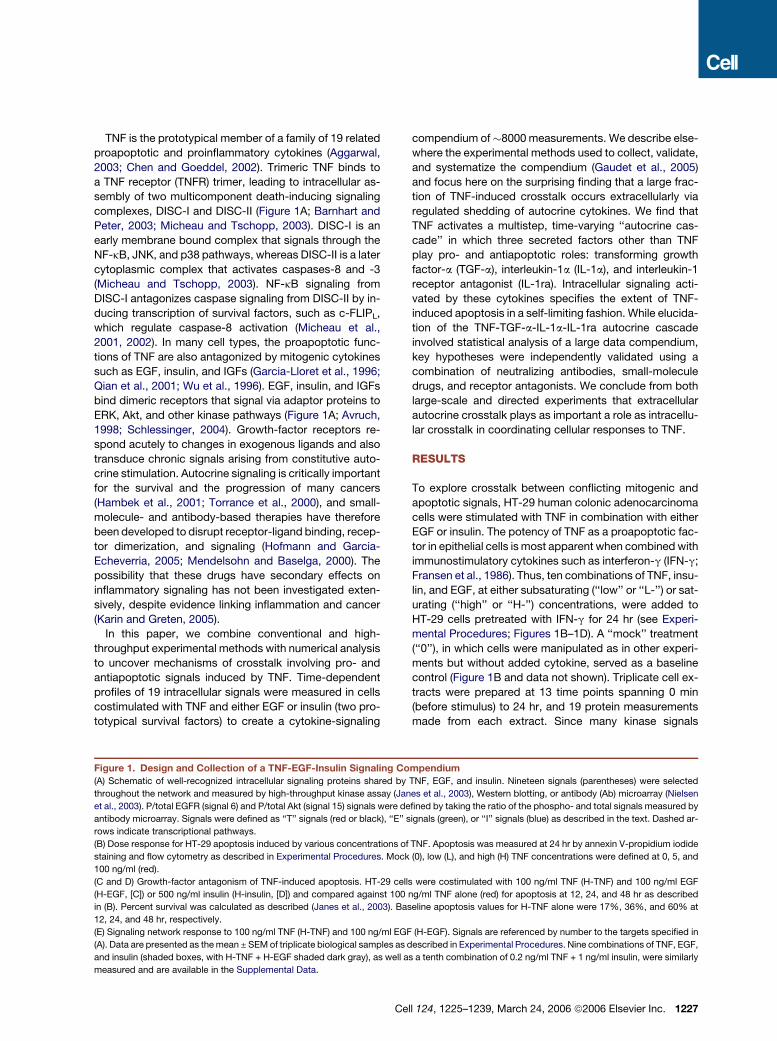
Figure 1. Design and Collection of a TNF-EGF-Insulin Signaling Compendium
(A) Schematic of well-recognized intracellular signaling proteins shared by TNF, EGF, and insulin. Nineteen signals (parentheses) were selected
throughout the network and measured by high-throughput kinase assay (Janes et al., 2003), Western blotting, or antibody (Ab) microarray (Nielsen
et al., 2003). P/total EGFR (signal 6) and P/total Akt (signal 15) signals were defined by taking the ratio of the phospho- and total signals measured by
antibody microarray. Signals were defined as ‘‘T’’ signals (red or black), ‘‘E’’ signals (green), or ‘‘I’’ signals (blue) as described in the text. Dashed ar-
rows indicate transcriptional pathways.
(B) Dose response for HT-29 apoptosis induced by various concentrations of TNF. Apoptosis was measured at 24 hr by annexin V-propidium iodide
staining and flow cytometry as described in Experimental Procedures. Mock (0), low (L), and high (H) TNF concentrations were defined at 0, 5, and
100 ng/ml (red).
(C and D) Growth-factor antagonism of TNF-induced apoptosis. HT-29 cells were costimulated with 100 ng/ml TNF (H-TNF) and 100 ng/ml EGF
(H-EGF, [C]) or 500 ng/ml insulin (H-insulin, [D]) and compared against 100 ng/ml TNF alone (red) for apoptosis at 12, 24, and 48 hr as described
in (B). Percent survival was calculated as described (Janes et al., 2003). Baseline apoptosis values for H-TNF alone were 17%, 36%, and 60% at
12, 24, and 48 hr, respectively.
(E) Signaling network response to 100 ng/ml TNF (H-TNF) and 100 ng/ml EGF (H-EGF). Signals are referenced by number to the targets specified in
(A). Data are presented as the mean ± SEM of triplicate biological samples as described in Experimental Procedures. Nine combinations of TNF, EGF,
and insulin (shaded boxes, with H-TNF + H-EGF shaded dark gray), as well as a tenth combination of 0.2 ng/ml TNF + 1 ng/ml insulin, were similarly
measured and are available in the Supplemental Data.
ell 124, 1225–1239, March 24, 2006 ª2006 Elsevier Inc. 1227

Figure 2. DPLSR Mapping of Intracellular Crosstalk in the Network Shared by TNF, EGF, and Insulin
Data were mapped as described in Experimental Procedures and in Figure S1 (Janes et al., 2004). The numbers, colors, and markers are identical to
those in Figure 1E, except for cleaved caspase-8 (signal 18), which has been changed from black to red for clarity. The gray box indicates the crosstalk
region shared by TNF, EGF, and insulin.
exhibit rapid changes between 0 and 30 min, whereas
caspases rise slowly in activity over many hours, the 13
time points were distributed unevenly, with seven ‘‘early’’
time points concentrated at t = 0–2 hr and six ‘‘late’’ time
points spread from t = 4–24 hr. The net result was a multi-
dimensional data set describing time-dependent changes
in protein activities following the stimulation of cells with
ten distinct cytokine cues.
Considerable effort was expended to ensure the repro-
ducibility and self-consistency of the data (Gaudet et al.,
2005). The median coefficient of variation for biological re-
peats across the full data set was �11%, making it possi-
ble to detect changes in protein state or activity as small
as ±15%–25%, even between experiments performed
on different days. Inspection of the complete 7980-
measurement data set revealed several distinct time
courses of signaling. We observed fast-acting ‘‘transient’’
signals, such as c-Jun N-terminal kinase 1 (JNK1) activity,
which peaked at 15 min and returned to baseline by 1 hr
(Figure 1E, signal 2). Many examples of ‘‘sustained’’ or
slow-rising signals were also observed, such as cleaved
caspase-8 levels, which began to increase at t > 2 hr
and remained high for the duration of the observation pe-
riod (Figure 1E, signal 18). Based on information in the lit-
erature, the 19 measurements acquired at each time point
were roughly characterized as TNF-dependent (‘‘T’’ sig-
nals), EGF-dependent (‘‘E’’ signals), or insulin-dependent
(‘‘I’’ signals; see Figure 1A). Importantly, each cytokine
treatment elicited multiple classes of signals: high TNF ac-
tivated both T and E signals, and high TNF + high EGF elic-
ited T, E, and I signals (Figure 1E and data not shown). We
therefore conclude that, as expected, TNF, EGF, and insu-
lin exhibit significant crosstalk at the level of intracellular
protein signals.
1228 Cell 124, 1225–1239, March 24, 2006 ª2006 Elsevier Inc.
Mapping Intracellular Crosstalk onto a Shared
Cytokine-Signal Space
To visualize connections between intracellular signals and
cytokine treatments in a simple and intuitive way, we con-
structed a compact representation of the entire compen-
dium by using discriminant partial least squares regres-
sion (DPLSR; Janes et al., 2004). A DPLSR map was
created such that the signaling proteins and cytokines
were projected onto a set of ‘‘principal components’’
that maximized covariation between the time-integrated
signaling profiles and the corresponding cytokine treat-
ment (see Figure S1 in the Supplemental Data available
with this article online for details). The first principal com-
ponent in the DPLSR map corresponded to a baseline that
distinguished all cytokine treatments from mock stimula-
tion, whereas the second and third principal components
discriminated among TNF, EGF, and insulin treatments
(Figure S1 and data not shown). The latter two principal
components therefore identified cytokine-specific signals
as well as instances where two or more cytokine pathways
had converged upon a common signaling protein.
By plotting cytokine treatments and integrated signals
along the two dimensions defined by the second and third
principal components, the extent of covariance among
signals and treatments could be evaluated. Some cases
of covariation were expected: For example, the cleavage
of caspase-8 mapped close to TNF (Figure 2, signal 18),
consistent with evidence that caspase-8 is activated by
TNFR via formation of DISC-II (Micheau and Tschopp,
2003). Similarly, three measures of Akt activity (signals
13–15) mapped close to insulin, which is a powerful
inducer of Akt signaling (Avruch, 1998). Sometimes, the
map position suggested unexpected biological regula-
tion: For example, the phosphorylation of IRS1 on Y896

Figure 3. TNF Activates an Early-Phase TGF-a Autocrine Circuit to Crosstalk through the EGFR-MEK-ERK Signaling Pathway(A) Comparison of P-MEK (upper) and ERK (lower) signaling dynamics induced by 100 ng/ml TNF (H-TNF, left), 5 ng/ml TNF (L-TNF, center), and
500 ng/ml insulin (H-insulin, right). EGFR phosphorylation was similarly increased in response to TNF (data not shown).
(B) Comparison of P-MEK (upper) and ERK (lower) signaling dynamics induced by 100 ng/ml EGF (H-EGF).
(C) TGF-a release in response to 5 ng/ml TNF (L-TNF) in the presence of 10 mg/ml C225 pretreatment for 1 hr before stimulation. The control treatment
was a mock stimulation with carrier only. Similar results were obtained in the absence of C225 (data not shown).
(D and E) TNF-induced E signaling through EGFR. pMEK (D) and ERK (E) signaling were measured after stimulation with 5 ng/ml TNF (L-TNF) in the
presence or absence of 10 mg/ml C225 pretreatment.
(F) Perturbation of TNF-induced ERK activation by pretreatment with 10 mg/ml C225, 1 mM AG1478, or 10 mM batimastat for 1 hr before stimulation
with 5 ng/ml TNF (L-TNF) for 15 min. 0.1% DMSO was added as a control pretreatment (No pretx). ERK activity from untreated cells was included as
a baseline, and ERK activation was defined as the increase in ERK activity compared to these untreated cells.
Data are presented as the mean ± SEM of triplicate biological samples as described in Experimental Procedures.
(P-IRS1(Y896); signal 10) was closely associated with EGF
but not insulin (Gaudet et al., 2005). Of greatest interest
was the clustering of signals midway between TNF,
EGF, and insulin, possibly implying covariance of the sig-
nals with two or more cytokines (Figure 2, gray box). When
the Euclidean distance between cytokine cues and signals
in this central cluster was calculated, E signals such as
EGFR (Figure 2, signal 4), ERK (signal 8), and MAPK-
ERK kinase (MEK, signal 7), were found to be roughly equi-
distant to TNF and EGF cytokine stimuli but significantly
farther from insulin (p < 0.05). Inspection of individual
cytokine time courses confirmed that TNF treatment acti-
vated EGFR, ERK, and MEK to a similar extent as EGF it-
self, whereas insulin did not (Figures 3A and 3B and data
not shown). Others have noted crosstalk between TNF
and EGF (Chen et al., 2004; Izumi et al., 1994), supporting
C
the paradoxical conclusion that TNF activates EGFR and
its downstream targets MEK and ERK with strength and
kinetics similar to EGF.
Rapid Activation of a TGF-a Autocrine Circuit by TNF
A direct intracellular link from activated TNFR to ERK has
not been established, but TNF can induce the shedding of
EGF-family ligands in mammary epithelial cells following
24 hr of stimulation (Chen et al., 2004). HT-29 cells also
shed ligands of the EGF family (Anzano et al., 1989), so
we asked whether TNF could stimulate this shedding with
the rapid kinetics observed for MEK, ERK, and EGFR
activation (Figure 3A and data not shown). EGF-family
ligands known to act as autocrine factors include TGF-a,
amphiregulin (AR), and heparin binding epidermal growth
factor (HB-EGF). Quantitative ELISA measurements of
ell 124, 1225–1239, March 24, 2006 ª2006 Elsevier Inc. 1229
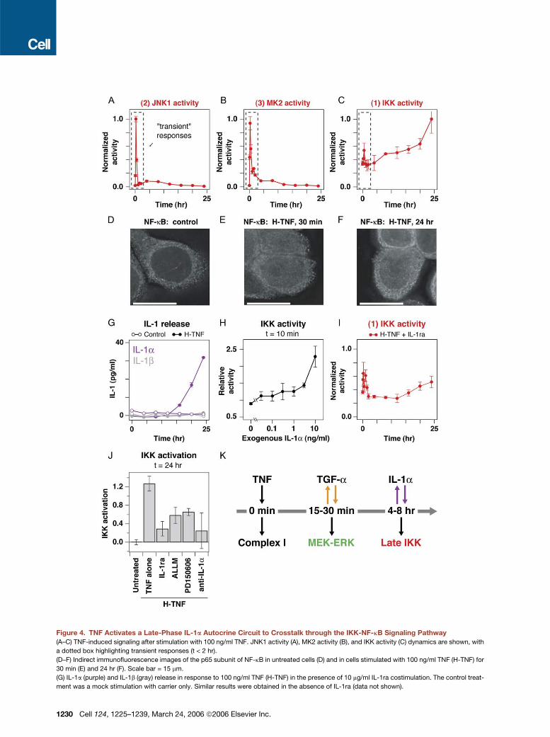
Figure 4. TNF Activates a Late-Phase IL-1a Autocrine Circuit to Crosstalk through the IKK-NF-kB Signaling Pathway
(A–C) TNF-induced signaling after stimulation with 100 ng/ml TNF. JNK1 activity (A), MK2 activity (B), and IKK activity (C) dynamics are shown, with
a dotted box highlighting transient responses (t < 2 hr).
(D–F) Indirect immunofluorescence images of the p65 subunit of NF-kB in untreated cells (D) and in cells stimulated with 100 ng/ml TNF (H-TNF) for
30 min (E) and 24 hr (F). Scale bar = 15 mm.
(G) IL-1a (purple) and IL-1b (gray) release in response to 100 ng/ml TNF (H-TNF) in the presence of 10 mg/ml IL-1ra costimulation. The control treat-
ment was a mock stimulation with carrier only. Similar results were obtained in the absence of IL-1ra (data not shown).
1230 Cell 124, 1225–1239, March 24, 2006 ª2006 Elsevier Inc.

EGF-family ligands in conditioned medium of HT-29 cells
before and after TNF addition showed all three to be pres-
ent, but only TGF-a was upregulated by TNF (Figure 3C
and Figure S2). TNF-induced TGF-a release in HT-29 cells
was much faster than previously reported (Chen et al.,
2004), peaking 1 hr after TNF addition (Figure 3C). Thus,
TNF stimulation is associated with near-immediate
release of an EGFR ligand, TGF-a, into the medium. Al-
though transcriptional upregulation of TGF-a by growth-
factor signaling pathways has been reported (Schulze
et al., 2001), the rapidity of TGF-a release after TNF treat-
ment suggests a posttranslational mechanism.
Addition of exogenous EGF rapidly activated MEK and
ERK in HT-29 cells (Figure 3B). We therefore asked
whether TNF-stimulated release of endogenous TGF-a
also activated MEK and ERK in an EGFR-dependent fash-
ion. When cells were treated with C225 antibody to block
the interaction of EGFR with its ligands (Figure S3), dra-
matic inhibition of both acute (t = 0–2 hr) and sustained
(t = 2–24 hr) MEK-ERK activation was observed after
TNF treatment (Figures 3D and 3E). The significant and
persistent reduction in MEK-ERK signaling following TNF
stimulation (4-fold lower at t = 30 min, p < 0.005 and
2-fold lower at t = 4–24 hr, p < 10�6) demonstrates a pre-
dominantly EGFR-dependent pathway. ERK activation
was also blocked by a small-molecule inhibitor of the
EGFR kinase (AG1478) and an inhibitor of the matrix me-
talloproteases that mediate TGF-a shedding from the
plasma membrane (batimastat; Peschon et al., 1998; Fig-
ure 3F). Taken together, these data show that an autocrine
circuit involving TGF-a and EGFR is the primary mecha-
nism by which TNF activates MEK-ERK signaling at
both short (t < 1 hr) and long (t > 4 hr) timescales in
HT-29 cells. The delay between the peak of direct ERK
activation by EGF at t = 5 min and indirect activation by
TNF at t = 15 min is an estimate of the minimum time
required to establish the TNF-induced TGF-a autocrine
circuit (Figure 3A).
TNF Activates a Late-Phase IL-1a Autocrine Circuit
Further inspection of the DPLSR map revealed that IKK
activity (Figure 2, signal 1) was also unexpectedly distant
from its presumed inducer, TNF. JNK1 and MK2 are two
well-recognized TNF-induced signals that, unlike IKK,
were close to TNF on the DPLSR map (Figure 2, signals
2 and 3; Wajant et al., 2003). We therefore compared the
dynamics of their activation to that of IKK. All three T sig-
nals were strongly induced 5–30 min after TNF addition,
C
but IKK was unique in exhibiting a second, sustained
phase of activation, rising from 4–24 hr (Figures 4A–4C).
In the classical TNF-induced pathway, IKK phosphory-
lates and inactivates IkB, an NF-kB inhibitor, which allows
NF-kB to translocate into the nucleus and induce gene ex-
pression (Karin and Ben-Neriah, 2000). By immunofluo-
rescence, we found that the p65 subunit of NF-kB—which
was largely cytoplasmic before TNF stimulation, reflecting
its sequestration by IkB (Figure 4D)—was present at high
levels in the nucleus in TNF-treated cells during early (t =
30 min) and late (t = 24 hr) peaks of IKK activation (Figures
4E and 4F). Thus, the two distinct phases of IKK signaling
both appear to activate NF-kB. However, since JNK1 and
MK2 activities had returned to low levels by t = 1 hr
(Figures 4A and 4B), it seemed unlikely that sustained
IKK and NF-kB signaling at t > 4 hr was mediated directly
by TNF.
One circumstance in which sustained NF-kB activation
is well described is in ultraviolet-irradiated keratinocytes,
where late NF-kB signaling is known to depend on sec-
ondary release of IL-1a (Bender et al., 1998). Because
IL-1 cytokines are potent IKK agonists (Dinarello, 1997),
we asked whether TNF treatment of HT-29 cells might
lead to IL-1 release and whether IL-1 would provoke sus-
tained IKK activation. We observed an 8-fold increase in
soluble IL-1a, but not IL-1b, in conditioned medium from
TNF-treated cells 12–24 hr after TNF addition (Figure 4G).
HT-29 cells are known to express the IL-1 receptor (IL-1R;
Panja et al., 1998), and we found that exogenous IL-1a
activated IKK at concentrations as low as 30 pg/ml (Fig-
ure 4H), consistent with the recognized ligand sensitivity
of IL-1R (Dinarello, 1997). Thus, HT-29 cells are IL-1 re-
sponsive and secrete IL-1a after TNF stimulation, sug-
gesting that IL-1a might constitute a TNF-dependent acti-
vator of IKK.
To determine whether IKK activation in TNF-treated
cells was dependent on secreted IL-1a, IKK activity was
measured in cells treated with high TNF in the presence
of saturating levels of IL-1ra, a naturally occurring IL-1R
antagonist (Figure S3; Dinarello, 2000). We observed
that activation of IKK 15–30 min after TNF addition was un-
altered by IL-1ra, consistent with a direct pathway via
TNFR (Figure 4I). However, sustained IKK activation
4–24 hr after TNF addition was significantly reduced by
IL-1ra addition (p < 0.001; Figures 4C and 4I). Substantial
reductions in TNF-induced IKK activity at 24 hr were also
observed with ALLM and PD150606, two structurally dis-
tinct calpain inhibitors that block processing and release
(H) IKK activity induced by recombinant IL-1a. HT-29 cells were stimulated with various concentrations of exogenous IL-1a for 10 min and analyzed
for IKK activity.
(I) IKK response to 100 ng/ml TNF (H-TNF) with 10 mg/ml IL-1ra cotreatment. Cells were treated and measured for IKK activity as shown in (C) but in the
presence of IL-1ra.
(J) Perturbation of TNF-induced IKK activation by cotreatment with 10 mg/ml IL-1ra, 25 mM ALLM, 25 mM PD150606, or 1 mg/ml anti-IL-1a during stim-
ulation with 100 ng/ml TNF (H-TNF) for 24 hr. IKK activity from untreated cells was included as a baseline, and IKK activation was defined as the in-
crease in IKK activity compared to these untreated cells.
(K) Timeline of TNF-induced TGF-a and IL-1a autocrine circuits and autocrine-dependent signals.
For (A)–(C) and (G)–(J), data are presented as the mean ± SEM of triplicate biological samples as described in Experimental Procedures.
ell 124, 1225–1239, March 24, 2006 ª2006 Elsevier Inc. 1231

Figure 5. The TNF-Induced TGF-a and IL-1a Circuits Are Coupled with an IL-1ra Circuit to Form an Autocrine Cascade
(A and B) Quantitative changes in TNF-induced caspase signaling after perturbation of the TGF-a and IL-1a autocrine circuits. Cleaved caspase-8
responses to 5 ng/ml TNF (L-TNF) with or without 10 mg/ml C225 pretreatment (A) or 100 ng/ml TNF (H-TNF) with or without 10 mg/ml IL-1ra cotreat-
ment (B). TNF-induced procaspase-3 signaling was also affected by IL-1ra (data not shown).
1232 Cell 124, 1225–1239, March 24, 2006 ª2006 Elsevier Inc.

of IL-1a from cells (Kobayashi et al., 1990), as well as with
IL-1a-neutralizing antibodies (Figure 4J). Together, these
data show that IKK signaling at t < 4 hr is independent of
IL-1a but that sustained IKK activation at t > 4 hr (which
is quantitatively more significant in HT-29 cells;
Figure 4C) is mediated primarily by the binding of IL-1a
to IL-1R (Figure 4K). Thus, the early and late phases of
IKK activation in TNF-treated HT-29 cells involve distinct
direct and autocrine-indirect mechanisms.
Coupling of Sequential Autocrine Circuits
into an Autocrine Cascade
The data described above establish that TGF-a and IL-1a
autocrine circuits play important roles in the activation of
MEK-ERK and IKK by TNF, but it remained unclear
whether autocrine-dependent signaling altered the activ-
ity of caspases directly implicated in apoptosis. To study
this, we blocked TGF-a- and IL-1a-dependent circuits in-
dividually and measured the cleavage of caspase-8, an
important initiator caspase downstream of TNFR (Chen
and Goeddel, 2002). Blocking IL-1R with IL-1ra signifi-
cantly reduced TNF-stimulated cleavage of caspase-8
compared to TNF alone (p < 10�9; Figure 5A). Conversely,
blocking EGFR with C225 significantly increased TNF-in-
duced caspase-8 cleavage (p < 0.001; Figure 5B). Thus,
TNF-induced TGF-a and IL-1a exert opposing control on
caspase-8 (Figure 5C). To determine how broad an effect
EGFR or IL-1R blockade had on TNF-induced signaling,
full time courses were collected for the 19 signals after
TNF stimulation in the presence of IL-1ra or C225. When
autocrine-perturbed time courses were compared to
data in the original cytokine-signaling compendium, we
found that the durations or magnitudes of roughly half of
the measured signals were altered (Figures 5D and 5E).
C
Thus, discrete perturbation of an autocrine circuit causes
a system-wide alteration in the underlying intracellular sig-
naling network.
The opposing contributions of TGF-a and IL-1a to TNF-
induced caspase-8 cleavage implied that blocking EGFR-
mediated signaling should decrease cell survival, whereas
blocking IL-1R-mediated signaling should decrease cell
death (Figure 5C). Consistent with this, we observed that
TNF was less effective as a prodeath stimulus when
IL-1a signaling was blocked with IL-1ra (Figure 5F). The
potency of IL-1ra as a prosurvival factor in TNF-treated
cells was comparable to that of saturating EGF or insulin
(Figures 1C and 1D). In contrast, blocking TGF-a signaling
with C225 antibody did not substantially increase TNF-
induced cell death (Figure 5G). This was paradoxical
because it implied that the ability of autocrine TGF-a to re-
duce caspase-8 cleavage (Figure 5C) did not translate into
reduced cell death. In an attempt to resolve this paradox,
the 19 signals measured for TNF + C225 treatment were
examined more closely (Figure 5E). We observed that
IKK activity was reduced to baseline levels throughout
the time course, exhibiting neither early- nor late-phase
activation (p < 10�10; Figure 5H). The attenuation of late-
phase IKK signaling (t = 4–24 hr) by C225 was particularly
interesting because our results indicated that late-phase
IKK signaling required an autocrine IL-1a circuit (Figures
4C and 4I). Therefore, it seemed possible that induction
of the IL-1a circuit (and thus late IKK signaling) might de-
pend upon prior release of TGF-a. To determine whether
TGF-a and IL-1a release were linked, we blocked TGF-a-
EGFR signaling with C225 and measured IL-1a levels fol-
lowing TNF stimulation. EGFR blockade with C225 com-
pletely prevented IL-1a release at low TNF concentrations
(Figure 5I) and significantly reduced IL-1a release at high
(C) Autocrine TGF-a (A-TGF-a) and autocrine IL-1a (A-IL-1a) exert opposing control on TNF-induced caspase-8 cleavage.
(D and E) Network-level changes in TNF-induced signaling in the presence of 10 mg/ml C225 pretreatment (D) or 10 mg/ml IL-1ra cotreatment. Selected
intracellular signals from C225- and IL-1ra-treated cells were integrated from 0–24 hr and compared to the integrated signals from 5 ng/ml TNF
(L-TNF) and 100 ng/ml TNF (H-TNF) treatments without autocrine perturbation, respectively. Uncertainty in the integrated signaling profiles was cal-
culated by bootstrapping with the original signaling replicates (Efron and Tibshirani, 1993).
(F) IL-1a-mediated synergism with TNF-induced apoptosis. HT-29 cells were cotreated with 100 ng/ml TNF (H-TNF) and 10 mg/ml IL-1ra and com-
pared against H-TNF alone (red) for apoptosis at 12, 24, and 48 hr as described in Figure 1B (Janes et al., 2005). Baseline apoptosis values for H-TNF
alone were 17%, 45%, and 67% at 12, 24, and 48 hr, respectively.
(G) TGF-a-mediated antagonism of TNF-induced apoptosis. HT-29 cells were pretreated with 10 mg/ml C225, stimulated with 5 ng/ml TNF (L-TNF),
and compared against L-TNF alone (red) for apoptosis at 12, 24, and 48 hr as described in Figure 1B (Janes et al., 2005). Baseline apoptosis values for
L-TNF alone were 11%, 24%, and 47% at 12, 24, and 48 hr, respectively.
(H) IKK response to 5 ng/ml TNF (L-TNF) with or without 10 mg/ml C225 pretreatment.
(I) IL-1a release induced by 0 or 5 ng/ml TNF with or without 10 mg/ml C225 pretreatment. The control treatment was a mock stimulation with carrier
only.
(J) TGF-a-mediated antagonism for TNF-induced apoptosis. HT-29 cells were treated with various concentrations of TGF-a and 5 ng/ml TNF (L-TNF)
and compared against L-TNF alone (red) for apoptosis at 24 hr as described in Figure 1B (Janes et al., 2005). Baseline apoptosis for L-TNF alone was
33%.
(K) IL-1ra release in response to 100 ng/ml TNF (H-TNF). The control treatment was a mock stimulation with carrier only.
(L) The IL-1a autocrine circuit is induced by TNF but not by TGF-a. HT-29 cells were treated with 100 ng/ml TNF (H-TNF) or 100 ng/ml TGF-a for 24 hr
and analyzed for IL-1a release.
(M) The TGF-a autocrine circuit is induced by TNF but not by IL-1a. HT-29 cells were treated with 100 ng/ml TNF (H-TNF) or 10 ng/ml IL-1a for 24 hr
and analyzed for TGF-a release.
(N) The molecular logic of the TNF-induced autocrine cascade. TNF, TGF-a, and IL-1a are coupled via an ‘‘AND’’ gate.
For (A)–(B) and (F)–(M), data are presented as the mean ± SEM of triplicate biological samples as described in Experimental Procedures. For (D) and
(E), data are presented as the mean percent change ± SD from 1000 resamplings of the original data set.
ell 124, 1225–1239, March 24, 2006 ª2006 Elsevier Inc. 1233

TNF concentrations (Figure S4). Thus, proper function of
the IL-1a autocrine circuit was dependent on the prior es-
tablishment of an EGFR-mediated TGF-a circuit.
The observed coupling between TGF-a and IL-1a auto-
crine circuits explains the inability of EGFR blockade to al-
ter TNF-induced cell death significantly. In TNF-treated
cells, C225 increased cleaved caspase-8 levels and re-
duced MEK-ERK signaling (which is generally regarded
as a prosurvival signal; Ballif and Blenis, 2001), but C225
also prevented IL-1a release (Figure 5I), which mediates
proapoptotic signals via IL-1R (Figure 5F). The ability of
C225 to block the offsetting TGF-a- and IL-1a-dependent
pathways implied that conditions could be found in which
EGFR and IL-1R signals were not perfectly balanced. At
some concentrations, TGF-a might act as a prosurvival
factor and at others as a proapoptotic factor. To test this
idea, cells were exposed to TNF in combination with exog-
enous TGF-a at 0.3–100 ng/ml, and the fraction of dying
cells was measured. We observed that, at low concentra-
tions, TGF-a was proapoptotic in combination with TNF,
whereas at higher concentrations, it was prosurvival (Fig-
ure 5J), confirming our prediction.
The link between the TGF-a and IL-1a autocrine circuits
raised the possibility that additional extracellular factors
might contribute to TNF signaling and cellular responses.
In recent microarray studies, we observed that transcrip-
tion of the IL-1ra gene was strongly upregulated by TNF
(K.A.J. and P.K.S., unpublished data). ELISA measure-
ments on conditioned medium of HT-29 cells revealed
that IL-1ra protein accumulated to significant levels 12 hr
after TNF stimulation (Figure 5K). However, unlike IL-1a,
TNF-induced IL-1ra release was unaffected by pretreat-
ment with C225 antibody, indicating that IL-1ra induction
does not require autocrine TGF-a (Figure S5). Thus,
IL-1ra constitutes a third TNF-inducible autocrine factor,
one with a prosurvival function (Figure 5F; Dinarello, 2000).
The successive waves of extracellular signaling circuits
induced by TNF suggest the existence of an autocrine
cascade involving TNF, TGF-a, IL-1a, and IL-1ra. The
most interesting features of the cascade are the connec-
tions between individual autocrine circuits: TGF-a is re-
quired for IL-1a release, and IL-1ra inhibits the action of
IL-1a. Is there a contingent logic underlying the order in
which these signals are released? We found that the link
between TGF-a and IL-1a (Figure 5I) was not reciprocal
because addition of exogenous IL-1a did not induce shed-
ding of TGF-a (Figure 5L). Moreover, exogenous TGF-a
was not sufficient to provoke IL-1a release (Figure 5M),
implying that signals from both TNFR and EGFR are re-
quired. The TGF-a and IL-1a circuits are therefore linked
together unidirectionally. TNF and TGF-a comprise inputs
to an ‘‘AND’’ function that induces IL-1a, which is subse-
quently inactivated by IL-1ra (Figure 5N).
Evidence for a TNF-Induced Autocrine Cascade
in Diverse Epithelial Cell Types
To examine whether TNF-induced autocrine cascades are
a conserved feature of TNF-responsive cells, we tested
1234 Cell 124, 1225–1239, March 24, 2006 ª2006 Elsevier Inc.
other epithelial cell lines for the essential features of the
TGF-a-IL-1a-IL-1ra cascade. Specifically, we asked
whether TNF would stimulate (1) release of TGF-a (Fig-
ure 6A), (2) release of IL-1a in an EGFR-dependent manner
(Figure 6B), and (3) release of IL-1ra (Figure 6C). We fo-
cused on two human cell lines known to have receptors
for TNF, EGF, and IL-1: A431 epidermoid carcinoma cells
and nontransformed 184A1 human mammary epithelial
cells (HMEC; Chen et al., 2004; Masui et al., 1993).
Both A431 and immortalized HMEC cells were treated
with TNF, and cytokine levels were measured at t = 0, 2,
and 24 hr by ELISA (Figures 6D–6I). We found that TGF-a,
IL-1a, and IL-1ra levels all increased in A431 cells after
TNF treatment. In particular, TGF-a release was nearly
as rapid as in HT-29 cells. TNF-stimulated IL-1a release
in A431 cells was inhibited significantly but not completely
by C225, probably reflecting incomplete blockade of high
EGFR levels on the surface of these cells (Masui et al.,
1993). In analogous experiments with TNF-simulated
HMEC cells, we found that TGF-a levels increased dra-
matically over a 24 hr period, IL-1a levels increased
>2.5-fold, and IL-1ra levels increased >4-fold. Moreover,
TNF-induced release of IL-1a into the medium was
blocked almost completely by C225 anti-EGFR antibody.
Thus, all three extracellular components of the TNF-stim-
ulated TGF-a-IL-1a-IL-1ra autocrine cascade are present
in HMEC cells, and induction of the IL-1a circuit is almost
completely dependent on prior establishment of the TGF-
a-EGFR circuit. The primary difference between HT-29
and HMEC cells is in the timing of TGF-a release, which
was considerably slower in HMECs, perhaps reflecting
a requirement for protein synthesis. Together, these data
indicate that a TNF-induced autocrine cascade consisting
of TGF-a, IL-1a, and IL-1ra cytokines exists in multiple ep-
ithelial cell types.
DISCUSSION
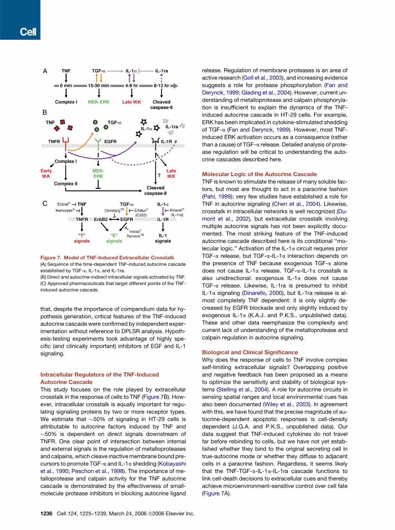
In this paper, we show that TNF induces three successive
autocrine circuits, which together form an autocrine cas-
cade that plays out over 24 hr (Figure 7A). Working in com-
bination with TNF itself, the cascade adds layers of pro-
and antiapoptotic signaling to set the level of apoptosis
in a self-limiting fashion. Shortly after TNF addition, pro-
apoptotic signals immediately downstream of TNFR bind-
ing are induced. About 10 min later, an autocrine TGF-a
circuit is established, leading to prosurvival signaling
through EGFR. The combination of TNF and TGF-a causes
release of IL-1a starting at 4 hr, which in turn activates pro-
death signaling through IL-1R. Finally, upregulation of the
IL-1R antagonist IL-1ra by 8–12 hr negatively regulates
IL-1R signaling and presumably constitutes a final anti-
apoptotic stimulus. Our work highlights the important
but underappreciated role of autocrine crosstalk in deter-
mining cell fate. Individual autocrine circuits have previ-
ously been studied in multiple cellular contexts. However,
extended connectivity between inducible autocrine cir-
cuits has not been reported previously, perhaps because

Figure 6. The Basic Elements of the TNF-Induced Autocrine Cascade Are Present in Other Epithelial Cell Types
(A–C) Main elements of the TNF-induced autocrine cascade: release of TGF-a (A), release of IL-1a in a TGF-a-dependent manner (B), and release of
IL-1ra (C).
(D and E) The TNF-induced autocrine cascade exists in A431 cells. A431 cells were stimulated with 100 ng/ml TNF + 10 mg/ml C225 (H-TNF) (D–F) or
100 ng/ml TNF alone (E) and analyzed for TGF-a at 2 and 24 hr (D), IL-1a at 24 hr (E), and IL-1ra at 24 hr (F). The control treatment was a mock stim-
ulation with carrier only.
(G–I) The TNF-induced autocrine cascade exists in human mammary epithelial cells (HMEC). HMEC cells were stimulated with 100 ng/ml TNF (H-TNF)
(G–I) or 100 ng/ml TNF (H-TNF) + 10 mg/ml C225 (H) and analyzed for TGF-a at 2 and 24 hr (G), IL-1a at 24 hr (H), and IL-1ra at 24 hr (I). The control
treatment was a mock stimulation with carrier only.
For (D)–(I), data are presented as the mean ± SEM of triplicate biological samples as described in Experimental Procedures. n.d., not detectable.
such linkages are only evident when large, multidimen-
sional signaling data sets are examined.
Data, Statistical Mining, and Hypotheses
The construction of a systematic time-dependent com-
pendium of cellular signals has been essential to our anal-
ysis of TNF. The 7980-measurement compendium con-
tains quantitative information on 19 activities and states
of kinases, caspases, transcription factors, adaptors, and
other signaling proteins distributed across signaling net-
works downstream of TNFR, EGFR, and insulin receptor
(Figure 1A). An advantage of constraining measurements
to a limited number of proteins is that it becomes practical
to examine many different cytokine combinations at mul-
tiple points in time. The experimental precision and repro-
ducibility of the measurements also made it possible to
C
study small (i.e., less than 2-fold) but biologically signifi-
cant differences in signals from one cytokine treatment
to the next. Our proteomic compendium is far from com-
prehensive, but, in the context of signal transduction, the
multicomponent, multicytokine time courses represent
a useful advance over more focused studies because
they place dynamic information within a broader physio-
logical context.
A major challenge with large experimental compendia is
generating experimentally testable biological hypotheses.
Here, we use DPLSR to derive a visually intuitive map from
time-integrated signals (Janes et al., 2004). In related
work, we construct alternative models that explicitly incor-
porate time-course data (Janes et al., 2005). Both analy-
ses show that a TNF-induced autocrine cascade is critical
in determining cell fate. However, it is important to note
ell 124, 1225–1239, March 24, 2006 ª2006 Elsevier Inc. 1235
that, despite the importance of compendium data for hy-
pothesis generation, critical features of the TNF-induced
autocrine cascade were confirmed by independent exper-
imentation without reference to DPLSR analysis. Hypoth-
esis-testing experiments took advantage of highly spe-
cific (and clinically important) inhibitors of EGF and IL-1
signaling.
Intracellular Regulators of the TNF-Induced
Autocrine Cascade
This study focuses on the role played by extracellular
crosstalk in the response of cells to TNF (Figure 7B). How-
ever, intracellular crosstalk is equally important for regu-
lating signaling proteins by two or more receptor types.
We estimate that �50% of signaling in HT-29 cells is
attributable to autocrine factors induced by TNF and
�50% is dependent on direct signals downstream of
TNFR. One clear point of intersection between internal
and external signals is the regulation of metalloproteases
and calpains, which cleave inactive membrane bound pre-
cursors to promote TGF-a and IL-1a shedding (Kobayashi
et al., 1990; Peschon et al., 1998). The importance of me-
talloprotease and calpain activity for the TNF autocrine
cascade is demonstrated by the effectiveness of small-
molecule protease inhibitors in blocking autocrine ligand
Figure 7. Model of TNF-Induced Extracellular Crosstalk
(A) Sequence of the time-dependent TNF-induced autocrine cascade
established by TGF-a, IL-1a, and IL-1ra.
(B) Direct and autocrine-indirect intracellular signals activated by TNF.
(C) Approved pharmaceuticals that target different points of the TNF-
induced autocrine cascade.
1236 Cell 124, 1225–1239, March 24, 2006 ª2006 Elsevier Inc.
release. Regulation of membrane proteases is an area of
active research (Goll et al., 2003), and increasing evidence
suggests a role for protease phosphorylation (Fan and
Derynck, 1999; Glading et al., 2004). However, current un-
derstanding of metalloprotease and calpain phosphoryla-
tion is insufficient to explain the dynamics of the TNF-
induced autocrine cascade in HT-29 cells. For example,
ERK has been implicated in cytokine-stimulated shedding
of TGF-a (Fan and Derynck, 1999). However, most TNF-
induced ERK activation occurs as a consequence (rather
than a cause) of TGF-a release. Detailed analysis of prote-
ase regulation will be critical to understanding the auto-
crine cascades described here.
Molecular Logic of the Autocrine Cascade
TNF is known to stimulate the release of many soluble fac-
tors, but most are thought to act in a paracrine fashion
(Pahl, 1999); very few studies have established a role for
TNF in autocrine signaling (Chen et al., 2004). Likewise,
crosstalk in intracellular networks is well recognized (Du-
mont et al., 2002), but extracellular crosstalk involving
multiple autocrine signals has not been explicitly docu-
mented. The most striking feature of the TNF-induced
autocrine cascade described here is its conditional ‘‘mo-
lecular logic.’’ Activation of the IL-1a circuit requires prior
TGF-a release, but TGF-a-IL-1a interaction depends on
the presence of TNF because exogenous TGF-a alone
does not cause IL-1a release. TGF-a-IL-1a crosstalk is
also unidirectional: exogenous IL-1a does not cause
TGF-a release. Likewise, IL-1ra is presumed to inhibit
IL-1a signaling (Dinarello, 2000), but IL-1ra release is al-
most completely TNF dependent: it is only slightly de-
creased by EGFR blockade and only slightly induced by
exogenous IL-1a (K.A.J. and P.K.S., unpublished data).
These and other data reemphasize the complexity and
current lack of understanding of the metalloprotease and
calpain regulation in autocrine signaling.
Biological and Clinical Significance
Why does the response of cells to TNF involve complex
self-limiting extracellular signals? Overlapping positive
and negative feedback has been proposed as a means
to optimize the sensitivity and stability of biological sys-
tems (Stelling et al., 2004). A role for autocrine circuits in
sensing spatial ranges and local environmental cues has
also been documented (Wiley et al., 2003). In agreement
with this, we have found that the precise magnitude of au-
tocrine-dependent apoptotic responses is cell-density
dependent (J.G.A. and P.K.S., unpublished data). Our
data suggest that TNF-induced cytokines do not travel
far before rebinding to cells, but we have not yet estab-
lished whether they bind to the original secreting cell in
true-autocrine mode or whether they diffuse to adjacent
cells in a paracrine fashion. Regardless, it seems likely
that the TNF-TGF-a-IL-1a-IL-1ra cascade functions to
link cell-death decisions to extracellular cues and thereby
achieve microenvironment-sensitive control over cell fate
(Figure 7A).
In the current work, we rely entirely on immortalized
cells grown in vitro. It will now be important to study
TNF-induced autocrine cascades in primary cells, such
as hepatocytes and adipocytes, in which TNF has estab-
lished physiological and pathological roles (Ruan and Lod-
ish, 2003; Streetz et al., 2000). However, the TNF-induced
autocrine cascade discovered in HT-29 cells clearly oper-
ates in a variety of other transformed and nontransformed
cell lines. Like HT-29 cells, A431 and HMEC cells have in-
ducible TNF-TGF-a-IL-1a-IL-1ra circuits, although the
timing and magnitude of each loop in the cascade appears
to vary. This suggests that the extent of crosstalk between
autocrine and intracellular networks, and their relative
physiological importance, may depend on cell type and
stimulus. The cell-type specificity of the TNF-induced au-
tocrine cascade clearly warrants further investigation.
Since its discovery three decades ago as an endotoxin-
induced serum factor with tumoricidal activity, TNF has re-
mained an important therapeutic target in a variety of hu-
man diseases (Carswell et al., 1975; Palladino et al., 2003).
Neutralizing anti-TNF antibodies (e.g., Remicade) and de-
coy receptors (e.g., Enbrel) are now used to treat inflam-
matory bowel disease and rheumatoid arthritis (Figure 7C;
Rutgeerts et al., 2004; Sfikakis and Kollias, 2003). How-
ever, clinical trials on sepsis and cancer, once promising
targets for TNF treatment, have been disappointing and
have highlighted the puzzling inefficacy and side effects
of cytokine-directed therapy (Anderson et al., 2004; Rein-
hart and Karzai, 2001). Our experiments provide some in-
sight into why therapeutics targeted against self-limiting
autocrine systems are so difficult to design. For example,
the treatment of TNF-stimulated HT-29 cells with the anti-
EGFR antibody C225 (known commercially as Erbitux)
does not increase the level of apoptosis, even though it
causes extensive changes in both intracellular and extra-
cellular signaling. It is not that Erbitux is inactive against
HT-29 cells, but rather that proapoptotic effects of block-
ing TGF-a are offset by a reduction in IL-1a levels. Under-
standing how autocrine cascades operate in normal and
diseased tissues seems likely to yield significant thera-
peutic insight, particularly with respect to multidrug thera-
pies. Indeed, combination treatments involving anti-TNF
biologics and IL-1ra are already being explored for the
treatment of rheumatoid arthritis (Zwerina et al., 2004).
EXPERIMENTAL PROCEDURES
Cell-Line Treatments and Lysis
HT-29 cells were grown, plated, and sensitized with IFN-g as de-
scribed previously (Janes et al., 2003). Sensitized cells were then
spiked with 0, 0.2, 5, 100 ng/ml TNF (Peprotech) + 0, 1, 100 ng/ml
EGF (Peprotech) or 0, 1, 5, 500 ng/ml insulin (Sigma) as a 20� stock
in serum-free medium. Triplicate plates were lysed at the indicated
times as described (Gaudet et al., 2005; Janes et al., 2003). For
TGF-a perturbation, C225, batimastat (both gifts from H.S. Wiley),
AG1478 (Calbiochem), and anti-TGF-a antibody (R&D Systems) were
added 1 hr before stimulation. For IL-1a perturbation, IL-1ra (R&D Sys-
tems), ALLM, PD150606 (both from Calbiochem), and anti-IL-1a
antibody (R&D Systems) were added at the same time as stimulation.
A431 cells (ATCC) and the HMEC line (Stampfer and Yaswen, 1993)
C
were maintained in recommended growth medium, plated at 20,000
cells/cm2, and sensitized and treated as described for HT-29 cells.
Network-Level Signaling Measurements
Kinase assays, Western blotting, and antibody microarrays were es-
sentially performed as described (Janes et al., 2003, 2004; Nielsen
et al., 2003). The full quantitative details of the network-level signaling
measurements are reported elsewhere (Gaudet et al., 2005). The final
processed data were normalized to the maximum value (across all
conditions) for that signal to aid comparison of signals with different
dynamic ranges.
DPLSR Mapping
Nine of ten cytokine treatments (all except 0.2 ng/ml TNF + 1 ng/ml in-
sulin) were used for the DPLSR mapping. The signaling network dy-
namics were integrated by trapezoidal rule to calculate a composite
metric for each signal within each time course. The integrated signals
and treatment classes were mapped by DPLSR as described previ-
ously (Janes et al., 2004) and in Figure S1.
Enzyme-Linked Immunosorbent Assays
Conditioned medium from HT-29, A431, and HMEC cultures was ana-
lyzed for TGF-a, AR, HB-EGF, IL-1a, IL-1b, and IL-1ra according to
manufacturer’s recommendations (R&D Systems).
Indirect Immunofluorescence
Cells were fixed with 4% paraformaldehyde in PBS for 10 min at room
temperature, then permeabilized with Perm/Wash (BD Biosciences)
and stained with anti-RelA (Santa Cruz, 1:100) and Alexa 594 anti-
rabbit IgG (Molecular Probes, 1:250). Coverslips were mounted with
VectaShield, and imaging was performed with a 63� Plan-Apochro-
mat objective and a Photometrics CoolSnap HQ camera on a Delta-
Vision RT Restoration microscope. 0.2 mm Z sections were acquired
and deconvolved using Applied Precision SoftWorx software. Images
are shown as individual slices from the reconstructed 3D image.
Apoptosis Measurements
Cells that scored double positive for cleaved cytokeratin (a caspase-3/
6/7 substrate) and cleaved-caspase-3 were measured by flow cytom-
etry or by annexin V-propidium iodide staining as described previously
(Janes et al., 2003). Changes in survival were calculated as before
(Janes et al., 2003).
Statistical Analysis
Covariations within the TNF-EGF-insulin network were calculated as
the Euclidean distance between the signals and the cytokine cues
on the DPLSR mapping (Figure 2); a Bonferroni-corrected Student’s
paired t test was used to compare Euclidean distances. For comparing
two individual means, a Student’s t test was used. For comparing two
time courses, a two-way analysis of variance was used. All tests were
performed at a significance level of a = 0.05 with Bonferroni correction
for multiple hypothesis testing when appropriate.
Supplemental Data
Supplemental Data include Supplemental References, five figures,
and three tables and can be found with this article online at http://
www.cell.com/cgi/content/full/124/6/1225/DC1/.
ACKNOWLEDGMENTS
We thank Michael Yaffe (MIT) and Steve Wiley (PNNL) for providing
critical reagents and for helpful discussions, Emily Pace (Merrimack)
for technical assistance with the antibody microarrays, and members
of the Sorger and Lauffenburger laboratory who helped with experi-
ments. This work was supported by NIH grant P50-GM68762 (P.K.S.
and D.A.L.) and the Whitaker Foundation (K.A.J.). P.K.S. is a founder
and chair of the Scientific Advisory Board of Merrimack
ell 124, 1225–1239, March 24, 2006 ª2006 Elsevier Inc. 1237

Pharmaceuticals. D.A.L. is a member of the Scientific Advisory Board
of Merrimack Pharmaceuticals.
Received: May 26, 2005
Revised: October 19, 2005
Accepted: January 5, 2006
Published: March 23, 2006
REFERENCES
Aggarwal, B.B. (2003). Signalling pathways of the TNF superfamily:
a double-edged sword. Nat. Rev. Immunol. 3, 745–756.
Anderson, G.M., Nakada, M.T., and DeWitte, M. (2004). Tumor necro-
sis factor-alpha in the pathogenesis and treatment of cancer. Curr.
Opin. Pharmacol. 4, 314–320.
Anzano, M.A., Rieman, D., Prichett, W., Bowen-Pope, D.F., and Greig,
R. (1989). Growth factor production by human colon carcinoma cell
lines. Cancer Res. 49, 2898–2904.
Avruch, J. (1998). Insulin signal transduction through protein kinase
cascades. Mol. Cell. Biochem. 182, 31–48.
Ballif, B.A., and Blenis, J. (2001). Molecular mechanisms mediating
mammalian mitogen-activated protein kinase (MAPK) kinase (MEK)-
MAPK cell survival signals. Cell Growth Differ. 12, 397–408.
Barnhart, B.C., and Peter, M.E. (2003). The TNF receptor 1: a split per-
sonality complex. Cell 114, 148–150.
Bender, K., Gottlicher, M., Whiteside, S., Rahmsdorf, H.J., and Herr-
lich, P. (1998). Sequential DNA damage-independent and -dependent
activation of NF-kappaB by UV. EMBO J. 17, 5170–5181.
Carswell, E.A., Old, L.J., Kassel, R.L., Green, S., Fiore, N., and William-
son, B. (1975). An endotoxin-induced serum factor that causes necro-
sis of tumors. Proc. Natl. Acad. Sci. USA 72, 3666–3670.
Chailler, P., and Menard, D. (1999). Ontogeny of EGF receptors in the
human gut. Front. Biosci. 4, D87–D101.
Chen, G., and Goeddel, D.V. (2002). TNF-R1 signaling: a beautiful
pathway. Science 296, 1634–1635.
Chen, W.N., Woodbury, R.L., Kathmann, L.E., Opresko, L.K., Zangar,
R.C., Wiley, H.S., and Thrall, B.D. (2004). Induced autocrine signaling
through the epidermal growth factor receptor contributes to the re-
sponse of mammary epithelial cells to tumor necrosis factor alpha.
J. Biol. Chem. 279, 18488–18496.
Dinarello, C.A. (1997). Interleukin-1. Cytokine Growth Factor Rev. 8,
253–265.
Dinarello, C.A. (2000). The role of the interleukin-1-receptor antagonist
in blocking inflammation mediated by interleukin-1. N. Engl. J. Med.
343, 732–734.
Downward, J. (2001). The ins and outs of signalling. Nature 411, 759–
762.
Dumont, J.E., Dremier, S., Pirson, I., and Maenhaut, C. (2002). Cross
signaling, cell specificity, and physiology. Am. J. Physiol. Cell Physiol.
283, C2–C28.
Efron, B., and Tibshirani, R.J. (1993). An Introduction to the Bootstrap
(London: Chapman and Hall).
Fan, H., and Derynck, R. (1999). Ectodomain shedding of TGF-alpha
and other transmembrane proteins is induced by receptor tyrosine ki-
nase activation and MAP kinase signaling cascades. EMBO J. 18,
6962–6972.
Fransen, L., Van der Heyden, J., Ruysschaert, R., and Fiers, W. (1986).
Recombinant tumor necrosis factor: its effect and its synergism with
interferon-gamma on a variety of normal and transformed human cell
lines. Eur. J. Cancer Clin. Oncol. 22, 419–426.
Garcia-Lloret, M.I., Yui, J., Winkler-Lowen, B., and Guilbert, L.J.
(1996). Epidermal growth factor inhibits cytokine-induced apoptosis
of primary human trophoblasts. J. Cell. Physiol. 167, 324–332.
1238 Cell 124, 1225–1239, March 24, 2006 ª2006 Elsevier Inc.
Gaudet, S., Janes, K.A., Albeck, J.G., Pace, E.A., Lauffenburger, D.A.,
and Sorger, P.K. (2005). A compendium of signals and responses trig-
gered by prodeath and prosurvival cytokines. Mol. Cell. Proteomics 4,
1569–1590.
Glading, A., Bodnar, R.J., Reynolds, I.J., Shiraha, H., Satish, L., Potter,
D.A., Blair, H.C., and Wells, A. (2004). Epidermal growth factor acti-
vates m-calpain (calpain II), at least in part, by extracellular signal-
regulated kinase-mediated phosphorylation. Mol. Cell. Biol. 24,
2499–2512.
Goll, D.E., Thompson, V.F., Li, H., Wei, W., and Cong, J. (2003). The
calpain system. Physiol. Rev. 83, 731–801.
Hambek, M., Solbach, C., Schnuerch, H.G., Roller, M., Stegmueller,
M., Sterner-Kock, A., Kiefer, J., and Knecht, R. (2001). Tumor necrosis
factor alpha sensitizes low epidermal growth factor receptor (EGFR)-
expressing carcinomas for anti-EGFR therapy. Cancer Res. 61,
1045–1049.
Hofmann, F., and Garcia-Echeverria, C. (2005). Blocking the insulin-
like growth factor-I receptor as a strategy for targeting cancer. Drug
Discov. Today 10, 1041–1047.
Izumi, H., Ono, M., Ushiro, S., Kohno, K., Kung, H.F., and Kuwano, M.
(1994). Cross talk of tumor necrosis factor-alpha and epidermal growth
factor in human microvascular endothelial cells. Exp. Cell Res. 214,
654–662.
Janes, K.A., Albeck, J.G., Peng, L.X., Sorger, P.K., Lauffenburger,
D.A., and Yaffe, M.B. (2003). A high-throughput quantitative multiplex
kinase assay for monitoring information flow in signaling networks: ap-
plication to sepsis-apoptosis. Mol. Cell. Proteomics 2, 463–473.
Janes, K.A., Kelly, J.R., Gaudet, S., Albeck, J.G., Sorger, P.K., and
Lauffenburger, D.A. (2004). Cue-signal-response analysis of TNF-
induced apoptosis by partial least squares regression of dynamic mul-
tivariate data. J. Comput. Biol. 11, 544–561.
Janes, K.A., Albeck, J.G., Gaudet, S., Sorger, P.K., Lauffenburger,
D.A., and Yaffe, M.B. (2005). A predictive systems model of signaling
identifies a molecular basis set for cytokine-induced apoptosis. Sci-
ence 310, 1646–1653.
Karin, M., and Ben-Neriah, Y. (2000). Phosphorylation meets ubiquiti-
nation: the control of NF-[kappa]B activity. Annu. Rev. Immunol. 18,
621–663.
Karin, M., and Greten, F.R. (2005). NF-kappaB: linking inflammation
and immunity to cancer development and progression. Nat. Rev. Im-
munol. 5, 749–759.
Kobayashi, Y., Yamamoto, K., Saido, T., Kawasaki, H., Oppenheim,
J.J., and Matsushima, K. (1990). Identification of calcium-activated
neutral protease as a processing enzyme of human interleukin 1 alpha.
Proc. Natl. Acad. Sci. USA 87, 5548–5552.
Masui, H., Castro, L., and Mendelsohn, J. (1993). Consumption of EGF
by A431 cells: evidence for receptor recycling. J. Cell Biol. 120, 85–93.
Mendelsohn, J., and Baselga, J. (2000). The EGF receptor family as
targets for cancer therapy. Oncogene 19, 6550–6565.
Micheau, O., and Tschopp, J. (2003). Induction of TNF receptor I-me-
diated apoptosis via two sequential signaling complexes. Cell 114,
181–190.
Micheau, O., Lens, S., Gaide, O., Alevizopoulos, K., and Tschopp, J.
(2001). NF-kappaB signals induce the expression of c-FLIP. Mol.
Cell. Biol. 21, 5299–5305.
Micheau, O., Thome, M., Schneider, P., Holler, N., Tschopp, J., Nich-
olson, D.W., Briand, C., and Grutter, M.G. (2002). The long form of FLIP
is an activator of caspase-8 at the Fas death-inducing signaling com-
plex. J. Biol. Chem. 277, 45162–45171.
Nicholson, D.W., and Thornberry, N.A. (1997). Caspases: killer prote-
ases. Trends Biochem. Sci. 22, 299–306.

Nielsen, U.B., Cardone, M.H., Sinskey, A.J., MacBeath, G., and
Sorger, P.K. (2003). Profiling receptor tyrosine kinase activation by us-
ing Ab microarrays. Proc. Natl. Acad. Sci. USA 100, 9330–9335.
Pahl, H.L. (1999). Activators and target genes of Rel/NF-kappaB tran-
scription factors. Oncogene 18, 6853–6866.
Palladino, M.A., Bahjat, F.R., Theodorakis, E.A., and Moldawer, L.L.
(2003). Anti-TNF-alpha therapies: the next generation. Nat. Rev.
Drug Discov. 2, 736–746.
Panja, A., Goldberg, S., Eckmann, L., Krishen, P., and Mayer, L. (1998).
The regulation and functional consequence of proinflammatory cyto-
kine binding on human intestinal epithelial cells. J. Immunol. 161,
3675–3684.
Peschon, J.J., Slack, J.L., Reddy, P., Stocking, K.L., Sunnarborg,
S.W., Lee, D.C., Russell, W.E., Castner, B.J., Johnson, R.S., Fitzner,
J.N., et al. (1998). An essential role for ectodomain shedding in mam-
malian development. Science 282, 1281–1284.
Qian, H., Hausman, D.B., Compton, M.M., Martin, R.J., Della-Fera,
M.A., Hartzell, D.L., and Baile, C.A. (2001). TNFalpha induces and
insulin inhibits caspase 3-dependent adipocyte apoptosis. Biochem.
Biophys. Res. Commun. 284, 1176–1183.
Reinhart, K., and Karzai, W. (2001). Anti-tumor necrosis factor therapy
in sepsis: update on clinical trials and lessons learned. Crit. Care Med.
29, S121–S125.
Ruan, H., and Lodish, H.F. (2003). Insulin resistance in adipose tissue:
direct and indirect effects of tumor necrosis factor-alpha. Cytokine
Growth Factor Rev. 14, 447–455.
Rutgeerts, P., Van Assche, G., and Vermeire, S. (2004). Optimizing
anti-TNF treatment in inflammatory bowel disease. Gastroenterology
126, 1593–1610.
Schlessinger, J. (2004). Common and distinct elements in cellular sig-
naling via EGF and FGF receptors. Science 306, 1506–1507.
Schulze, A., Lehmann, K., Jefferies, H.B., McMahon, M., and Down-
ward, J. (2001). Analysis of the transcriptional program induced by
Raf in epithelial cells. Genes Dev. 15, 981–994.
C
Sfikakis, P.P., and Kollias, G. (2003). Tumor necrosis factor biology in
experimental and clinical arthritis. Curr. Opin. Rheumatol. 15, 380–386.
Singh, P., and Rubin, N. (1993). Insulinlike growth factors and binding
proteins in colon cancer. Gastroenterology 105, 1218–1237.
Stampfer, M.R., and Yaswen, P. (1993). Culture systems for study of
human mammary epithelial cell proliferation, differentiation and trans-
formation. Cancer Surv. 18, 7–34.
Stelling, J., Sauer, U., Szallasi, Z., Doyle, F.J., 3rd, and Doyle, J. (2004).
Robustness of cellular functions. Cell 118, 675–685.
Streetz, K., Leifeld, L., Grundmann, D., Ramakers, J., Eckert, K., Spen-
gler, U., Brenner, D., Manns, M., and Trautwein, C. (2000). Tumor ne-
crosis factor alpha in the pathogenesis of human and murine fulminant
hepatic failure. Gastroenterology 119, 446–460.
Torrance, C.J., Jackson, P.E., Montgomery, E., Kinzler, K.W., Vogel-
stein, B., Wissner, A., Nunes, M., Frost, P., and Discafani, C.M.
(2000). Combinatorial chemoprevention of intestinal neoplasia. Nat.
Med. 6, 1024–1028.
Wajant, H., Pfizenmaier, K., and Scheurich, P. (2003). Tumor necrosis
factor signaling. Cell Death Differ. 10, 45–65.
Wiley, H.S., Shvartsman, S.Y., and Lauffenburger, D.A. (2003). Com-
putational modeling of the EGF-receptor system: a paradigm for sys-
tems biology. Trends Cell Biol. 13, 43–50.
Wu, Y., Tewari, M., Cui, S., and Rubin, R. (1996). Activation of the
insulin-like growth factor-I receptor inhibits tumor necrosis factor-
induced cell death. J. Cell. Physiol. 168, 499–509.
Zwerina, J., Hayer, S., Tohidast-Akrad, M., Bergmeister, H., Redlich,
K., Feige, U., Dunstan, C., Kollias, G., Steiner, G., Smolen, J., and
Schett, G. (2004). Single and combined inhibition of tumor necrosis
factor, interleukin-1, and RANKL pathways in tumor necrosis factor-
induced arthritis: effects on synovial inflammation, bone erosion, and
cartilage destruction. Arthritis Rheum. 50, 277–290.
ell 124, 1225–1239, March 24, 2006 ª2006 Elsevier Inc. 1239