the resistance of the septum of the median giant …...the resistance of the septum of the median...
TRANSCRIPT
The Resistance of the Septum of the
Median Giant Axon of the Earthworm
PETER BRINK and LLOYD BARR
From the Department of Physiology and Biophysics, University of Illinois, Urbana, Illinois, 61801. Dr. Brink's present address is the Department of Anatomical Sciences, Health Sciences Center, State University of New York at Stony Brook, Stony Brook, New York 11704.
A B S T R A C T It is generally thought that nexuses constitute low-resistance path- ways between cell interiors in epithelial, neural, muscular, and even connective tissues. However, there are no reliable estimates of the specific resistance of a nexus. The reason for this is that in most cases the surfaces of nexuses between cells are geometrically complex and therefore it has been very hard to accurately estimate nexal areas. However, the septa of the median giant axon have a relatively simple shape. Moreover, in this preparation, it was possible to make a measuring current flow parallel to the axon axis so that from the voltage difference appearing between intracellular electrodes dur ing current flow, the specific septal membrane resistance could be calculated. The average specific nexal resistance obtained was 5.9 1~ cm 2 if one assumes that 100% of the septum is nexus. The steady state I-V curve for the septum is linear ( -10 mV). Placement of electrodes was validated by ionophoresing fluorescein into one of the cells. This allowed visualization of the septa even though the septa were found to be permeable to fluorescein and TEA. Exposure of the axon to hypertonic saline impedes the movement of fluorescein across the septa. By analogy with other tissues it is concluded that hypertonic solutions disrupt nexuses.
A mathematical model was derived which predicts the steady-state t ransmem- brahe potential vs. distance from a point source of intracellular current . When the specific nexal membrane resistance is 5.9 fl cm 2, the prediction closely approxi- mates the fall of t ransmembrane potential vs. distance in an ordinary infinite cable. This is commensurate with the electrophysiological behavior of this multicellular "axon ."
I N T R O D U C T I O N
Electrical c o u p l i n g b e t w e e n myocard ia l cells a n d smooth musc le cells has b e e n shown to be suf f ic ien t for i m p u l s e p r o p a g a t i o n (Bar r et al. , 1065; B a r r et al. , 1068). Direct c o m m u n i c a t i o n by d i f fu s ion b e t w e e n cell in t e r io r s has also b e e n impl i ca t ed as a m e c h a n i s m for metabol ic c o u p l i n g ( S uba k - S ha r pe et al. , 1060). T h e only m e m b r a n e j u n c t i o n a l special izat ion c o m m o n to v e r t e b r a t e t issues where electrical or b iochemica l c o u p l i n g has b e e n shown is the n e x u s ( Mc N ut t a n d W e i n s t e i n , 1070, 1073). T h e i n t e n t o f this s tudy was to es t imate the specific res is tance o f the nexa l m e m b r a n e a n d to es t imate the pe rmeab i l i t y of the n e x u s to t e t r a e t h y l a m m o n i u m (TEA) a n d f luoresce in . T h e effects o f T E A , ca lc ium, a n d hype r ton ic i t y were also inves t iga ted to he lp de l inea te the ro le o f nexa l m e m b r a n e res is tance in e lec t ron ic sp read .
THE JOURNAL OF GENERAL PHYSIOLOGY " VOLUME 69, 1977 " p a g e s 5 1 7 - 5 3 6 5 1 7

5 1 8 T H E J O U R N A L OF G E N E R A L P H Y S I O L O G Y ' V O L U M E 69 • 1 9 7 7
Although organisms f rom the simplest metazoan up to mammals all possess nexus-like `junctional membranes between specific cell types, preparat ions from most tissues contain large numbers o f small cells. The junct ions are not evenly distributed on the cells and the geometry o f ceils is often complex. To make an accurate calculation o f nexal resistance, a system with large areas o f nexus and no o ther junction present would be advantageous. The system would preferably be cable-like to allow one-dimensional analysis. A preparat ion that suits these needs is the median giant "axon" o f the ear thworm nerve cord.
In the ear thworm the median giant axon is on the dorsal side of the ventral nerve cord. This axon actually is composed of many cells, cylindrical in shape, which are connected by specialized membrane structures. These specialized areas o f relatively simple geometry were first seen by Stough (1926) who called them septa. Rushton (1945), Bullock (1945), Eccles et al. (1933), and later Kao and Grundfes t (1957) showed that an action potential could travel along the axon in either direction. The ability to conduct in either direction, the small distances between the septa, and the fast conduct ion velocity made chemical synapses at the septa seem improbable. The septa are usually found where the segmental branches leave the nerve cord (Gunther , 1971) and are very often accompanied by a slight bulge. Stough (1926) showed that septa traverse the axon at an obtuse angle o f about 150 ° going f rom the anterior to the posterior end. The area of the - septum would then be approximately double the cross-sectional area of the axon. Closer examination o f the septa by Dewey and Barr (1964) by electron microscopy revealed that a large fraction o f the septa was nexus and that no other junctional type was present (see also Hamma , 1959; Coggeshall, 1965).
M A T E R I A L S A N D M E T H O D S
Theory A technique developed by Weidmann (1970) was employed to analyze the septa and axoplasmic resistances. The length of the nerve cord used was many times the space constant of the median giant axon (Goldman, 1964; Dierolf and Brink, 1973). The cord was surrounded by oil, with a thin film of saline left between the cord and the oil bath. Current pulses were applied via two agar-saline filled electrodes placed at the ends of the cord. It is shown that the current flow in this system can be described by the behavior of a one-dimensional cable (see Appendices A and B). As follows from cable theory, for preparations that are long relative to the length constant, there exists a region where the inside and outside currents run parallel with little or no transmembrane current. In this region there is no IR drop across the membrane, i.e. the potential difference between inside and outside due to current flow is zero.
In such a case, the determination of the current flowing longitudinally inside and the measurement of the voltage variation inside the axon with distance during a pulse, will allow the calculation of septa and axoplasmic resistance. This can be done easily because the total current is known and in the middle zone the intracellular and extracellular components distribute inversely to the respective resistivities.
Definition of Symbols Because of the symmetry of the system we shall use the middle of the extracellular fluid layer as the reference point.
V0(x) = the electrical potential difference between any extracellular point and the

BroNX AND BARR Resistance of Septum of Median Giant Axon 519
r e fe rence point ; Vt(x) = the electrical potent ia l d i f fe rence be tween any intraceUular point and the r e fe rence point minus the res t ing potential ; Vm(x) = the t r ansmernbrane poten- tial d i f f e rence = Vl(x) - V0(x); Vm(x) = Imrm; V0m'(x) = V0(x) d u r i n g a p r o p a g a t i n g action potential ; VIAP(x) = Vl(x) d u r i n g a p ropaga t ing action potent ial ; V0P"l~(x) = steady state of V0(x) d u r i n g a long constant cu r r en t pulse; VlP"Lse(x) = steady state value o f Vl(x) d u r i n g a long constant cu r r en t pulse; I0(x) = cu r r en t in the extracel lu lar space; It(x) = cu r ren t in the intracel lular space; Ira(x) = t r a n s m e m b r a n e cu r r en t pe r uni t length; IoAP(x) = I0(x) d u r i n g a p ropaga t ing action potent ial ; ItAP(x) = Is(x) d u r i n g a p r o p a g a t i n g action p o t e n t i a l ; Iopulse(x) = I0(x) d u r i n g a pulse; I iPulse(x) = Is(x) d u r i n g a pulse; rm = resistance o f m e m b r a n e per unit length ; r0 = resistance o f ex t race l lu lar space pe r uni t l ength ; rt = resistance o f intracel lular space per uni t length; r~ ax = resistance in in t racel lular space
az = resistance in in t racel lular space be tween two points be tween two points Ax apart ; raxo Ax apar t and with no in te rven ing sep tum; rs = resistance o f the nexus(es) at a sep tum; 0 = conduct ion velocity; Ren = resistance be tween an inside point and an outs ide poin t away f rom which an axon extends indef ini te ly (see A p p e n d i x B). F rom O h m ' s Law, in genera l
(dVo(x)/dx)/ro = Io(x), (1)
(dVl(x)/dx)/ri = Is(x). (2)
For the case o f an action potent ia l at any point IoAP(xx) = ItAP(xt) and for a wave p ropaga t ing at a constant velocity (Katz, 1966):
dVoAP(x)/dx = l /0dVo'AP'(x)/dt , (3)
dVtAP(x)/dx = 1/0dV(AP)(x)/dt, (4)
Equat ing the cur ren ts in (1) and (2) and subst i tut ing for the spatial der ivat ive f rom (3) and (4) results af ter r e a r r a n g e m e n t in:
dV°AP(x)/dt = ~ (5) dViAp(x)/dt rl
Now that we can calculate the resistance rat io, the cu r r en t ratio d u r i n g an appl ied pulse may be f o u n d to calculate the in ternal cur ren t . As a result o f an appl ied pulse (Ipulse), cu r ren t flow in the central r eg ion be tween electrodes separa ted by a large distance can be descr ibed as follows:
IlpulSe(x) + I0Pulse(x) = Ipulse. (6)
Because there is no t r a n s m e m b r a n e cu r r en t in the midd le zone, for small x
Vm(X) = O, (7)
dViV"lSe(x)/dx = dVoPUlse(x)/dx, (8)
f rom (1), (2) and (5)
f rom (5) and r e a r r a n g e m e n t
dVopUlSe(x)/iopulse(x) dx /
dVtPUtse(x)/itpuls~(x) dx /
= r~ (9) r l '
which
dVtAO(x)/dt I0pUlSe(x) dVoAP(x)/d t ilpul,e(x) , (10)
reflects the d e p e n d e n c e on the internal to ex te rna l resistance ratio o f both

5 2 0 T H E . JOURNAL OF G E N E R A L P H Y S I O L O G Y " V O L U M E 69 • 1 9 7 7
observed AP ratio and the pulse current ratio. Substituting (6) into (10) and rearranging
Ij(x) = Ivutse/[1 + (dViA~(x)/dt)(dVoA'(x)/dt)-l]. (11)
Substituting for the derivatives, the relevant approximations of finite differences to correspond to the actual measurements, we have from Ohm's Law in the middle zone
r~ ax = [AV~pUlSe/P 'uls~] [1 + (AViaP(x)/At)(AV0aP(x)/At)-'], (12)
if Ax is taken to include only one septum,
ri ax ---- rax o + rs ax. (13)
Equation (12) is essentially a statement of Ohm's law for internal current using variables which can be measured. Measurement of AVipuJse/Ax in an area away from a septum allows the calculation of raaxXo alone. The same measurement with a septum allows the calculation of rs. Maximum rates of rise of the intracellular and extracellular action potentials were used for calculation of AViAn/At and AVoAP/At.
In order properly to analyze the septal resistances the assumption that a region exists where little current flows across the plasma membrane (i.e. where dV/dx equals a constant) had to be verified. Fig. 1 shows the steady-state voltage in the extracellular space and intracellular space as a function of distance in response to an applied current pulse recorded between a reference electrode, centered between the stimulating electrodes, and the recording electrode. The circles are the voltages recorded in the extracellular space, and the x's are the voltages recorded intracellularly. The measurements of septal and axoplasmic resistances were made in the region wherein the steady-state voltage outside the axon is the same as the steady-state voltage inside the axon.
Pyrex microelectrodes used for voltage measurements had resistances ranging from 10 to 20 Mlq when filled with 3 M KC1. Microelectrodes used for ionophoresis of fluorescein were filled with 0.1 M K citrate and then allowed to stand in a fluorescein solution. The fluorescein solution was made by adding to 80 ml of 1 M KOH 10 g of fluorescein and 3 g of K citrate, making the solution 0.38 M fluorecein and 0.1 M K citrate. Slow titration with H3PO4 brought the pH to 7.6 and kept the fluorescein in solution. Microelectrodes used for ionophoresis or hydraulic injection of 2.0 M TEA and 2.5 M calcium chloride were back filled.
For monitoring the fluorescence of fluorescein, a Brinkmann tungsten source was used with Brinkmann k530 barrier filter as well as an excitor blue FITC filter type 910031B (Brinkmann Instruments, Inc., Westbury, N. Y.). All densitometer measurements were made on a Weston photographic analyzer, model no. 877 (Weston, Inc., Newark, N.J.).
The hydraulic injections of TEA, calcium chloride, and fluorescein solutions followed the method of Grundfest et al., 1954.
Earthworms from Mogul-Ed (Oshkosh, Wis.) were identified as LumbriczL~ terrestris L.
by the criterion o f J . Stephenson (1930). They were maintained in a box 28 inches by 22 inches by 6 inches deep filled with a mixture of six parts soil, three parts mulch or leaves, and one part coffee grounds at 10°C for up to 2 wk. Earthworm saline (Prosser and Brown, 1950) was buffered by addition of 0.3 g/liter of tr ihydroxymethylamine methane (Tris). The pH was adjusted to 7.4 by addition of hydrochloric acid. Hypertonic saline was made by the addition of solid sucrose to isotonic saline (osmolarity = 780 mosmol, isotonic saline = 260 mosmol).
Earthworms were anesthetized in a 2% solution of MS 222 (tricaine methanesulfanate) for 2 or 3 rain and the worms' nerve cords were dissected (Kao and Grundfest, 1957). The saline solutions used in the experiments all contained 27 /~M carbachol to stop sponta- neous contractions. No charge in membrane resistance, axoplasmic resistance, or the space constant could be detected in the presence of carbachol.

BRINK AND BARR Resistance of Septum of Median Giant Axon 521
A l t h o u g h the length o f the una t tached nerve cord in saline was app rox ima te ly 1.5 cm, af ter the cord had been p inned , it was s t re tched to 2,5 cm which was about its in vivo length . A sep tum was usually found nea r branches in the ne rve cord and of ten a sl ight bulge in the giant axon could be seen in that region. Only septa found in the reg ion where d V / d x was l inear were used for m e a s u r e m e n t . T w o microe lec t rodes were inser ted 200- 500/~m apar t on both sides o f a sep tum. T h e two ext race l lu lar r eco rd ing e lec t rodes were placed on the ne rve cord about 4-7 mm apart . Mineral oil was added to the p repa ra t ion , and the saline was r e m o v e d by vacuum until a thin film of saline r e m a i n e d a r o u n d the nerve cord. A st imulus was appl ied and the biphasic ext racel lu lar action potentials and
7 O
5 O
l L ]L4o
. 2 0
m m - I 0
,~: I I I I I I I I I I ICI~. 12 15 14 15 16 17 18 19 20
-20
- 7 0 -
FIGURE l . Steady-state voltage r eco rded as a funct ion o f dis tance when a cu r r en t pulse was appl ied. Vol tage was r eco rded be tween a r e fe rence e lec t rode , c en t e r ed be tween the s t imulat ing e lect rodes , and a r e c o r d i n g e lect rode. Circles r ep re sen t the voltages r eco rded in the ext racel lu lar space; x = the voltages r eco rded in the intracel lular space. Arrows indicate posit ion o f septa.
intracel lular action potentials were r eco rded . T h e dV/d t was taken f rom both intracel lu- lar and ext race l lu lar action potentials and thei r rat io gave the ratio o f parallel longi tudi - nal resistances.
T h e change in the voltages be tween the two microe lec t rodes on e i ther side o f a s e p t u m in response to cu r r en t pulses o f var ious ampl i tudes was r eco rded . O n e mic roe lec t rode was r emoved and placed outs ide next to the o the r microe lec t rode still inside the axon , and the t r a n s m e m b r a n e voltage was r eco rded in response to the same cur ren t intensit ies. T h e t r a n s m e m b r a n e voltage change was never g rea te r than a fract ion o f a millivolt in the region where d V / d x was l inear when a cu r ren t pulse was appl ied . I f one assumes a un i fo rm t r a n s m e m b r a n e potent ia l in a cell 1 mm long and 50 /.tin wide and a plasma m e m b r a n e resistivity o f 104 ~ cm 2 (Goldman , 1964; Die ro l f and Brink, 1973), a measu red t r a n s m e m b r a n e voltage o f 0.3 mV (largest value observed exper imenta l ly) indicates some

522 THE JOURNAL OF GENERAL PHYSIOLOGY" VOLUME 6 9 ' 1977
9.4 × 10 -ix A flowing across the plasma membrane. The longitudinal current flow of 20 nA is therefore three orders of magni tude greater than that crossing the plasma mem- brane.
A microelectrode filled with fluorescein solution was always inserted into the cell where the septum was thought to be after axoplasmic measurements were made. The dye was ionophoresed into the cell with hyperpolar iz ing pulses of 20-30 nA and 20-30 ms in durat ion every 100 ms for 15-30 min. Since permeabili ty of the septum to fluorescein is low, the septum appears clearly as a temporary barr ier to the dye.
Conduction velocity measurements were made in a chamber which had four neighbor- ing compar tments in a row with small channels connecting adjacent compar tments . Three compar tments had diameters of 0.5 cm while one had a diameter of 1.5 cm (Fig. 2). Solutions were introduced on the floor of the large central chamber. A drain tube was placed at a level higher than the input line. In the median giant axon, the conduction
VAC.
L// / FIGURE 2. Nerve chamber used for measurement of conduction velocity in the presence of various saline solutions. VAC, vacuum; VB, Vaseline bridge; SI, solu- tion input; V, the voltage recording apparatus; I, stimulus source.
velocity is slow enough to allow the action potential to be recorded across both Vaseline seals quite distinctly on a single oscilloscope sweep.
R E S U L T S
Septal Resistance Fig . 3 i l lus t ra tes t he c u r r e n t vo l t age r e l a t i o n s h i p o f t he s e p t u m in t he g i a n t a x o n . V a r y i n g in tens i t i e s o f s t e ady - s t a t e c u r r e n t pu l s e s we re a p p l i e d a n d the vo l t age d r o p across the s e p t u m was p l o t t e d as a f u n c t i o n o f t he l o n g i t u d i n a l c u r r e n t f low in t he a x o n . In 12 e x p e r i m e n t s d o n e in n o r m a l sa l ine t h e c u r r e n t vo l t age c u r v e was c o m p u t e d fo r the s ep t a . I n all cases t he g r a p h s were l i ne a r . T h e c o r r e l a t i o n coe f f i c i en t v a r i e d f r o m 0.97 to 0.99.
T a b l e I s u m m a r i z e s t he r e su l t s o f 18 e x p e r i m e n t s in n o r m a l sa l ine in which sep ta l r e s i s t ance was ca l cu l a t ed . T h e a v e r a g e va lue o f the sep ta l m e m b r a n e r e s i s t ance (r~) was 38 k ~ . T h e sep ta l m e m b r a n e r e s i s t ance was c a l c u l a t e d by a s s u m i n g t ha t a s e p t u m r u n s across t he a x o n at an a n g l e o f a b o u t 150 ° ( S t o u g h ,

BmNK AND BARR Resistance of Septum of Median Giant Axon 523
I C -
e -
6 -
4 -
2 -
0- -
-8--
-10-
I I I I I I I [ I l I I I I I q 2 0 - i 0 0 - 8 0 - 6 0 - 4 0 - 2 0 0 20 4O 6O O0 IO0 i20
Nonoomp
FIGURE 3. I-V curve fo r a s ep t u m in a giant axon in the ne rve cord o f the e a r t h w o r m . L o n g - d u r a t i o n c u r r e n t pulses were appl ied . T h e septal vol tage a n d longi tud ina l c u r r e n t in the axon were m e a s u r e d and p lo t ted .
T A B L E I
S E P T A L M E M B R A N E R E S I S T A N C E
d Vl/dt dVddt Cell length r, R, rs R.
m V /ms m V /ra.~ ram kD f I era k,n f l c,a t
Range 330-665 6.2-33.0 0.7-2.00 140-950 149-420 19-85 1.5-12.5 Mean 457 18.7 1.21 365 229 38 5.9 SEM ---24 -+1.8 -+0.08 -+3.2 -+12 -+0.41 -+0.71
dVt/dt = the slope measured from the fast rate of rise of the intracellularly recorded action potential. dV0/dt = the slope measured from the fast rate of rise of the extracellularly recorded action potential. Cell length was measured as the distance from septum to septum from the cell in which axoplasmic and septal voltage measurements were made. The positions of the septa were revealed by dye injection. r a = the total cytoplasmic resistance in ohms of each cell where measurements were made. Ra = the axoplasmic resistivity. r s = the resistance of a septum in ohms. R, = the septal membrane resistance in ohms cm 2.
1926). T h e average a rea o f a s e p t u m was 1.5 x 10 -4 cm 2. I f the total sur face a rea o f t h e s e p t a l m e m b r a n e w e r e n e x u s , t h e n t h e s p e c i f i c n e x a l m e m b r a n e r e s i s t -
a n c e w o u l d b e 5.9 f l c m z. T h e cel l l e n g t h w a s m e a s u r e d f r o m s e p t u m to s e p t u m
as r e v e a l e d by t h e d y e i n j e c t i o n .

524 THE JOURNAL OF GENERAL PHYSIOLOGY " VOLUME 6 9 • 1977
T h e effect o f T E A of septal resistance was measu red and the data are given in Tab le II . T E A was found to diffuse well beyond the sep tum o f the injected cells. T E A was ionophoresed into the axon by app ly ing 15-nA depolar iz ing pulses, 20 ms long, every 70 ms for 2 h and 45 rain. At the end o f the injection, the effect o f T E A on the action potent ial was taken as the change in the dura t ion as measured at one-ha l f the height o f the action potential . T h e injection and record ing points were within 500 /zm of the sep tum which was later r evea l ed by fluorescein injection. 250 tzm away f rom the sep tum on the noni~jected side, the dura t ion of the action potential was increased by a factor o f 3, while, in the injected cell the dura t ion was 3.5 times g rea te r than the action potential dura t ion before T E A application. T h e dura t ion of the action potential was increased by a factor o f 1.3 at a distance of 1.5 m m away f rom the sep tum, and at 5 m m away no increase in dura t ion of the action potent ial could be observed. No di f ference in the septal resistance before and af ter T E A applicat ion was observed. T h e injected cell was
T A B L E I I
EFFECT OF TEA AND FLUORESCEIN ON SEPTAL RESISTANCE
dVJdt dvo/dt Cell length ra R, r, R,
raV/ms mVIms mm kf~ f l cm k f l f l era 2
TEA injection control 500 15 1.8 700 310 30 4.7
TEA ionophoresis (2 h 45 min) 450 14 - 690 305 29 4.5
Fluorescein injection control 475 20 0.9 230 210 43 6.7
Fluorescein ionophoresis (30 min) 460 19 - 240 220 43 6.7
For definitions see legend to Table I.
slightly depolar ized af ter injection. Recently, Weingar t (1974) using the me thod o f We idmann (1966) showed that radioactive T E A diffuses easily down bundles of ungula te myoca rd ium.
Septal and axoplasmic resistances were calculated before and af ter a 30-min per iod o f ionophoresis o f f luorescein into the cell and no change in Rs or Ra was observed, as shown in Table II . No change in rest ing potential was observed ( - 6 8 mV). T h e r e were no changes in the t r a n s m e m b r a n e voltages r eco rded in response to cu r ren t pulses injected via microelect rodes between controls and T E A or f luorescein injections.
Calcium Injection
In four exper imen t s af ter the m e a s u r e m e n t of septal and axoplasmic voltages, a microelec t rode filled with 2.5 M calcium chloride was inserted into the cell where the axoplasmic m e a s u r e m e n t s were made . 20-nA depolar iz ing pulses 20 ms long were appl ied every 100 ms. Within minutes the injected cell was depolar ized and an action potential could not be elicited f rom that cell. T h e plasma m e m b r a n e

BRINK AND BARR Resistance of Septum of Median Giant Axon 525
resistance decreased cont inuously as calcium was injected. For these reasons the septal resistance could not be es t imated in these exper iments . Because calcium has been shown by H o d g k i n and Keynes (1957) to bind in the axoplasm o f squid axon , we could not know intracel lular calcium concentra t ion at the sep tum o f the injected cell even if an effect on septal resistance could be observed. Cells adjacent to the calcium-injected cells showed some depolar izat ion within a few minutes af ter t e rmina t ion o f calcium ionophores is , thus calcium may be able to pass t h r o u g h the nexal m e m b r a n e s .
Anisotonic Solutions
In some exper iments , a f te r the septal and axoplasmic resistance calculations were made , the normal saline was exchanged for hyper tonic saline (3x) . T h e
I E -
I0 o - - - - - ' . ( 3
4 •
2
0 I I I I I I I I J_ I0 20 30 40 50 60
(3X Soline) Minules
FIGURE 4. Conduction velocity as a function of time in hypertonic saline (3×). The arrow indicates when the hypertonic saline was added. Conduction velocity was measured via the Vaseline bridge method as described in Materials and Methods.
exchange o f solutions u n d e r oil was difficult and in some cases caused sponta- neous contract ions o f the nerve cord. Applicat ion of hyper tonic solutions caused depolar izat ion o f the axon m e m b r a n e and conduct ion failure. T h e rest ing potential a long the axon also varied f rom - 2 0 mV to - 4 5 mV in hyper tonic solution af ter a 60-rain exposure . Because conduct ion velocity failed and m e m - b rane potentials were decreased, it was impossible to measure septal and axo- plasmic resistances. Fig. 4 shows the effect o f hyper tonic saline (3 x) on conduc- tion velocity with time. Conduct ion is decreased and finally fails. M e m b r a n e depolar izat ion could be causing this effect, but d isrupt ion o f nexal m e m b r a n e s is also a possibility.
Fig. 5 shows the external voltage as a funct ion o f distance between a re fe rence electrode, centered between the st imulation electrodes, and a record ing elec- t rode when a steady-state cu r ren t pulse was appl ied while the p repa ra t ion was in hyper tonic saline. T h e t r a n s m e m b r a n e voltage equals that o f the extracel lular

5 2 6 T~E JOURNAL OF GENERAL PHYSIOLOGY ' VOLUME 69 • 1977
space even at the ex t r em e ends o f the p repa ra t ion near the s t imulat ing elec- t rodes. This is contras ted to the normal situation in isotonic saline where there is a d i f ference between VI and V0 in close proximi ty to s t imulat ing electrodes due to t r a n s m e m b r a n e cu r ren t flow (Fig. 1). In t e rmed ia t e situations, where an action potential was slowed, were so difficult to obtain that the effects on resistance of hyper tonic saline were not analyzable.
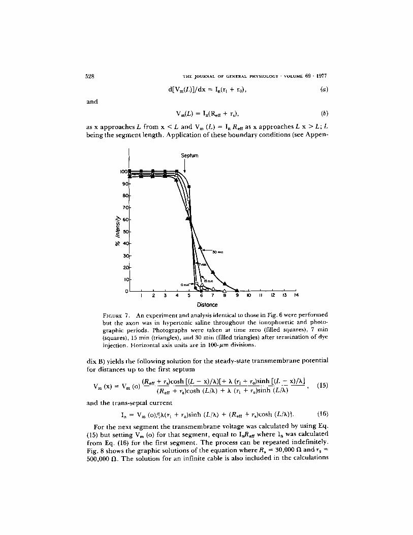
Since fluorescein passes the sep tum with a t ime constant o f approx ima te ly 1 h, it can be used to detect changes in septal permeabi l i ty . Fig. 6 is a g r aph of the
70
60
5 0 -
4 0 -
3 0 -
2 0 -
I 0 -
- 4 0 -
I I t t I I I I I L
- 6 0 -
- 7 0
- 5 0 -
FIGUX~E 5. The steady-state voltage recorded as a function of distance when a current pulse was applied after the axon had been standing in hypertonic saline (3×) for 30 min. Voltage was recorded between a reference electrode centered between the stimulating electrodes and a recording electrode. Circles are the voltages recorded in the extracellular space; x = voltages recorded in the intracellu- lar space.
intensity o f f luorescence of ionophoresed f luorescein in a cell vs. distance a long a giant axon in normal solutions. Pho tographs of the injected cell were made at various t ime intervals. With each negative, background m e a s u r e m e n t s were made and subtracted f rom the measu red intensities in the axon. T h e intensity measu red at the t ime zero in the injected cell o f the axon was taken as 100% intensity. Dur ing the ent i re expe r imen t , that is dye injection and p h o t o g r a p h - ing, the nerve cord was s u r r o u n d e d by hyper tonic saline (3 x) . Compar i son of Figs. 6 and 7 shows that f luorescein trans-septal m o v e m e n t is blocked by hyper- tonic saline. One explanat ion for the block of f luorescein m o v e m e n t is that the nexuses which in par t make up the septal m e m b r a n e are d i s rup ted by hyper -

BRINK AND BARR Resistance of Septum of Median Giant Axon 527
tonic solutions. At very long times ( - 3 h) some fluorescein can be seen appar - ently t ravers ing the s ep tum, but this may be expla ined by m o v e m e n t o f dye in the extracel lular space or by the possibility that not all nexal area is d i s rupted .
A Model for the Earthworm Median Giant Axon
We have c o m p a r e d the predict ion o f a s imple model , which incorpora tes ou r results, with the exponent ia l sp read of cu r r en t a long a cable. This compar i son demons t ra tes the effect o f the existence o f the extra , nexal resistances at the
IOC
9C
8C
7C
6C
5O
4O
3O
20
I0
0 -I
Seplum
I I I I I
0 I, 2 3 4
Se~)tum
~--45 m m
0 ~ 1 7 mm
7 mm ~ 0 "~ , , , , .
5 6 7 8 9 I0 II 12 13 14
Distance
FIGURE 6. Fluorescence of fluorescein in an axon in isotonic saline measured as a function of distance at varying time intervals after termination of the injection period. The time intervals were time zero (triangles), 7 min (squares), 17 min (circles), 30 min (filled triangles), and 45 min (filled squares). The vertical axis represents per cent original intensity. The arrows indicate the positions of the septa along the axon (see text). Distance was measured from the injection electrode. Horizontal axis units are in 100-p.m divisions.
septa in the internal pa thway. Since the e a r t h w o r m median giant axon is made up o f many cylindrical cells a t tached to one ano the r end to end by nexuses at the septa, it can be cons idered as many finite cables in series connected to each o ther by finite resistances (see A p p e n d i x B). T h e general equat ion descr ibing the first segment is:
d2V h 2 = V , (14)
dx 2
where we take V as Vm as described above. I f cur ren t is passed between an internal point and ano t he r immedia te ly outside an EMGA, the internal cu r ren t Ii ap roaches the t ransnexal cu r ren t In at the end o f an axonal segment . T h e r e - fore,
528
and
T H E J O U R N A L OF G E N E R A L P H Y S I O L O G Y • V O L U M E 6 0 " 1977
d[Vm(L)]/dx --- in(ri + ro), (a)
Vm(L ) = In(Re, + rs) , (b)
as x approaches L f rom x < L and Vm (L) = In Reff as x approaches L x > L; L being the segment length. Applicat ion of these b o u n d a r y condit ions (see Appen-
Septum
I 2 3 4 5 6 7 8 9 I0 I I 12 13 14
Distance
FIGURE 7. An experiment and analysis identical to those in Fig. 6 were performed but the axon was in hypertonic saline throughout the ionophoretic and photo- graphic periods. Photographs were taken at time zero (filled squares), 7 min (squares), 15 min (triangles), and 30 min (filled triangles) after termination of dye injection. Horizontal axis units are in 100-/zm divisions.
dix B) yields the following solution for the steady-state t r a n s m e m b r a n e potential for distances up to the first s ep tum
Vm (x) = V m (o) (Reef + rs)cosh [(L - x)/~,)[+ k (ri + ro)sinh [(L - x)/X] (Reff + %)cosh (L/k) + k (ri + ro)sinh ( L / X ) - - ' (15)
and the trans-septal cu r ren t
I n = V m (o)/[k(r~ + ro)sinh (L/k) + (Reff + rs)cosh (L/X)]. (16)
For the next segment the t r a n s m e m b r a n e voltage was calculated by using Eq. (15) but setting Vm (o) for that segment , equal to I,Reff where In was calculated f rom Eq. (16) for the first segment . T h e process can be repea ted indefinitely. Fig. 8 shows the graphic solutions of the equat ion where Rs = 30,000 1~ and rs = 500,000 1~. T h e solution for an infinite cable is also included in the calculations

B R I N K AND B A R R Resistance of Septura of Median Giant Axon 529
made for Fig. 8: the space constant equalled 3.55 mm, the septa occurred every 1 mm (L), and Vo equalled 30 mV. The similarity of the 30 kf/ curve to the exponential would seem to preclude any experimental distinction between them. This would confo rm to our results.
D I S C U S S I O N
Our range of septal specific resistance was 1.5-12.5 f / c m 2. I f the nexus covered 100% of the septum, then the values are equivalent to nexal specific resistance. I f
I I i I ' l , I I
k -- 3.55 mm 3 o ~ _ L = 1.0 mm -
Rn=O -
~ 2 0 - -
o
Rn
i
0i i i i I I , , I I 25 .50 .75 1.25 1.50 1.75 2.25 2.50
IL 2L
FIGURE 8. Graphic solutions of transmembrane potential vs. distance when a constant current pulse is applied to a model septate axon.
the average area of nexus was only 10% the total surface of a septum, the nexal resistance would range f rom 0.15 to 1.25 1"1 cm 2. I f the nexal membrane area were larger due to folding, the nexal resistance would be higher. This range of values for nexal membrane resistance is within the range of nexal membrane resistances previously repor ted (Kriebel, 1967; Weidmann, 1966). The conduct- ance o f nexal membranes is three orders o f magni tude higher than the plasma membrane , and it can be concluded that the nexus is a low-resistance pathway between cells in the giant axon.
It has been suggested that a pore 1.0-2.0 nm in diameter is located in the cen- ter of the particles which make up a nexus (McNutt and Weinstein, 1970). I f one assumes that the conductance o f the pore is the same as an equivalent amoun t o f

530 T H E J O U R N A L OF G E N E R A L P H Y S I O L O G Y • V O L U M E 69 " 1977
axoplasm, calculations can be made to est imate the lower limit on the n u m b e r o f pores needed to give specific septal resistance o f 5.9 l-I cm 2. I f r p = r a L/A where r a equals the resistivity o f cytoplasm (200 f l -cm), and rp equals resistance o f a pore in ohms, and A equals cross-sectional a rea o f the pore , and L equals length of the pore . I f one assumes that the pore is 18 nm in length and has a cross- sectional area o f 3.14 x 10 -14 cm 2, the hypothet ical resistance o f a pore is then rp = 1 . 1 5 x 10 l° ~ .
Given that the particles are a r r anged in an hexagonal a r ray where the center to center distance is 9 nm, the n u m b e r o f pores per 1 cm 2 is 1.4 x 10 TM, and the specific nexal resistance would be 8 × 10 -3 f l cm 2. This value is three orders o f magn i tude less than the observed value o f 5.9 fl cm 2. On the basis o f his morphologica l data on rat myoca rd ium, Mat ter (1973) has calculated a value of 5.1 × 10 -2 f~ cm 2 which contrasts similarly with available physiological data. Obviously, no account was made in the previous calculation for the effect o f frictional forces or fixed charges that may or may not exist in the pore . I f app rox ima te ly 70% of the septal m e m b r a n e area were nexus, the total area of nexus would be 1.2 × 10 -4 cm 2 and there would be 1.7 x 10⁸ pores . T o achieve a specific nexal m e m b r a n e resistance (Rs) o f 5.9 f l cm 2 or septal resistance (rs) or 30,000 ~ , each pore would have to have a resistance o f rp = 5.7 x 10 TM fl. Consequent ly , the axoplasmic resistance (Ra) in such a pore would have to be 100,000 l'l cm or 500 times the bulk axoplasmic resistance. A pore axoplasmic resistance o f 100,000 1~ cm is conceivable only if there are frictional forces or fixed charges at work r e t a rd ing the m o v e m e n t o f ions as they pass t h rough a pore . Ano the r explanat ion for increased axoplasmic resistance in the pore might be smaller pore sizes. But to attain a pore resistance o f 5.7 x 10 TM f / when R a
equals 200 f / c m , the pore d iamete r would have to be 0.09 nm. This low value would not permi t the diffusion even of potass ium ions much less f luorescein. Clearly the diffusion of larger molecules such as Kolodny's (1971) macromolecu- lar RNA, microperox idase (Reese et al., 1971), and Procion yellow ( Imanaga , 1974) would be impossible.
Since potassium is the principal ion on ei ther side of a nexus and cytoplasmic calcium is very low, the bear ing of the T E A exper iments on the possibility that the nexus is t raversed by potass ium channels is significant. T E A was iono- phoresed because of its well-known inhibition o f the activation of potass ium channels , since by analogy with the l inear I-V curve for squid plasma m e m b r a n e (Rojas and Ehrenste in , 1965) one might p r e sume the high nexus conductance is due to activated potass ium channels . T h e r e f o r e , TEA might decrease the nexal conductance . This was not realized. Moreover , T E A crossed septal m e m b r a n e s much more readily than it does p lasma m e m b r a n e .
T h e results o f the hyper tonic exper imen t s were consistent with results that show nexal d isrupt ion in hyper tonic media but could be also expla ined as a decrease in plasma m e m b r a n e resistance. However , the fact that the flow of f luorescein is essentially s t oppped at a s ep tum in the axon in hyper tonic saline probably means that nexal m e m b r a n e s have separa ted in te r fer ing with the longitudinal pa th for the dye. Reduct ion in p lasma m e m b r a n e resistance in hyper tonic saline might allow grea te r loss of dye th rough the plasma m e m b r a n e , thereby decreas ing the a m o u n t o f dye moving along the long axis. However , if

BRINK AND BARR Resistance of Septum of Median Giant Axon 531
this were the case, then the f luorescent intensity o f the injected cell in hyper tonic saline would be expected to fall more rapidly than that of normal saline when, in fact, just the opposite occurs.
The value of axoplasmic resistance (Ra) is in good agreement with previous estimates o f axoplasmic resistance (Goldman, 1964; Dierolf and Brink, 1973). T h e cell length measurement was made to allow computat ion of the total cytoplasmic resistance (ra) of a cell so that comparison of the cytoplasmic resistance with the septal resistance (rs) could be made. Unfor tunately , there is no internal s tandard similar to the sarcomere to determine the p roper length of stretch. To combat this problem f rom prepara t ion to preparat ion, efforts were made to dissect nerve cords of similar length. The average cell length should be considered a very rough estimate of the average distance between septa.
In summary , the nexal membrane complex has been shown to have a low resistance relative to the plasma membrane . It is a nonrect ifying structure over a voltage range of - 10 inV. Since anionic fluorescein and cationic TEA will cross the septum, the role of the nexus in the formation of metabolic coupling needs fur ther clarification by use of techniques such as autoradiography. However , in the ear thworm as well as in o ther examples f rom widely separated species, the nexal pores seem to be large enough to allow intermixing of metabolically impor tant molecules (Gilula, 1974). Thus it is as impor tant to cellular communi- cation as the plasma membrane is to the maintenance of the internal milieu of cells. Al though the nexus may have been one of the first evolutionary steps going f rom unicellular to multicellular organisms, there is evidence that nexuses f rom different tissues or organisms may vary considerably in fine s tructure (Gilula, 1974).
A P P E N D I X A
To analyze our experimental situation in more general terms, consider a segment of a nonmyelinated axon sealed at each end and submerged in paraffin oil so that the outside of the axon is covered by a thin layer of aqueous solution. In addition, small punctate electrodes are touched to the external layer at the very ends of the axon. In order to derive a relatively simple mathematical description of the electrical behavior of this system we will stipulate as is commonly done (Hodgkin and Rushton, 1946; Cole, 1968) that:
(a) all resistances are constant; (b) the external layer and the axon are thin enough and the membrane resistance is
high enough so that the internal and external electrical potentials are functions of distance along the axon only;
(c) a steady state is present; i.e., capacitive transients can be ignored; (d) h z = rm/(r o + r0; (e) I o (x) and Ii (x) are positive when going from right to left and Im (x) is positive when
going outward; 0 c) other symbol are as given earlier; (g) the electrodes are point sources and sinks of current at exactly the ends of a
segment of the axon. The lumped parameter representation of this finite one-dimensional cable is shown in
Fig. 1 A. The equation for the continuous case is
h~d 2 [Vm (x)]/dx 2 - Vm (X) = 0, (1)

532
which has the genera l solut ion
V m (x) = C sinh (x/k) + C1 cosh (x/k),
because o f symmet ry Vm (O) ---- 0, t he re fo re C1 = 0, and thus
Vm (x) = C sinh (x/h),
since Ii ( - L ) = 0.
f] f] Ii (x) = Imdx = (C/rm)sinh (x/k)dx L L
= (Ck/rm) [cosh (x/h) - cosh (L/k)] ,
fu r the r , Iputs e = I o ( - L ) and dVi ( - L ) / d x = O, t he re fo re
dV m ( - L ) / d x = - d V o ( - L ) / d x
(C/k)cosh ( - L/h) = - r o Ipul~e C = -k ro Ipul~ sech (L/k),
T H E J O U R N A L OF G E N E R A L P H Y S I O L O G Y " V O L U M E 69 • 1977
%(o)=0
(2)
(3)
(4)
(5)
t he re fo re
V m ( X ) = - k r o Ipuzse sech (L/X)sinh (x/X). (6)
T h e condi t ions u n d e r which the c o m m o n assumpt ion (Hodgk in and Rushton , 1946),
I i (x) = Ipu]s e ro/(ro + ri),
holds may be seen f rom not ing that this is equivalent to set t ing dVi (x)/dx = dVo (x)/dx
[dV o (x)/dx]/[dV~ (x) /dx] = [dV l (x) /dx - dV m (x) /dx] / [dVi (x)/dx], (7)
u p o n substi tut ion and r e a r r a n g e m e n t
[dVo (xJ/[dV~ (, ,)/d,, (8)
= [ri + ro cosh (x/k)/cosh ( L / k ) ] / [ r i - ri cosh (x/k)/cosh ( L s h ) ] ,
for the e a r t h w o r m axon X may be taken as less than 4 mm. I f p repara t ions 4 cm long are used, cosh ( L / h ) / ~ 75 and the assumpt ion will hold qui te well for the midd le cen t ime te r or so, I f the p repara t ion is longer , the accuracy of the assumpt ion increases very rapidly because o f the d e p e n d e n c e o f cosh (L/k). T h a t the p repa ra t ion behaves in the cen ter reg ion like two parallel resistances can be seen f rom cons ider ing the first o f the two terms descr ib ing the effect ive resistance be tween the e lect rodes
/ * L
= 2Vo (L)/Ip.lse = 2[ri [ Ij (x) dx - Vm (L)]/Ipuise, (9) R e f f .to
Vo(-L) r o ro Vo(L)
rl X=O rl
FIGURE 1 A. Equivalent circuit for an axon o f length 2L. St imula t ing electrodes a re placed at both ends as descr ibed in Materials and Methods. rm, the m e m b r a n e resistance; r0, ext racel lu lar fluid resistance; ri axoplasmic resistances.

BRINK AND BARR Resistance of Septum of Median Giant Axon
R e ff -- 2rl ro k L + 2k ro 2 tanh (L/h). r i r o r i r 0
533
(10)
A P P E N D I X B
T h e inf luence o f the nexal resistances at the septa o f the e a r t h w o r m median giant axon may be cons ide red by seeing how they en te r into the express ion for changes o f trans- m e m b r a n e voltage as a funct ion o f dis tance when a constant cu r ren t is injected into an axon via a mic roe lec t rode . In a very long, nonsep ta te , nonmye l ina t ed axon this o f course would be descr ibed by the famil iar
V m (X) = V m (o ) e -x t~ . (1)
In o r d e r to e x a m i n e the more compl ica ted septate case we stipulate: (a) the first six st ipulat ions o f A p p e n d i x A; (b) the axon is made up o f one -d imens iona l cable segments o f length L which are
connec ted direct ly in the ex te rna l path and connec ted via a nexal resistance Rn in the in ternal path at the septa;
(c) cur ren t , Ipulse, is passed be tween in terna l and externa l point e lectrodes placed at one end of a s egmen t away f rom which the axon ex tends indefini tely;
(d) the cu r r en t across the first s ep tum is I n (Fig. 1 B).
i PULSE ( ) X=O L 2L
FIGURE 1 B. Schemat ic represen ta t ion o f segmenta l axon. L = the posit ion o f the septa. Each cell is connec ted to an adjacent cell by a septal resistance. Ipuls e is the value o f the total appl ied cur ren t .
For the first s egmen t the si tuation is equiva len t to a f ini te cable t e rmina ted by a f ini te resistance, (r s + Ren), where Ren is equal to the effect ive resistance be tween the two point e lectrodes. A genera l solution for the region x < L is
V m (x) = C1 cosh (x/h) + C2 sinh (x/X). (2)
F r o m dV m (x)/dx = dVl (x)/dx - dV 0 (x)/dx and Ohm' s law
dVm (L) dx In (ri + ro), (3)
and f rom O h m ' s law
combin ing to e l iminate I.
f rom (2) at x = L
V m (L) = I, (% + Refr), (4)
V~ (L) = -(r~ + Ren)/(ri + ro) dV~ (L) dx ' (5)
V m (L) = C1 cosh (L/k) + C~ sinh (L/k),
dVm(L) dx -= (Cdh) sinh (L/h) + (C2/h) cosh (L/k),
and f rom (2) at x = 0
(6)
(7)
vm (o) = c , , (8)

534 T H E J O U R N A L OF G E N E R A L P H Y S I O L O G Y " V O L U M E 69 • 1 9 7 7
subst i tut ing into (5)
Vm (0) cosh (L/h) + C2 sinh (L/h)
_ (r~ + Reft) [(Vm(o)/h)sinh (L/h) + (C2/h) cosh L/hi, rL+ro
r e a r r a n g i n g
(9)
sett ing
and
the re fo re ,
fo L V m (X) In = Ipulse -- dx , rm
l foL K = rmVm (o~ V m (x) dx
_ hZ(r~ + to) [cosh (L/h) - 1] + h(rs + Rett)sinh (L/h) rm [(rs + RefOeosh (L/h) + (rt + ro)sinh (L/h)J '
r s + R e f f
F = V m (L)/Vm (o) = [(r~ + Rett)cosh (L/h) + h (r~ + ro)sinh (L/h)] '
upon subst i tut ing into (13)
Ref, = [Vm (o) F - (Ipuls e - - V m (o)K)rs] Ipulse - Vm (o)K - - ' (15)
subst i tut ing f rom (12) for V m (o) and r e a r r a n g i n g
Reft K 2 + (rxK + F - l)Reff - r s = 0. (16)
I f this is mul t ip l ied out , a cubic equa t ion o f the fo rm
AR~n + BR~ff + CReff + D = O, (17)
V o {cosh L/ h + [(rs + Ren)/h (rl + ro)]sinh L/h} (10) C2 = - {[(rs + Ren)/h (rl + ro)]cosh (L/X)} + sinh (L/h) '
Vm (x) = Vm (o) (rs + Ren) cosh [(L - x) /h] + k (ri + ro) sinh [(L - x) /h] (11) (rs + Reu) cosh (L/X) + h (rl + to) sinh (L/h)
An express ion for Ren as a funct ion o f L, r~, r l, ro, and rm is still r equ i r ed in o r d e r to c o m p u t e Vm (x) using (11). By def ini t ion
Rett = Vm (o)/Ip~lse, (12)
and also since the axon is indef ini te ly long
Retf = [Vm (L) - I n rs]/In, (13)
where , the cu r r en t out o f the first s e g m e n t is:
f L
In = I p u l s e - - / Im dx, (14) Jo

BRINK AND BARR Resistance of Septura of Median Giant Axon 535
results, where: A = ),sinh(L/k); B = 2krs sinh(L/k); C = hrs 2 sinh(L/k)-Xrm (ri + ro)sinh (L/k) - rsrm cosh(L/h); D = rsrm [rscosh(L/h) + h(r~ + ro)sinh(L/h)].
The only real positive root of this cubic equation may be easily computed. When r s = 3
× 1041q,rm = 3 × 1051-/cm2,r[ = 3 × 10efl/cm,ro = 0 a n d L = 0.1 cm,Ren = 1.03 × 1061]. This is only 1.08 times r,,x/7~~, which is the equivalent for an equivalent nonseptate axon.
The authors would like to thank Drs. Erik Jakobsson and Maynard Dewey for their valuable discussions. This work was supported in part by National Institutes of Health grant HE 14125.
Received for publication 24 March 1976.
R E F E R E N C E S
BARR, L., W. BERGER, and M. DEweY. 1968. Electrical transmission at nexus between smooth muscle cells.J. Gen. Physiol. 51:347-368.
BARR, L., M. DEWEY, and W. BERGZR. 1965. Propagation of action potential and structure of the nexus in cardiac muscle. J. Gen. Physiol. 48:797-823.
BULLOCK, T. H. 1945. Functional organization of the giant fiber system of Lumbricus. J . Neurophysiol. 8:55-71.
COGGESHALL, R. 1965. A fine structure analysis of the ventral nerve cord of the associated sheath of L. terrestris. J . Comp. Neurol. 125:393-407.
COLE, K. S. 1968. Membranes, Ions and Impulses. University of California Press. 64 et seq.
DrwEv, M., and L. BARR. 1964. A study of the structure and distribution of the nexus.J .
Cell Biol. 23:553-585~ DIrROLF, B., and P. BRINK. 1973. Effects of thermal acclimation on cable constants of the
earthworm median giant axon. Comp. Biochem. Physiol. 44A:401-406. ECCLES, J . , R. GRANtT, and J. Z. YOUNG. 1933. Impulses in the giant nerve fibres of
ea r thworm.J . Physiol. (Lond.). 77:23P-25P. GILULA, N. 1974. Junctions between cells. In Cell Communication. R. P. Cox, editor.
John Wiley & Sons, New York. 1-29. GOLDMAN, L. 1964. The effects of stretch on cable and spike parameters of single nerve
fibers; some implications for the theory of impulse propagation. J. Physiol. (Lond.). 175:425-444.
GRUNDFEST, H., C. Y. KAO, and M. ALTAMIRANO. 1954. Bioelectric effects of ions microinjected into the giant axon of leligo. J. Gen. Physiol. $8:(2, Pt. 2):245-282.
GUNTHER, J. 1971. Der cytologische Aufbau der dorsalen Riensenfaser yon Lumbricus terrestris. Z. Wiss. Zool. 183:51-70.
HAMMA, K. 1959. Some observations on the fine structure of the giant nerve fibers of the earthworm Eisenia foetida. J. Biophys. Biochem. Cytol. 6:61-66.
HODGKIN, A. L. and W. A. H. RUSHTON. 1946. The electrical constants of a crustacean nerve fibre. Proc. R. Soc. Lond. Ser. B Biol. Sci. 133:444-479.
HODGKIN, A. L., and R. D. KEYNES. 1957. Movements of labelled calcium in squid giant axons. J. Physiol. (Lond.). 138:253-281.
IMANAGA, I. 1974. Cell to cell diffusion of Procion yellow in sheep and calf Purkinje fiber. J. Membr. Biol. 16:381-388.

536 THE JOURNAL OF GENERAL PHYSIOLOGY'VOLUME 69 ' 1977
KAO, C. Y., and H. GRUNDFEST. 1957. Postsynaptic electrogenesis in septate giant axons. I. Earthworm median giant axon. J. Neurophysiol. 20:553-573.
KATZ, B. 1966. Nerve, Muscle and Synapse. McGraw-Hill Book Co., New York. KOLOONV. G. M. 1971. Evidence for transfer of macromolecular RNA between mamma-
lian cells in culture. Exp. Cell Res. 65:313-324. KRmBEL, M. E. 1967. Electrical characteristics of tunicate heart cell membranes and
nexuses. J. Gen. Physiol. 52:46-59. McNUTT N. S., and R. WEINSTEIN. 1970. The ultrastructure of the nexus. J. Cell Biol.
47:666-688. McNOTT N. S., and R. S. WEINSTEIN. 1973. Membrane ultrastructure and mammalian
intercellular junctions. Prog. Biophys. Mol. Biol. 26:45-97. MATTER A. 1973. A morphometric study on the nexus of rat cardiac muscle. J. Cell Biol.
56:690-696. PROSSER, C. L., and F. A. BROWN. 1950. Comparative Animal Physiology. W. B. Saun-
ders & Co., Philadelphia, Pa. 75. REESE, T., M. V. L. BENNrTT, and W. FEOrR. 1971. Cell to cell movement of peroxidase
injected into the septate axon of crayfish. Anat. Rec. 169:409. RojAs, E., and G. EHRENSTEIN. 1965. Voltage clamp experiments on axons with potas-
sium as the only internal and external calcium ion.J. Cell Comp. Physiol. 66:71-78. Supp.
2. RUSHTON, W. A. H. 1945. Action potentials from the isolated nerve cord of the earth-
worm. Proc. R. Soc. Lond. Ser. B Biol. Sci. 132:423-437. STEVHENSON, J. 1930. The Oligochaeta. Reprint 1972. J. Cramer and H. K. Swann,
editors. Vol. 92. Historiae Naturalis Classica Ediderunt. Stechant-Hafner Serv. Agency, New York.
STOUGH, H. 1926. Giant nerve fibers of the earthworm. J. Comp. Neurol. 40:409-464. SUBAK-SHARPE, J. H., R. R. BURK, and J. D. PITTS. 1969. Metabolic cooperation between
biochemically marked mammalian cells in tissue culture. J. Cell Sci. 4:353-367. WEIDMANN, S. 1970. Electrical constants of trabecular muscle from mammalian heart .J .
Physiol. (Lond.). 210:1041-1054. WEIDMANN, S. 1966. The diffusion of radiopotassium across intercalated discs of mam-
malian cardiac muscles. J. Physiol. (Lond.). 187:323-342. WEINGART, R. 1974. The permeability to TEA ions of the surface membrane and the
intercalated discs of sheep and calf myocardium. J. Physiol. (Lond.). 240:741-762.