the rate of il-1beta secretion in different … · human myeloid cells activate the nlrp3 ... fu nc...
TRANSCRIPT
1
THE RATE OF IL-1BETA SECRETION IN DIFFERENT MYELOID CELLS VARIES WITH THE EXTENT OF REDOX RESPONSE TO TOLL-LIKE RECEPTOR TRIGGERING *
Sonia Carta1, Sara Tassi1, Ilaria Pettinati1, Laura Delfino1, Charles A. Dinarello2, and Anna Rubartelli1
From Cell Biology Unit, National Cancer Research Institute, 16132 Genova, Italy1 and Department of Medicine, Division of Infectious Diseases, University of Colorado Denver, Aurora, Colorado 80045,
USA2
Running head: Prooxidant and antioxidant events in inflammasome activation Address correspondence to: Anna Rubartelli, MD, Largo Rosanna Benzi, 10; 16132 Genova, Italy. Phone: +39 010 5737379. Fax: +39 010 5737560. E-mail: [email protected] Human myeloid cells activate the NLRP3 inflammasome and secrete IL-1β in response to various Toll-like Receptor (TLR) ligands, but the rate of secretion is much higher in primary human monocytes than in cultured macrophages or THP-1 cells. The different myeloid cells also display different redox status under resting conditions and redox response to TLR activation. Resting monocytes display a balanced redox state, with low production of Reactive Oxygen Species (ROS) and antioxidants. TLR engagement induces an effective redox response with increased ROS generation followed by a sustained antioxidant response, parallelled by efficient IL-1β secretion. Drugs blocking ROS production or the antioxidant response prevent the secretion of mature IL-1β but not the biosynthesis of pro-IL-1β , indicating that redox remodelling is responsible for IL-1β processing and release. Unlike monocytes, THP-1 cells and cultured macrophages have upregulated antioxidant systems that buffer the oxidative hit provided by TLR triggering and suppress the consequent redox response. This aborted redox remodelling is paralleled by low efficiency IL-1β processing and secretion. High doses (5 mM) of H2O2 overcome the high antioxidant capacity of THP-1 cells, restore an efficient redox response and increase the rate of IL-1β secretion. Together these data indicate that a tightly controlled redox homeostasis in resting cells is a prerequisite for a robust redox response to TLR ligands, in turn necessary for the efficient inflammasome activation. Inflammasome activation by bacterial DNA is not modulated by redox responses, suggesting that redox dependent regulation of IL-1β secretion is
restricted to some inflammasomes including NLRP3 but excluding AIM-2.
Interleukin-1β (1) is endowed with two features infrequent in secretory proteins. First, it lacks signal peptides and is externalized through a leaderless pathway of secretion, alternative to the endoplasmic reticulum-Golgi classical one (2). Second, it is produced as an inactive precursor (pro-IL-1β) that is proteolytically converted to the mature bioactive form by caspase-1, in turn activated upon the assembly of the multiprotein complex inflammasome (3). The process of inflammasome activation is ill-defined. The involvement of redox-related events has been proposed for NLR family, pyrin domain containing 3 (NLRP3)-inflammasome activation (4). The underlying mechanism is however controversial. Some studies support a direct contribution of Reactive Oxygen Species (ROS) produced by NADPH-oxidase, but do not provide a molecular mechanism for ROS-mediated inflammasome activation (5-7). Others studies propose a role for the antioxidant response (8,9) that is induced by, and follows, ROS increase (10). The latter hypothesis is based on the pharmacologic evidence that specific inhibition of ROS-induced antioxidant systems prevents IL-1β processing and secretion (9) and on the finding that pro-caspase-1 needs to be reduced by superoxide dismutase-1 (SOD1) to undergo activation (8). Recently, the requirement of ROS for inflammasome activation has been questioned in conditions such as Chronic Granulomatous Disease, where the impaired ROS production due to a genetic defect does not affect IL-1β secretion (11-13).
Many in vitro cell systems are available for the study of inflammasome activation, including mouse macrophages (14), human
http://www.jbc.org/cgi/doi/10.1074/jbc.M110.203398The latest version is at JBC Papers in Press. Published on May 31, 2011 as Manuscript M110.203398
Copyright 2011 by The American Society for Biochemistry and Molecular Biology, Inc.
by guest on July 27, 2018http://w
ww
.jbc.org/D
ownloaded from
2
primary monocytes or cultured macrophages (15, 16), mouse and human continuous myelomonocytic cell lines (17,7). Although any of these cell systems offers some advantages, they frequently differ in the regulation of IL-1β processing and secretion. Mouse macrophages requires Pattern Recognition Receptor (PRR) engagement as a first signal to trigger pro-IL-1β expression and synthesis, and a second stimulus, such as extracellular ATP, crystals (18,19) or pathogenic dusts (7) to induce inflammasome assembly and cleavage and secretion of the cytokine (3). Differently, in human monocytes second signals enhance and accelerate IL-1β processing and secretion but are not strictly required since PRR stimulation drives the release of endogenous ATP that autocrinally triggers the cascade of events leading to inflammasome activation (20). Also cells from the human acute monocytic leukemia cell line THP-1 secrete IL-1β in vitro following a single or a double stimulus (7). However, divergences between inflammasome assembly and IL-1β secretion in THP-1 cells and primary monocytes have been noted. In particular, exposure to reducing agents and downmodulation of thioredoxin have been reported to cause opposite effects on the rate of IL-1β secretion in monocytes (9) and THP-1 cells (7,21).
Long-term tumour cell cultures display many differences from the primary cultures (22). In particular, cell lines generally exhibit a more reduced phenotype than primary cells, due to overexpression of oxido-reductases such as thioredoxin (23). Moreover, the cystine/cysteine cycle is upregulated in many cell lines, with accumulation of free thiols in the culture medium (24). While these redox alterations ensure faster growth, resistance to apoptosis and other benefits to immortalized cells, they may cause erroneous interpretation of results when redox dependent events are studied.
In the attempt to clarify the mechanism of redox regulation of inflammasome, we compared the process of IL-1β processing and secretion in primary human monocytes, cultured human macrophages and THP-1 cells, with respect to the basal redox and the redox changes induced by Toll-like Receptor (TLR) stimulation. In particular, we focused on the activity of the cystine/cysteine redox cycle (24), a major
antioxidant mechanism in inflammatory cells (25-28) that mediates uptake of oxidized cystine from the extracellular environment, intracellular conversion to cysteine and secretion of the reduced aminoacid, resulting in reduction of the extracellular redox. The main molecular components of this redox cycle are xCT, the functional subunit of the Xc- transporter, that mediates the internalization of oxidized cystine
(29), and thioredoxin (30) that participates to the intracellular conversion of cystine to cysteine (31,32).
Our data indicate that a tightly controlled redox homeostasis in resting cells is a prerequisite for an effective redox response to Pathogen Associated Molecular Pattern molecules (PAMPs), in turn necessary for an efficient process of IL-1β maturation and secretion. Due to an unbalance in the basal redox state, with predominance of antioxidant systems, THP-1 cells and cultured macrophages display a weak redox response to PAMPs, resulting in low IL-1β processing and secretion. Remarkably, not all inflammasomes are under redox control: IL-1β processing and secretion in response to transfection of bacterial DNA, that is known to induce AIM-2 inflammasome (33-35), is neither accompanied by redox changes nor modulated by redox-directed inhibitors, both in monocytes and THP-1 cells.
Experimental Procedures Chemicals- L-Buthionine-(S,R)-sulfoximine
(BSO), 1,3-bis(2-chloroethyl)-1-nitrosourea (BCNU), diphenylene iodonium (DPI), 5,5’-dithiobis-(2-nitrobenzoic acid) (DTNB), DTT, hydrogen peroxide (H2O2), LPS, PMA, and zymosan were from Sigma Aldrich; Pam3Cys-Ser-(Lys)4 (PAM3) was from Alexis, ac-YVAD-cmk was from Bachem, 2’,7’-dichlorofluorescein diacetate (H2DCF-DA) and monochlorobimane (MCB) were from Molecular Probes.
Cell cultures- Human monocytes were purified from freshly drawn peripheral blood of healthy donors and stimulated with PAMPs as described (9) or cultured 24 or 48 h in RPMI 1640 medium supplemented with 10% FCS in the presence of 50 ng/ml of GM-CSF (Peprotech) before PAMP stimulation. Murine peritoneal macrophages were obtained from peritoneal lavage of Balb/c mice (Charles River) after 4 days from injection of 4% sterile thioglycollate (Becton
by guest on July 27, 2018http://w
ww
.jbc.org/D
ownloaded from

3
Dickinson). THP-1 cells and RAW 264.7 cells (kind gift of Dr. P. Naquet, CIML, Marseille) were cultured in RPMI 1640 medium supplemented with 10% FCS. THP-1 cells were differentiated 30 min in the presence of PMA (0.3 µg/ml). Monocytes (0.5x10⁶), GM-CSF-cultured monocytes (0.5x10⁶), THP-1 cells (106), RAW 264.7 cells (0.15x10⁶), murine macrophages (106) were then plated in 0.5 ml in 24 well plates and exposed to 1 µg/ml LPS, 50 µg/ml zymosan or 1 µg/ml PAM3 at 37°C in RPMI 1640 medium supplemented with 10% FCS for different times as described (9).
When indicated, the following redox-active compounds were added to the cultures and left for the duration of the experiment: DPI (20 µM), BCNU (50 µM) (both added 15 min after PAMPs); BSO (150 µM, added 15 min before PAMPs); H2O2 (from 0.1 to 10 mM); DTT (from 0.05 to 2 mM). For each substance a dose-response was performed. When single doses are used, they correspond to the concentrations displaying the highest efficacy in the absence of cell toxicity evaluated by measuring cell death by trypan blue exclusion or LDH release. To reduce the experimental variability all the experiments were performed with THP-1 cells and monocytes processed and analyzed in parallel.
ELISA analyses- Pro-IL-1β content of cell lysates and IL-1β content in supernatants were quantified by ELISA (R&D Systems, catalogue number DLBP00 and DY201, respectively) (9).
Determination of intracellular ROS and reduced GSH- Cells were stimulated with the various PAMPs for different times as indicated and 10 µM H2DCF-DA (to assess intracellular ROS, 9, 36) or 100 µM MCB (to assess intracellular GSH, 9, 36) were added to cultures 30 min before the end of the incubation. Fluorescence was measured in cell lysates with a microplate fluorimeter with excitation 480 nm and emission 530 nm for H2DCF-DA. Data were normalized versus cell number or the protein content of cell lysates, measured by the Lowry method (9, 36).
Determination of cysteine in culture media- Supernatants (0.1 ml) from monocytes and THP-1 cells cultured as above were reacted with 10 mM DTNB and the absorption measured at 412 nm. Cysteine was used as standard (9, 37).
Western Blot Analysis- Triton X-100 cell lysates were resolved on 12,5% SDS-PAGE, and electrotransferred. Filters were probed with the following Abs: anti-IL-1β 3ZD mAb (obtained from the NCI Biological Resources Branch, Bethesda, USA), anti-human thioredoxin mAb (clone 2B1, kind gift of Dr. F. Clarke, Brisbane, Australia), anti SOD1 (Stressgene) or anti mouse IL-1β (R&D) followed by the relevant secondary antibody (DAKO) and developed with ECL-plus (GE Healthcare) (9, 36). Anti-β-tubulin (Sigma) or anti-GAPDH (Novus Biologicals) mAb were used as loading control.
NLRP3 silencing- Primary monocytes (10 x106) or THP-1 cells (2x10⁶) were nucleofected with 5 µg of scrambled or NLRP3 siRNA (Invitrogen) according to the manufacturer’s instructions (Amaxa Nucleofector Technology). Transfected monocytes were incubated for 24 h with 20 ng/ml IFN-γ while nucleofected THP-1 cells were primed 30 min with PMA. Both cell types were then stimulated with 1 µg/ml LPS, or 50 µg/ml zymosan for additional 18 h. At the end of incubation, supernatants were collected for ELISA and cells were processed for western blot or gene expression analyses. Real time-PCR- Total RNA was isolated from cell by TriPure Isolation Reagent (Roche) and reverse-transcribed by Superscript III Reverse Transcriptase (Invitrogen) according to manufacturer’s instructions. Real-time PCR determination of xCT, thioredoxin, SOD1, NLRP-3 and pro-IL-1β cDNA was performed using SYBR greenER qPCR Super Mix for iCycler reagent (Invitrogen). The specific primers for xCT and GAPDH were described in (37), and for thioredoxin and SOD1 in (9). The specific primers used for NLRP3 were: 5' GAG GCA ACA CTC TCG GAG AC 3' forward; 5' TCT GGC TGG AGG TCA GAA GT 3' reverse. The specific primers used for pro-IL-1β were: 5' TCC AGG GAC AGG ATA TGG AG 3' forward; 5' TCT TTC AAC ACG CAG GAC AG 3' reverse. Relative expression was determined using ΔCt method (38). Transfection of bacterial DNA- pcDNA3 plasmid (Invitrogen) was isolated from E. coli. To remove contaminating LPS from the DNA preparation, DNA was incubated with polymyxin B (50 µg/ml) at room temperature for 30 min.
by guest on July 27, 2018http://w
ww
.jbc.org/D
ownloaded from

4
DNA was ethanol precipitated twice and resuspended in endotoxin-free water. Transfections were performed using Lipofectamine 2000 according to the manufacturer’s protocol (Invitrogen). All transfections were performed using 1 µg of DNA (39).
Statistical Analysis- The data were statistically analyzed by using one-way ANOVA test, followed by Bonferroni posttest, using GraphPad software.
RESULTS
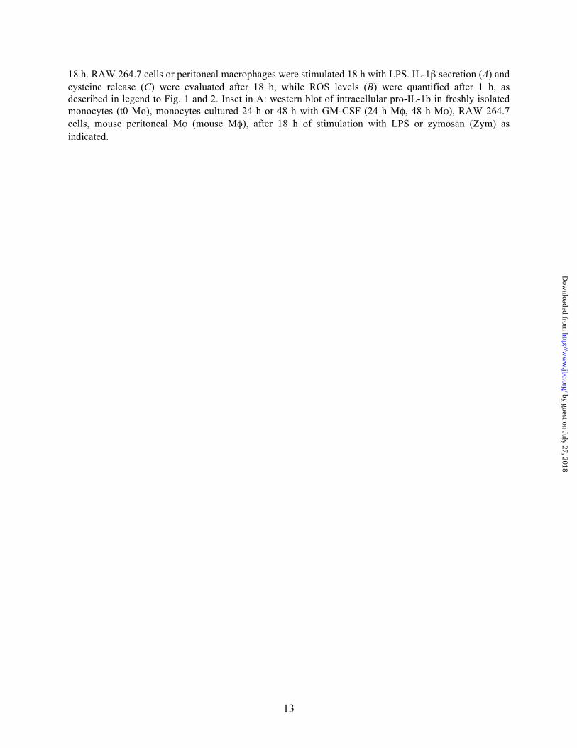
IL-1β secretion by PAMP-stimulated THP-1 cells and primary monocytes. Synthesis, processing and secretion of IL-1β by primary human monocytes and THP-1 cells stimulated by PAMPs display a number of differences (Fig. 1). First, even if a high variability in the synthesis of pro-IL-1β is found in different donors (15), monocytes consistently produce more pro-IL-1β than THP-1 (Fig. 1A and B). In the different experiments the amount of intracellular pro-IL-1β after 18 h of stimulation with PAMPs was 3 to 7 fold bigger in monocytes than in THP-1 cells. The differences in the rate of secretion of mature IL-1β are more dramatic (Fig. 1C and D). PAMP-stimulated monocytes release 15 to 40 times more mature IL-1β than THP-1 cells in 18 h of culture. The portion of secreted IL-1β with respect to the intracellular pro-IL-1β varied from 4 to 10% among the different healthy donors, while it was only 0.2 to 0.35% in THP-1 cells, confirming that the processing/secretory machinery was ≥20 fold more efficient in monocytes than in THP-1. In keeping with previous observations (2, 40), no 17 kDa IL-1β was detected intracellularly (Fig. 1A, see also Supplemental Fig. 1), indicating that, at least in human myeloid cells, pro-IL-1β processing is temporarily associated to IL-1β secretion.
Exposure to the caspase-1 inhibitor ac-YVAD-cmk prevented the secretion of IL-1β (Fig. 1E), but not the synthesis of pro-IL-1β (Fig. 1F) in both primary monocytes and THP-1 cells. siRNA-induced NLRP3 knock-down also resulted in a dramatic inhibition of IL-1β secretion (Fig. 1G), with no effects on pro-IL-1β mRNA expression (not shown), confirming that NLRP3
inflammasome activation is required for PAMP-induced IL-1β secretion (3).
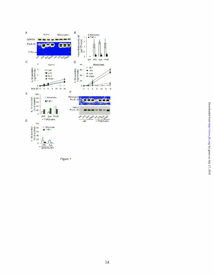
THP-1 cells exhibit a more reduced phenotype than primary monocytes. The redox status of monocytes and THP-1 cells under resting conditions and the redox changes induced by PAMP stimulation were then compared. The amount of oxidants was quantified as ROS production, while the degree of antioxidant activity was evaluated in terms of amount of reduced GSH (41) and of expression and activity of the cystine/cysteine redox cycle (24-28). As shown in Fig. 2A, unstimulated monocytes and THP-1 cells displayed similar levels of basal ROS. In contrast, GSH levels were higher in THP-1 cells than in monocytes (Fig. 2B). The basal activity of the cystine-cysteine cycle was strongly upregulated in THP-1 cells compared to monocytes, resulting in a spontaneous release of 45±12 mm of cysteine in 18h, versus 15±7 mm released by monocytes (Fig. 2C). Interestingly, while xCT mRNA is similar in unstimulated THP-1 cells and monocytes (Fig. 2D), the expression of thioredoxin is much higher in THP-1 cells (Fig. 2E), indicating that the activity of the cystine/cysteine cycle can be modulated by thioredoxin expression in the absence of xCT upregulation. The expression of the anti-oxidant enzyme SOD1 was also investigated and found higher in THP-1 cells than in monocytes (Fig. 2F).
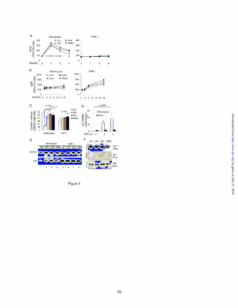
THP-1 cells undergo a milder redox remodelling than monocytes in response to PAMPs. Following TLR triggering, ROS are strongly induced in monocytes, peaking after 1 h and returning to the baseline by 6 h (Fig. 3A, left panel). In contrast, PAMP-induced ROS increase in THP-1 cells was under the treshold of detection (Fig 3A, right panel). The analysis of the antioxidant response revealed that GSH was moderately or no induced by PAMPs in both cell types (Fig. 3B). Differently, TLR triggering caused a stronger upregulation of the cystine/cysteine cycle activity in primary monocytes than in THP-1 cells (Fig. 3C-E). The release of cysteine, evaluated at 18 h from exposure to the various PAMPs, increased four-fold in monocytes, while it was only slightly enhanced in THP-1 cells (Fig. 3C). In agreement, both xCT (Fig. 3D) and thioredoxin (Fig. 3E) are strongly upregulated by PAMPs in monocytes but slightly or no affected in THP-1 cells. Like
by guest on July 27, 2018http://w
ww
.jbc.org/D
ownloaded from
5
thioredoxin, SOD1 is not or barely detectable in monocytes but is overexpressed in THP-1 cells (Fig. 3F). However, while in monocytes a slight induction is observed after TLR triggering, in THP-1 cells PAMP stimulation does not affect the intracellular content of the enzyme. THP-1 cells require PMA priming in order to differentiate to macrophages. The effects of exposure to PMA or PAMPs alone on redox state and IL-1β synthesis and secretion were investigated (Supplemental Fig. 2). PMA, but not LPS, displayed a mild oxidative effect (panels A-D), with a slight decrease in GSH and cysteine release (Panel B and C), but unaffected thioredoxin content (Panel D). Only the association of the two stimuli triggered IL-1β synthesis and secretion, while PMA or LPS alone induced very low, if any, intracellular pro-IL-1β, and no IL-1β secretion (panel E).
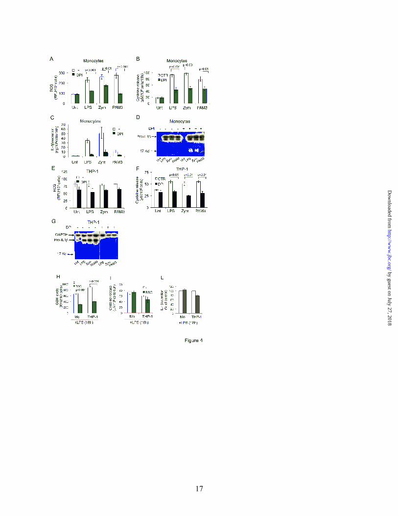
Effects of redox-directed drugs on PAMP-induced pro-IL-1β synthesis and IL-1β secretion. To evaluate the effect of ROS and antioxidant response on inflammasome activation, specific inhibitors of NADPH oxidase (DPI, 42) or of GSH synthase (BSO, 43) were used. In monocytes, DPI inhibited the PAMP-induced ROS increase (Fig. 4A), the consequent antioxidant response (Fig. 4B) and IL-1β secretion (Fig. 4C). ELISA quantification revealed that pro-IL-1β biosynthesis and intracellular accumulation were only slightly affected by DPI treatment, that caused an inhibition of ≤ 20% (mean of 3 different experiments). This result was confirmed by western blot analysis, which also revealed the absence of intracellular 17 kDa mature IL-1β in untreated and DPI-treated cells (Fig. 4D and Supplemental Fig. 1). These results indicate that in monocytes DPI acts post-translationally and inhibits IL-1β processing and secretion. Also in THP-1 cells, DPI treatment lowered both ROS production (Fig. 4E) and the antioxidant response (Fig. 4F). However, PAMP-induced pro-IL-1β synthesis was also blocked (Fig. 4G), making impossible to demonstrate any effect of DPI on the post-translational IL-1β processing. Similarly, BCNU, that blocks cysteine release and IL-1β secretion in monocytes without affecting pro-IL-1β synthesis (9), was found to profoundly inhibit pro-IL-1β synthesis in THP-1 cells (not shown).
Treatment with BSO caused GSH depletion in PAMP-stimulated cells (Fig. 4H), but did not
affect cysteine release (Fig. 4I) and IL-1β secretion (Fig. 4L) by both monocytes and THP-1 cells, ruling out a role for the GSH system in inflammasome activation.
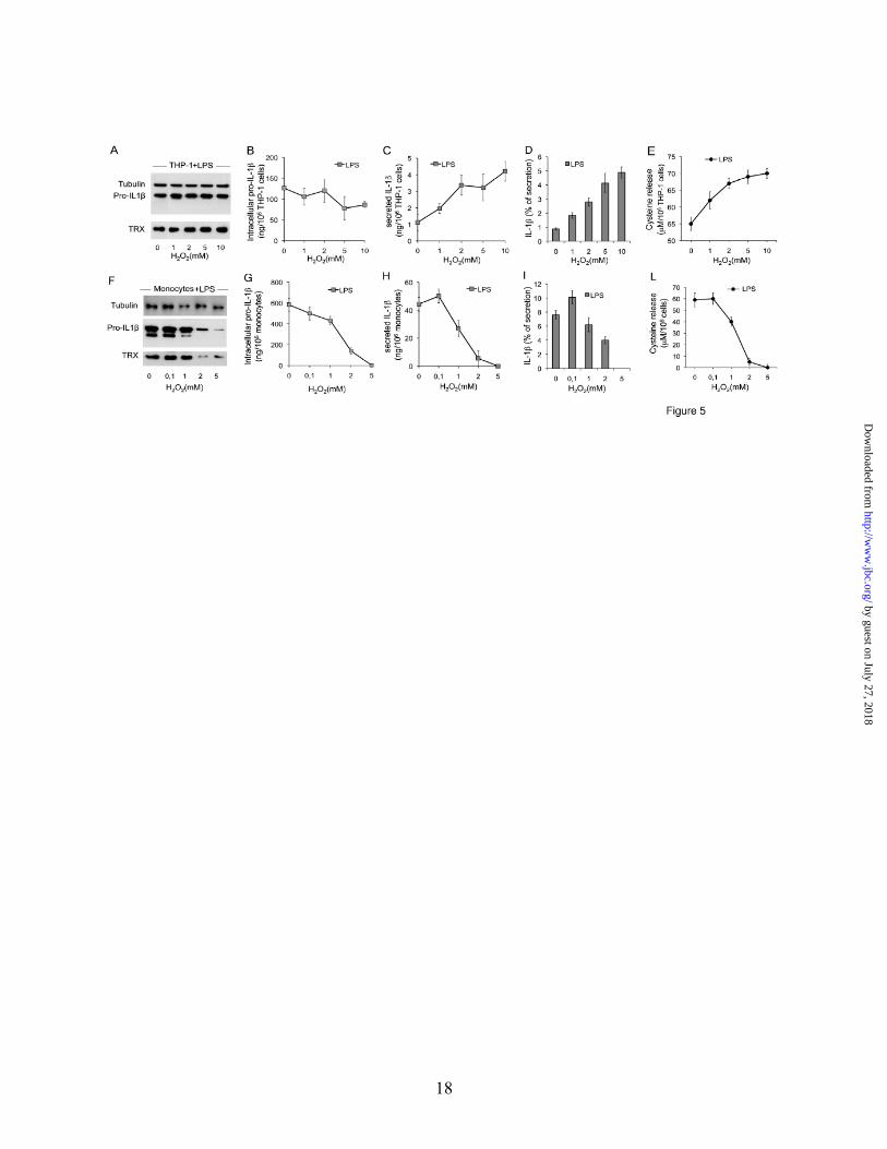
Oxidizing agents induce IL-1β processing and secretion in THP-1 cells. Drugs affecting ROS production and thioredoxin activity are not good tools to study the role of redox on inflammasome activation in THP-1 cells, due to their inhibitory effect on pro-IL-1β synthesis (see Fig. 4). We then investigated whether modulating the redox state of cell cultures with oxidizing or reducing compounds has any effect on IL-1β processing and secretion. THP-1 cells and monocytes were stimulated with LPS in the presence of increasing amounts of H2O2 (Fig. 5). In THP-1 cells, 1 and 2 mM H2O2 strongly induced IL-1β secretion (Fig. 5C and D) without affecting the intracellular content of pro-IL-1β (Fig. 5A and B). Also higher doses (5 and 10 mM) of H2O2 induced secretion of IL-1β (Fig. 5C & D), paralleled by a small decrease in the intracellular pro-IL-1β (Fig. 5B), without affecting cell viability. Consequently, the ratio of IL-1β secreted is strongly increased (Fig. 5D). At all the H2O2 concentrations used, an effective antioxidant response was induced (Fig. 5E).
Unlike in THP-1 cells, both synthesis and secretion of IL-1β were severely affected in primary monocytes exposed to H2O2 at concentrations ≥5 mM (Fig. 5F-I and data not shown), likely due, at least in part, to a cytotoxic effect (>70% of dying cells were found after 18 h of culture). At 2 mM, the toxic effect of H2O2 was instead negligible, but LPS-induced synthesis (Fig. 5F and G) and secretion (Fig. 5H and I) of IL-1β were dramatically prevented. At these concentrations, also the antioxidant response was impaired (Fig. 5L).
The above data suggest that the overexpressed antioxidant systems in THP-1 cells buffer TLR-induced ROS generation, thereby hindering the ROS-induced antioxidant response. A stronger oxidative hit, such as that provided by H2O2 ≥1mM is required to overcome the antioxidant state, allowing an effective redox remodeling and consequently, IL-1β processing and secretion (Fig. 5A-E).
Interestingly, primary monocytes and THP-1 cells displayed a different sensitivity also to
by guest on July 27, 2018http://w
ww
.jbc.org/D
ownloaded from

6
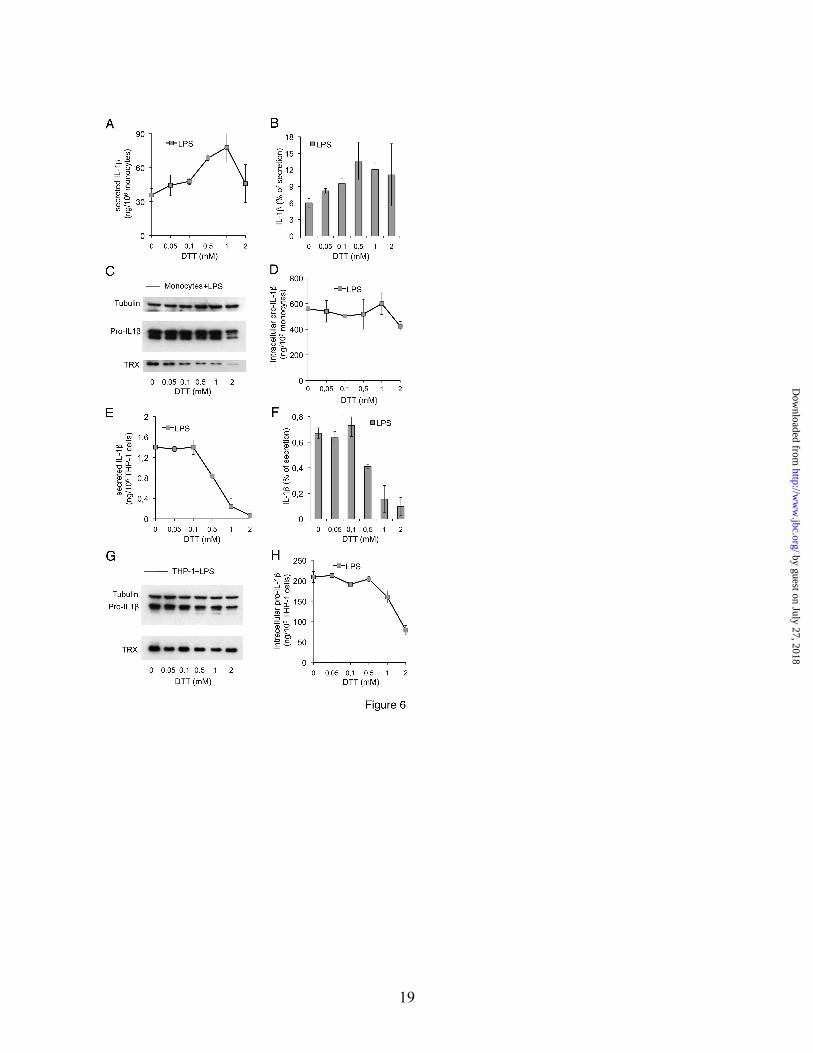
reducing agents (Fig. 6). DTT increased the rate of PAMP-induced IL-1β secretion in monocytes in a dose-dependent manner between 0.1 and 1 mM, (Fig. 6A and B), while the same concentrations exhibited an inhibitory effect on IL-1β secretion by THP-1 cells (Fig. 6E and F). These effects were operative at the post-translational level, since at DTT concentration <2 mM no significant variations in the amount of intracellular pro-IL-1β were observed in monocytes (Fig. 6C & D), and a decrease was detected in THP-1 cells only at 1 and 2 mM DTT (Fig. 6G and H). Interestingly, DTT prevented the increase of intracellular thioredoxin induced by PAMPs in monocytes (Fig. 6C, see also Fig. 3E), indicating that the generation of a reducing environment, rather than the specific increase of thioredoxin, is required for inflammasome activation.
IL-18 like IL-1β is produced as a precursor which undergoes processing by caspase-1 in an inflammasome dependent way (1). Interestingly, similarly to IL-1β, IL-18 secretion is induced by H2O2 in THP-1 cells and by DTT in human monocytes (data not shown) confirming that the inflammasome is the target of the redox related events leading to secretion.
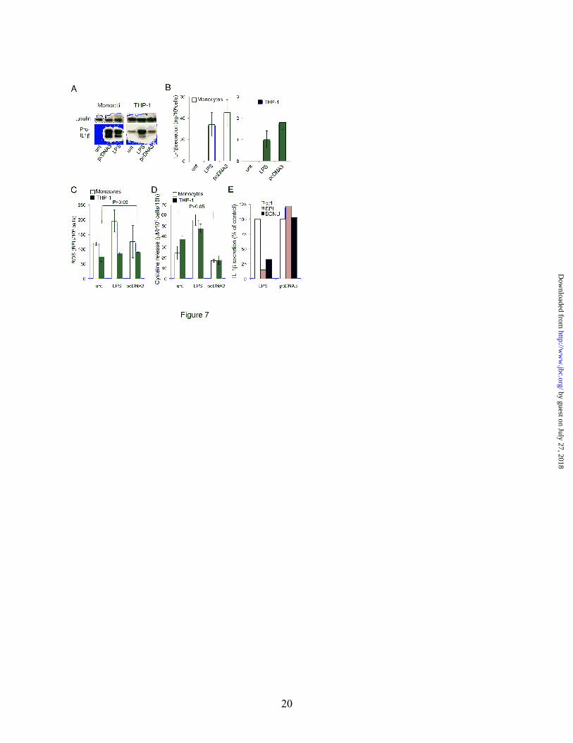
Bacterial DNA induces IL-1β processing and secretion in a redox-independent way. To investigate whether redox changes modulate inflammasomes other that NLRP3, THP-1 cells and monocytes were transfected with bacterial DNA, that induces IL-1β secretion in a NLRP3-independent, AIM2 inflammasome-dependent manner (33-35, 39). IL-1β synthesis and secretion, and redox remodeling were then assessed. As shown in Fig. 7, after liposome-mediated transfection of plasmidic DNA (pcDNA3), pro-IL-1β was induced in monocytes at levels comparable with those induced by LPS, while very low or no induction over the basal level was detected in THP-1 cells (Fig. 7A). In monocytes, also the rate of IL-1β secretion was comparable to that induced by LPS (Fig. 7B). Differently in THP-1 cells, in spite of the low induction of pro-IL-1β by plasmidic DNA, the levels of secreted IL-1β were higher than those induced by LPS (Fig. 7B). Neither ROS production (Fig. 7C) nor cysteine release (Fig. 7D) was significantly modulated by DNA transfection. In addition, in monocytes, DPI and BCNU, that affect ROS production and
thioredoxin activity respectively (9), strongly decreased LPS-induced IL-1β secretion but have no effects on the secretion of IL-1β triggered by cytosolic DNA (Fig. 7E). These findings indicate the activation of IL-1β processing and secretion by cytosolic DNA is independent of redox remodelling. Therefore, in THP-1 cells where redox remodeling is weak and requires a strong hit, the redox independent, cytosolic DNA-mediated activation of inflammasome (most likely AIM2 inflammasome, 33-35) is much more efficient than the redox-dependent NLRP3 inflammasome activation. Monocytes cultured in GM-CSF display a reduced phenotype and an impaired response to PAMPs. We have previously shown that in monocyte-derived macrophages the rate of IL-1β processing and secretion following LPS stimulation is very low (16). We then investigated whether, like in THP-1 cells, the impaired IL-1β secretion is related to an impaired redox state. As shown in Fig. 8A, monocytes cultured 24 h or 48 h with GM-CSF, and then stimulated with LPS or zymosan for additional 18 h, secreted less than 10% the amount of IL-1β secreted by monocytes from the same donor immediately stimulated with PAMPs for 18 h after blood withdrawal. This drop in IL-1β secretion was unrelated to the biosynthesis of pro-IL-1β that was barely decreased with respect to fresh monocytes (Fig. 8A, inset). The ROS levels in unstimulated GM-CSF-cultured monocytes were only a little lower than in unstimulated fresh monocytes, but were not further induced by PAMPs (Fig. 8B). Likewise, in GM-CSF-cultured cells the antioxidant response, evaluated as cysteine release, was weak or absent (Fig. 8C). A similarly low rate of IL-1β secretion paralleled by a weak redox remodeling after TLR triggering was observed in other myeloid cells, such as peritoneal murine macrophages and the mouse leukaemic monocyte macrophage cell line RAW 264.7 (Fig. 8A-C), independently of the basal redox phenotype.
DISCUSSION
Here we have compared freshly isolated
primary monocytes, GM-CSF-cultured monocytes and THP-1 cells and found striking differences in
by guest on July 27, 2018http://w
ww
.jbc.org/D
ownloaded from
7
the redox processes involved in NLRP3 inflammasome activation that result in a dramatically different rate of IL-1β secretion.
Under resting conditions, the redox homeostasis in primary monocytes is ensured by a low amount of ROS counterbalanced by a low expression of antioxidant systems. This balanced redox state allows an efficient redox remodelling after TLR stimulation, with ROS production followed by an adaptive response aimed at restoring the redox homeostasis. In contrast, unstimulated THP-1 cells and GM-CSF-cultured monocytes, in spite of a low level of basal ROS, display upregulated antioxidant systems, with high activity of the cystine/cysteine cycle and secretion of large amount of cysteine in the absence of TLR triggering. Moreover, unlike in freshly drawn monocytes, in THP-1 cells and cultured monocytes the induction of ROS by PAMPs is low or undetectable and the antioxidant response is weak. A likely explanation of this finding is that in resting THP-1 cells and GM-CSF-cultured monocytes the oxidative hit provided by PAMPs is under the threshold required to increase ROS, due to the expanded antioxidant systems that quench the induced ROS. In turn, ROS at low levels are not able to induce a sustained antioxidant response. This weak redox remodelling is not sufficient to efficiently activate the inflammasome, with the net result of a low rate of IL-1β processing and secretion.
Support to this interpretation comes from the observation that high doses of H2O2 are well tolerated and induce antioxidant response and IL-1β secretion in THP-1 cells, while at the same doses monocytes undergo oxidative stress and die. This result indicates that an oxidative hit stronger than that provided by PAMPs is required to overcome the high reducing capacity of THP-1 cells and to induce an antioxidant response, while the same stronger hit is lethal for resting monocytes, due to their low reducing capacity. The different redox state of the two cell types is also the reason why the same concentrations of the reducing agent DTT profoundly inhibit PAMP-induced IL-1β processing and secretion in THP-1 cells but enhance it in primary monocytes. Conceivably, a reducing environment is well tolerated by monocytes, thanks to their balanced redox state with low antioxidant expression, and
favours inflammasome activation. In contrast, THP-1 cells, due to their dramatically expanded antioxidant apparatus are unable to further resist exposure to reducing agents, and undergo a “reductive stress”. A similar reductive stress with high levels of antioxidants and decreased levels of ROS occurs in mice overexpressing Hsp27 and, remarkably, results in a weaker systemic response to LPS (44,45). In other myeloid cell lines, such as the promyelocytic leukemia cell line HL-60, exposure to DTT induces apoptosis, indicating that cells with a highly reducing potential are unable to resist to an additional reducing hit (46). The different redox state of resting monocytes and THP-1 cells is therefore responsible for their different response to pro- or antioxidant agents both in terms of survival and of modulation of IL-1β secretion. While the involvement of redox related events in NLRP3 inflammasome activation is largely accepted, the underlying molecular mechanism(s) have still to be defined, either in the case of second signal-induced activation, that provide a stronger oxidative hit, or in response to TLR triggering, that induces a milder redox response. In particular, ROS have been proposed to activate the inflammasome but the molecular target(s) and the consequence of oxidation are unknown (4). On the other hand, the anti-oxidant response to ROS is required for inflammasome activation (8,9) but how antioxidants mediate inflammasome activation is similarly unclear. A possible mechanism has been proposed by Meissner et al. (8), who reported that upregulation of the antioxidant enzyme SOD1 results in reduction of two gluthationylated cysteine residues on pro-caspase-1, allowing the activation of the enzyme, otherwise inactive (8). Accordingly, SOD1 silencing prevents PAMP-induced IL-1β secretion in human monocytes (9). We show here that both SOD1 and thioredoxin are expressed at very low level in resting monocytes and are upregulated by PAMPs. In contrast, both SOD1 and thioredoxin are overexpressed by THP-1 cells but are not further induced by PAMPs. If reducing conditions would be sufficient to activate the inflammasome, the redox state of resting THP-1 cells should result in constitutive inflammmasome activation. In contrast, we have shown here that the inflammasome activity is low under the reducing conditions that characterize THP-1 cells
by guest on July 27, 2018http://w
ww
.jbc.org/D
ownloaded from
8
and is enhanced when a stronger oxidative hit allows ROS increase and the consequent antioxidant response. Together these findings indicate that an effective redox remodelling after PAMP stimulation, with ROS increase followed by antioxidant response, is required for inflammasome activation. Pre-existing reducing conditions do not favour inflammasome activity but rather prevent PAMP-induced redox remodelling, by buffering the mild oxidative hit provided by TLR engaments. This conclusion is supported by the observation that different myeloid cells (human and mouse macrophages, THP-1 and RAW 264.7 cells), that under resting conditions have levels of antioxidants different but in all cases high enough to smooth the redox remodelling to TLR stimulation, display a very low efficiency of IL-1β processing and secretion in response PAMPs.
Cytosolic DNA has been shown to bind AIM2 that assembles with ASC and caspase-1 leading to pro-IL-1β processing (33-35). An important finding of this study is that inflammasome activation by transfected bacterial DNA is not modulated by redox responses. Moreover, in THP-1 cells the cytosolic DNA-mediated activation of AIM2 inflammasome is much more efficient than the TLR ligand-mediated activation of NLRP3 inflammasome, resulting in higher rate of IL-1β processing and secretion. Since THP-1 cells display a weak redox remodeling due to expanded antioxidant systems under basal conditions, it is conceivable that the stronger activation of AIM2 inflammasome than of NLRP3 inflammasome is related to the different requirement of a redox response by the two inflammasomes: not necessary for the former, while mandatory for the latter. In any case, these data indicate that the redox-mediated regulation is not a property of all inflammasomes and suggest that the redox status of cells and
microenvironment influences inflammasome assembly induced by extracellular but not intracellular activators.
The redox alterations of THP-1 cells, RAW 264.7 cells and macrophages are different from those observed in monocytes from patients affected by Cryopyrin Associated Periodic Fever syndromes, where mutations in the NLRP3 gene cause increased IL-1β secretion (1). These cells under resting conditions display elevated levels not only of antioxidants, like cells used in this study, but also of ROS (37). The redox response to TLR triggering is abnormal also in NLRP3-mutated monocytes: however, while in THP-1 cells the redox response is frustrated, in mutated monocytes is quickened and then rapidly exhausted. As a result, secretion of IL-1β is accelerated, but reaches a plateau much earlier than in healthy controls (37).
In conclusion, we have shown that the different redox state under resting conditions and the redox remodelling following PAMP stimulation in primary monocytes, cultured macrophages and THP-1 cells dictate the different efficiency of inflammasome activation displayed by the three cell types as well as their different response to compounds affecting the microenvironmental redox. Therefore, many of the discrepant data reported in the literature can be ascribed to the different redox state of primary and continuous monocytic cells. Not only our results recommend caution on the interpretation of data obtained from in vitro studies of redox related events on tumor cell lines, but also suggest that, in vivo, different redox alterations, as found in many chronic diseases, may perturb the innate immune response by altering inflammasome activation and IL-1β secretion.
REFERENCES
1. Dinarello, C. A. (2009) Annu. Rev. Immunol. 27, 519-550 2. Rubartelli, A., Cozzolino, F., Talio, M., and Sitia, R. (1990) EMBO J. 9, 1503-1510. 3. Schroder, K., and Tschopp, J. (2010) Cell 140, 821-832. 4. Martinon, F. (2010) Eur. J. Immunol. 40, 616-619. 5. Cruz, C. M., Rinna, A., Forman, H. J., Ventura, A. L., Persechini, P. M., and Ojcius, D. M.
(2007) J. Biol. Chem. 282, 2871-2879. 6. Hewinson, J., Moore, S. F., Glover, C., Watts, A. G., and MacKenzie A. B. (2008) J. Immunol.
by guest on July 27, 2018http://w
ww
.jbc.org/D
ownloaded from

9
180, 8410-8420. 7. Dostert, C., Pétrilli, V., Van Bruggen, R., Steele, C., Mossman, B. T. and Tschopp J. (2008)
Science 320, 674-677. 8. Meissner, F., Molawi, K., and Zychlinsky, A. 2008. Nat. Immunol. 9, 866-872. 9. Tassi, S., Carta, S., Vené, R., Delfino, L., Ciriolo, M. R., and Rubartelli, A. (2009) J. Immunol.
183, 1456-1462. 10. Carta, S., Castellani, P., Delfino, L., Tassi, S., Venè, R. and Rubartelli, A. (2009) J. Leuk. Biol.
86, 549-555. 11. Meissner, F., Seger, R. A., Moshous, D., Fischer, A., Reichenbach, J., and Zychlinsky, A. (2010)
Blood 116, 1570-1573. 12. van Bruggen, R., Koker, M.Y., Jansen, M., van Houdt, M., Roos, D., Kuijpers, T.W., and van den
Berg, T.K. (2010) Blood 115, 5398-5400. 13. van de Veerdonk, F. L., Smeekens, S. P., Joosten, L. A., Kullberg, B. J., Dinarello, C. A., van der
Meer, J. W., and Netea, M.G. (2010) Proc. Natl. Acad. Sci. U S A 107, 3030-3033. 14. Mariathasan, S., Weiss, D.S., Newton, K., McBride, J., O'Rourke, K., Roose-Girma, M., Lee,
W.P., Weinrauch, Y., Monack, D.M., and Dixit, V.M. (2006) Nature 440, 228-32. 15. Gattorno, M., Tassi, S., Carta, S., Delfino, L., Ferlito, F., Pelagatti, M.A., D'Osualdo, A.,
Buoncompagni, A., Alpigiani, M.G., Alessio, M., Martini, A., and Rubartelli, A. (2007) Arthritis Rheum. 56, 3138-48.
16. Netea, M.G., Nold-Petry, C.A., Nold, M.F., Joosten, L.A., Opitz, B., van der Meer, J.H., van de Veerdonk, F.L., Ferwerda, G., Heinhuis, B., Devesa, I., Funk, C.J., Mason, R.J., Kullberg, B.J. Rubartelli, A., van der Meer, J.W., and Dinarello, C.A. (2009) Blood 113, 2324-35.
17. Ngai, S., Batty, S., Liao, K.C., and Mogridge, J. (2010) FEBS J. 277, 119-27. 18. Martinon, F., Burns, K., and Tschopp, J. (2002) Mol. Cell 10, 417-26. 19. Duewell, P., Kono, H., Rayner, K. J., Sirois, C. M., Vladimer, G., Bauernfeind, F. G., Abela, G.
S., Franchi, L., Nunez, G., Schnurr, M., Espevik, T., Lien, E., Fitzgerald, K. A., Rock, K. L., Moore, K. J., Wright, S. D., Hornung, V., and Latz, E. (2010) Nature 464, 1357-1361.
20. Piccini, A., Carta, S., Tassi, S., Lasiglie’, D., Fossati, G., and Rubartelli, A. (2008) Proc. Natl. Acad. Sci. U S A 105, 8067-8072.
21. Zhou, R., Tardivel, A., Thorens, B., Choi, I., and Tschopp, J. (2010) Nat. Immunol. 11, 136-140. 22. Hwang, C., and Sinskey, A. J. (1991) In: Production of Biologicals from animal cells in culture,
eds Spier, R.E., Griffiths, J. B., and Meignier, B., Halley Court, Oxford. p. 548-567. 23. Powis, G., Kirkpatrick, D.L., Angulo, M., and Baker, A. (1998) Chemico-Biological Interactions
111-112, 23-34. 24. Banjac, A., Perisic, T., Sato, H., Seiler, A., Bannai, S., Weiss, N., Kölle, P., Tschoep, K., Issels,
R. D., Daniel, P. T., Conrad, M., and Bornkamm, G. W. (2008) Oncogene 27, 1618-1628. 25. Sido, B., Braunstein, J., Breitkreutz, R., Herfarth, C., and Meuer, S.C. (2000) J. Exp. Med. 192,
907-912. 26. Angelini, G., Gardella, S., Ardy, M., Ciriolo, M. R., Filomeni, G., Di Trapani, G., Clarke, F.,
Sitia, R., and Rubartelli, A. (2002) Proc. Natl. Acad. Sci. U S A 99, 1491-1496. 27. Sido, B., Lasitschka, F., Giese, T., Gassler, N., Funke, B., Schroder-Braunstein, J., Brunnemer,
U., Meuer, S. C., and Autschbach, F. (2008) Gastroenterology 134, 179-191. 28. Yan, Z., Garg, S. K., and Banerjee, R. (2009) Nat. Chem. Biol. 5, 721-723. 29. Sato, H., Tamba, M., and Bannai, S. (1999) J. Biol. Chem. 274, 11455-11458. 30. Lillig, C. H., and Holmgren, A. (2007) Antioxid. Redox Signal. 9, 25-47. 31. Holmgren, A. (1977) J. Biol. Chem. 252, 4600-4606. 32. Mandal, P. K., Seiler, A., Perisi, T., Kolle, P., Banjac Canak, A., Forster, H., Weiss, N.,
Kremmer, E., Lieberman, M. W., Bannai, S., Kuhlencordt, P., Sato, H., Bornkamm, G. W., and Conrad, M. (2010) J. Biol. Chem. 285, 22244-22253.
33. Fernandes-Alnemri, T., Yu, J.W., Datta, P., Wu, J., and Alnemri, E.S. (2009) Nature 458, 509–
by guest on July 27, 2018http://w
ww
.jbc.org/D
ownloaded from

10
513. 34. Hornung, V., Ablasser, A., Charrel-Dennis, M., Bauernfeind, F., Horvath, G., Caffrey, D.R., Latz,
E., and Fitzgerald, K.A. (2009) Nature 458, 514–518. 35. Burckstummer, T. Baumann, C., Blüml, S., Dixit, E., Dürnberger, G., Jahn, H., Planyavsky, M.,
Bilban, M., Colinge, J., Bennett, K.L., and Superti-Furga, G. (2009) Nat. Immunol. 10, 266–272. 36. Vené, R., Delfino, L., Castellani, P., Balza, E., Bertolotti, M., Sitia, R., and Rubartelli, A. (2010)
Antioxid. Redox Signal. 13, 1145-1155. 37. Tassi, S., Carta, S., Delfino, L., Corsi, R., Martini, A., Gattorno, M., and Rubartelli, A. (2010)
Proc. Natl. Acad. Sci. U S A 107, 9789-9794. 38. Livak, K. J., and Schmittgen, T. D. (2001) Methods 24, 402–408. 39. Muruve DA, Pétrilli V, Zaiss AK, White LR, Clark SA, Ross PJ, Parks RJ, Tschopp J. Nature.
2008 Mar 6;452(7183):103-7. 40. Andrei, C., Dazzi, C., Lotti, L., Torrisi, M.R., Chimini, G., and Rubartelli, A. (1999) Mol. Biol.
Cell. 10, 1463-1475. 41. Meister, A. (1995) Methods Enzymol. 251, 3–7. 42. Ellis, J. A., Mayer, S. J., and Jones, O. T. (1988) Biochem. J. 251, 887–891. 43. Griffith, O. W. (1982) J. Biol. Chem. 257, 13704–13712. 44. You, W., Min, X., Zhang, X., Qian, B., Pang, S., Ding, Z., Li, C., Gao, X., Di, R., Cheng, Y., and
Liu, L. (2009) Shock 32, 108-117. 45. Zhang, X., Min, X., Li, C., Benjamin, I. J., Qian, B., Zhang, X., Ding, Z., Gao, X., Yao, Y., Ma,
Y., Cheng, Y., and Liu, L. (2010) Hypertension 55, 1412-1417. 46. Tartier, L., McCarey, Y. L., Biaglow, J. E., Kochevar, I. E., and Held, K. D. (2000) Cell Death
Differ.7, 1002-1010.
FOOTNOTES *We thank Dr A. Omenetti and R. Vené for helpful discussion, Dr. E. Balza for the help with mice experiments, Dr P. Naquet for the kind gift of RAW 264.7 cells, the National Cancer Institute (Biological Resources Branch) for generously providing the anti-IL-1β 3ZD mAb and Prof. F. Clarke for the gift of anti thioredoxin mAb. The work was supported in part by grants from Telethon, Ministero Salute, Compagnia San Paolo. The Abbreviations used are: BCNU, 1,3-bis(2-chloroethyl)-1-nitrosourea; BSO, L-buthionine-(S,R)-sulfoximine; DPI, diphenylene iodonium; DTNB, 5,5'-dithiobis-(2-nitrobenzoic acid); GSH, glutathione; H2DCF-DA, 2',7'-dichlorofluorescein diacetate; MCB, monochlorobimane; PAM3, Pam3Cys-Ser-(Lys)4; PAMP, pathogen-associated molecular pattern molecule; PRR, Pattern Recognition Receptor; ROS, reactive oxygen species; SOD1, superoxide dismutase-1; TLR, Toll-like Receptor.
FIGURE LEGENDS Fig. 1. PAMP-stimulated THP-1 cells secrete low amount of IL-1β. PMA-activated THP-1 cells and monocytes from healthy donors (HD, n=10) were cultured for different times in the absence or presence of LPS, zymosan (Zym) or PAM3. A. Western blot of intracellular pro-IL-1β in THP-1 cells and in monocytes from one representative healthy donor, analyzed in parallel after 18 h of exposures to the different PAMPs. One representative experiment out of 4 is shown. B. ELISA quantification of intracellular pro-IL-1β in monocytes (grey columns) and THP-1 cells (black columns) after 18 h of incubation in the absence or presence of PAMPs, as indicated. Data are expressed as µg/106 cells (mean ±SD, n=3).
by guest on July 27, 2018http://w
ww
.jbc.org/D
ownloaded from

11
C and D. Kinetics of IL-1β secretion by resting or PAMP-activated THP-1 cells (C) and monocytes (D). Secreted IL-1β was quantified by ELISA. Data are expressed as ng/106 cells (mean ± SD, n=3). E and G. IL-1β secreted by monocytes (open column) or THP-1 (closed columns) exposed to LPS, Zymosan or PAM3 for 18 h as indicated in the presence or absence of 200 µM ac-YVAD-cmk (E) or after NLRP3 silencing (G). Secreted IL-1β was quantified by ELISA. Data are expressed as percent of IL-1β secreted by cells stimulated by PAMPs in the absence of ac-YVAD-cmk (E) or after nucleofection with scramble siRNA (G). F. Western blot of intracellular pro-IL-1β in PAMP-stimulated monocytes and THP-1 cells, cultured 18 h in the absence (ctrl) or presence (+ YVAD-cmk) of ac-YVAD-cmk (n=3). Fig. 2. Differences in the redox state of resting monocytes and THP-1 cells A-D. Intracellular ROS and GSH levels, cysteine release, expression of xCT, thioredoxin and SOD1 were compared in unstimulated monocytes (Mo) and THP-1 cells. ROS and GSH were evaluated on a per cell basis. Same results were obtained by normalizing ROS toward the cell protein content (not shown). A. ROS levels were quantified by H2DCFDA fluorometric methods. Data are expressed as Relative Fluorescence Units (RFU), mean ± SD (n=3). B. GSH levels were quantified by MCB fluorometric methods. Data are expressed as RFU, mean ± SD (n=3). C. Extracellular cysteine was quantified by DTNB assay in 18 h supernatants. Data are expressed as µM/106 cells (mean ±SD, n=4). D-F. The expression of mRNA for xCT (D), thioredoxin (Trx, E) and SOD1 (F) was analyzed by real-time PCR. Data are expressed as fold changes in THP-1 cells with respect to unstimulated monocytes. In each case, one representative experiment out of 3 performed is shown. Fig. 3. A weak redox remodelling is induced by PAMPs in THP-1 cells. Monocytes and THP-1 cells were cultured in the absence or presence of LPS, zymosan or PAM3 for various times as indicated. A and B. ROS (A) and GSH (B) levels were evaluated as in Fig. 2 before stimulation (0) and at different times, from PAMP exposure in monocytes (left panel) and THP-1 cells (right panel), as indicated. At all time points, ROS and GSH levels were normalized against protein content. C. Extracellular cysteine was quantified by DTNB assay in 18 h supernatants of monocytes and THP-1 cells, cultured without or with the different PAMPs. Data are expressed as µM/106 cells (mean ±SD, n=4). D. xCT expression in monocytes (3 different donors, white columns) and THP-1 cells (grey columns) before (0) or after 3 and 6h from exposure to LPS was assayed by real-time PCR. Data are expressed as fold changes with respect to unstimulated monocytes (mean ± SD, n=3). E. Western blot analysis of thioredoxin (Trx) in cell lysates from monocytes and THP-1 cells unstimulated (lanes 1 and 5) or stimulated 18 h with LPS (lanes 2 and 6) zymosan (lanes 3 and 7), PAM3 (lanes 4 and 8). Top panel: the same blot was hybridized with anti-GAPDH as loading control. F. Western blot analysis of SOD1 in cell lysates from THP-1 (20 µg, top panel) or monocytes (20 µg, middle panel, or 80 µg, lower panel). For E and F, results from monocytes from one representative healthy donor out of the six analyzed, and one experiment on THP-1 cells out of 4 performed are shown. Fig. 4. Effects of redox active drugs (DPI and BSO) on monocytes and THP-1 redox remodelling and IL-1β secretion. The effects of DPI (A-G) or BSO (H-L) on PAMP-induced redox remodelling and IL-1β secretion were investigated in monocytes and THP-1 cells. ROS levels (A and E), cysteine release (B and F), IL-1β secretion (C) were analyzed in monocytes (A-C) and THP-1 cells (E and F) exposed (closed columns) or not (open columns) to DPI 30 min after the addition of the various PAMPs. A, E. ROS levels by untreated or DPI-treated cells were measured after 1 h from PAMP exposure. B, C, F. cysteine release (B,F) and IL-1β secretion (C) by untreated or DPI-treated cells were measured
by guest on July 27, 2018http://w
ww
.jbc.org/D
ownloaded from

12
after 18 h from PAMP exposure. D & G. pro-IL-1β present in cell lysates from untreated (-) or DPI-treated (+) monocytes (D) and THP-1 cells (G) after 18 h of culture with the various PAMPs as indicated was analyzed by western blot. H-L. Monocytes and THP-1 cells were cultured 18 h without or with LPS in the absence or presence of BSO and GSH levels (H), cysteine release (I) and secreted IL-1β (L) were quantified as in legend to Fig. 1 and 2. Mean of 3 different experiments ± SD is shown. Fig. 5. H2O2 induces antioxidant response and IL-1β secretion in THP-1 cells but not in monocytes. THP-1 cells (A-E) and monocytes (F-L) were exposed 18 h to LPS in the presence or absence of increasing concentrations H2O2 as indicated. A, F Western blot analyses of intracellular pro-IL-1β and thioredoxin (Trx) (top panel: β-tubulin as a loading control) in cell lysates from THP-1 cells (A) and monocytes (F). One representative experiment out of three is shown. B, C, G and H. Intracellular pro-IL-1β (B and G) and secreted IL-1β (C and H) were quantified by ELISA in cell lysates and supernatants respectively (B and C: THP-1 cells; G and H, monocytes). Data are expressed as ng/106 cells (mean±SD of 2 independent experiments). D & I. The ratio of IL-1β secretion was calculated according to the formula: [secreted IL-1β/(intracellular pro-IL-1β + secreted IL-1β)] x 100. E & L. Extracellular cysteine present in 18 h supernatants of THP-1 cells (E) and monocytes (L) was quantified by DTNB assay . Data are expressed as µM/106 cells (mean±SD of 3 independent experiments). Fig. 6. DTT induces IL-1β secretion in monocytes but not in THP-1 cells Monocytes (A-D) and THP-1 cells (E-H) were cultured 18 h with LPS, in the absence or presence of increasing concentrations of DTT as indicated. A and E. Secreted IL-1β was quantified by ELISA (n=2). B and F. The ratio of IL-1β secretion is calculated as in legend to Fig. 5. C and G. Western blot analysis of intracellular pro-IL-1β and thioredoxin (Trx) from one representative healthy donor and from THP-1 cells (n=3). Upper panel: the same blot was hybridized with anti-β-tubulin. D and H. Intracellular pro-IL-1β was quantified by ELISA in cell lysates of monocytes (D) and THP-1 cells (H). Data (mean±SD) from 2 independent experiments are shown. Fig. 7. Cytosolic DNA induces IL-1β processing and secretion in a redox-independent way. A. Western blot analyses of intracellular pro-IL-1β in monocytes (lanes 1-3) and THP-1 cells (lanes 4-6) after 18 h in culture in the absence (untreated, unt) or presence of LPS (LPS) or after lipofectamine mediated transfection of pcDNA3 (pcDNA3). Upper panel: the same blot was hybridized with anti-β-tubulin. B. IL-1β secreted by the same cells shown in A, measured by ELISA. C, D. ROS levels (C) and cysteine release (D) in cells treated as in A, measured after 1 h (C) or 18 h (D) respectively from exposure to LPS (LPS) or from transfection (pcDNA3). E. Monocytes were exposed to LPS (LPS) or transfected (pcDNA3) and then cultured in the absence or presence of DPI or BCNU. IL-1β secreted after 18 h was quantified by ELISA. Data are expressed as percent of IL-1β secretion by control cells (LPS-treated or pcDNA3 transfected cells, not exposed to DPI or BCNU). Fig. 8. GM-CSF cultured monocytes, mouse macrophages and RAW 264.7 cells display mild redox changes to PAMP stimulation and a poor IL-1β secretion. Monocytes freshly drawn for peripheral blood were immediately processed and exposed to LPS or zymosan (Zym) or cultured 24 h or 48 h with GM-CSF before exposure to LPS or zymosan for additional
by guest on July 27, 2018http://w
ww
.jbc.org/D
ownloaded from
13
18 h. RAW 264.7 cells or peritoneal macrophages were stimulated 18 h with LPS. IL-1β secretion (A) and cysteine release (C) were evaluated after 18 h, while ROS levels (B) were quantified after 1 h, as described in legend to Fig. 1 and 2. Inset in A: western blot of intracellular pro-IL-1b in freshly isolated monocytes (t0 Mo), monocytes cultured 24 h or 48 h with GM-CSF (24 h Mφ, 48 h Mφ), RAW 264.7 cells, mouse peritoneal Mφ (mouse Mφ), after 18 h of stimulation with LPS or zymosan (Zym) as indicated.
by guest on July 27, 2018http://w
ww
.jbc.org/D
ownloaded from

RubartelliSonia Carta, Sara Tassi, Ilaria Pettinati, Laura Delfino, Charles A. Dinarello and Anna
response to Toll-like receptor triggering secretion in different myeloid cells varies with the extent of redoxβThe rate of IL-1
published online May 31, 2011J. Biol. Chem.
10.1074/jbc.M110.203398Access the most updated version of this article at doi:
Alerts:
When a correction for this article is posted•
When this article is cited•
to choose from all of JBC's e-mail alertsClick here
Supplemental material:
http://www.jbc.org/content/suppl/2011/05/31/M110.203398.DC1
by guest on July 27, 2018http://w
ww
.jbc.org/D
ownloaded from