the ras signaling . pathway in drosophilaweb.stanford.edu/class/archive/cbio/cbio241/cbio... ·...
TRANSCRIPT
The Ras signaling pathway in Drosophila . David A Wassarman, Marc Therrien and Gerald M Rubin
Howard Hughes Medical Institute and University of California at Berkeley, Berkeley, USA
During Drosophila eye development, a Ras cascade mediates the decision between neuronal and non-neuronal differentiation of the R7 photoreceptor precursor. Recent genetic and molecular studies have identified a set of protein kinases as components of the Ras cascade and nuclear targets of the cascade, including Yan, Pointed, Jun, and Phyllopod. The Ras cascade
functions in other Drosophila signal transduction pathways, eliciting a distinct response in each. case, presumably through phosphorylation of
specific transcription factors.
Current Opinion in Genetics and Development 1995, 5:44-50
Introduction
Ras is a key component of signal transduction path- ways initiated by receptor tyrosine kinases (RTKs). Ge- netic studies of RTK pathways in Drosophila melanogaster and Caenorhabditis elegans (see the review in this issue by PS Kayne and PW Sternberg Ipp 38431) and biochemi- cal studies in mammalian systems (see the review in this issue by F McCormick [pp 51-551) support this premise and further suggest that a set of factors functioning upstream and downstream of Ras is shared by RTKs, collectively forming a ‘Ras cascade’. The Ras cascade transfers a signal fi-om an activated RTK at the plasma membrane through the cytoplasm and into the nucleus, where it regulates the expression and/or the fimction of genes required to carry out the instructions of the initial signal.
Here, we will review recent progress toward understand- ing the role of Rasl, the Drosophila homolog of the transforming genes Ha-rar, Ki-ras, and N-rat, in eye development [l]. Particular attention will be paid to the role of Rasl in a signal transduction pathway that determines the fate of a single cell type in the eye. Current evidence suggests that this pathway may serve as a paradigm for Ras function in RTK pathways in Drosophila and other organisms.
Eye development: an overview
Ras? is required for the development of all photorecep- tors in the Drosophila eye [2]. The Drosophila compound
eye is composed of approximately 800 identical units called ommatidia, each of which contains eight photore- ceptor cells (Rl-R8), f our non-neuronal cone cells, and eight accessory cells arranged in a highly ordered pattern. In third instar larvae, this pattern develops from an undif- ferentiated field of pluripotent cells, the eye imaginal disc (reviewed in [3]). The establishment of cellular identity in the developing eye disc is independent of cell lineage; instead, it occurs through a series of cell-cell interac- tions by which undifferentiated cells receive signals from neighboring cells, inducing them to adopt a specific fate.
Ommatidial assembly is initiated by neuronal differen- tiation of the R8 precursor cell, followed by R2/R5, R3/R4, Rl/R6, and R7 precursors. Addition of cone cells and accessory cells to the photoreceptor cluster pro- duces the final ommatidial complement. Cells within ommatidia can be identified on the basis of morphol- ogy and position. The ‘outer’ photoreceptors, Rl-R6, have large rhabdomeres, the light-sensitive apparatus, and surround the ‘inner’ photoreceptors, R7 and R8, which have smaller rhabdomeres.
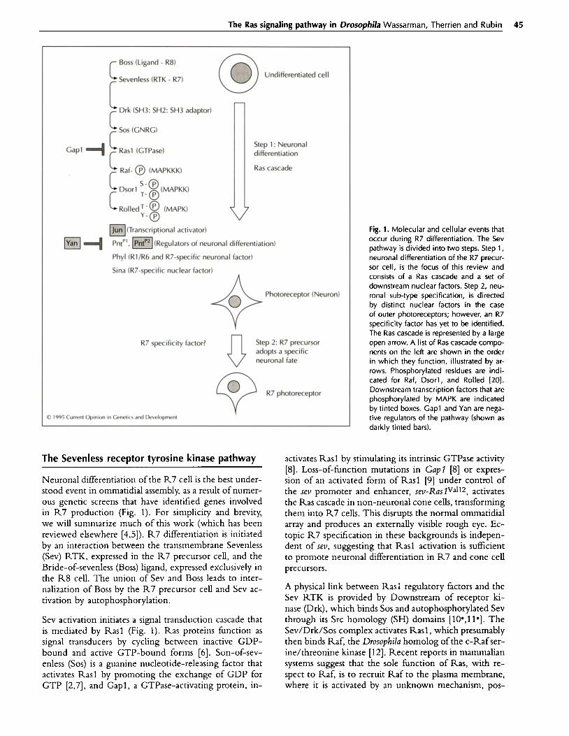
From a mechanistic standpoint, the process of photore- ceptor recruitment probably occurs in two steps, each directed by signals transmitted via cell-cell interactions (Fig. 1). First, a particular undifferentiated cell is chosen to enter the developing ommatidium and is instructed whether or not to undergo neuronal differentiation. Sec- ond, if a neuronal pathway is chosen, the fate is narrowed down to a specific type of neuron, Rl-R8. It should be noted that neuronal sub-type specification (i.e. step 2) may occur before neuronal differentiation (i.e. step 1). Currently, no data are available that distinguish the or- der of these steps.
44
Abbreviations Boss-Bride-f-sevenless; Drk-Downstream of receptor kinase; Egfr-epidermal growth factor receptor; FIpNlipse;
MAPK-mitogen-activated protein kinase; MAPKK-mitogen-activated protein kinase kinase; MAPKKK-mitogen-activated protein kinase kinase kinase; R-photoreceptor cell; RTK-receptor tyrosine kinase;
Sev-Sevenless; SH-Src homology; Sina-Seven-in-absentia; Sos-Son-of sevenless; Tor-Torso.
0 Current Biology Ltd ISSN 0959-437X
The Ras signaling pathway in Drosophila Wassarman, Therrien and Rubin 45
c Boss (Ligand - R8)
Sevenless (RTK - R7) Undifferentiated cell
r L c Drk (SH3: SH2: SH3 adaptor)
C SOS (GNRC)
Gap1 + Step 1: Neuronal
c
Rasl (CTPase) differentiation
Raf- @ (MAPKKK) Ras cascade
c
c
Dsorl ‘- @ (MAPKK) T- @
Rolled T-@ (MAPK) ‘-8
pg _ fl* (Transcriptional activator)
p&J+ PntP’, MfRegulators of neuronal differentiation)
Phyl fRl/R6 and R7specific neuronal factor)
Sina fR7-specific nuclear factor) ,
Photoreceptor (Neuron)
R7 specificity factor? Step 2: R7 precursor adopts a specific neuronal fate
R7 photoreceptor
B 1995 Current Opinion in Genelicr and Developmenl
The Sevenless receptor tyrosine kinase pathway
Neuronal differentiation of the R7 cell is the best under- stood event in ommatidial assembly, as a result of numer- ous genetic screens that have identified genes involved in R7 production (Fig. 1). For simplicity and brevity, we will summarize much of this work (which has been reviewed elsewhere [4,5]). R7 differentiation is initiated by an interaction between the transmembrane Sevenless (Sev) RTK, expressed in the R7 precursor cell, and the Bride-of-sevenless (Boss) ligand, expressed exclusively in the R8 cell. The union of Sev and Boss leads to inter- nalization of Boss by the R7 precursor cell and Sev ac- tivation by autophosphorylation.
Sev activation initiates a signal transduction cascade that is mediated by Rasl (Fig. 1). Ras proteins function as signal transducers by cycling between inactive GDP- bound and active GTP-bound forms [6]. Son-of-sev- enless (SOS) is a guanine nucleotide-releasing factor that activates Rasl by promoting the exchange of GDP for GTP [2,7], and Gapl, a GTPase-activating protein, in-
Fig. 1. Molecular and cellular events that occur during R7 differentiation. The Sev pathway is divided into two steps. Step 1, neuronal differentiation of the R7 precur- sor cell, is the focus of this review and consists of a Ras cascade and a set of downstream nuclear factors. Step 2, neu- ronal sub-type specification, is directed by distinct nuclear factors in the case of outer photoreceptors; however, an R7 specificity factor has yet to be identified. The Ras cascade is represented by a large open arrow. A list of Ras cascade compo- nents on the left are shown in the order in which they function, illustrated by ar- rows. Phosphorylated residues are indi- cated for Raf, Dsorl, and Rolled [20]. Downstream transcription factors that are phosphorylated by MAPK are indicated by tinted boxes. Cap1 and Yan are nega- tive regulators of the pathway (shown as darkly tinted bars).
activates Rasl by stimulating its intrinsic GTPase activity [8]. Loss-of-f uric Ion mutations in Gap1 [8] or expres- t’ sion of an activated form of Rasl [9] under control of the rev promoter and enhancer, sev-I7ias?Va112, activates the Ras cascade in non-neuronal cone cells, transforming them into R7 cells. This disrupts the normal ommatidial array and produces an externally visible rough eye. Ec- topic R7 specification in these backgrounds is indepen- dent of SW, suggesting that Rasl activation is sufficient to promote neuronal differentiation in R7 and cone cell precursors.
A physical link between Rasi regulatory factors and the Sev RTK is provided by Downstream of receptor ki- nase (Drk), which binds SOS and autophosphorylated Sev through its Src homology (SH) domains [lO*,ll*]. The Sev/Drk/Sos complex activates Rasl, which presumably then binds Raf, the Dromphila homolog of the c-Raf ser- ine/threonine kinase [12]. Recent reports in mammalian systems suggest that the sole function of Ras, with re- spect to Raf, is to recruit Raf to the plasma membrane, where it is activated by an unknown mechanism, pos-

46 Oncogenes and cell proliferation
sibly phosphorylation [13,14]. In agreement with this hypothesis, membrane localization of Drusopltila Raf, artificially achieved by fusing the Raf kinase domain to the Torso (Tor) RTK transmembrane and extracel- lular domains, SE-raftor, results in an activated Raf that bypasses the requirement for Rasl in R7 determination 1151. Raf, a mitogen-activated protein kinase kinase ki- nase (MAPKKK), phosphorylates the Dsorl tyro- sine/threonine kin&e, a mitogen-activated protein ki- nase kinase (MAPKK) [16*,17], which ultimately phos- phorylates the Rolled serine/threonine kinase, a mitogen-activated protein kinase (MAPK) [18,19*]. Al- though no biochemical evidence exists for this chain of events in Drosophila, data f?om other systems are suffl- cient to suggest that the Drosophila Ras cascade functions similarly [20], and by genetic criteria, through analysis of double mutant strains, these genes have been shown to function in this order (Fig. 1) [15,16*,19*]; for exam- ple, loss-of-flnction mutations in rolled were identified as dominant suppressors of the rough eye phenotype produced by constitutively active Rasl or Raf (sev- RaslVdl2 or SE-raftor, respectively), indicating that rolled is required for R7 development and that it acts down- stream of Ras? and rof[19*,21].
Like Drk, SOS, Rasl [2], and Raf[l5], Rolled is probably required for development of additional photoreceptors, as flies homozygous for a weak allele of rolled contain fewer photoreceptor cells per ommatidium [19*]. A gain- of-function allele of rolled, rollefievenmaker, resulting from a single amino acid change within the kinase domain, dominantly transforms cone cells into R7 cells in the absence of Sev activation, demonstrating that activation of Rolled is both necessary and sufficient to stimulate the Sev pathway and promote neuronal differentiation (step 1 in Fig. 1) [22**].
Rolled/MAPK targets
Activated Rolled/MAPK is thought to propagate the Ras cascade signal by phosphorylating a series of tar- gets that presumably control expression and/or fimc- tion of downstream genes required to execute the R7 developmental fate. MAPK proteins have been shown to phosphorylate serine and threonine residues within the consensus recognition site Pro-X-Ser/Thr-Pro (in the three-letter amino acid code, where X can be any amino acid) [20]. Genetic dissection of R7 cell determi- nation has begun to shed light on the events downstream of Rolled/MAPK.
It has been shown recently that two members of the Ets family of transcription factors, Yan and Pointed, func- tion in the Sev pathway and are targets for phospho- rylation by Rolled (Fig. 1) [23**,24*]. Flies homozy- gous for a hypomorphic yan mutation have extra R7 and outer photoreceptor cells, even in the absence of
sev function [25,26], and loss-of-function yan mutations dominantly enhance the rough eye phenotypes of Gupl, sev-Ras lvdl2, and sE-raf or, suggesting that it functions as a negative regulator of the Sev pathway [21,25]. In con- trast, pointed mutations suppress Gap I and sev-Ras lVal12 induced rough eye phenotypes, suggesting that it acts as a positive regulator of the Sev pathway [23**].
The pointed gene is transcribed horn two promoters, Pl and P2, which produce transcripts encoding two related proteins that are expressed in all cells prior to neuro- nal differentiation [23**,24*,27]. The PointedP2 protein contains a single MAPK phosphorylation site, and in vitro kinase assays have shown that it can be phosphorylated by MAPK and that this phosphorylation is dependent on the MAPK site [24*]. Co-transfection experiments in Drosophila tissue culture cells have demonstrated that activation of the Ras cascade stimulates the ability of PointedPZ to activate transcription, and that this activ- ity is also dependent on the presence of the phospho- acceptor residue [23**]. Therefore, these results suggest that PointedP2 function is stimulated by Rolled/MAPK phosphorylation.
Yan contains eight consensus MAPK phosphorylation sites, at least one of which is phosphorylated in re- sponse to activation of the Ras cascade in tissue cul- ture cells [23**]. In the co-transfection assays described above, Yan was shown to repress transcriptional activa- tion by PointedPI. This repression is alleviated by activa- tion of the Ras cascade, suggesting that Rolled/MAPK phosphorylation of Yan reduces its ability to repress transcription. Different scenarios can explain this ob- servation. The simplest one would be that Pointed and Yan compete for the same DNA-binding site(s) or protein partner(s) and that a Rolled/MAPK-phospho- rylated Yan would have reduced afinity for its target(s), thus allowing Pointed to activate transcription. Alterna- tively, phosphorylation could induce a destabilization or a relocalization of Yan. It is also possible that Yan func- tions as a positive regulator of transcription whose target genes would antagonize Pointed-mediated transcription. In this case, phosphorylation of Yan by Rolled/MAPK would abrogate its transcriptional properties.
Recently, another transcription factor, Jun, was impli- cated in the determination of photoreceptor fate [28*-l. Jun is a member of the AP-1 family of transcription fac- tors, which in mammalian cells function in growth con- trol and regulation of differentiation [29]. Interestingly, the Drosophila homolog of Jun is expressed in a spatial and temporal pattern that matches photoreceptor dif- ferentiation, but its expression precedes the appearance of neuronal antigens. Expression of a dominant-negative form of Jun under control of the sev enhancer results in missing photoreceptors that correspond to cells in which the mutant protein is expressed. Furthermore, dominant-negative Jun suppresses the cone cell + R7 cell transformation triggered by constitutive activation of either Sev, Rasl, Raf, or Rolled. These results sug- gest that Jun functions downstream of the Ras cascade

The Ras signaling pathway in Drosophila Wassarman, Therrien and Rubin 47
in R7 cell determination (Fig. 1). Since the expression pattern ofJun is unaltered in a sev background, Sev acti- vation ofJun is probably post-transcriptional. In fact, the kinase sites that are implicated in activation of human c- Jun by the Ras cascade are conserved in Drosophila Jun and may be targets for Rolled/MAPK or a Drosophila homolog of the recently identified Jun-specific kinase, JNKl [30-321.
In screens for dominant suppressors of the extra R7 cell phenotype produced by sev-RaslVdl* or sE-rajlor, two research groups have independently identified a gene, phyllopod, which encodes a novel nuclear factor that functions downstream of the Ras cascade in the Sev pathway (Fig. 1) [33**,34**]. In the developing eye, phyl- lopod is expressed in a subset of photoreceptors, namely Rl/R6 and R7, and is required in those cells for proper differentiation. Using the sev enhancer/promoter system, it has been shown that misexpression ofphyllopod in cone cells converts them into R7-like cells, suggesting that Phyllopod can specify neuronal fate. In fact, ectopic ex- pression of Phyllopod in accessory pigment cells, which do not ordinarily express Phyllopod, induces expression of neuronal antigens. It has thus been postulated that one role of the Ras cascade in the presumptive R7 cell is to induce phyllopod expression.
In agreement with multiple studies from mammalian systems, genetic studies in Drosophila have identified a connecting point between the Ras cascade and nuclear factors required for developmental decisions. This link is provided, at least in part, by Rolled/MAPK, which directly modulates, by phosphorylation, the activity of transcription factors involved in cell fate determina- tion. In addition, the Drosophila model has allowed the identification of the novel gene, phyllopod, required for neuronal determination and the expression of which ap- pears to depend on the Ras cascade. It will be interest- ing to verify whether phyllopod transcription is induced by Rolled/MAPK-modified transcription factors, and to investigate the possibility that the Phyllopod protein and its role in Ras-dependent neuronal differentiation have been conserved throughout evolution.
Other receptor tyrosine kinase pathways
mutations cause pleiotropic phenotypes affecting pat- terning of follicle cells during oogenesis, wing vein dif- ferentiation, and photoreceptor development, whereas a gain-of-function Egf allele, Ellipse (Elp), causes a reduc- tion in the number of ommatidia and additional veins in the wing [41,42]. Breathless is required during em- bryonic development for the migration of tracheal and midline glial cells [43].
Mutations in drk, SOS, Rasl, raf, and rolled domi- nantly suppress phenotypes produced by gain-of-func- tion mutations of sev (sevslt), Efi (Elp). and for (to&-3) [2,19’,21], and activated Rasl and raf sup- press the breathless tracheal migration defect [38]. Fur- thermore, rollesjevenmaker, the gain-of-function rolled al- lele, produces phenotypes that mimic se@rl, Elp, and to&L3 [22**]. Therefore, it appears that the Ras cas- cade functions as a naive transmitter of all RTK signals. The identification of MAPK targets in these pathways may clarify how the Ras cascade produces such diverse responses.
A Ras cascade in other photoreceptors
Does a Ras cascade promote neuronal differentiation in photoreceptor precursor cells other than R7? This ap- pears to be the case, as the development of all photore- ceptors, not just R7, is affected in homozygous clones of loss-of-function mutations in Rasl or other compo- nents of the cascade [2,15,19’]. Furthermore, targets of Rolled/MAPK (i.e. Yan and Pointedpz) are expressed in all undifferentiated cells in the eye disc and are required for neuronal differentiation of other photoreceptors in addition to R7 [23**,24*].
To date, specific RTKs, analogous to Sev, that activate the Ras cascade in R2/R5, R4/R4, Rl/R6, or R8 have not been identified. Egfr may fulfill this role in some photoreceptor precursors, as it appears to be required for neuronal differentiation of all photoreceptors; however, this has been difficult to assess, as Eg& is required for both proliferation and differentiation during eye development 1451.
In addition to Sev, the Ras cascade functions down- stream of other Drorophila RTKs, including Tor [35,36], and Drosophila homologs of epidermal growth factor re- ceptor (Egf?) [37] and fibroblast growth factor receptor (DFGF-RI/Breathless) [38]. Tor functions in the em- bryonic terminal system, which is responsible for speci- fication of the tail and unsegmented head regions [39]. Loss-of-function tar mutations result in deletion of these terminal structures, whereas a gain-of-function muta- tion, lotRL3, shows the opposite phenotype, an absence of segmentation in the middle region and an expansion of the terminal structures [39,40]. Loss-of-function Esf
Specification of photoreceptor sub-type
If the Ras cascade is a common inducer of neuronal dif- ferentiation in all photoreceptors, then specification of neuronal sub-type (i.e. Rl-R8) must require another signal input (step 2 in Fig. 1). Presumably, the devel- opmental history of a photoreceptor precursor cell di- rects the expression of genes that determine sub-type. The expectation is that loss-of-function mutations in these determination genes will affect sub-type specifica- tion and not neuronal differentiation. Two genes, rough and seven-up, fit this model.

48 Oncogenes and cell proliferation
Rough is a homeodomain protein that’ is expressed in R2/R5 and R3/R4, and it appears to specify the R2/R5 outer photoreceptor cell fate [46,47]. In loss-of-function rough mutations, cells that would nor- mally develop as R2/R5 are transformed toward the R3/R4/Rl/R6 fate [48]. The ability of Rough to specify outer photoreceptor fate is demonstrated most clearly by ectopic expression of rough using the seu en- hancer/promoter system, which results in transformation of the R7 precursor cell into an outer, Rl-Rblike, pho- toreceptor neuron [46,49]. Seven-up is a member of the steroid receptor superfamily that is expressed and re- quired in R3/R4 and Rl/R6 for normal ommatidial development [50]. In the absence of Seven-up, these precursor cells are transformed to an R7 fate, suggest- ing that Seven-up specifies outer photoreceptor fate in R3/R4 and Rl/R6.
An additional nuclear protein that fulfills some of the cri- teria for a specificity factor is Seven-in-absentia (Sina). Sina is expressed in many cells in the developing eye, but it is required only in the R7 precursor for nor- mal cell-fate specification [Sl]. In the absence of sina, the presumptive R7 cell fails to express neuronal anti- gens and adopts the fate of a non-neuronal cone cell. Thus, loss-of-function mutations in sina, like rough and seven-up, alter cell-type specification, but, unlike rough and seven-up, sina’ mutations prevent neuronal differen- tiation.
Conclusions and future directions
Genetic dissection of the Sev RTK signal transduction pathway has led to the identification of a Ras cascade, the function of which is to promote neuronal differenti- ation of photoreceptor precursor cells. The Ras cascade functions downstream of other RTKs in Drosophila, elic- iting different responses in each case, presumably through regulation of specific transcription factors.
Several issues concerning the Ras cascade remain to be resolved. First, how is Raf activated? The interaction of Raf with Ras and a newly identified family of proteins, 14-3-3 proteins, may be sufficient to stimulate its activity, but it is possible that activation requires additional fac- tors, such as a kinase that phosphorylates Raf [52,53]. Second, are there negative regulators of the Ras cas- cade, other than Gapl? Obvious candidates are pro- tein phosphatases, which could inactivate components of the cascade that are positively regulated by phos- phorylation. Phosphatases that are specific for MAPK have been identified in mammalian systems- CL100 and PACl in humans and 3CH134 in mouse [54]. These dual-specificity phosphatases catalyze dephospho- rylation of both tyrosine and serine/threonine residues. Although enzymes of this class have not been identi- fied in Drosophila, serine/threonine phosphatases (such as protein phosphatase 2A) which dephosphorylate MAPK
in vitro have been identified. Third, what are the gene targets for transcription factors regulated by the Ras cas- cade, and are additional factors regulated by the Ras cascade? Further genetic analysis of RTK pathways in Drosophila may provide answers to these questions.
Acknowledgements
We thank members of the I\ubin lab for insightful discussions
and thoughtful comments on the manuscript. DA Wassarman is a fellow of the Helen Hay Whitney Foundation, M Therrien
is a fellow of the National Cancer Institute of Canada, and GM Kubin is an investigator of the Howard Hughes Medical Institute.
References and recommended reading
Papers of particular interest, published within the annual period of review, have been highlighted as: . . .
1.
2.
3.
4.
5.
6.
7.
a.
9.
10. .
of special interest of outstanding interest
Neumann-Silberberg FS, Scheiter E, Hoffmann FM, Shilo B: The Drosophila ras on&genes: structure and nucleotide sequence. Cell 1984, 37:1027-1033.
Simon MA, Bowtell DDL, Dodson CS, Laverty TR, Rubin GM: Rasl and a putative guanine nucleotide exchange factor per- forms crucial steps in signaling by the Sevenless tyrosine ki- nase. Cell 1991, 67:701-716.
Wolff T, Ready DF: Pattern formation in the Drosophila retina. In The development of Drosophila melanogasfer. Edited by Bate M, Martinez-Arias A. Cold Spring Harbor: Cold Spring Harbor Press; 1993:1277-1325.
Dickson B, Hafen E: Generic dissection of eye development in Drosophila. Edited by Bate M, Martinez-Arias A. Cold Spring Harbor: Cold Spring Harbor Press; 1993:1327-l 362.
Zipursky SL, Rubin GM: Determination of neuronal cell fate: lessons’from the R7 neuron of Drosophila. Annu Rev Neurosci 1994, 17:373-397.
Boguski MS, McCormick F: Proteins regulating Ras and its rel- atives. Narure 1993, 366:643-654.
Rogge RD, Karlovich CA, Banerjee U: Genetic dissection of a neurodevelopmental pathway: Son of sevenless function down- stream of the sevenless and ECF receptor tyrosine kinase. Cell 1991, 64:39-48.
Gaul U, Mardon C, Rubin GM: A putative Ras CTPase acti- vating protein acts as a negative regulator of signaling by the Sevenless receptor tyrosine kinase. Cell 1992, 68:1007-l 019.
Fortini ME, Simon MA, Rubin GM: Signalling by the Sevenless protein tyrosine kinase is mimicked by Rasl activation. Nature 1992, 355:559-561.
Simon MA, Dodson GS, Rubin GM: An SH3-SHZ-SH3 protein is required for p21bt activation and binds to Sevenless and SOS proteins in vitro. Cell 1993, 73:169-l 77.
This study provides biochemical evidence that Drk, an SH3:SH2:SH3 protein, binds to Sev, an RTK, and SOS, a guanine nucleotide releasing factor, and genetic evidence that Drk functions upstream of Rasl in the Sev pathway. (Also see [ll l ] annotation.)
11. Olivier JP, Raabe T, Henkemeyer M, Dickson B, Mbamalu G, . Margotis B, Schlessinger J, Hafen E, Pawson T A Drosophila
SHZ-SH3 adaptor protein implicated in coupling the Seven- less tyrosine kinase to an activator of Ras guanine nucleotide exchange, SOS. Cell 1993, 73:179-191.
The Ras signaling pathway in Drosophila Wassarman, Therrien and Rubin 49
Drk, the Drosophila homolog of C. elegans Sem-5 and human CRB2, was shown to bind autophosphorylated RTKs with its SH2 domain and SOS through its SH3 domains. Futhermore, its activity was shown to correlate with localization to the plasma membrane. (Also see [lo’] annotation.)
12.
13.
14.
15.
16. .
Moodie SA, Wolfman: The 3Rs of life: Ras, Raf, and growth regulation. Trends Cenet 1994, 10:44-48.
Stokoe D, Macdonald SC, Cadwaller K, Symons M, Hancock JF: Activation of Raf as a result of recruitment to the plasma membrane. Science 1994, 264:1463-l 467.
Leevers 51, Paterson HF, Marshall CJ: Requirement for Ras and Raf activation is overcome by targeting Raf to the plasma membrane. Nature 1994, 369:41 l-41 4.
Dickson B, Sprenger F, Morrison D, Hafen E: Raf functions downstream of Rasl in the Sevenless signal transduction pathway. Nature 1992, 360:600-603.
Tsuda L, lnoue YH, Yoo MA, Mizuno M, Hata M, Lim YM, Adachi-Yamada T, Ryo H, Masamune Y, Nishida Y: A protein kinase similar to MAP kinase activator acts downstream of the Raf kinase in Drosophila. Cell 1993, 72:407-414.
Dsorl, a MAPKK, was shown to function downstream of Raf in the Tor pathway.
17. Lu X, Melnick MB, Hsu J-C, Perrimon N: Genetic and molec- ular analyses of mutation involved in Drosophila raf signal transduction. EMBO I 1994, 13:2592-2599.
18. Biggs WH, Zipursky SL: Primary structure, expression, and signal-dependent tyrosine phosphorylation of a Drosophila homolog of extracellular signal-regulated kinase. Proc Nat/ Acad Sci USA 1992, 89:62954299.
19. Biggs WH, Zavitz KH, Dickson B, van der Srraten A, Brunner . D, Hafen E, Zipursky SL: The Drosophila rolled locus encodes
a MAP kinase required in the Sevenless signal transduction pathway. EM60 / 1994, 13: 1628-l 635.
Rolled, a MAPK, was shown lo function downstream of Raf in the Sev pathway.
20. Marshall Cj: MAP kinase kinase kinase, MAP kinase kinase and MAP kinase. Curr Opin Gener Dev 1994, 4:82-89.
21. Chang HC, Karim FD, O’Neill EM, Rebay I, Solomon NM, Therrien M, Wassarman DA, Wolff T, Rubin GM: The Ras signal transduction pathway in Drosophda eye development. Cold Spring Harb Symp Quanr Biol 1995, in press.
22. Brunner D, Oellers N, Szabad J, 8iggs WH, Zipursky SL, Hafen . . E: A gain-of-function mutation in Drosophila MAP kinase ac-
tivates multiple receptor tyrosine kinase signaling pathways. Cell 1994, 76:875-888.
The ro//e&venmaker gainaf-function allele of ro//ec#MAPK induces R7 development in rhe absence of Sev function, suggesting that activation of Rolled/MAPK is both necessary and sufficient for R7 induction. In addi- tion, this mutation activates pathways initiated by the Tor and Egfr RTKs.
23. O’Neill EM, Rebay I, Tjian R, Rubin GM: The activities of two . . E&related transcription factors required for Drosophila eye
development are modulated by the Ras/MAPK pathway. Cell 1994, 78:137-l 47.
This paper and 124.1 present the first genetic and biochemical demon- strations of specific MAPK targets in a Drosophila RTK pathway. In tissue culture assays, PointedP, is a constitutively strong transcriptional activa- tor and is unresponsive to Rasl activation, whereas Poin@lp2 activity is stimulated by the Rasl/MAPK pathway. The activity of Pointedpz is de- pendent on its single MAPK site. Yan, which is phosphorylated by MAPK in tissue culture cells, represses the activity of Pointedpl and this repres- sion is alleviated by Rasl/MAPK activation. This suggests that Yan and Pointed have antagonizing functions in the Sev pathway that are regu- lated by MAPK phosphorylalion.
24. Brunner D, Ducker K, Oeliers N, Hafen E, Scholz H, Klambt . C: The ETS domain protein Pointed-P2 is a target of MAP
kinase in the Sevenless signal transduction pathway. Nature 1994, 370:386-389.
This work reiterates and complements that presented in 123**] by demon- strating that Pointedf’z is phosphorylated by MAPK in virro. Furthermore, mutating the phosphoacceptor site in Pointed P2 eliminates its ability to rescue the poinred phenotype in transgenic animals.
25. Lai ZC, Rubin GM: Negative control of photoreceptor devel- opment in Drosophila by the product of the yan gene, an L% domain protein. Cell 1992, 70:609-620.
26. Tei H, Nihonmatsu I, Yokokura T, Ueda R, Sano Y, Okuda T, Sata K, Hirata K, Fujita SC, Yamamoto D: pokkuri, a Drosophila gene encoding an E-2Cspecific (Ets) domain protein, prevents overproduction of the R7 photoreceptor. Proc Narl Acad Sci USA 1992, 89:6856-6860.
27. Klambt C: The Drosophi/a gene pointed encodes two Ets-like proteins which are involved in the development of the midline glial cells. Development 1993, 117: 163-l 76.
28. Bohmann D, Ellis MC, Staszewski LM, Mlodzik M: DrosopMa . . Jun mediates Rasdependent photoreceptor determination. Cell
1994, 78:973-986. This study demonstrates genetically, as anticipated from mammalian studies, that in Drosophila, Jun functions downstream of the Ras cas- cade. Jun appears lo be required for thedevelopment of all photoreceptor cells, acting downstream of Rasl lo mediate the decision between neu- ronal and non-neuronal cell fates.
29. Vogt PK, Bos TJ: jun: oncogene and transcription factor. Adv Cancer Res 1990, 55:1-35.
30. Binelruy B, Smeal T, Karin M: Ha-ras augments c-Jun activ- ity and stimulates phosphorylation of its activation domain. Narure 1991, 351:122-l 27.
31. Pulverer BJ, Kyriakis JM, Aruch J, Nikolakaki E, Woodgett JR: Phosphorylation of c-Jun mediated by MAP kinase. Na- ture 1991, 353:67&674.
32. Derijard B, Hibi M, Wu I, Barrett T, Su 6, Deng T, Karin M, Davis RJ: JNKl: a protein kinase stimulated by UV-light and Ha-Ras that binds and phosphorylates the c-Jun activation do- main. Cell 1994, 76:1025-l 037.
33. Chang HC, Solomon NM, Wassarman DA, Karim FD, Therrien . . M, Rubin GM, Wolff T: phy//opod functions downstream of
Rasl in the fate determination of a subset of photoreceptors in Drosophila. Cell 1995, in press.
See [34**1 annotation.
34. . .
Dickson BJ, Dominguez M, van der Straten A, Hafen E: Con- trol of Drosophila photoreceptor fates by Phyllopod, a novel nuclear protein acting downstream of Raf. Cell 1995, in press.
This paper and [33**] provide compelling evidence that phybpod is a photoreceptor-specific target of the Ras cascade. Phyllopod expression appears to depend on activation of the Ras cascade, and its activity is required in Rl/R6 and R7 precursor cells for their neuronal specification.
35.
36.
37.
38.
39.
40.
41.
42.
Doyle HI, Bishop JM: Torso, a receptor tyrosine kinase required for embryonic pattern formation, shares substrates with the Sevenless and ECF-R pathways in Drosophila. Genes Dev 1993, 7:633-646.
Hsu J-C, Perrimon N: A temperature-sensitive MEK muta- tion demonstrates the conservation of the signaling path- ways activated by receptor tyrosine kinases. Genes Dev 1994, 8:2176-2187.
Diaz-Benjumea FJ, Hafen E: The Sevenless signalling cassette mediates Drosophila ECF receptor function during epidermal development. Development 1994, 120:569-578.
Reichman-Freid M, Dickson B, Hafen E, Shilo BZ: Elucidation of the role of breathless, a Drosophila FCF receptor homolog, in tracheal cell migration. Genes Dev 1994, 8:42w39.
Perrimon N: The Torso receptor protein-tyrosine kinase signal- ing pathway: an endless story. Cell 1993, 74219-222.
Klingler M, Erdelyi M, Szabad J, Nusslein-Volhard C: Function of torso in determining the terminal anlagen of the Drosophila embryo. Nature 1988, 335275-277.
Shilo BZ, Raz E: Developmental control by the Drosophila EGF receptor homolog DER. Trends Genet 1991, 7:388-392.
Baker NE, Rubin GM: Effect on eye development of domi- nant mutations in Drosophila homologue of the ECF receptor. Narure 1989, 340:150-l 53.

50 Oncogenq and cell proliferation
43.
44.
45.
46.
47.
40.
49.
Klambt C, Glazer 1, Shilo BZ: breathless, d Drosophila FCF receptor homolog is essential for migration of tracheal and specific midline glial cells. Genes Dev 1992, 6:166&1672.
Basfer K, Christen B, Hafen E: Ligand-independent activation of the Sevenless receptor tyrosine kinase changes the fate of cells in the developing Drosophila eye. Cell 1991, 64:1069-l 081.
Xu T, Rubin CM: Analysis of genetic mosiacs in developing and adult Drosophifa tissues. Developmenr 1993, 117:1223-l 237.
Kimmel BE, Heberfein U, Rubin GM: The homeodomain pro- tein rough is expressed in a subset of cells in the developing DrosopMa eye where it can spe&y photoreceptor cell sub type. Genes Dev 1990, 4712-727.
Tomlinson A, Kimmel BE, Rubin GM: rough, a Drosophila homeobox gene required in photoreceptors R2 and R5 for inductive interactions in the developing eye. Cell 1988, 55771-704.
Heberlein U, Mlodrik M, Rubin GM: Cell-fate determination in the developing Drosophila eye: role of the rot& gene. Development 1991, 112:703-712.
Basler K, Yen D, Tomlinson A, Hafen E: Reprogramming cell fate in the developing Drosophila retina: transformation of
50.
51.
52.
53.
54.
R7 cells by ectopic expression of rough. Genes Dev 1990, 41728-739.
Mlodzik M, Hiromi Y, Weber U, Goodman CS, Rubin GM: The Drosophi/a seven-up gene, a member of the steriod receptor gene superfamily, controls photoreceptor cell fates. Cell 1990, 60:21 l-224.
Carthew RW, Rubin GM: seven-in-absentia, a gene required for specification of R7 cell fate in the DrosopMa eye. Cell 1990, 63:561-577.
Freed E, Symons M, Macdonald SC, McCormick F, Ruggieri R: Binding of 14-3-3 proteins to the protein kinase Raf and effects on its activation. Science 1994, 265:1713-l 716.
lrie K, Gotoh Y, Yashar BM, Errede B, Nishida E, Matsumoto K: Stimulatory effects of yeast and mammalian 14-3-3 proteins on the Raf protein kinase. Science 1994, 265:1716-l 719.
Nebreda AR: Inactivation of MAP kinases. Trends Eiochem 1994, 19:1-2.
DA Wassarman, M Therrien and GM Rubin. Department of Molecular and Cell Biology, Howard Hughes Medical Institute, University of California at Berkeley, Berkeley, California 9472% 3200, USA.