the primary function of the respiratory system is to
TRANSCRIPT

1
UNIT FOUR
RESPIRATION SYSTEM IN ANIMALS
Introduction
Breathing is an involuntary event. How often a breath is taken and how much air is inhaled or
exhaled are tightly regulated by the respiratory center in the brain. Under normal breathing
conditions, humans will breathe approximately 15 times per minute on average. A respiratory
cycle consists of an inhalation and an exhalation: with every normal inhalation, oxygenated air
fills the lungs, while with every exhalation, deoxygenated air rushes back out. The oxygenated
air crosses the lung tissue, enters the bloodstream, and travels to organs and tissues. Oxygen (O2)
enters the cells where it is used for metabolic reactions that produce ATP, a high-energy
compound. At the same time, these reactions release carbon dioxide (CO2) as a by-product. CO2
is toxic and must be eliminated; thus, CO2 exits the cells, enters the bloodstream, travels back to
the lungs, and is expired out of the body during exhalation.
The primary function of the respiratory system is to deliver oxygen to the cells of the body’s
tissues and remove carbon dioxide. The main structures of the human respiratory system are the
nasal cavity, the trachea, and the lungs. All aerobic organisms require oxygen to carry out their
metabolic functions.
Along the evolutionary tree, different organisms have devised different means of obtaining
oxygen from the surrounding atmosphere. The environment in which the animal lives greatly
determines how an animal respires. The complexity of the respiratory system correlates with the
size of the organism. As animal size increases, diffusion distances increase and the ratio of
surface area to volume drops. In unicellular (single-celled) organisms, diffusion across the cell
membrane is sufficient for supplying oxygen to the cell. Diffusion is a slow, passive transport
process. In order to be a feasible means of providing oxygen to the cell, the rate of oxygen
uptake must match the rate of diffusion across the membrane. In other words, if the cell were
very large or thick, diffusion would not be able to provide oxygen quickly enough to the inside
of the cell. Therefore, dependence on diffusion as a means of obtaining oxygen and removing
carbon dioxide remains feasible only for small organisms or those with highly-flattened bodies,

2
such as flatworms (platyhelminthes). Larger organisms have had to evolve specialized
respiratory tissues, such as gills, lungs, and respiratory passages, accompanied by a complex
circulatory system to transport oxygen throughout their entire body.
The Respiratory System and Direct Diffusion
Respiratory processes that help organisms exchange O2 and CO2 range from simple direct
diffusion to complex respiratory systems.
Direct diffusion: This flatworm’s process of respiration works by diffusion across the outer
membrane.
Direct Diffusion
For small multicellular organisms, diffusion across the outer membrane is sufficient to meet their
oxygen needs. Gas exchange by direct diffusion across surface membranes is efficient for
organisms less than 1 mm in diameter. In simple organisms, such as cnidarians and flatworms,
every cell in the body is close to the external environment. Their cells are kept moist so that
gases diffuse quickly via direct diffusion. Flatworms are small, literally flat worms, which
‘breathe’ through diffusion across the outer membrane. The flat shape of these organisms

3
increases the surface area for diffusion, ensuring that each cell within the body is close to the
outer membrane surface and has access to oxygen. If the flatworm had a cylindrical body, then
the cells in the center would not be able to get oxygen.
Most animal respiration involves four steps:
1. Taking air in (inspiration) and pushing air out (expiration). The term breathing refers to
the processes of inspiration and expiration in humans and many other animals.
2. Circulating gases throughout the body.
3. Exchanging needed gases for unnecessary gases.
4. Using the needed gases.
Depending on the complexity of their bodies and the environment in which they live, animals
evolved respiratory surfaces to achieve respiration. Four basic types of gas-exchange systems
occur in animals:
Integumentary exchange occurs through the outer respiratory surface of some small
animals that constantly stay moist.e.g. Earthworms exchange oxygen and carbon dioxide
directly through their skin. The oxygen diffuses into tiny blood vessels in the skin
surface, where it combines with the red pigment hemoglobin. Hemoglobin binds loosely
to oxygen and carries it through the animal’s bloodstream. Carbon dioxide is transported
back to the skin by the hemoglobin.
Gills are respiratory structures that extend outward from an animal’s body to exchange
gases in watery environments. E.g. Fishes use outward extensions of their body surface
called gills for gas exchange. Gills are flaps of tissue richly supplied with blood vessels.
As a fish swims, it draws water into its mouth and across the gills. Oxygen diffuses out of
the water into the blood vessels of the gill, while carbon dioxide leaves the blood vessels
and enters the water passing by the gills.
Tracheal exchange systems rely on a network of respiratory tubes that end in holes to
move oxygen and carbon dioxide throughout the bodies of certain types of insects. E.g.
Terrestrial arthropods have a series of openings called spiracles at the body surface.

4
Spiracles open into tiny air tubes called tracheae, which expand into fine branches that
extend into all parts of the arthropod body.
Lungs are respiratory structures that extend into an animal’s body, creating moist internal
surfaces that use diffusion to transport gases into and out of the body. e.g. Terrestrial
vertebrates such as amphibians, reptiles, birds, and mammals have well-developed
respiratory systems with lungs.
Frogs swallow air into their lungs, where oxygen diffuses into the blood to join with
hemoglobin in the red blood cells. Amphibians can also exchange gases through their
skin.
Reptiles have folded lungs to provide increased surface area for gas exchange. Rib
muscles assist lung expansion and protect the lungs from injury.
Birds have large air spaces called air sacs in their lungs. When a bird inhales, its rib cage
spreads apart and a partial vacuum is created in the lungs. Air rushes into the lungs and
then into the air sacs, where most of the gas exchange occurs. This system is birds’
adaptation to the rigors of flight and their extensive metabolic demands.
The lungs of mammals are divided into millions of microscopic air sacs called alveoli
(the singular is alveolus). Each alveolus is surrounded by a rich network of blood vessels
for transporting gases. In addition, mammals have a dome-shaped diaphragm that
separates the thorax from the abdomen, providing a separate chest cavity for breathing
and pumping blood. During inhalation, the diaphragm contracts and flattens to create a
partial vacuum in the lungs. The lungs fill with air, and gas exchange follows.
Gas Exchange Mechanisms in Fish, Toad, Mammals and Plants
Skin, Gills, and Tracheal Systems
Respiration can occur using a variety of respiratory organs in different animals, including skin,
gills, and tracheal systems.

5
Skin and Gills
There are various methods of gas exchange used by animals. As seen in mammals, air is taken in
from the external environment to the lungs. Other animals, such as earthworms and amphibians,
use their skin (integument) as a respiratory organ. A dense network of capillaries lies just below
the skin, facilitating gas exchange between the external environment and the circulatory system.
The respiratory surface must be kept moist in order for the gases to dissolve and diffuse across
cell membranes.
Organisms that live in water also need a way to obtain oxygen. Oxygen dissolves in water, but at
a lower concentration in comparison to the atmosphere, which has roughly 21 percent oxygen.
Fish and many other aquatic organisms have evolved gills to take up the dissolved oxygen from
water. Gills are thin tissue filaments that are highly branched and folded. When water passes
over the gills, the dissolved oxygen in the water rapidly diffuses across the gills into the
bloodstream. The circulatory system can then carry the oxygenated blood to the other parts of the
body. In animals that contain coelomic fluid instead of blood, oxygen diffuses across the gill
surfaces into the coelomic fluid. Gills are found in mollusks, annelids, and crustaceans.
This fish, like many other aquatic organisms, has gills that allow it to obtain oxygen from water.
The folded surfaces of the gills provide a large surface area to ensure that fish obtain sufficient
oxygen. Diffusion is a process in which material travels from regions of high concentration to

6
low concentration until equilibrium is reached. In this case, blood with a low concentration of
oxygen molecules circulates through the gills. The concentration of oxygen molecules in water is
higher than the concentration of oxygen molecules in gills. As a result, oxygen molecules diffuse
from water (high concentration) to blood (low concentration). Similarly, carbon dioxide
molecules diffuse from the blood (high concentration) to water (low concentration).
Oxygen transport and gills: As water flows over the gills, oxygen is transferred to blood via the
veins.
Tracheal Systems
Insect respiration is independent of its circulatory system; therefore, the blood does not play a
direct role in oxygen transport. Insects have a highly-specialized type of respiratory system
called the tracheal system, which consists of a network of small tubes that carries oxygen to the
entire body. The tracheal system, the most direct and efficient respiratory system in active
animals, has tubes made of a polymeric material called chitin.
Insect bodies have openings, called spiracles, along the thorax and abdomen. These openings
connect to the tubular network, allowing oxygen to pass into the body, regulating the diffusion of
CO2 and water vapor. Air enters and leaves the tracheal system through the spiracles. Some
insects can ventilate the tracheal system with body movements.

7
Insect respiration: Insects perform respiration via a tracheal system, in which openings called
spiracles allow oxygen to pass into the body.
Amphibian and Bird Respiratory Systems
Birds and amphibians have different oxygen requirements than mammals, and as a result,
different respiratory systems.
Amphibian Respiration
Amphibians have evolved multiple ways of breathing. Young amphibians, like tadpoles, use gills
to breathe, and they do not leave the water. As the tadpole grows, the gills disappear and lungs
grow (though some amphibians retain gills for life). These lungs are primitive and are not as
evolved as mammalian lungs. Adult amphibians are lacking or have a reduced diaphragm, so
breathing through the lungs is forced. The other means of breathing for amphibians is diffusion
across the skin. To aid this diffusion, amphibian skin must remain moist. It has vascular tissues
to make this gaseous exchange possible. This moist skin interface can be a detriment on land, but
works well under water.
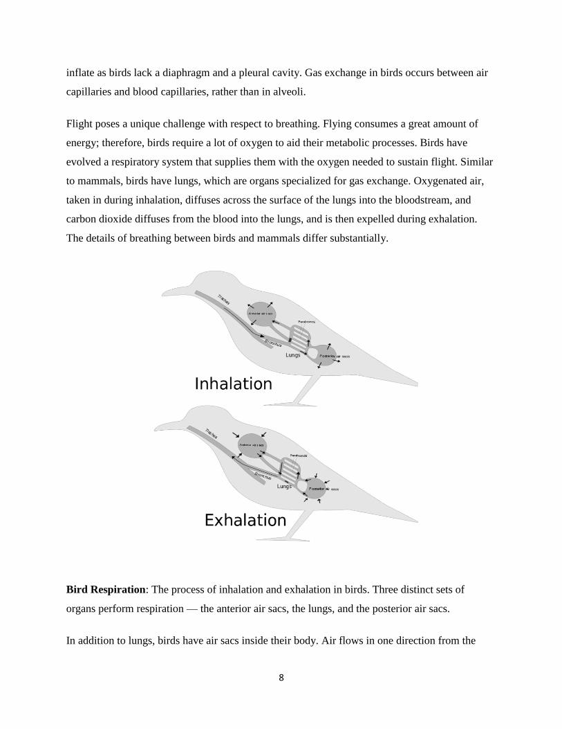
Avian (Birds) Respiration
Birds are different from other vertebrates, with birds having relatively small lungs and nine air
sacs that play an important role in respiration. The lungs of birds also do not have the capacity to
8
inflate as birds lack a diaphragm and a pleural cavity. Gas exchange in birds occurs between air
capillaries and blood capillaries, rather than in alveoli.
Flight poses a unique challenge with respect to breathing. Flying consumes a great amount of
energy; therefore, birds require a lot of oxygen to aid their metabolic processes. Birds have
evolved a respiratory system that supplies them with the oxygen needed to sustain flight. Similar
to mammals, birds have lungs, which are organs specialized for gas exchange. Oxygenated air,
taken in during inhalation, diffuses across the surface of the lungs into the bloodstream, and
carbon dioxide diffuses from the blood into the lungs, and is then expelled during exhalation.
The details of breathing between birds and mammals differ substantially.
Bird Respiration: The process of inhalation and exhalation in birds. Three distinct sets of
organs perform respiration — the anterior air sacs, the lungs, and the posterior air sacs.
In addition to lungs, birds have air sacs inside their body. Air flows in one direction from the

9
posterior air sacs to the lungs and out of the anterior air sacs. The flow of air is in the opposite
direction from blood flow, and gas exchange takes place much more efficiently. This type of
breathing enables birds to obtain the requisite oxygen, even at higher altitudes where the oxygen
concentration is low. This directionality of airflow requires two cycles of air intake and
exhalation to completely get the air out of the lungs.
Mammalian Respiratory System
The mammalian respiratory system equilibrates air to the body, protects against foreign
materials, and allows for gas exchange.
In mammals, pulmonary ventilation occurs via inhalation when air enters the body through the
nasal cavity. Air passes through the nasal cavity and is warmed to body temperature and
humidified. The respiratory tract is coated with mucus that is high in water to seal the tissues
from direct contact with air. As air crosses the surfaces of the mucous membranes, it picks up
water. This equilibrates the air to the body, reducing damage that cold, dry air can cause.
Particulates in the air are also removed in the nasal passages. These processes are all protective
mechanisms that prevent damage to the trachea and lungs.
From the nasal cavity, air passes through the pharynx and the larynx to the trachea. The function
of the trachea is to funnel the inhaled air to the lungs and the exhaled air out of the body. The
human trachea, a cylinder about 10-12cm long, 2cm in diameter found in front of the esophagus,
extends from the larynx into the chest cavity. It is made of incomplete rings of hyaline cartilage
and smooth muscle that divides into the two primary bronchi at the mid thorax. The trachea is
lined with mucus-producing goblet cells and ciliated epithelia that propel foreign particles
trapped in the mucus toward the pharynx. The cartilage provides strength and support to the
trachea to keep the passage open. The smooth muscle can contract, causing a decrease in the
trachea’s diameter, which propels expired air upwards from the lungs at a great force. The forced
exhalation helps expel mucus when we cough.
Trachea and bronchi structure: The trachea and bronchi are made of incomplete rings of
cartilage.

10
Route of inhalation: Air enters the respiratory system through the nasal cavity and pharynx. It
then passes through the trachea and into the bronchi, which bring air into the lungs.

11
Lungs: Bronchi and Alveoli
The end of the trachea bifurcates to the right and left lungs, which are not identical. The larger
right lung has three lobes, while the smaller left lung has two lobes. The muscular diaphragm,
which facilitates breathing, is inferior to the lungs, marking the end of the thoracic cavity.
Lung structure: The trachea bifurcates into the right and left bronchi in the lungs. The larger
right lung is made of three lobes. To accommodate the heart, the left lung is smaller, having only
two lobes.
As air enters the lungs, it is diverted through bronchi beginning with the two primary bronchi.
Each bronchus divides into secondary, then into tertiary bronchi, which further divide to create
smaller diameter bronchioles that split and spread through the lung. The bronchi are made of
cartilage and smooth muscle; at the bronchioles, the cartilage is replaced with elastic fibers.
Bronchi are innervated by nerves of both the parasympathetic and sympathetic nervous systems
that control muscle contraction or relaxation, respectively. In humans, bronchioles with a
diameter smaller than 0.5 mm are the respiratory bronchioles. Since they lack cartilage, they rely
on inhaled air to support their shape. As the passageways decrease in diameter, the relative
amount of smooth muscle increases.
The terminal bronchioles then subdivide into respiratory bronchioles which subdivide into

12
alveolar ducts. Numerous alveoli (sing. alveolus) and alveolar sacs surround the alveolar ducts.
The alveolar ducts are attached to the end of each bronchiole; each duct ends in approximately
100 alveolar sacs. Each sac contains 20-30 alveoli that are 200-300 microns in diameter. Alveoli
are made of thin-walled, parenchymal cells that are in direct contact with capillaries of the
circulatory system. This ensures that oxygen will diffuse from alveoli into the blood and that
carbon dioxide produced by cells as a waste product will diffuse from the blood into alveoli to be
exhaled. The anatomical arrangement of capillaries and alveoli emphasizes the relationship of
the respiratory and circulatory systems. As there are so many alveoli (around 300 million per
lung) within each alveolar sac and so many sacs at the end of each alveolar duct, the lungs have a
sponge-like consistency. This organization produces a very large surface area that is available for
gas exchange.
Alveolar structure: Terminal bronchioles are connected by respiratory bronchioles to alveolar
ducts and alveolar sacs. Each alveolar sac contains 20 to 30 spherical alveoli and has the
appearance of a bunch of grapes. Air flows into the atrium of the alveolar sac, then circulates
into alveoli where gas exchange occurs with the capillaries. Mucus glands secrete mucus into the
airways, keeping them moist and flexible.
13
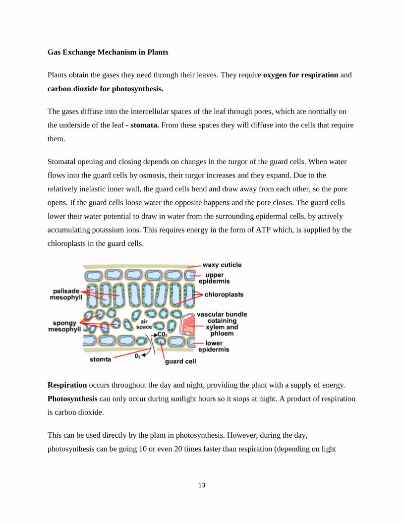
Gas Exchange Mechanism in Plants
Plants obtain the gases they need through their leaves. They require oxygen for respiration and
carbon dioxide for photosynthesis.
The gases diffuse into the intercellular spaces of the leaf through pores, which are normally on
the underside of the leaf - stomata. From these spaces they will diffuse into the cells that require
them.
Stomatal opening and closing depends on changes in the turgor of the guard cells. When water
flows into the guard cells by osmosis, their turgor increases and they expand. Due to the
relatively inelastic inner wall, the guard cells bend and draw away from each other, so the pore
opens. If the guard cells loose water the opposite happens and the pore closes. The guard cells
lower their water potential to draw in water from the surrounding epidermal cells, by actively
accumulating potassium ions. This requires energy in the form of ATP which, is supplied by the
chloroplasts in the guard cells.
Respiration occurs throughout the day and night, providing the plant with a supply of energy.
Photosynthesis can only occur during sunlight hours so it stops at night. A product of respiration
is carbon dioxide.
This can be used directly by the plant in photosynthesis. However, during the day,
photosynthesis can be going 10 or even 20 times faster than respiration (depending on light

14
intensity), so the stomata must stay open so that the plant has enough carbon dioxide, most of
which diffuses in from the external atmosphere.
For practicals, study the structures and functions of the gas exchange mechanisms in fish, birds
plants mammals and amphibians, their similarities and differences, and the need for such
exchange mechanism.
END OF UNIT