the photoluminescence mechanism in carbon dots (graphene ... · the photoluminescence mechanism in...
TRANSCRIPT
Nano Res
1
The photoluminescence mechanism in carbon dots
(graphene quantum dots, carbon nanodots and
polymer dots): current state and future perspective
Shoujun Zhu, Yubin Song, Xiaohuan Zhao, Jieren Shao, Junhu Zhang and Bai Yang()
Nano Res., Just Accepted Manuscript • DOI: 10.1007/s12274-014-0644-3
http://www.thenanoresearch.com on November 19 2014
© Tsinghua University Press 2014
Just Accepted
This is a “Just Accepted” manuscript, which has been examined by the peer-review process and has been
accepted for publication. A “Just Accepted” manuscript is published online shortly after its acceptance,
which is prior to technical editing and formatting and author proofing. Tsinghua University Press (TUP)
provides “Just Accepted” as an optional and free service which allows authors to make their results available
to the research community as soon as possible after acceptance. After a manuscript has been technically
edited and formatted, it will be removed from the “Just Accepted” Web site and published as an ASAP
article. Please note that technical editing may introduce minor changes to the manuscript text and/or
graphics which may affect the content, and all legal disclaimers that apply to the journal pertain. In no event
shall TUP be held responsible for errors or consequences arising from the use of any information contained
in these “Just Accepted” manuscripts. To cite this manuscript please use its Digital Object Identifier (DOI®),
which is identical for all formats of publication.
Nano Research
DOI 10.1007/s12274-014-0644-3
The photoluminescence mechanism in carbon dots
(graphene quantum dots, carbon nanodots and
polymer dots): current state and future perspective
Shoujun Zhu, Yubin Song, Xiaohuan Zhao, Jieren Shao,
Junhu Zhang and Bai Yang*
State Key Laboratory of Supramolecular Structure and
Materials, College of Chemistry, Jilin University,
Changchun, 130012, P. R. China.
E-mail: [email protected]
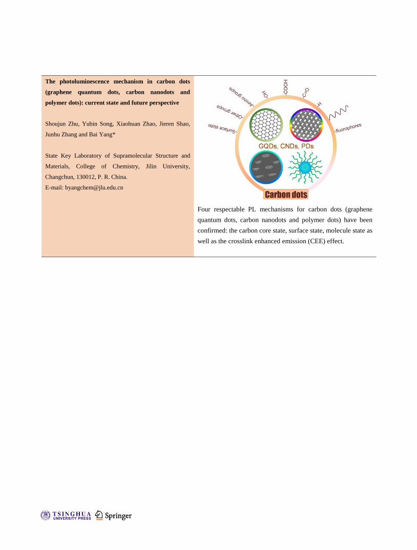
Four respectable PL mechanisms for carbon dots (graphene
quantum dots, carbon nanodots and polymer dots) have been
confirmed: the carbon core state, surface state, molecule state as
well as the crosslink enhanced emission (CEE) effect.

The photoluminescence mechanism in carbon dots
(graphene quantum dots, carbon nanodots and
polymer dots): current state and future perspective
Shoujun Zhu, Yubin Song, Xiaohuan Zhao, Jieren Shao, Junhu Zhang and Bai Yang()
Received: day month year
Revised: day month year
Accepted: day month year
(automatically inserted by
the publisher)
© Tsinghua University Press
and Springer-Verlag Berlin
Heidelberg 2014
KEYWORDS
Carbon dots, graphene
quantum dots, carbon
nanodots, polymer dots,
photoluminescence
mechanism
ABSTRACT
At present, the clear PL mechanism of carbon dots (CDs) is still open debate for
the related researchers. Because of the variety of CDs, it is highly important to
summarize the PL mechanism for these kinds of carbon materials, which can
guide the effective synthesis routes and novel applications. This review will
focus on the PL mechanism of the CDs. Three kinds of fluorescent CDs were
involved: graphene quantum dots (GQDs), carbon nanodots (CNDs) and
polymer dots (PDs). Four respectable PL principles have been confirmed: the
quantum confinement effect or conjugated π-domains determined by carbon
core, surface state determined by hybridization of the carbon backbone and the
connected chemical groups, the molecule state determined by the solely
fluorescent molecules connected on the surface or inner of the CDs, as well as
the crosslink enhanced emission (CEE) effect. To give a thorough summary, the
category and synthesis routes as well as the chemical/physical properties for
CDs were shortly introduced in advance.
1. Introduction
Carbon materials are already well known for many
years, which include graphite, diamond, fullerenes,
carbon nanotube (CNT) and graphene. To make these
kinds of materials fluorescent, their size and surface
chemical groups should be carefully modulated. The
as-prepared fluorescent carbon materials are always
consisted of sp2/sp3 carbon, oxygen/nitrogen based
groups and post-modified chemical groups. Up to
now, many kinds of fluorescent carbon-based
nanomaterials have been synthesized, including
carbon nanodots (CNDs) [1-2], fluorescent CNT [3],
graphene oxide (GO) [4-5], graphene quantum dots
(GQDs) [6-12], polymer dots (PDs) [13-15],
nanodiamond [16-17] and so on. In this review,
carbon dots (CDs) prepared by the chemical
synthetic strategies are chosen as the main object to
discuss, containing GQDs, CNDs and PDs. These
three CDs possess similar photoluminescence (PL),
while they are distinguished by the intrinsic inner
structure and surface chemical groups. In detail, the
synthesis of CDs can be divided into top-down
nano-cutting method and bottom-up organic
Nano Research
DOI (automatically inserted by the publisher)
Review Article
| www.editorialmanager.com/nare/default.asp
Nano Res.
approaches. Top-down nano-cutting method
generally includes cutting different carbon resources
like GO, carbon fiber, CNT, fullerene and graphite
electrode. Bottom-up organic approaches always
contain carbonization of carbohydrate, self-assembly
of polycyclic aromatic hydrocarbon (PAH) as well as
organic synthesis from small molecules.
At present, the clear PL mechanism of CDs is
still an open debate for the related researchers [18].
Because of the variety of CDs, it is highly important
to summarize the PL mechanism for these kinds of
carbon materials, which can guide the effective
synthesis routes and novel applications. This review
will focus on the PL mechanism of the CDs; three
kinds of fluorescent CDs were involved: graphene
quantum dots (GQDs), carbon nanodots (CNDs) and
polymer dots (PDs) (Fig. 1) [19]. Four respectable PL
principles have been confirmed: the quantum
confinement effect or conjugated π-domains
determined by carbon core, surface state determined
by hybridization of the carbon backbone and
connected chemical groups, the molecule state
determined by the solely fluorescent molecules
connected on the surface or inner of the CDs as well
as the crosslink enhanced emission (CEE) effect. To
give a thorough summary, the category, synthesis
routes and the chemical/physical properties for CDs
were shortly introduced in advance. Due to the
abundant and increasing reports about CDs, we
apologize to the researchers whose important
publications may be left out.
2. Category and synthesis routes
2.1 A classification of reported CDs
CDs are the comprehensive definition for various
nanosized carbon materials. In a broad sense, all
nanosized materials, which are mainly comprised of
carbon can be called as CDs. CDs always possess at
least one dimension less than 10 nm and fluorescence
as their instinct properties. The structure of CDs
consisted of sp2/sp3 carbon and oxygen/nitrogen
based groups or polymeric aggregations. In detail,
CDs mainly contained GQDs, CNDs and PDs (Fig. 1).
The GQDs possess single or few layers graphene and
connected chemical groups on the edge. They are
anisotropic with lateral dimension larger than the
height. CNDs are always spherical and they are
divided into carbon naonparticles without crystal
lattice and carbon quantum dots (CQDs) with
obvious crystal lattice. As a result, the PL center is
very different for different kinds of CNDs. The PDs is
aggregated or cross-linked polymer, which is
prepared from linear polymer or monomers. In
addition, carbon core and the connected polymer
chains can also assemble to form PDs. Due to the
diversity of the CDs, there were lots of approaches to
these CDs, mainly including the “top-down” cutting
from different carbon source, and “bottom-up”
synthesis from organic molecules or polymers as well
as surface functionality or passivation. We called the
“top-down” and “bottom-up” routes as
“nano-methods”, which are distinguished from the
precisely organic routes [20-21].
2.2 “Top-down” cutting from different carbon source
Generally, the CDs were obtained from oxide-cutting
different carbon resources, such as graphite power
[22], carbon rods [23], carbon fiber [24], carbon
nanotube [25-26], carbon black [27] and even candle
soot [28] (Fig. 2). These carbon materials possess
perfect sp2 carbon structure and lack efficient band
gap to give the fluorescence. To make these kinds of
carbon source photoluminescence, the sizes and
surface chemistry have to be carefully modulated. As
a result, the most common methods were cutting
with concentrated acid oxidizing (HNO3 or
H2SO4/HNO3 mixture) [29]. In these processes, the
bulk carbon materials were cut into small pieces,
while the surface was modified by oxygen based
groups. The resulted small carbon product was
so-called GQDs, CQDs or CNDs. It should be noted
that the two-step cutting routes have always be used
to prepare GQDs. The first step is to convert a
graphite based material to GO sheets (usually the
modified Hummers method), and the second step is
cutting the GO into GQDs with various methods.
[30-31].
Other “Top-down” cutting routes contained
electrochemistry [32-33],
hydrothermal/solvothermal/special oxidation [30, 34],
metal-graphite intercalation [35] as well as strong
physical routes, such as arc discharge [36], laser
ablation [37] and nanolithography by reactive ion
etching (RIE) [38-39]. In the electrochemistry method,
the graphite rod electrode broke up to form CQDs or
GQDs during the electrochemical cutting processes.
The applied electrolyte contains ethanol [23], ionic
www.theNanoResearch.com∣www.Springer.com/journal/12274 | Nano Research
Nano Res.
liquid [32], NaH2PO4 [40], tetrabutylammonium
per-chlorate (TBAP) [41] or even PBS/water [42-43],
etc. Electric field peeled off nanosized carbon from
electrode through graphite layer intercalation and/or
radical reaction.
In hydrothermal/solvothermal/special
oxidation methods, some oxidized carbon resources,
such as GO and oxidized CNT, which possessed
defect-based chemical group (oxygen-based groups),
can be cut into pieces by hydrothermal/solvothermal
at high temperature and pressure [30, 34]. Some
special oxidation methods, such as photo-fenton
reaction of GO to GQDs [44] can also break up GO.
Specially, the GQDs prepared by mask-assisted RIE
was an efficient approach to precisely control the size
and surface chemistry of the CDs [38], which is an
ideal model system to clarify the PL mechanism.
2.3 “Bottom-up” synthesis from organic molecules or
polymers
The “bottom-up” methods were efficient routes to
produce fluorescent CDs in large scale (Fig. 2). For
example, small molecules and polymer may undergo
dehydration and further carbonization to form the
CNDs and PDs. The applied molecules always
possessed -OH, -COOH, -C=O and -NH2 groups,
which can dehydrate in elevated temperature. There
were lots of approaches to perform the dehydration
and carbonization processes, such as hydrothermal
[45], microwave [46], combustion [47], pyrolysis in
concentrated acid [48], carbonization in microreactor
[49], enhanced hydrothermal
(microwave-hydrothermal [50],
plasma-hydrothermal [51]) and so on. These
formation processes are usually uncontrollable,
resulting in CDs with polydispersity, but using
designed precursors may accurately obtain the GQDs
with desired molecules weight and size, for example,
intramolecular oxidative polycyclic aromatic
hydrocarbons (PAHs). Although the
organic-synthesized GQDs are the perfect model to
understand the PL mechanism of fluorescent carbon
materials, the complicated synthesis method and the
difference compared with common fluorescent CDs
reduce the possibility for this issue [52].
2.4 Surface functionality or passivation
The prepared CDs always possess lots of reactive
groups, which afford the possibility to be modified
by other chemical groups. The functionality and
passivation can enhance the quantum yields (QY) of
the CDs, change the PL emission and meet the special
applications. The QY of raw CDs used to be very low,
which is hard for application and even detection.
Sun’s groups pioneered to utilize NH2-PEG
passivation to elevate the PL QY of the CNDs [53].
Zhu et al. also used the similar method to enhance
the PL properties of the GQDs [54]. Yang’s groups
used the cross-linked route to enhance the PL
properties of the PDs; The bare PEI possess little
fluorescence while the cross-linked PDs have
elevated PL QY [55]. The surface or edge
modification has been also used to tune the PL
emission of the CDs. For example, the green emission
can be changed to blue one by the surface reduction
[56].
3. Chemical and physical properties
3.1 Chemical structure
As mentioned in the category and synthesis routes
section, there were diversiform fluorescent CDs and
various synthesis routes to obtain these materials. As
a result, the chemical structure of the CDs possesses
diversity according to the different synthesis
approaches. In detail, the GQDs possess single or few
graphene layers and connected chemical groups on
the edge. They are anisotropic with lateral dimension
larger than the height. Due to the existence of carbon
core, the GQDs possess certain crystallinity with
average lattices 0.24 nm (Fig. 3a), which corresponds
to (100) spacing of single graphene dots lied on the
lacey support films [35]. CNDs are always spherical
and they are divided into carbon naonparticles
without crystal lattice and CQDs with obvious
crystal lattice [57]. The typical interlayer distance of
CQDs is ca. 0.34 nm, which corresponding to (002)
spacing of the crystalline graphite (Fig. 3b). The PDs
is aggregation/assemble or cross-linked polymer
from linear non-conjugated polymers. In addition,
the carbon core and the grafted polymer chains can
also form the PDs. All of the CDs possess the
connected or modified chemical groups on the
surface, such as oxygen-based, amino-based groups
or polymer chains, etc. The direct characterization
methods for carbon core include high-resolution
TEM (HRTEM), Raman spectroscope and XRD. For
the grafting chemical groups, the FTIR, XPS, NMR,
MALDI-TOF (Fig. 3c) and element analysis were
| www.editorialmanager.com/nare/default.asp
Nano Res.
used to determine the general structure [58]. As a
result, these kinds of fluorescent CDs were not
“pure” carbon materials, the hybridization and
coefficient between the carbon core and surrounding
chemical groups play the leading role in the PL
behavior of the CDs.
3.2 Optical properties
Despite the diversity of the structures, the CDs
possess some similar optical properties on the
absorption and fluorescence. Herein we would just
summarize the common optical properties rather
than consider some specific examples. The
absorption of the CDs typically shows strong optical
absorption in the UV region (230-320 nm), with a tail
extending into the visible range. For carbon core, a
maximum peak at ca. 230 nm is ascribed to π-π*
transition of aromatic C-C bonds, while a shoulder at
300 nm attributes to n-π* transition of C=O bonds or
other connected groups [59]. Besides, the connected
chemical groups may contribute the absorption at
UV-visible regions. The observed deviations in
absorption spectra data, at least to some extent,
indicate the differences of compositions or structures
in different hybridization derivatives.
The PL properties were the most concerned
issue for CDs in view of investigation of the PL
mechanism and novel applications. Generally, the
emission spectra of the CDs are roughly symmetrical
on the wavelength scale. The emission peak of CDs is
usually wide with large stocks shift when compared
with that of organic dyes. The emission peak position
is always related to the excitation wavelength, which
is call wavelength-dependence behaviors. It may
result from the wide distributions of differently sized
dots and surface chemistry, different emissive traps
(salvation effect), or a mechanism currently
unresolved [60]. Fortunately, the
excitation-dependent PL behaviors can be applied in
multi-color imaging applications [61-62]
The PL of CDs is kind of property like that of
semiconductor quantum dots (QDs), but these
fluorescent nanoparticles possess many differences.
Firstly, it seemed not efficient to tune the PL color by
control the size of CDs. In most situations, the PL
color of CDs is relative to the surface group rather
than the size. The most common CDs have the strong
PL from blue to green color, and a few CDs can
possess optimal emission in long wavelength section
[23, 28, 63]. Another main difference between CDs
and QDs is that the PL bandwidth of CDs is much
wider. The wide peak may result from the
inhomogeneity chemical structure and diverse PL
centers.
QY is the number of emitted photons relative
to the number of absorbed photons. CDs possess
rather low QY (even lower than 1%), when CDs was
just discovered. After surface modification or
passivation, the QYs can be increased dramatically.
The enhace PL properties were attributed to the
strongly PL centers on the surface, synergy by both
carbon core and chemical goups or solely by
fluorophores [64]. From then on, the QY of the CDs
improved year by year, Generally, QY depences on
the synthesis routes and the surface chemistry.
Most of CDs possess good photostability,
which is benefited from the carbon core based PL
center. Neither blinking nor meaningful reduction in
PL intensity were observed in such CDs after
continuous exposure to excitation (Fig. 4a) [37].
However, for the CDs with molecule state emission,
the PL intensity decreased dramatically after high
power UV exposure [65]. Some special luminescence
behaviors of CDs can be observed in some situation,
for example electrochemical luminescence (Fig. 4b)
[33], the ECL mechanism of the CDs was suggested
to involve the formation of excited-state CDs via
electron-transfer annihilation of negatively charged
and positively charged CDs. Although the
up-conversion PL (two-photon absorption and
anti-Stokes PL) was reported [23, 54], it is quite
important to establish a proper characterization
system to investigate this kind of properties, because
some so-called “up-conversion PL” in CDs could be
due to the excitation of second-order diffraction light
(wavelength λ/2) from the monochromators in the
fluorescence spectrophotometer [66-67]. The
amplified spontaneous green emission and lasing
emission was also observed from CNDs (Fig. 4c) [68],
the high PL quantum yield and small overlap
between absorption and emission of CDs ethanol
solution are the key factors in achieving lasing
emission. The special optical properties may lead to
novel application of different CDs.
Besides direct characterization, there are
several indirect approach prove the PL origin. The
pH-dependent and solvent-dependent PL is very
www.theNanoResearch.com∣www.Springer.com/journal/12274 | Nano Research
Nano Res.
important to investigate the emission behaviors of
CDs (Fig. 4d-e) [30, 34]. The molecule state was
affected under both the strongly acidic and base
atmospheres, while PL intensity of the carbon
core-edge state may increase due to protonation or
deprotonation of the functional groups. The PL
quench behaviors of CDs is another important tool to
understand the PL mechanism (Fig. 4f) [69].
3.3 Biological toxicity
The toxicity of CDs is an extraordinary concern
because of their potential for bio-based applications
[12, 19, 70-71]. Bioimaging-based applications of
diagnostics in vitro/vivo must be non-toxic and
biocompatible [72]. In the last several years,
metal-based QDs bioimaging methodologies
appeared, together with toxicity concerns for their
intrinsically toxic elements like cadmium. Compared
with metal-based QDs, GQDs are constituted by
intrinsically non-toxic element, carbon, which makes
them a particularly useful and promising
bio-analytical tool. Toxicity studies have been
conducted by various research groups, and CDs
appear to have low toxicity in vitro and in vivo. So
far, the inherent toxicities of CDs have been
evaluated by the cell-viability assay. The results
indicated that GQDs, CNDs and PDs possess
excellent biocompatibility and low cytotoxicity [15,
29-30, 53, 73-75]. The carboxylated GQDs do not
cause apparent toxicities in rats at different dosage (5
and 10 mg/kg) for 22 days as evidenced by blood
biochemistry and hematological analysis [76]. No
severe symptoms of inflammation were observed in
the liver, kidney, spleen, heart, or lung at 22 days
after the administration of the carboxylated GQDs
nanoparticles. All the evidences point to the great
potential of CDs for in vitro and in vivo imaging
studies.
4. PL mechanism of GQDs
GQDs were the simplest CDs possessing the
structure of single layer carbon core with connected
chemical groups on the surface or edge. As a result,
GQDs were the ideal model to investigate the PL
mechanism of CDs. To explain the PL mechanism of
GQDs, the PL behavior of chemically derived GO
should be introduced firstly, because GO is an
important raw material for GQDs preparation and
they possess similar chemical structures. GO contains
oxygen-based functional groups either on the basal
plane or at the edges. Therefore, the 2-3 nm aromatic
sp2 domains are surrounded by linearly aligned
epoxy and hydroxyl-boned sp3 C-O matrix [5, 77]. In
such a structure of GO, the fluorescent property is
determined by the π states of the sp2 sites. The π and
π* electronic levels of the sp2 clusters, which is
influenced by the bandgap of σ and σ* states of the
sp3 matrix, are strongly confined. Radiative
recombination of electron-hole (e-h) pairs in such sp2
clusters can arouse the fluorescence [78]. Because of
the existence of wide size distribution of sp2 domains
in GO, the bandgaps of different sizes of sp2 cover a
wide range, leading to the wide PL emission
spectrum from visible to near infrared (Fig. 5). Many
groups investigated the fluorescence of GO and
reduced GO (r-GO). For example, Luo et al proposed
that the bond distortions may contribute the
fluorescence of GO and r-GO [79]. Gokus et al. have
observed visible luminescence in the oxygen
plasma-treated graphene and attribute the emission
to CO-related localized electronic states at the
oxidation sites [4]. Furthermore, Galande et al. have
studied the pH-dependent fluorescence of GO and
suggest the emission of quasi-molecular fluorophores
in such kinds of materials [80]. They found that the
excited state of the fluorophore species protonated in
acidic media, which makes the PL spectra different in
acidic and basic solutions. This kind of
quasi-molecular fluorophore is caused by the
carboxylic acid groups that are electronically coupled
with the surrounding graphene core sheets [81].
Similar to GO, GQDs possess more defect,
oxygen groups and functional groups on the surface.
The current fluorescence results of GO could be
referred to understand these emissions in GQDs.
Excitons in graphene have an infinite Bohr diameter.
Thus, graphene fragments of any size will show
quantum confinement effects. As a result, GQDs have
a non-zero bandgap and PL on excitation. This
bandgap is tunable by modifying the size and surface
chemistry [6]. Considerable development in the
preparation of GQDs has been witnessed in the last
five years, researchers have figured out reasonable
PL mechanism, which could be referred to
surface/edge state and conjugated π-domains.
4.1 Surface/edge state in GQDs
The surface/edge state contained triple carbene at the
| www.editorialmanager.com/nare/default.asp
Nano Res.
zigzag edges, oxygen based groups on the graphene
core, and resonance of amine moieties and graphene
core.
When the graphene sheets are cut along
different crystallographic directions, diverse types of
edges (armchair and zigzag edges) can be obtained.
The edge type plays an important role in
determining the electronic, magnetic, and optical
properties. Ritter et al. have stated that
predominantly zigzag-edge GQDs with 7-8 nm
average dimensions are metallic owing to the
presence of zigzag edge states, and GNRs with a
higher fraction of zigzag edges exhibit a smaller
energy gap than a predominantly armchair-edge
ribbon of similar width [82]. Radovic and Bockrath
reported that the zigzag sites are carbene-like, with a
triplet ground state being most common, whereas the
armchair sites are carbyne-like, with a singlet ground
state most common [83]. Pan et al. suggested that
blue PL of hydrothermally GQDs might be attributed
to free zigzag sites with a carbine-like triplet ground
state described as σ1π1 [34]. Under acidic conditions,
the free zigzag sites of the GQDs are protonated,
forming a reversible complex between the zigzag site
and H+, and leading to the breaking of the emissive
triple carbene state and the PL quenching (Fig. 4b).
To the contrary, the PL recovers because the free
zigzag sites are restored under alkaline condition.
Lin et al. prepared GQDs with size about 20 nm from
exfoliating and disintegrating CNTs or graphite
flakes [35]. The obvious single layer and clear zigzag
edge were proved in AFM and bright field high
resolution TEM. The new opened band gap arises
from the triple carbenes at the zigzag edges,
corresponding to the transitions from the HOMO to
σ and π orbital of LOMO in triple carbenes. These
two kinds of GQDs with size of 9.6 nm and 20 nm
respectively possess the similar absorption and
emission behavior, which proved that the PL
mechanism was determined by the triple carbene at
the zigzag edges instead of the quantum confinement
effects (Fig. 6a).
Pan et al. also used single-particle
spectroscopic measurements to investigate the PL
behaviors of the GQDs [84]. As schematically shown
in Fig. 6b, photo-excited electrons through the π-π*
transitions were proposed to relax into either the sp2
energy levels or the defect states (actually we called
surface state), giving rise to the blue or
long-wavelength PL, respectively. The former
emission might bear the discrete feature due to
quantum confinement effect (QCE) of electrons
inside the sp2 carbon domains. The latter emission is
related to the hybrid structure by both the oxygen
functional groups (at the edges and/or on the basal
planes) and graphene core. Despite noticeable
differences in the size and the number of layers from
particle to particle, all of the GQDs studied possess
almost the same spectral line shapes and peak
positions. As a result, it suggested the PL of these
GQDs were caused by surface state.
Besides the oxygen based groups, the
amine-based groups were also important
composition for surface state in GQDs. In the work of
Tetsuka et al, the ammonia-assisted hydrothermal
method was used to prepare GQDs. The product is
edge-terminated by a primary amine, allowing the
electronic structure to be modified with the effective
orbital resonance of amine moieties and graphene
core [58]. For the GQDs with the same sizes, the
emission wavelength increased with the contents of
amine-groups. Furthermore, the GQDs possess high
QY because of the reduction of carboxylic and
epoxide groups which act as the non-radiative
electron-hole recombination center. Combined the
experimental results and ab initio calculations, the
primary amine at the edge of GQDs have higher
HOMO orbital than that with hydrogen-terminated
due to the strong orbital interaction with the -NH2
groups. The resonance feature between the
delocalized π orbital and the molecule orbital in the
-NH2 groups results in a narrowing of the optical
band gap. Such amino-contained GQDs have also
been reported by other groups. Jin et al. reported that
the functionalized GQDs exhibit a redshift in the PL
emission spectrum, compared to the pre-existing
GQDs (the PL emissions of the amine-functionalized
GQDs also shifted with changes of the pH due to the
protonation and deprotonation of the functional
groups) [85]. Calculations from DFT illustrated that
PL shifts resulted from charge transfers between the
functional groups and GQDs, which can tune the
band gap of the GQDs. The band gap of the GQDs
decreases to 2.254 eV, when a GQD is functionalized
by one amino. And the band gap will gradually
decrease with the increasing number of -NH2 groups
www.theNanoResearch.com∣www.Springer.com/journal/12274 | Nano Research
Nano Res.
(Fig. 6c). Kumar et al. also investigated the PL
behaviors of amino-functionalized GQDs. First
principles calculations suggested that primary amine
edge termination (NH2) resulted in formation of an
additional interband ca. 3.28 eV within the energy
gap due to p orbital hybridization of C-N atoms at
the edge sites [86]. Feng and co-workers proved that
1, 2-ethylenediamine functionalization on the surface
of GQDs can form a specific cyclic structure which
facilitates the proton transfer from the ammonium
moiety to the conjugated structure, and thus lead to
the largest enhancement of fluorescence [87].
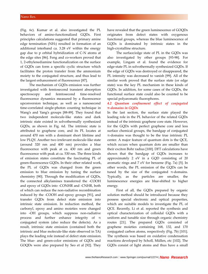
The mechanism of GQDs emission was further
investigated with femtosecond transient absorption
spectroscopy and femtosecond time-resolved
fluorescence dynamics measured by a fluorescence
upconversion technique, as well as a nanosecond
time-correlated single-photon counting technique in
Wang’s and Yang’s groups [88-89]. They found that
two independent molecule-like states and dark
intrinsic state existed in solvothermally synthesized
GQDs, as shown in Fig. 6d. The intrinsic state is
attributed to graphene core, and its PL locates at
around 470 nm with a dominant short lifetime and
low PLQY. Another two irrelevant molecule-like state
(around 320 nm and 400 nm) provides a blue
fluorescence with peak at ca. 430 nm and green
fluorescence with peak at ca. 530 nm. The three kinds
of emission states constitute the fascinating PL of
green-fluorescence GQDs. In their other related work,
the PL of GQDs was changed from the green
emission to blue emission by tuning the surface
chemistry [90]. Through the modification of GQDs,
the connected alkylamines transferred the -COOH
and epoxy of GQDs into -CONHR and -CNHR, both
of which can reduce the non-radiative recombination
induced by the -COOH and epoxy groups [91], and
transfer GQDs from defect state emission into
intrinsic state emission. In reduction method, the
carbonyl, epoxy and amino moieties were changed
into -OH groups, which suppress non-radiative
process and further enhance integrity of π
conjugated system (also reduce the defects). As a
result, intrinsic state emission (contained both the
intrinsic and blue molecule-like state observed in TA)
plays the leading role instead of defect state emission.
The blue- and green-color emissions of GQDs and
GOQDs were also prepared by Seo et al [92]. They
have revealed that the green luminescence of GOQDs
originates from defect states with oxygenous
functional groups, whereas the blue luminescence of
GQDs is dominated by intrinsic states in the
high-crystalline structure.
The surface/edge state of PL in the GQDs was
also investigated by other groups [93-98]. For
example, Lingam et al. found the evidence for
edge-state PL in solvothermally synthesized GQDs. If
the edge of GQDs was destroyed or disappeared, the
PL intensity was decreased to vanish [99]. All of the
similar work proved that the surface state (or edge
state) was the key PL mechanism in these kinds of
GQDs. In addition, for some cases of the GQDs, the
functional surface state could also be counted to be
special polyaromatic fluorophores.
4.2 Quantum confinement effect of conjugated
π-domains in GQDs
In the last section, the surface state played the
leading role in the PL behavior of the related GQDs
instead of the intrinsic graphene core state. However,
for the GQDs with perfect graphene core and less
surface chemical groups, the bandgap of conjugated
π-domains was thought to be the true intrinsic PL
center. A major feature of quantum dots is the QCE,
which occurs when quantum dots are smaller than
their exciton Bohr radius [100]. DFT calculations have
shown that the bandgap of GQDs increases to
approximately 2 eV in a GQD consisting of 20
aromatic rings and 7 eV for benzene (Fig. 7a) [5]. In
other words, the PL emission of the GQDs can be
tuned by the size of the conjugated π-domains.
Typically, as the particles are smaller, the
luminescence energies are blue-shifted to higher
energy.
First of all, the GQDs prepared by organic
solution method should be introduced because they
possess special electronic and optical properties,
which are suitable models to investigate the PL of
QCE. Recently, Li et al. reported the synthesis and
optical characterization of colloidal GQDs with a
uniform and tunable size through organic chemistry
routes [21]. The prepared GQDs consisted of
graphene moieties containing 168, 132, and 170
conjugated carbon atoms, respectively (Fig. 7b) [101].
The synthesis was based on oxidative condensation
reactions developed by Scholl, Müllen, etc [102]. The
GQDs consist of light atoms and thus have a small
| www.editorialmanager.com/nare/default.asp
Nano Res.
dielectric constant and weak spin-orbit coupling.
These lead to strong carrier-carrier interactions and
electronic states with well-defined spin multiplicity.
As a result, GQDs have much larger energy band
than other inorganic semiconductor QDs with similar
sizes. That was why most of the GQDs possess PL at
range of blue to green section. Furthermore, the
size-dependent, discrete excitonic levels could
significantly slow down the relaxation of high
excited states in GQDs due to a phonon bottleneck.
In addition, strong carrier-carrier interactions could
lead to generation of more than one exciton with one
photon absorbed, a process particularly useful for
improving the efficiency of photon-generated carrier
[103]. The singlet-triplet splitting of GQDs was
determined to be ca. 175 meV, and intersystem
crossing was so efficient that it competed with
internal conversion among the states with the same
multiplicity. As a result, the GQDs emitted both
fluorescence and phosphorescence [101]. Since triplet
states have a significantly longer lifetime, they could
profoundly affect the chemical reactivity and other
processes such as charge transfer or exciton
migration in the GQDs-based system. Yang et al.
investigated the photophysics of the organic
synthesized GQDs (C42H18, C96H30, C132H34 and
C222H42), and found that the intrinsic state depended
on size, while the energy level offset between
intrinsic state and edge state decided their optical
properties (Fig. 7c) [104]. As a result, the green
fluorescence of the C42H18, C96H30 not only depends
on the size, but also results from bright edge state. If
the energy level offset between intrinsic state and
edge state was large enough, the fluorescence is
dominant. If the energy level offset was small enough
(meeting thermal activation condition, ca. kBT), the
long carrier lifetime in intrinsic state will give a
possibility for intersystem crossing from singlet
excited state to triple excited state of edge state, such
as above mentioned C132. In the cases of C132H34 and
C222H42, the intrinsic state possibly decreased and was
lower than the edge state; as a result, they lost the
expected fluorescence.
Then, the size dependent PL of GQDs prepared
by “nano-methods” was introduced (The PL of these
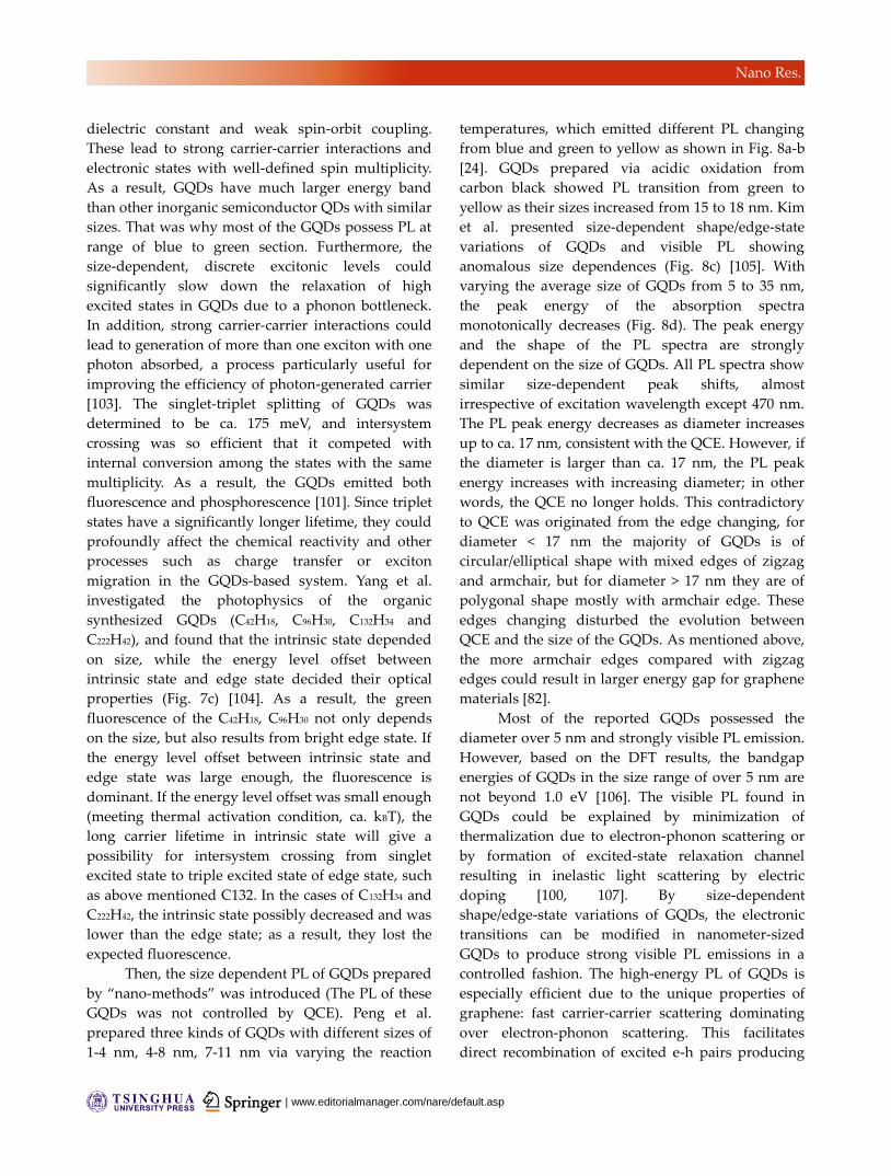
GQDs was not controlled by QCE). Peng et al.
prepared three kinds of GQDs with different sizes of
1-4 nm, 4-8 nm, 7-11 nm via varying the reaction
temperatures, which emitted different PL changing
from blue and green to yellow as shown in Fig. 8a-b
[24]. GQDs prepared via acidic oxidation from
carbon black showed PL transition from green to
yellow as their sizes increased from 15 to 18 nm. Kim
et al. presented size-dependent shape/edge-state
variations of GQDs and visible PL showing
anomalous size dependences (Fig. 8c) [105]. With
varying the average size of GQDs from 5 to 35 nm,
the peak energy of the absorption spectra
monotonically decreases (Fig. 8d). The peak energy
and the shape of the PL spectra are strongly
dependent on the size of GQDs. All PL spectra show
similar size-dependent peak shifts, almost
irrespective of excitation wavelength except 470 nm.
The PL peak energy decreases as diameter increases
up to ca. 17 nm, consistent with the QCE. However, if
the diameter is larger than ca. 17 nm, the PL peak
energy increases with increasing diameter; in other
words, the QCE no longer holds. This contradictory
to QCE was originated from the edge changing, for
diameter < 17 nm the majority of GQDs is of
circular/elliptical shape with mixed edges of zigzag
and armchair, but for diameter > 17 nm they are of
polygonal shape mostly with armchair edge. These
edges changing disturbed the evolution between
QCE and the size of the GQDs. As mentioned above,
the more armchair edges compared with zigzag
edges could result in larger energy gap for graphene
materials [82].
Most of the reported GQDs possessed the
diameter over 5 nm and strongly visible PL emission.
However, based on the DFT results, the bandgap
energies of GQDs in the size range of over 5 nm are
not beyond 1.0 eV [106]. The visible PL found in
GQDs could be explained by minimization of
thermalization due to electron-phonon scattering or
by formation of excited-state relaxation channel
resulting in inelastic light scattering by electric
doping [100, 107]. By size-dependent
shape/edge-state variations of GQDs, the electronic
transitions can be modified in nanometer-sized
GQDs to produce strong visible PL emissions in a
controlled fashion. The high-energy PL of GQDs is
especially efficient due to the unique properties of
graphene: fast carrier-carrier scattering dominating
over electron-phonon scattering. This facilitates
direct recombination of excited e-h pairs producing
www.theNanoResearch.com∣www.Springer.com/journal/12274 | Nano Research
Nano Res.
such high-energy PL before thermalization of the
carriers with the lattice [108]. The high Coulomb
scattering rate of graphene, which is attributed to the
strongly reduced dielectric screening in the
two-dimensional structure, is also essential for
producing high-density non equilibrium carriers
responsible for the strong e-h recombination.
Moreover, because the surface/edge
state-derived PL emissions are relatively brighter,
there may be a general risk for their contaminating
the observed bandgap fluorescence in the GQDs [86].
As a result, lots of experimentally observed PL of the
GQDs was surface/edge state. For clear investigation
on the QCE of the GQDs, the organic synthesis and
nanolithography based routes to perfect sample will
be highly desired in the near future. Besides, the
chromatographic separation was also a powerful tool
to investigate the PL mechanism of GQDs. For
example, Zhu et al. pioneered the column
chromatography to separate the GQDs with tuned
oxidation degree, as a result, the separated GQDs
possessed PL from blue to green [31]. Matsuda and
co-worker developed the size-exclusion high
performance liquid chromatography (HPLC) to
separate the prepared GQDs [109]. Drastic change in
PL spectra of GQDs from UV to red light region is
observed by difference in their overall sizes. Discrete
changes in emission wavelength indicate that the PL
change comes from the differences in the population
of small sp2 fragments with various sizes or shapes
embedded in the GQDs. It’s also highly important to
clarify the PL mechanism of GQDs by theoretical
calculation, for example, Alam Sk et al.
systematically investigated the PL properties of
GQDs using DFT and TDDFT calculations [106]. It is
revealed that the emission of zigzag-edged GQDs can
cover the entire visible light spectrum by varying the
diameter from 0.89 to 1.80 nm. Armchair edged and
pyrrolic N-doping induced blue-shift, while the
chemical functionalities and defects can cause the
red-shift PL. Furthermore, the isolated
inhomogenous sp2 domains can widen the PL peaks
of GQDs.
5. PL mechanism of CNDs
5.1 Quantum size effect in carbon quantum dots
(CQDs)
There are a few studies concerned on the quantum
size effect in CNDs. We would rather called these
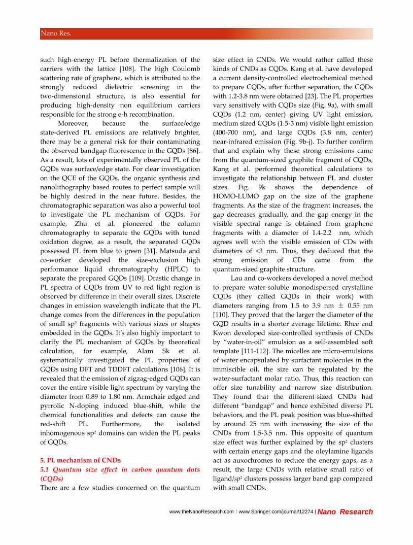
kinds of CNDs as CQDs. Kang et al. have developed
a current density-controlled electrochemical method
to prepare CQDs, after further separation, the CQDs
with 1.2-3.8 nm were obtained [23]. The PL properties
vary sensitively with CQDs size (Fig. 9a), with small
CQDs (1.2 nm, center) giving UV light emission,
medium sized CQDs (1.5-3 nm) visible light emission
(400-700 nm), and large CQDs (3.8 nm, center)
near-infrared emission (Fig. 9b-j). To further confirm
that and explain why these strong emissions came
from the quantum-sized graphite fragment of CQDs,
Kang et al. performed theoretical calculations to
investigate the relationship between PL and cluster
sizes. Fig. 9k shows the dependence of
HOMO-LUMO gap on the size of the graphene
fragments. As the size of the fragment increases, the
gap decreases gradually, and the gap energy in the
visible spectral range is obtained from graphene
fragments with a diameter of 1.4-2.2 nm, which
agrees well with the visible emission of CDs with
diameters of <3 nm. Thus, they deduced that the
strong emission of CDs came from the
quantum-sized graphite structure.
Lau and co-workers developed a novel method
to prepare water-soluble monodispersed crystalline
CQDs (they called GQDs in their work) with
diameters ranging from 1.5 to 3.9 nm ± 0.55 nm
[110]. They proved that the larger the diameter of the
GQD results in a shorter average lifetime. Rhee and
Kwon developed size-controlled synthesis of CNDs
by “water-in-oil” emulsion as a self-assembled soft
template [111-112]. The micelles are micro-emulsions
of water encapsulated by surfactant molecules in the
immiscible oil, the size can be regulated by the
water-surfactant molar ratio. Thus, this reaction can
offer size tunability and narrow size distribution.
They found that the different-sized CNDs had
different “bandgap” and hence exhibited diverse PL
behaviors, and the PL peak position was blue-shifted
by around 25 nm with increasing the size of the
CNDs from 1.5-3.5 nm. This opposite of quantum
size effect was further explained by the sp2 clusters
with certain energy gaps and the oleylamine ligands
act as auxochromes to reduce the energy gaps, as a
result, the large CNDs with relative small ratio of
ligand/sp2 clusters possess larger band gap compared
with small CNDs.
| www.editorialmanager.com/nare/default.asp
Nano Res.
5.2 Surface state in CNDs
The functional groups have various energy levels,
which may result in a series of emissive traps. When
the light of a certain excitation wavelength
illuminates the CNDs, a surface state emissive trap
will dominate the emission. And a higher degree of
surface oxidation or other effective modification can
result in more surface defects, resulting in the
red-shifed emission. The surface state was not single
chemical groups but the hybridization of the carbon
backbone and connected chemical groups. From the
first report of CNDs [25], which were prepared via
electrophoretic analysis and purification of
fluorescent CNT fragments in 2004 (the CNDs were
oxidation-cutting product with oxygen-based
chemical groups on the surfaces), most PL centers of
the reported CNDs were proved as surface state. Sun
and co-workers systematically investigated the
surface passivation of CNDs by amino-polyethylene
glycol (PEG) (The CDs were firstly produced via
laser ablation of a carbon target in the presence of
water vapor with argon as carrier gas) [37]. The
authors proved that many organic molecules could
serve as the purpose of surface passivation, which
further proved the surface energy traps controlled
the PL mechanism. Although the excitation may
induce the band-gap absorption band, the resulted
fluorescence was also controlled by the surface state
emission (Fig. 10a) [113].
From then on, lots of oxidation-based surface
state was directly attached during the process of
preparing CNDs, in another words, the
oxygen-based groups on the carbon core were the
primary surface state of the CNDs. Mao and
co-workers obtained colorful CNDs derived from
oxidizing the candle soot and further separation
(Top-down route), the PL ranged from violet to red
may be induced by different surface oxidation [28].
The CNDs with surface state emission were also
achieved by bottom-up carbonization method, and
the PL can be tuned from blue to green by different
reaction conditions. It’s very interesting that most
surface state emission was focused on the blue-green
range. Pang and co-workers proved that the surface
states were the key factor to the luminescence on the
electrochemical CNDs [41]. Red-shifted emissions
were observed for the CNDs with a high surface
oxidation degree. Zheng and other researchers have
also proved the CNDs with green emission can be
changed to the blue ones by surface reduction [56].
Furthermore, Richards and co-workers used
single-particle fluorescence technology to investigate
the PL fluctuations the oxidized/reduced CNDs [114].
They suggested the possibility that single dots can
possess multiple fluorophore units associated with
the CND core and oxygenated defect related
emissive traps. The majority of the reduced CND
particles showed multiple levels, while the oxidized
particles predominantly showed a single level. A
possible explanation for this is that after the initial
excitation the energy is transferred from the higher
energy absorbing site to a lower energy emissive site
in the oxidized CND particles. In contrast, when
CNDs are reduced the low-energy emissive traps
were entirely or partially removed, blocking
energy-transfer pathways. In addition, the authors
have also proved that the CNDs possess
photo-bleaching at single particle level, which was in
contrast to the reported steady-state fluorescence
characterization (Fig. 10b).
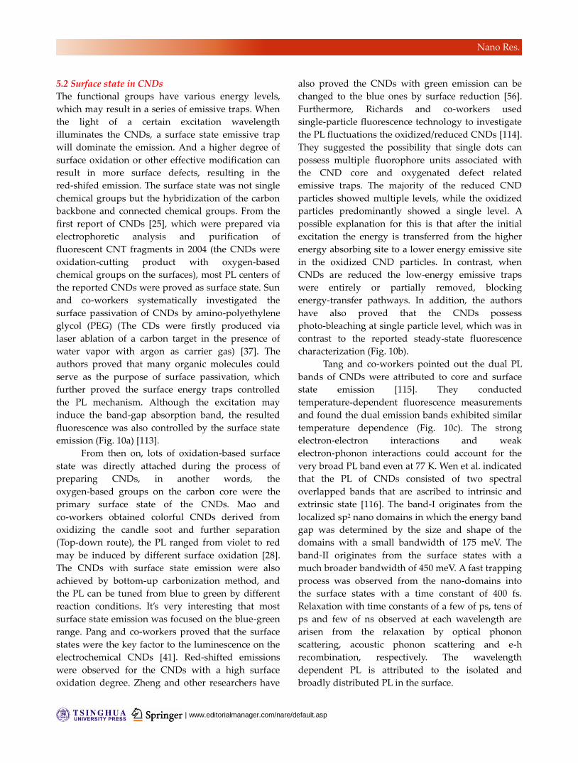
Tang and co-workers pointed out the dual PL
bands of CNDs were attributed to core and surface
state emission [115]. They conducted
temperature-dependent fluorescence measurements
and found the dual emission bands exhibited similar
temperature dependence (Fig. 10c). The strong
electron-electron interactions and weak
electron-phonon interactions could account for the
very broad PL band even at 77 K. Wen et al. indicated
that the PL of CNDs consisted of two spectral
overlapped bands that are ascribed to intrinsic and
extrinsic state [116]. The band-I originates from the
localized sp2 nano domains in which the energy band
gap was determined by the size and shape of the
domains with a small bandwidth of 175 meV. The
band-II originates from the surface states with a
much broader bandwidth of 450 meV. A fast trapping
process was observed from the nano-domains into
the surface states with a time constant of 400 fs.
Relaxation with time constants of a few of ps, tens of
ps and few of ns observed at each wavelength are
arisen from the relaxation by optical phonon
scattering, acoustic phonon scattering and e-h
recombination, respectively. The wavelength
dependent PL is attributed to the isolated and
broadly distributed PL in the surface.
www.theNanoResearch.com∣www.Springer.com/journal/12274 | Nano Research
Nano Res.
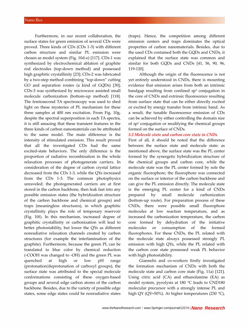
Furthermore, in our recent collaboration, the
surface states for green emission of several CDs were
proved. Three kinds of CDs (CDs 1-3) with different
carbon structure and similar PL emission were
chosen as model system (Fig. 10d-e) [117]. CDs-1 was
synthesized by electrochemical ablation of graphite
rod electrodes (top-down method) and possessed
high graphitic crystallinity [23]; CDs-2 was fabricated
by a two-step method combining “top-down” cutting
GO and separation routes (a kind of GQDs) [30];
CDs-3 was synthesized by microwave assisted small
molecule carbonization (bottom-up method) [118].
The femtosecond TA spectroscopy was used to shed
light on these mysteries of PL mechanism for these
three samples at 400 nm excitation. From Fig. 10g,
despite the spectral superposition in each TA spectra,
it is still amazing that these transient features in the
three kinds of carbon nanomaterials can be attributed
to the same model. The main difference is the
intensity of stimulated emission. This result proved
that all the investigated CDs had the same
excited-state behaviors. The only difference is the
proportion of radiative recombination in the whole
relaxation processes of photogenerate carriers. In
consideration of the degree of carbon crystal lattice
decreased from the CDs 1-3, while the QYs increased
from the CDs 1-3. The common photophysics
unraveled: the photogenerated carriers are at first
stored in the carbon backbone, then leak fast into any
possible emission states (the hybridization structure
of the carbon backbone and chemical groups) and
traps (meaningless structures), in which graphitic
crystallinity plays the role of temporary reservoir
(Fig. 10f). In this mechanism, increased degree of
graphitic crystallinity or carbonization will lead to
better photostability, but lower the QYs as different
nonradiative relaxation channels created by carbon
structures (for example the interlamination of the
graphite). Furthermore, because the green PL can be
translated to blue color by chemical reduction
(-COOH was changed to -OH) and the green PL was
quenched at high or low pH range
(protonation/deprotonation of carboxyl groups), the
surface state was attributed to the special molecule
conformations consisting of these oxygen-based
groups and several edge carbon atoms of the carbon
backbone. Besides, due to the variety of possible edge
states, some edge states could be nonradiative states
(traps). Hence, the competition among different
emission centers and traps dominates the optical
properties of carbon nanomaterials. Besides, due to
the used CDs contained both the GQDs and CNDs, it
explained that the surface state was common and
similar for both GQDs and CNDs [41, 56, 90, 96,
119-120].
Although the origin of the fluorescence is not
yet entirely understood in CNDs, there is mounting
evidence that emission arises from both an intrinsic
bandgap resulting from confined sp2 conjugation in
the core of CNDs and extrinsic fluorescence resulting
from surface state that can be either directly excited
or excited by energy transfer from intrinsic band. As
a result, the tunable fluorescence emission of CDs
can be achieved by either controlling the domain size
of sp2 conjugation or modifying the chemical groups
formed on the surface of CNDs.
5.3 Molecule state and carbon core state in CNDs
First of all, it should be noted that the difference
between the surface state and molecule state: as
mentioned above, the surface state was the PL center
formed by the synergetic hybridization structure of
the chemical groups and carbon core, while the
molecule state was the PL center formed by sole the
organic fluorophore; the fluorophore was connected
on the surface or interior of the carbon backbone and
can give the PL emission directly. The molecule state
is the emerging PL center for a kind of CNDs
prepared by small molecule carbonization
(bottom-up route). For preparation process of these
CNDs, there were possible small fluorophore
molecules at low reaction temperature, and as
increased the carbonization temperature, the carbon
core formed by dehydration of the initiative
molecules or consumption of the formed
fluorophores. For these CNDs, the PL related with
the molecule state always possessed strongly PL
emission with high QYs, while the PL related with
the carbon core state possessed weak PL behavior
with high photostability.
Giannelis and co-workers firstly investigated
the formation mechanism of CNDs with both the
molecule state and carbon core state (Fig. 11a) [121].
Using citric acid (CA) and ethanolamine (EA) as
model system, pyrolysis at 180 °C leads to CND180
molecular precursor with a strongly intense PL and
high QY (QY=50%). At higher temperatures (230 °C),
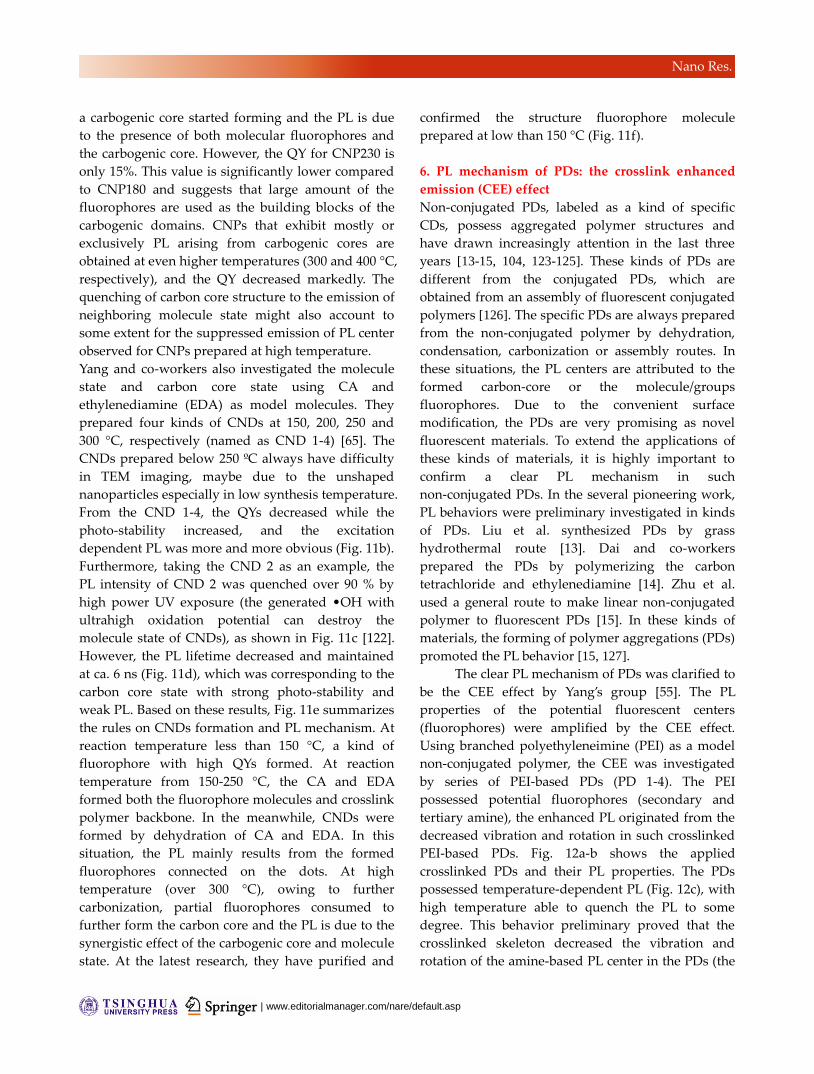
| www.editorialmanager.com/nare/default.asp
Nano Res.
a carbogenic core started forming and the PL is due
to the presence of both molecular fluorophores and
the carbogenic core. However, the QY for CNP230 is
only 15%. This value is significantly lower compared
to CNP180 and suggests that large amount of the
fluorophores are used as the building blocks of the
carbogenic domains. CNPs that exhibit mostly or
exclusively PL arising from carbogenic cores are
obtained at even higher temperatures (300 and 400 °C,
respectively), and the QY decreased markedly. The
quenching of carbon core structure to the emission of
neighboring molecule state might also account to
some extent for the suppressed emission of PL center
observed for CNPs prepared at high temperature.
Yang and co-workers also investigated the molecule
state and carbon core state using CA and
ethylenediamine (EDA) as model molecules. They
prepared four kinds of CNDs at 150, 200, 250 and
300 °C, respectively (named as CND 1-4) [65]. The
CNDs prepared below 250 ºC always have difficulty
in TEM imaging, maybe due to the unshaped
nanoparticles especially in low synthesis temperature.
From the CND 1-4, the QYs decreased while the
photo-stability increased, and the excitation
dependent PL was more and more obvious (Fig. 11b).
Furthermore, taking the CND 2 as an example, the
PL intensity of CND 2 was quenched over 90 % by
high power UV exposure (the generated •OH with
ultrahigh oxidation potential can destroy the
molecule state of CNDs), as shown in Fig. 11c [122].
However, the PL lifetime decreased and maintained
at ca. 6 ns (Fig. 11d), which was corresponding to the
carbon core state with strong photo-stability and
weak PL. Based on these results, Fig. 11e summarizes
the rules on CNDs formation and PL mechanism. At
reaction temperature less than 150 °C, a kind of
fluorophore with high QYs formed. At reaction
temperature from 150-250 °C, the CA and EDA
formed both the fluorophore molecules and crosslink
polymer backbone. In the meanwhile, CNDs were
formed by dehydration of CA and EDA. In this
situation, the PL mainly results from the formed
fluorophores connected on the dots. At high
temperature (over 300 °C), owing to further
carbonization, partial fluorophores consumed to
further form the carbon core and the PL is due to the
synergistic effect of the carbogenic core and molecule
state. At the latest research, they have purified and
confirmed the structure fluorophore molecule
prepared at low than 150 °C (Fig. 11f).
6. PL mechanism of PDs: the crosslink enhanced
emission (CEE) effect
Non-conjugated PDs, labeled as a kind of specific
CDs, possess aggregated polymer structures and
have drawn increasingly attention in the last three
years [13-15, 104, 123-125]. These kinds of PDs are
different from the conjugated PDs, which are
obtained from an assembly of fluorescent conjugated
polymers [126]. The specific PDs are always prepared
from the non-conjugated polymer by dehydration,
condensation, carbonization or assembly routes. In
these situations, the PL centers are attributed to the
formed carbon-core or the molecule/groups
fluorophores. Due to the convenient surface
modification, the PDs are very promising as novel
fluorescent materials. To extend the applications of
these kinds of materials, it is highly important to
confirm a clear PL mechanism in such
non-conjugated PDs. In the several pioneering work,
PL behaviors were preliminary investigated in kinds
of PDs. Liu et al. synthesized PDs by grass
hydrothermal route [13]. Dai and co-workers
prepared the PDs by polymerizing the carbon
tetrachloride and ethylenediamine [14]. Zhu et al.
used a general route to make linear non-conjugated
polymer to fluorescent PDs [15]. In these kinds of
materials, the forming of polymer aggregations (PDs)
promoted the PL behavior [15, 127].
The clear PL mechanism of PDs was clarified to
be the CEE effect by Yang’s group [55]. The PL
properties of the potential fluorescent centers
(fluorophores) were amplified by the CEE effect.
Using branched polyethyleneimine (PEI) as a model
non-conjugated polymer, the CEE was investigated
by series of PEI-based PDs (PD 1-4). The PEI
possessed potential fluorophores (secondary and
tertiary amine), the enhanced PL originated from the
decreased vibration and rotation in such crosslinked
PEI-based PDs. Fig. 12a-b shows the applied
crosslinked PDs and their PL properties. The PDs
possessed temperature-dependent PL (Fig. 12c), with
high temperature able to quench the PL to some
degree. This behavior preliminary proved that the
crosslinked skeleton decreased the vibration and
rotation of the amine-based PL center in the PDs (the
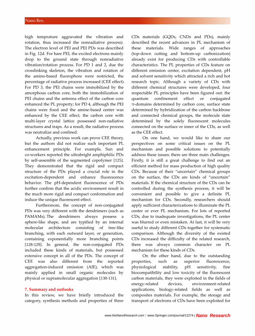
www.theNanoResearch.com∣www.Springer.com/journal/12274 | Nano Research
Nano Res.
high temprature aggravated the vibration and
rotation, thus increased the nonradiative process).
The electron level of PEI and PEI PDs was described
in Fig. 12d. For bare PEI, the excited electrons mainly
drop to the ground state through nonradiative
vibration/rotation process. For PD 1 and 2, due the
crosslinking skeleton, the vibration and rotation of
the amino-based fluorophore were restricted, the
percentage of radiative process increased (CEE effect).
For PD 3, the PEI chains were immobilized by the
amorphous carbon core, both the immobilization of
PEI chains and the antenna effect of the carbon core
enhanced the PL property; for PD 4, although the PEI
chains were fixed and the amine-based center was
enhanced by the CEE effect, the carbon core with
multi-layer crystal lattice possessed non-radiative
structures and traps. As a result, the radiative process
was neutralize and confined.
Actually, previous work can prove CEE theory,
but the authors did not realize such important PL
enhancement principle. For example, Sun and
co-workers reported the ultrabright amphiphilic PDs
by self-assemble of the segmented copolymer [125].
They demonstrated that the rigid and compact
structure of the PDs played a crucial role in the
excitation-dependent and enhance fluorescence
behavior. The pH-dependent fluorescence of PDs
further confirm that the acidic environment result in
the much more rigid and compact conformation and
induce the unique fluorescent effect.
Furthermore, the concept of non-conjugated
PDs was very different with the dendrimers (such as
PAMAMs). The dendrimers always possess a
sphere-like shape, and are typified by an internal
molecular architecture consisting of tree-like
branching, with each outward layer, or generation,
containing exponentially more branching points
[128-129]. In general, the non-conjugated PDs
included these kinds of materials, but possessed
extensive concept in all of the PDs. The concept of
CEE was also different from the reported
aggregation-induced emission (AIE), which was
mainly applied in small organic molecules by
physical or supramolecular aggregation [130-131].
7. Summary and outlooks
In this review, we have briefly introduced the
category, synthesis methods and properties of three
CDs materials (GQDs, CNDs and PDs), mainly
described the recent advances in PL mechanism of
these materials. Wide ranges of approaches
(top-down cutting and bottom-up carbonization)
already exist for producing CDs with controllable
characteristics. The PL properties of CDs feature on
different emission center, excitation dependent, pH
and solvent sensitivity which attracted a rich and hot
research topic. Although a variety of CDs with
different chemical structures were developed, four
respectable PL principles have been figured out: the
quantum confinement effect or conjugated
π-domains determined by carbon core, surface state
determined by hybridization of the carbon backbone
and connected chemical groups, the molecule state
determined by the solely fluorescent molecules
connected on the surface or inner of the CDs, as well
as the CEE effect.
On one hand, we would like to share our
perspectives on some critical issues on the PL
mechanism and possible solutions to potentially
address these issues: there are three main challenges.
Firstly, it is still a great challenge to find out an
efficient method for mass production of high quality
CDs. Because of their “uncertain” chemical groups
on the surface, the CDs are kinds of “uncertain”
materials. If the chemical structure of the CDs can be
controlled during the synthesis process, it will be
convenient and possible to give a definite PL
mechanism for CDs. Secondly, researchers should
apply sufficient characterizations to illuminate the PL
center or ever PL mechanism. For lots of reported
CDs, due to inadequate investigations, the PL center
was blurred or even mistaken. At last, it will be very
useful to study different CDs together for systematic
comparison. Although the diversity of the existed
CDs increased the difficulty of the related research,
there was always common character on PL
mechanism for these kinds of CDs.
On the other hand, due to the outstanding
properties, such as superior fluorescence,
physiological stability, pH sensitivity, fine
biocompatibility and low toxicity of the fluorescent
carbon materials, they were exploited in the fields of
energy-related devices, environment-related
applications, biology-related fields as well as
composites materials. For example, the storage and
transport of electrons of CDs have been exploited for
| www.editorialmanager.com/nare/default.asp
Nano Res.
solar cell [42, 132-133], organic light emitting diode
(OLED) [50, 134], photo-detector [135], photocatalysis
[23] and supercapacitor [136]. The quench based PL
(on-off and off-on) of CDs have been developed in
substance sensor (biomolecules, metal ions and
toxic/dangerous substance) [74, 137-138]. The stable
PL and low toxicity of CDs make them perfect
candidate in bioimaging [30, 53], biosensor [139],
drug delivery [140] and medical diagnosis [141]. The
rich chemical groups on the surface and facile
modification of CDs are beneficial to fluorescent
nanocomposites [127], functional hybrids [142] as
well high refractive index materials [143]. In the
future, we expect the advent of more facile and
robust synthetic methods and novel applications to
better realize the potential of the increasingly CDs
materials. Moreover, it could be safe,
environment-benign for whatever the synthesis
process, CDs themselves and relative applications,
which show powerful potential in above mentioned
fields. For the reasons above, it is extremely
important to promote the development of this kind
of material, which could be a promising candidate to
take place of the organic dyes or inorganic quantum
dots in relative fields. The rapid developments in
synthesizing CDs with controllable sizes, tailorable
chemical structures and clear PL mechanism, will
promote their applications.
Acknowledgements
This work was supported by the National Science
Foundation of China (Grand No. 51373065, 21221063,
81320108011, 91123031), the National Basic Research
Program of China (973 Program, Grant No.
2012CB933800), and the Specialized Research Fund
for the Doctoral Program of Higher Education (no.
20130061130010).
References
[1] Baker, S. N.; Baker, G. A. Luminescent carbon nanodots:
emergent nanolights. Angew. Chem. Int. Ed. 2010, 49,
6726-6744.
[2] Li, H.; Kang, Z.; Liu, Y.; Lee, S.-T. Carbon nanodots:
synthesis, properties and applications. J. Mater. Chem. 2012, 22,
24230.
[3] Welsher, K.; Liu, Z.; Sherlock, S. P.; Robinson, J. T.; Chen,
Z.; Daranciang, D.; Dai, H. A route to brightly fluorescent carbon
nanotubes for near-infrared imaging in mice. Nat. Nanotech.
2009, 4, 773-780.
[4] Gokus, T.; Nair, R. R.; Bonetti, A.; Bohmler, M.; Lombardo,
A.; Novoselov, K. S.; Geim, A. K.; Ferrari, A. C.; Hartschuh, A.
Making graphene luminescent by oxygen plasma treatment. ACS
Nano 2009, 3, 3963-3968.
[5] Eda, G.; Lin, Y. Y.; Mattevi, C.; Yamaguchi, H.; Chen, H. A.;
Chen, I. S.; Chen, C. W.; Chhowalla, M. Blue
photoluminescence from chemically derived graphene oxide. Adv.
Mater. 2010, 22, 505-509.
[6] Zhu, S.; Tang, S.; Zhang, J.; Yang, B. Control the size and
surface chemistry of graphene for the rising fluorescent materials.
Chem. Commun. 2012, 48, 4527-4539.
[7] Shen, J.; Zhu, Y.; Yang, X.; Li, C. Graphene quantum dots:
emergent nanolights for bioimaging, sensors, catalysis and
photovoltaic devices. Chem. Commun. 2012, 48, 3686-3699.
[8] Zhang, Z.; Zhang, J.; Chen, N.; Qu, L. Graphene quantum
dots: an emerging material for energy-related applications and
beyond. Energy Environ. Sci. 2012, 5, 8869.
[9] Li, L.; Wu, G.; Yang, G.; Peng, J.; Zhao, J.; Zhu, J. J.
Focusing on luminescent graphene quantum dots: current status
and future perspectives. Nanoscale 2013, 5, 4015-4039.
[10] Bacon, M.; Bradley, S. J.; Nann, T. Graphene quantum dots.
Part. Part. Syst. Charact. 2014, 31, 415-428.
[11] Zhou, X.; Guo, S.; Zhang, J. Solution-processable graphene
quantum dots. Chemphyschem 2013, 14, 2627-2640.
[12] Lin, L.; Rong, M.; Luo, F.; Chen, D.; Wang, Y.; Chen, X.
Luminescent graphene quantum dots as new fluorescent
materials for environmental and biological applications. TrAC
Trends Anal. Chem. 2014, 54, 83-102.
[13] Liu, S.; Tian, J.; Wang, L.; Zhang, Y.; Qin, X.; Luo, Y.;
Asiri, A. M.; Al-Youbi, A. O.; Sun, X. Hydrothermal treatment
of grass: a low-cost, green route to nitrogen-doped, carbon-rich,
photoluminescent polymer nanodots as an effective fluorescent
sensing platform for label-free detection of Cu (II) ions. Adv.
Mater. 2012, 24, 2037-2041.
[14] Qiao, Z. A.; Huo, Q.; Chi, M.; Veith, G. M.; Binder, A. J.;
Dai, S. A "ship-in-a-bottle" approach to synthesis of polymer
dots@silica or polymer dots@carbon core-shell nanospheres.
Adv. Mater. 2012, 24, 6017-6021.
[15] Zhu, S.; Zhang, J.; Wang, L.; Song, Y.; Zhang, G.; Wang, H.;
Yang, B. A general route to make non-conjugated linear
polymers luminescent. Chem. Commun. 2012, 48, 10889-10891.
[16] Yu, S. J.; Kang, M. W.; Chang, H. C.; Chen, K. M.; Yu, Y.
C. Bright fluorescent nanodiamonds: no photobleaching and low
cytotoxicity. J. Am. Chem. Soc. 2005, 127, 17604-17605.
[17] Mochalin, V. N.; Shenderova, O.; Ho, D.; Gogotsi, Y. The
properties and applications of nanodiamonds. Nat. Nanotech.
2012, 7, 11-23.
[18] Cao, L.; Meziani, M. J.; Sahu, S.; Sun, Y. P.
Photoluminescence properties of graphene versus other carbon
nanomaterials. Acc. Chem. Res. 2013, 46, 171-180.
[19] Song, Y.; Zhu, S.; Yang, B. Bioimaging based on
fluorescent carbon dots. RSC Adv. 2014, 4, 27184.
[20] Feng, X.; Wu, J.; Ai, M.; Pisula, W.; Zhi, L.; Rabe, J. P.;
Mullen, K. Triangle-shaped polycyclic aromatic hydrocarbons.
Angew. Chem. Int. Ed. 2007, 46, 3033-3036.
[21] Yan, X.; Cui, X.; Li, L. S. Synthesis of large, stable colloidal
graphene quantum dots with tunable size. J. Am. Chem. Soc.
2010, 132, 5944-5945.
[22] Qiao, Z.-A.; Wang, Y.; Gao, Y.; Li, H.; Dai, T.; Liu, Y.;
Huo, Q. Commercially activated carbon as the source for
www.theNanoResearch.com∣www.Springer.com/journal/12274 | Nano Research
Nano Res.
producing multicolor photoluminescent carbon dots by chemical
oxidation. Chem. Commun. 2010, 46, 8812.
[23] Li, H.; He, X.; Kang, Z.; Huang, H.; Liu, Y.; Liu, J.; Lian, S.;
Tsang, C. H.; Yang, X.; Lee, S. T. Water-soluble fluorescent
carbon quantum dots and photocatalyst design. Angew. Chem. Int.
Ed. 2010, 49, 4430-4434.
[24] Peng, J.; Gao, W.; Gupta, B. K.; Liu, Z.; Romero-Aburto, R.;
Ge, L.; Song, L.; Alemany, L. B.; Zhan, X.; Gao, G.; Vithayathil,
S. A.; Kaipparettu, B. A.; Marti, A. A.; Hayashi, T.; Zhu, J. J.;
Ajayan, P. M. Graphene quantum dots derived from carbon fibers.
Nano Lett. 2012, 12, 844-849.
[25] Xu, X.; Ray, R.; Gu, Y.; Ploehn, H. J.; Gearheart, L.; Raker,
K.; Scrivens, W. A. Electrophoretic analysis and purification of
fluorescent single-walled carbon nanotube fragments. J. Am.
Chem. Soc. 2004, 126, 12736-12737.
[26] Shinde, D. B.; Pillai, V. K. Electrochemical preparation of
luminescent graphene quantum dots from multiwalled carbon
nanotubes. Chem. Eur. J. 2012, 18, 12522-12528.
[27] Dong, Y.; Chen, C.; Zheng, X.; Gao, L.; Cui, Z.; Yang, H.;
Guo, C.; Chi, Y.; Li, C. M. One-step and high yield simultaneous
preparation of single- and multi-layer graphene quantum dots
from CX-72 carbon black. J. Mater. Chem. 2012, 22, 8764-8766.
[28] Liu, H.; Ye, T.; Mao, C. Fluorescent carbon nanoparticles
derived from candle soot. Angew. Chem. Int. Ed. 2007, 46,
6473-6475.
[29] Tao, H.; Yang, K.; Ma, Z.; Wan, J.; Zhang, Y.; Kang, Z.;
Liu, Z. In vivo NIR fluorescence imaging, biodistribution, and
toxicology of photoluminescent carbon dots produced from
carbon nanotubes and graphite. Small 2012, 8, 281-290.
[30] Zhu, S.; Zhang, J.; Qiao, C.; Tang, S.; Li, Y.; Yuan, W.; Li,
B.; Tian, L.; Liu, F.; Hu, R.; Gao, H.; Wei, H.; Zhang, H.; Sun,
H.; Yang, B. Strongly green-photoluminescent graphene
quantum dots for bioimaging applications. Chem. Commun. 2011,
47, 6858-6860.
[31] Zhu, S.; Zhang, J.; Liu, X.; Li, B.; Wang, X.; Tang, S.;
Meng, Q.; Li, Y.; Shi, C.; Hu, R.; Yang, B. Graphene quantum
dots with controllable surface oxidation, tunable fluorescence and
up-conversion emission. RSC Adv. 2012, 2, 2717.
[32] Lu, J.; Yang, J. X.; Wang, J.; Lim, A.; Wang, S.; Loh, K. P.
One-pot synthesis of fluorescent carbon nanoribbons,
nanoparticles, and graphene by the exfoliation of graphite in
ionic liquids. ACS Nano 2009, 3, 2367-2375.
[33] Zheng, L.; Chi, Y.; Dong, Y.; Lin, J.; Wang, B.
Electrochemiluminescence of water-soluble carbon nanocrystals
released electrochemically from graphite. J. Am. Chem. Soc.
2009, 131, 4564-4565.
[34] Pan, D.; Zhang, J.; Li, Z.; Wu, M. Hydrothermal route for
cutting graphene sheets into blue-luminescent graphene quantum
dots. Adv. Mater. 2010, 22, 734-738.
[35] Lin, L.; Zhang, S. Creating high yield water soluble
luminescent graphene quantum dots via exfoliating and
disintegrating carbon nanotubes and graphite flakes. Chem.
Commun. 2012, 48, 10177-10179.
[36] Bottini, M.; Balasubramanian, C.; Dawson, M. I.;
Bergamaschi, A.; Bellucci, S.; Mustelin, T. Isolation and
characterization of fluorescent nanoparticles from pristine and
oxidized electric arc-produced single-walled carbon nanotubes. J.
Phys.Chem. B 2006, 110, 831-836.
[37] Sun, Y. P.; Zhou, B.; Lin, Y.; Wang, W.; Fernando, K. A.;
Pathak, P.; Meziani, M. J.; Harruff, B. A.; Wang, X.; Wang, H.;
Luo, P. G.; Yang, H.; Kose, M. E.; Chen, B.; Veca, L. M.; Xie, S.
Y. Quantum-sized carbon dots for bright and colorful
photoluminescence. J. Am. Chem. Soc. 2006, 128, 7756-7757.
[38] Lee, J.; Kim, K.; Park, W. I.; Kim, B. H.; Park, J. H.; Kim, T.
H.; Bong, S.; Kim, C. H.; Chae, G.; Jun, M.; Hwang, Y.; Jung, Y.
S.; Jeon, S. Uniform graphene quantum dots patterned from
self-assembled silica nanodots. Nano Lett. 2012, 12, 6078-6083.
[39] Fan, L.; Zhu, M.; Lee, X.; Zhang, R.; Wang, K.; Wei, J.;
Zhong, M.; Wu, D.; Zhu, H. Direct synthesis of graphene
quantum dots by chemical vapor deposition. Part. Part. Syst.
Charact. 2013, 30, 764-769.
[40] Zhao, Q. L.; Zhang, Z. L.; Huang, B. H.; Peng, J.; Zhang, M.;
Pang, D. W. Facile preparation of low cytotoxicity fluorescent
carbon nanocrystals by electrooxidation of graphite. Chem.
Commun. 2008, 5116-5118.
[41] Bao, L.; Zhang, Z. L.; Tian, Z. Q.; Zhang, L.; Liu, C.; Lin,
Y.; Qi, B.; Pang, D. W. Electrochemical tuning of luminescent
carbon nanodots: from preparation to luminescence mechanism.
Adv. Mater. 2011, 23, 5801-5806.
[42] Li, Y.; Hu, Y.; Zhao, Y.; Shi, G.; Deng, L.; Hou, Y.; Qu, L.
An electrochemical avenue to green-luminescent graphene
quantum dots as potential electron-acceptors for photovoltaics.
Adv. Mater. 2011, 23, 776-780.
[43] Deng, J.; Lu, Q.; Mi, N.; Li, H.; Liu, M.; Xu, M.; Tan, L.;
Xie, Q.; Zhang, Y.; Yao, S. Electrochemical synthesis of carbon
nanodots directly from alcohols. Chem. Eur. J. 2014, 20,
4993-4999.
[44] Zhou, X.; Zhang, Y.; Wang, C.; Wu, X.; Yang, Y.; Zheng,
B.; Wu, H.; Guo, S.; Zhang, J. Photo-Fenton reaction of graphene
oxide: a new strategy to prepare graphene quantum dots for DNA
cleavage. ACS Nano 2012, 6, 6592-6599.
[45] Yang, Z. C.; Wang, M.; Yong, A. M.; Wong, S. Y.; Zhang,
X. H.; Tan, H.; Chang, A. Y.; Li, X.; Wang, J. Intrinsically
fluorescent carbon dots with tunable emission derived from
hydrothermal treatment of glucose in the presence of
monopotassium phosphate. Chem. Commun. 2011, 47,
11615-11617.
[46] Zhu, H.; Wang, X.; Li, Y.; Wang, Z.; Yang, F.; Yang, X.
Microwave synthesis of fluorescent carbon nanoparticles with
electrochemiluminescence properties. Chem. Commun. 2009,
5118-5120.
[47] Bourlinos, A. B.; Stassinopoulos, A.; Anglos, D.; Zboril, R.;
Karakassides, M.; Giannelis, E. P. Surface functionalized
carbogenic quantum dots. Small 2008, 4, 455-458.
[48] Peng, H.; Travas-Sejdic, J. Simple aqueous solution route to
luminescent carbogenic dots from carbohydrates. Chem. Mater.
2009, 21, 5563-5565.
[49] Zong, J.; Zhu, Y.; Yang, X.; Shen, J.; Li, C. Synthesis of
photoluminescent carbogenic dots using mesoporous silica
spheres as nanoreactors. Chem. Commun. 2011, 47, 764-766.
[50] Tang, L.; Ji, R.; Cao, X.; Lin, J.; Jiang, H.; Li, X.; Teng, K.
S.; Luk, C. M.; Zeng, S.; Hao, J.; Lau, S. P. Deep ultraviolet
photoluminescence of water-soluble self-passivated graphene
quantum dots. ACS Nano 2012, 6, 5102-5110.
[51] Wang, J.; Wang, C. F.; Chen, S. Amphiphilic egg-derived
carbon dots: rapid plasma fabrication, pyrolysis process, and
multicolor printing patterns. Angew. Chem. Int. Ed. 2012, 51,
9297-9301.
[52] Zhang, C.; Liu, Y.; Xiong, X. Q.; Peng, L. H.; Gan, L.; Chen,
C. F.; Xu, H. B. Three-dimensional nanographene based on
triptycene: synthesis and its application in fluorescence imaging.
Org. Lett. 2012, 14, 5912-5915.
[53] Cao, L.; Wang, X.; Meziani, M. J.; Lu, F.; Wang, H.; Luo, P.
G.; Lin, Y.; Harruff, B. A.; Veca, L. M.; Murray, D.; Xie, S. Y.;
Sun, Y. P. Carbon dots for multiphoton bioimaging. J. Am. Chem.
Soc. 2007, 129, 11318-11319.

| www.editorialmanager.com/nare/default.asp
Nano Res.
[54] Shen, J.; Zhu, Y.; Chen, C.; Yang, X.; Li, C. Facile
preparation and upconversion luminescence of graphene quantum
dots. Chem. Commun. 2011, 47, 2580-2582.
[55] Zhu, S.; Wang, L.; Zhou, N.; Zhao, X.; Song, Y.; Maharjan,
S.; Zhang, J.; Lu, L.; Wang, H.; Yang, B. The crosslink enhanced
emission (CEE) in non-conjugated polymer dots: from the
photoluminescence mechanism to the cellular uptake mechanism
and internalization. Chem. Commun. 2014, 50, 13845-13848.
[56] Zheng, H.; Wang, Q.; Long, Y.; Zhang, H.; Huang, X.; Zhu,
R. Enhancing the luminescence of carbon dots with a reduction
pathway. Chem. Commun. 2011, 47, 10650-10652.
[57] Nie, H.; Li, M.; Li, Q.; Liang, S.; Tan, Y.; Sheng, L.; Shi,
W.; Zhang, S. X.-A. Carbon dots with continuously tunable
full-color emission and their application in ratiometric pH
sensing. Chem. Mater. 2014, 26, 3104-3112.
[58] Tetsuka, H.; Asahi, R.; Nagoya, A.; Okamoto, K.; Tajima, I.;
Ohta, R.; Okamoto, A. Optically tunable amino-functionalized
graphene quantum dots. Adv. Mater. 2012, 24, 5333-5338.
[59] Wang, Y.; Kalytchuk, S.; Zhang, Y.; Shi, H.; Kershaw, S. V.;
Rogach, A. L. Thickness-dependent full-color emission tunability
in a flexible carbon dot ionogel. J. Phys. Chem. Lett. 2014, 5,
1412-1420.
[60] Wang, Y.; Li, Y.; Yan, Y.; Xu, J.; Guan, B.; Wang, Q.; Li, J.;
Yu, J. Luminescent carbon dots in a new magnesium
aluminophosphate zeolite. Chem. Commun. 2013, 49, 9006-9008.
[61] Ray, S. C.; Saha, A.; Jana, N. R.; Sarkar, R. Fluorescent
carbon nanoparticles: synthesis, characterization, and bioimaging
application. J. Phys. Chem. C 2009, 113, 18546-18551.
[62] Wang, X.; Qu, K.; Xu, B.; Ren, J.; Qu, X. Multicolor
luminescent carbon nanoparticles: Synthesis, supramolecular
assembly with porphyrin, intrinsic peroxidase-like catalytic
activity and applications. Nano Res. 2011, 4, 908-920.
[63] Bhunia, S. K.; Saha, A.; Maity, A. R.; Ray, S. C.; Jana, N. R.
Carbon nanoparticle-based fluorescent bioimaging probes. Sci.
Rep. 2013, 3, 1473.
[64] Qu, D.; Zheng, M.; Zhang, L.; Zhao, H.; Xie, Z.; Jing, X.;
Haddad, R. E.; Fan, H.; Sun, Z. Formation mechanism and
optimization of highly luminescent N-doped graphene quantum
dots. Sci. Rep. 2014, 4, 5294.
[65] Zhu, S.; Meng, Q.; Wang, L.; Zhang, J.; Song, Y.; Jin, H.;
Zhang, K.; Sun, H.; Wang, H.; Yang, B. Highly
photoluminescent carbon dots for multicolor patterning, sensors,
and bioimaging. Angew. Chem. Int. Ed. 2013, 52, 3953-3957.
[66] Gan, Z.; Wu, X.; Zhou, G.; Shen, J.; Chu, P. K. Is there real
upconversion photoluminescence from graphene quantum dots?
Adv. Opt. Mater. 2013, 1, 554-558.
[67] Wen, X.; Yu, P.; Toh, Y. R.; Ma, X.; Tang, J. On the
upconversion fluorescence in carbon nanodots and graphene
quantum dots. Chem. Commun. 2014, 50, 4703-4706.
[68] Qu, S.; Liu, X.; Guo, X.; Chu, M.; Zhang, L.; Shen, D.
Amplified spontaneous green emission and lasing emission from
carbon nanoparticles. Adv. Funct. Mater. 2014, 24, 2689-2695.
[69] Fan, L.; Hu, Y.; Wang, X.; Zhang, L.; Li, F.; Han, D.; Li, Z.;
Zhang, Q.; Wang, Z.; Niu, L. Fluorescence resonance energy
transfer quenching at the surface of graphene quantum dots for
ultrasensitive detection of TNT. Talanta 2012, 101, 192-197.
[70] Luo, P. G.; Sahu, S.; Yang, S.-T.; Sonkar, S. K.; Wang, J.;
Wang, H.; LeCroy, G. E.; Cao, L.; Sun, Y.-P. Carbon “quantum”
dots for optical bioimaging. J. Mater. Chem. B 2013, 1, 2116.
[71] Esteves da Silva, J. C. G.; Gonçalves, H. M. R. Analytical
and bioanalytical applications of carbon dots. TrAC Trends Anal.
Chem. 2011, 30, 1327-1336.
[72] Sun, X.; Liu, Z.; Welsher, K.; Robinson, J. T.; Goodwin, A.;
Zaric, S.; Dai, H. Nano-graphene oxide for cellular imaging and
drug delivery. Nano Res. 2008, 1, 203-212.
[73] Goh, E. J.; Kim, K. S.; Kim, Y. R.; Jung, H. S.; Beack, S.;
Kong, W. H.; Scarcelli, G.; Yun, S. H.; Hahn, S. K. Bioimaging
of hyaluronic acid derivatives using nanosized carbon dots.
Biomacromolecules 2012, 13, 2554-2561.
[74] Kong, B.; Zhu, A.; Ding, C.; Zhao, X.; Li, B.; Tian, Y.
Carbon dot-based inorganic-organic nanosystem for two-photon
imaging and biosensing of pH variation in living cells and tissues.
Adv. Mater. 2012, 24, 5844-5848.
[75] Liu, C.; Zhang, P.; Zhai, X.; Tian, F.; Li, W.; Yang, J.; Liu,
Y.; Wang, H.; Wang, W.; Liu, W. Nano-carrier for gene delivery
and bioimaging based on carbon dots with PEI-passivation
enhanced fluorescence. Biomaterials 2012, 33, 3604-3613.
[76] Nurunnabi, M.; Khatun, Z.; Huh, K. M.; Park, S. Y.; Lee, D.
Y.; Cho, K. J.; Lee, Y. K. In vivo biodistribution and toxicology
of carboxylated graphene quantum dots. ACS Nano 2013, 7,
6858-6867.
[77] Qian, J.; Wang, D.; Cai, F. H.; Xi, W.; Peng, L.; Zhu, Z. F.;
He, H.; Hu, M. L.; He, S. Observation of multiphoton-induced
fluorescence from graphene oxide nanoparticles and applications
in in vivo functional bioimaging. Angew. Chem. Int. Ed. 2012, 51,
10570-10575.
[78] Chien, C. T.; Li, S. S.; Lai, W. J.; Yeh, Y. C.; Chen, H. A.;
Chen, I. S.; Chen, L. C.; Chen, K. H.; Nemoto, T.; Isoda, S.;
Chen, M.; Fujita, T.; Eda, G.; Yamaguchi, H.; Chhowalla, M.;
Chen, C. W. Tunable photoluminescence from graphene oxide.
Angew. Chem. Int. Ed. 2012, 51, 6662-6666.
[79] Luo, Z.; Vora, P. M.; Mele, E. J.; Johnson, A. T. C.;
Kikkawa, J. M. Photoluminescence and band gap modulation in
graphene oxide. Appl. Phys. Lett. 2009, 94, 111909.
[80] Galande, C.; Mohite, A. D.; Naumov, A. V.; Gao, W.; Ci, L.;
Ajayan, A.; Gao, H.; Srivastava, A.; Weisman, R. B.; Ajayan, P.
M. Quasi-molecular fluorescence from graphene oxide. Sci. Rep.
2011, 1, 85.
[81] Shang, J.; Ma, L.; Li, J.; Ai, W.; Yu, T.; Gurzadyan, G. G.
The origin of fluorescence from graphene oxide. Sci. Rep. 2012,
2, 792.
[82] Ritter, K. A.; Lyding, J. W. The influence of edge structure
on the electronic properties of graphene quantum dots and
nanoribbons. Nat. Mater. 2009, 8, 235-242.
[83] Radovic, L. R.; Bockrath, B. On the chemical nature of
graphene edges: origin of stability and potential for magnetism in
carbon materials. J. Am. Chem. Soc. 2005, 127, 5917-5927.
[84] Xu, Q.; Zhou, Q.; Hua, Z.; Xue, Q.; Zhang, C.; Wang, X.;
Pan, D.; Xiao, M. Single-particle spectroscopic measurements of
fluorescent graphene quantum dots. ACS Nano 2013, 7,
10654-10661.
[85] Jin, S. H.; Kim da, H.; Jun, G. H.; Hong, S. H.; Jeon, S.
Tuning the photoluminescence of graphene quantum dots
through the charge transfer effect of functional groups. ACS
Nano 2013, 7, 1239-1245.
[86] Kumar, G. S.; Roy, R.; Sen, D.; Ghorai, U. K.; Thapa, R.;
Mazumder, N.; Saha, S.; Chattopadhyay, K. K.
Amino-functionalized graphene quantum dots: origin of tunable
heterogeneous photoluminescence. Nanoscale 2014, 6,
3384-3391.
[87] Qian, Z.; Ma, J.; Shan, X.; Shao, L.; Zhou, J.; Chen, J.; Feng,
H. Surface functionalization of graphene quantum dots with
small organic molecules from photoluminescence modulation to
bioimaging applications: an experimental and theoretical
investigation. RSC Adv. 2013, 3, 14571.
www.theNanoResearch.com∣www.Springer.com/journal/12274 | Nano Research
Nano Res.
[88] Wang, L.; Wang, H. Y.; Wang, Y.; Zhu, S. J.; Zhang, Y. L.;
Zhang, J. H.; Chen, Q. D.; Han, W.; Xu, H. L.; Yang, B.; Sun, H.
B. Direct observation of quantum-confined graphene-like states
and novel hybrid states in graphene oxide by transient
spectroscopy. Adv. Mater. 2013, 25, 6539-6545.
[89] Wang, L.; Zhu, S.-J.; Wang, H.-Y.; Wang, Y.-F.; Hao,
Y.-W.; Zhang, J.-H.; Chen, Q.-D.; Zhang, Y.-L.; Han, W.; Yang,
B.; Sun, H.-B. Unraveling bright molecule-like state and dark
intrinsic state in green-fluorescence graphene quantum dots via
ultrafast spectroscopy. Adv. Opt. Mater. 2013, 1, 264-271.
[90] Zhu, S.; Zhang, J.; Tang, S.; Qiao, C.; Wang, L.; Wang, H.;
Liu, X.; Li, B.; Li, Y.; Yu, W.; Wang, X.; Sun, H.; Yang, B.
Surface chemistry routes to modulate the photoluminescence of
graphene quantum dots: from fluorescence mechanism to
up-conversion bioimaging applications. Adv. Funct. Mater. 2012,
22, 4732-4740.
[91] Mei, Q.; Zhang, Z. Photoluminescent graphene oxide ink to
print sensors onto microporous membranes for versatile
visualization bioassays. Angew. Chem. Int. Ed. 2012, 51,
5602-5606.
[92] Liu, F.; Jang, M. H.; Ha, H. D.; Kim, J. H.; Cho, Y. H.; Seo,
T. S. Facile synthetic method for pristine graphene quantum dots
and graphene oxide quantum dots: origin of blue and green
luminescence. Adv. Mater. 2013, 25, 3657-3662.
[93] Li, X.; Lau, S. P.; Tang, L.; Ji, R.; Yang, P. Multicolour
light emission from chlorine-doped graphene quantum dots. J.
Mater. Chem. C 2013, 1, 7308.
[94] Luo, P.; Ji, Z.; Li, C.; Shi, G. Aryl-modified graphene
quantum dots with enhanced photoluminescence and improved
pH tolerance. Nanoscale 2013, 5, 7361-7367.
[95] Sun, H.; Gao, N.; Wu, L.; Ren, J.; Wei, W.; Qu, X. Highly
photoluminescent amino-functionalized graphene quantum dots
used for sensing copper ions. Chem. Eur. J. 2013, 19,
13362-13368.
[96] Feng, Y.; Zhao, J.; Yan, X.; Tang, F.; Xue, Q. Enhancement
in the fluorescence of graphene quantum dots by hydrazine
hydrate reduction. Carbon 2014, 66, 334-339.
[97] Sun, Y.; Wang, S.; Li, C.; Luo, P.; Tao, L.; Wei, Y.; Shi, G.
Large scale preparation of graphene quantum dots from graphite
with tunable fluorescence properties. PCCP 2013, 15,
9907-9913.
[98] Jiang, F.; Chen, D.; Li, R.; Wang, Y.; Zhang, G.; Li, S.;
Zheng, J.; Huang, N.; Gu, Y.; Wang, C.; Shu, C. Eco-friendly
synthesis of size-controllable amine-functionalized graphene
quantum dots with antimycoplasma properties. Nanoscale 2013,
5, 1137-1142.
[99] Lingam, K.; Podila, R.; Qian, H.; Serkiz, S.; Rao, A. M.
Evidence for edge-state photoluminescence in graphene quantum
dots. Adv. Funct. Mater. 2013, 23, 5062-5065.
[100] Chen, C. F.; Park, C. H.; Boudouris, B. W.; Horng, J.;
Geng, B.; Girit, C.; Zettl, A.; Crommie, M. F.; Segalman, R. A.;
Louie, S. G.; Wang, F. Controlling inelastic light scattering
quantum pathways in graphene. Nature 2011, 471, 617-620.
[101] Li, L.-s.; Yan, X. Colloidal Graphene Quantum Dots. J.
Phys. Chem. Lett. 2010, 1, 2572-2576.
[102] Tomovic, Z.; Watson, M. D.; Müllen, K.
Superphenalene-based columnar liquid crystals. Angew. Chem.
Int. Ed. 2004, 43, 755-758.
[103] Mueller, M. L.; Yan, X.; Dragnea, B.; Li, L. S. Slow
hot-carrier relaxation in colloidal graphene quantum dots. Nano
Lett. 2011, 11, 56-60.
[104] Zhu, S.; Wang, L.; Li, B.; Song, Y.; Zhao, X.; Zhang, G.;
Zhang, S.; Lu, S.; Zhang, J.; Wang, H.; Sun, H.; Yang, B.
Investigation of photoluminescence mechanism of graphene
quantum dots and evaluation of their assembly into polymer dots.
Carbon 2014, 77, 462-472.
[105] Kim, S.; Hwang, S. W.; Kim, M. K.; Shin, D. Y.; Shin, D.
H.; Kim, C. O.; Yang, S. B.; Park, J. H.; Hwang, E.; Choi, S. H.;
Ko, G.; Sim, S.; Sone, C.; Choi, H. J.; Bae, S.; Hong, B. H.
Anomalous behaviors of visible luminescence from graphene
quantum dots: interplay between size and shape. ACS Nano 2012,
6, 8203-8208.
[106] Sk, M. A.; Ananthanarayanan, A.; Huang, L.; Lim, K. H.;
Chen, P. Revealing the tunable photoluminescence properties of
graphene quantum dots. J. Mater. Chem. C 2014, 2, 6954-6960.
[107] Lui, C. H.; Mak, K. F.; Shan, J.; Heinz, T. F. Ultrafast
photoluminescence from graphene. Phys. Rev. Lett. 2010, 105,
127404.
[108] Kim, R.; Perebeinos, V.; Avouris, P. Relaxation of
optically excited carriers in graphene. Phys. Rev. B 2011, 84,
075449.
[109] Fuyuno, N.; Kozawa, D.; Miyauchi, Y.; Mouri, S.; Kitaura,
R.; Shinohara, H.; Yasuda, T.; Komatsu, N.; Matsuda, K. Drastic
Change in photoluminescence properties of graphene quantum
dots by chromatographic separation. Adv. Opt. Mater. 2014,
10.1002/adom.201400200.
[110] Tang, L.; Ji, R.; Li, X.; Teng, K. S.; Lau, S. P.
Size-dependent structural and optical characteristics of
glucose-derived graphene quantum dots. Part. Part. Syst.
Charact. 2013, 30, 523-531.
[111] Kwon, W.; Rhee, S. W. Facile synthesis of graphitic
carbon quantum dots with size tunability and uniformity using
reverse micelles. Chem. Commun. 2012, 48, 5256-5258.
[112] Kwon, W.; Lee, G.; Do, S.; Joo, T.; Rhee, S.-W.
Size-controlled soft-template synthesis of carbon nanodots
toward versatile photoactive materials. Small 2014, 10, 506-513.
[113] Wang, X.; Cao, L.; Yang, S. T.; Lu, F.; Meziani, M. J.;
Tian, L.; Sun, K. W.; Bloodgood, M. A.; Sun, Y. P. Bandgap-like
strong fluorescence in functionalized carbon nanoparticles.
Angew. Chem. Int. Ed. 2010, 49, 5310-5314.
[114] Das, S. K.; Liu, Y.; Yeom, S.; Kim, D. Y.; Richards, C. I.
Single-particle fluorescence intensity fluctuations of carbon
nanodots. Nano Lett. 2014, 14, 620-625.
[115] Yu, P.; Wen, X.; Toh, Y.-R.; Tang, J.
Temperature-Dependent Fluorescence in Carbon Dots. J. Phys.
Chem. C 2012, 116, 25552-25557.
[116] Wen, X.; Yu, P.; Toh, Y.-R.; Hao, X.; Tang, J. Intrinsic
and extrinsic fluorescence in carbon nanodots: ultrafast
time-resolved fluorescence and carrier dynamics. Adv. Opt.
Mater. 2013, 1, 173-178.
[117] Wang, L.; Zhu, S. J.; Wang, H. Y.; Qu, S. N.; Zhang, Y. L.;
Zhang, J. H.; Chen, Q. D.; Xu, H. L.; Han, W.; Yang, B.; Sun, H.
B. Common origin of green luminescence in carbon nanodots and
graphene quantum dots. ACS Nano 2014, 8, 2541-2547.
[118] Qu, S.; Wang, X.; Lu, Q.; Liu, X.; Wang, L. A
biocompatible fluorescent ink based on water-soluble
luminescent carbon nanodots. Angew. Chem. Int. Ed. 2012, 51,
12215-12218.
[119] Sun, H.; Wu, L.; Gao, N.; Ren, J.; Qu, X. Improvement of
photoluminescence of graphene quantum dots with a
biocompatible photochemical reduction pathway and its
bioimaging application. ACS Appl. Mater. Inter. 2013, 5,
1174-1179.
[120] Li, L.-L.; Ji, J.; Fei, R.; Wang, C.-Z.; Lu, Q.; Zhang, J.-R.;
Jiang, L.-P.; Zhu, J.-J. A facile microwave avenue to

| www.editorialmanager.com/nare/default.asp
Nano Res.
electrochemiluminescent two-color graphene quantum dots. Adv.
Funct. Mater. 2012, 22, 2971-2979.
[121] Krysmann, M. J.; Kelarakis, A.; Dallas, P.; Giannelis, E. P.
Formation mechanism of carbogenic nanoparticles with dual
photoluminescence emission. J. Am. Chem. Soc. 2012, 134,
747-750.
[122] Song, Y.; Zhu, S.; Xiang, S.; Zhao, X.; Zhang, J.; Zhang,
H.; Fu, Y.; Yang, B. Investigation into the fluorescence
quenching behaviors and applications of carbon dots. Nanoscale
2014, 6, 4676-4682.
[123] Ding, D.; Goh, C. C.; Feng, G.; Zhao, Z.; Liu, J.; Liu, R.;
Tomczak, N.; Geng, J.; Tang, B. Z.; Ng, L. G.; Liu, B.
Ultrabright organic dots with aggregation-induced emission
characteristics for real-time two-photon intravital vasculature
imaging. Adv. Mater. 2013, 25, 6083-6088.
[124] Lai, T.; Zheng, E.; Chen, L.; Wang, X.; Kong, L.; You, C.;
Ruan, Y.; Weng, X. Hybrid carbon source for producing
nitrogen-doped polymer nanodots: one-pot hydrothermal
synthesis, fluorescence enhancement and highly selective
detection of Fe (III). Nanoscale 2013, 5, 8015-8021.
[125] Sun, Y.; Cao, W.; Li, S.; Jin, S.; Hu, K.; Hu, L.; Huang, Y.;
Gao, X.; Wu, Y.; Liang, X. J. Ultrabright and multicolorful
fluorescence of amphiphilic polyethyleneimine polymer dots for
efficiently combined imaging and therapy. Sci. Rep. 2013, 3,
3036.
[126] Wu, C.; Chiu, D. T. Highly fluorescent semiconducting
polymer dots for biology and medicine. Angew. Chem. Int. Ed.
2013, 52, 3086-3109.
[127] Zhu, S.; Zhang, J.; Song, Y.; Zhang, G.; Zhang, H.; Yang,
B. Fluorescent nanocomposite based on PVA polymer dots. Acta
Chim. Sinica 2012, 70, 2311.
[128] Sun, M.; Hong, C. Y.; Pan, C. Y. A unique aliphatic
tertiary amine chromophore: fluorescence, polymer structure, and
application in cell imaging. J. Am. Chem. Soc. 2012, 134,
20581-20584.
[129] Zhu, Q.; Qiu, F.; Zhu, B.; Zhu, X. Hyperbranched
polymers for bioimaging. RSC Adv. 2013, 3, 2071.
[130] Hong, Y.; Lam, J. W.; Tang, B. Z. Aggregation-induced
emission: phenomenon, mechanism and applications. Chem.
Commun. 2009, 4332-4353.
[131] Hong, Y.; Lam, J. W.; Tang, B. Z. Aggregation-induced
emission. Chemical Society Reviews 2011, 40, 5361-5388.
[132] Mirtchev, P.; Henderson, E. J.; Soheilnia, N.; Yip, C. M.;
Ozin, G. A. Solution phase synthesis of carbon quantum dots as
sensitizers for nanocrystalline TiO2 solar cells. J. Mater. Chem.
2012, 22, 1265.
[133] Gupta, V.; Chaudhary, N.; Srivastava, R.; Sharma, G. D.;
Bhardwaj, R.; Chand, S. Luminscent graphene quantum dots for
organic photovoltaic devices. J. Am. Chem. Soc. 2011, 133,
9960-9963.
[134] Zhang, X.; Zhang, Y.; Wang, Y.; Kalytchuk, S.; Kershaw,
S. V.; Wang, Y.; Wang, P.; Zhang, T.; Zhao, Y.; Zhang, H.; Cui,
T.; Wang, Y.; Zhao, J.; Yu, W. W.; Rogach, A. L.
Color-switchable electroluminescence of carbon dot
light-emitting diodes. ACS Nano 2013, 7, 11234-11241.
[135] Shen, J.; Zhu, Y.; Yang, X.; Zong, J.; Zhang, J.; Li, C.
One-pot hydrothermal synthesis of graphene quantum dots
surface-passivated by polyethylene glycol and their photoelectric
conversion under near-infrared light. New J. Chem. 2012, 36, 97.
[136] Liu, W.-W.; Feng, Y.-Q.; Yan, X.-B.; Chen, J.-T.; Xue,
Q.-J. Superior micro-supercapacitors based on graphene quantum
dots. Adv. Funct. Mater. 2013, 23, 4111-4122.
[137] Lin, Z.; Xue, W.; Chen, H.; Lin, J. M.
Peroxynitrous-acid-induced chemiluminescence of fluorescent
carbon dots for nitrite sensing. Anal.Chem. 2011, 83, 8245-8251.
[138] Liu, J. J.; Zhang, X. L.; Cong, Z. X.; Chen, Z. T.; Yang, H.
H.; Chen, G. N. Glutathione-functionalized graphene quantum
dots as selective fluorescent probes for phosphate-containing
metabolites. Nanoscale 2013, 5, 1810-1815.
[139] Li, X.; Zhu, S.; Xu, B.; Ma, K.; Zhang, J.; Yang, B.; Tian,
W. Self-assembled graphene quantum dots induced by
cytochrome c: a novel biosensor for trypsin with remarkable
fluorescence enhancement. Nanoscale 2013, 5, 7776-7779.
[140] Tang, J.; Kong, B.; Wu, H.; Xu, M.; Wang, Y.; Wang, Y.;
Zhao, D.; Zheng, G. Carbon nanodots featuring efficient FRET
for real-time monitoring of drug delivery and two-photon
imaging. Adv. Mater. 2013, 25, 6569-6574.
[141] Markovic, Z. M.; Ristic, B. Z.; Arsikin, K. M.; Klisic, D.
G.; Harhaji-Trajkovic, L. M.; Todorovic-Markovic, B. M.; Kepic,
D. P.; Kravic-Stevovic, T. K.; Jovanovic, S. P.; Milenkovic, M.
M.; Milivojevic, D. D.; Bumbasirevic, V. Z.; Dramicanin, M. D.;
Trajkovic, V. S. Graphene quantum dots as autophagy-inducing
photodynamic agents. Biomaterials 2012, 33, 7084-7092.
[142] Xie, Z.; Wang, F.; Liu, C. Y. Organic-inorganic hybrid
functional carbon dot gel glasses. Adv. Mater. 2012, 24,
1716-1721.
[143] Zhang, G.; Zhang, H.; Zhang, X.; Zhu, S.; Zhang, L.; Meng,
Q.; Wang, M.; Li, Y.; Yang, B. Embedding graphene
nanoparticles into poly(N,N′-dimethylacrylamine) to prepare
transparent nanocomposite films with high refractive index. J.
Mater. Chem. 2012, 22, 21218-21224.
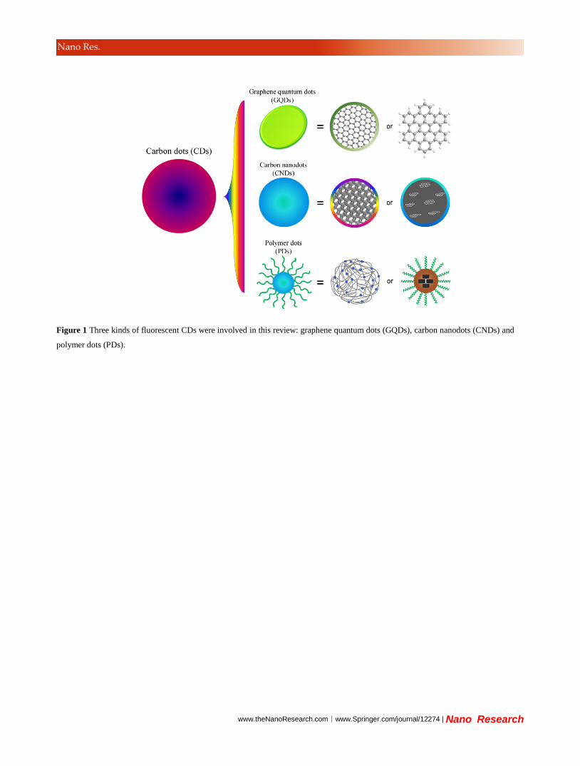
www.theNanoResearch.com∣www.Springer.com/journal/12274 | Nano Research
Nano Res.
Figure 1 Three kinds of fluorescent CDs were involved in this review: graphene quantum dots (GQDs), carbon nanodots (CNDs) and
polymer dots (PDs).
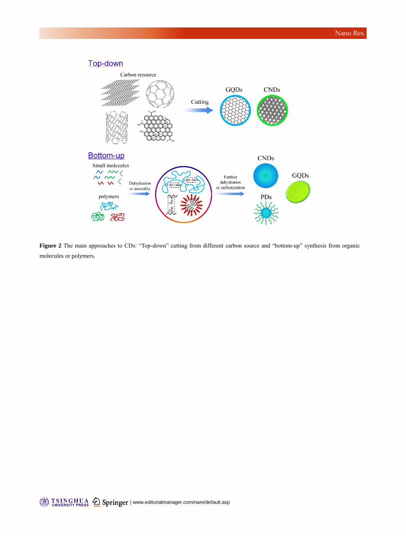
| www.editorialmanager.com/nare/default.asp
Nano Res.
Figure 2 The main approaches to CDs: “Top-down” cutting from different carbon source and “bottom-up” synthesis from organic
molecules or polymers.
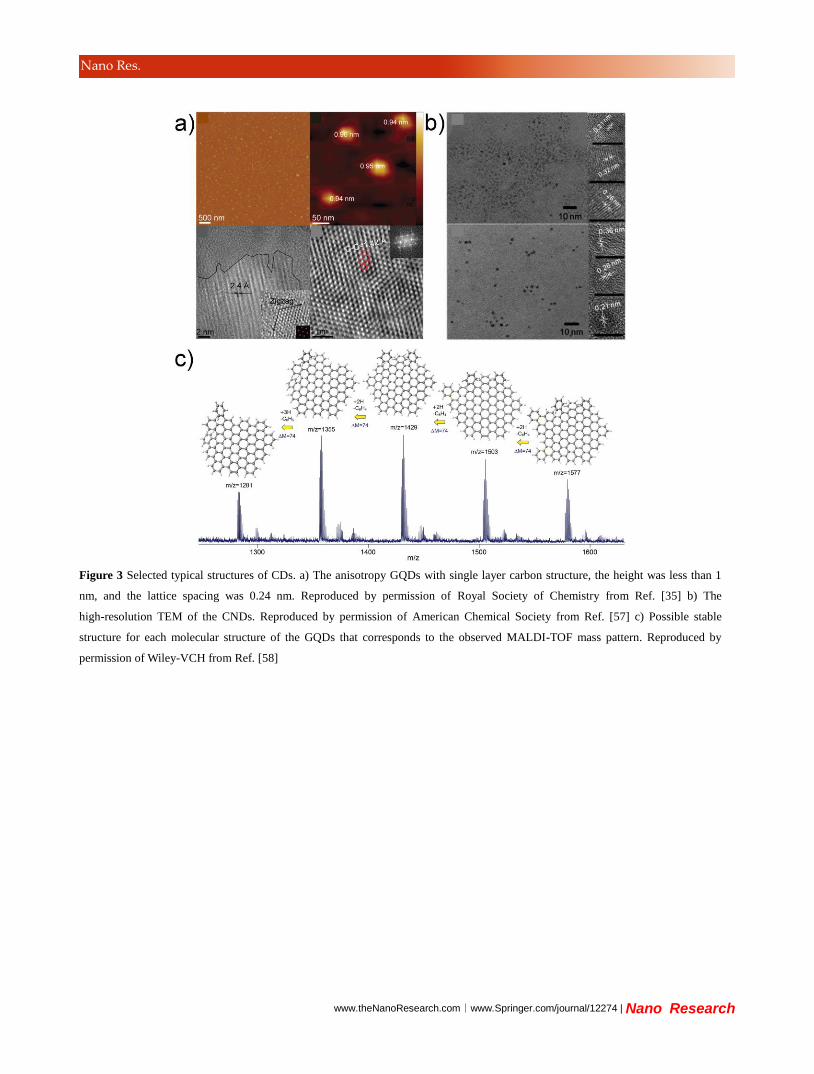
www.theNanoResearch.com∣www.Springer.com/journal/12274 | Nano Research
Nano Res.
Figure 3 Selected typical structures of CDs. a) The anisotropy GQDs with single layer carbon structure, the height was less than 1
nm, and the lattice spacing was 0.24 nm. Reproduced by permission of Royal Society of Chemistry from Ref. [35] b) The
high-resolution TEM of the CNDs. Reproduced by permission of American Chemical Society from Ref. [57] c) Possible stable
structure for each molecular structure of the GQDs that corresponds to the observed MALDI-TOF mass pattern. Reproduced by
permission of Wiley-VCH from Ref. [58]
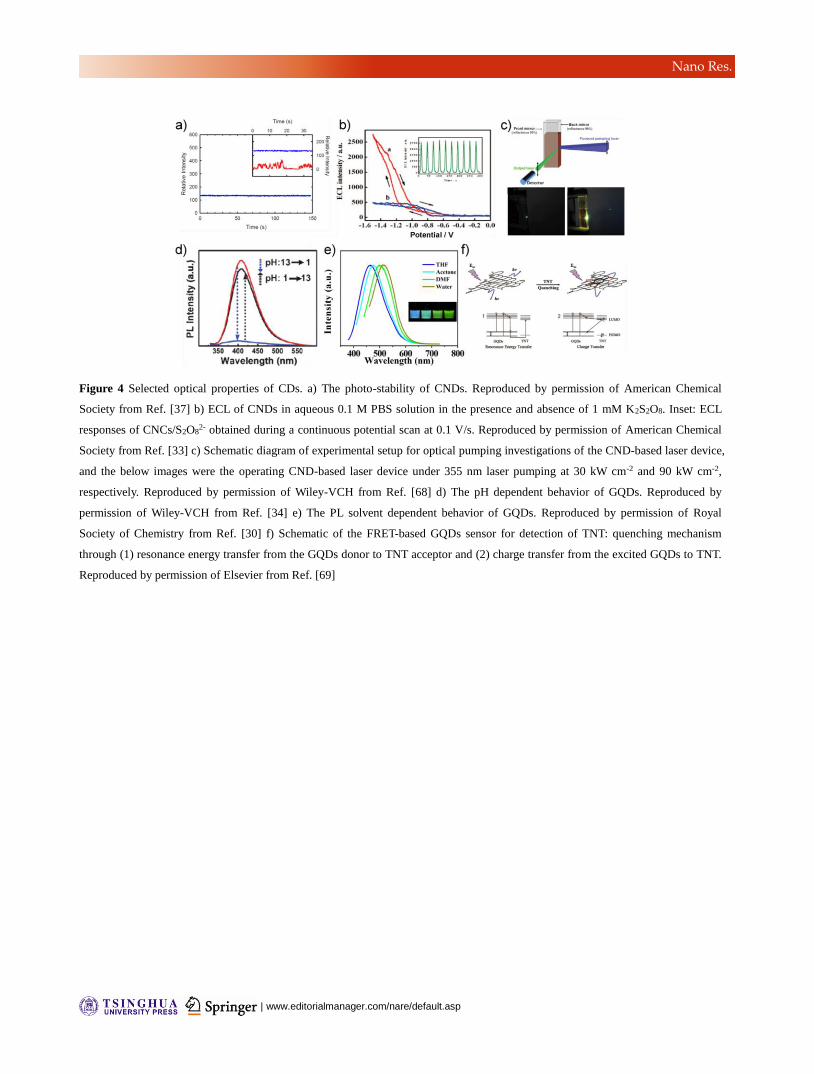
| www.editorialmanager.com/nare/default.asp
Nano Res.
Figure 4 Selected optical properties of CDs. a) The photo-stability of CNDs. Reproduced by permission of American Chemical
Society from Ref. [37] b) ECL of CNDs in aqueous 0.1 M PBS solution in the presence and absence of 1 mM K2S2O8. Inset: ECL
responses of CNCs/S2O82- obtained during a continuous potential scan at 0.1 V/s. Reproduced by permission of American Chemical
Society from Ref. [33] c) Schematic diagram of experimental setup for optical pumping investigations of the CND-based laser device,
and the below images were the operating CND-based laser device under 355 nm laser pumping at 30 kW cm-2 and 90 kW cm-2,
respectively. Reproduced by permission of Wiley-VCH from Ref. [68] d) The pH dependent behavior of GQDs. Reproduced by
permission of Wiley-VCH from Ref. [34] e) The PL solvent dependent behavior of GQDs. Reproduced by permission of Royal
Society of Chemistry from Ref. [30] f) Schematic of the FRET-based GQDs sensor for detection of TNT: quenching mechanism
through (1) resonance energy transfer from the GQDs donor to TNT acceptor and (2) charge transfer from the excited GQDs to TNT.
Reproduced by permission of Elsevier from Ref. [69]
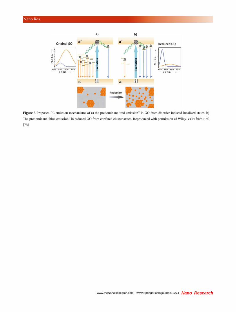
www.theNanoResearch.com∣www.Springer.com/journal/12274 | Nano Research
Nano Res.
Figure 5 Proposed PL emission mechanisms of a) the predominant “red emission” in GO from disorder-induced localized states. b)
The predominant “blue emission” in reduced GO from confined cluster states. Reproduced with permission of Wiley-VCH from Ref.
[78]
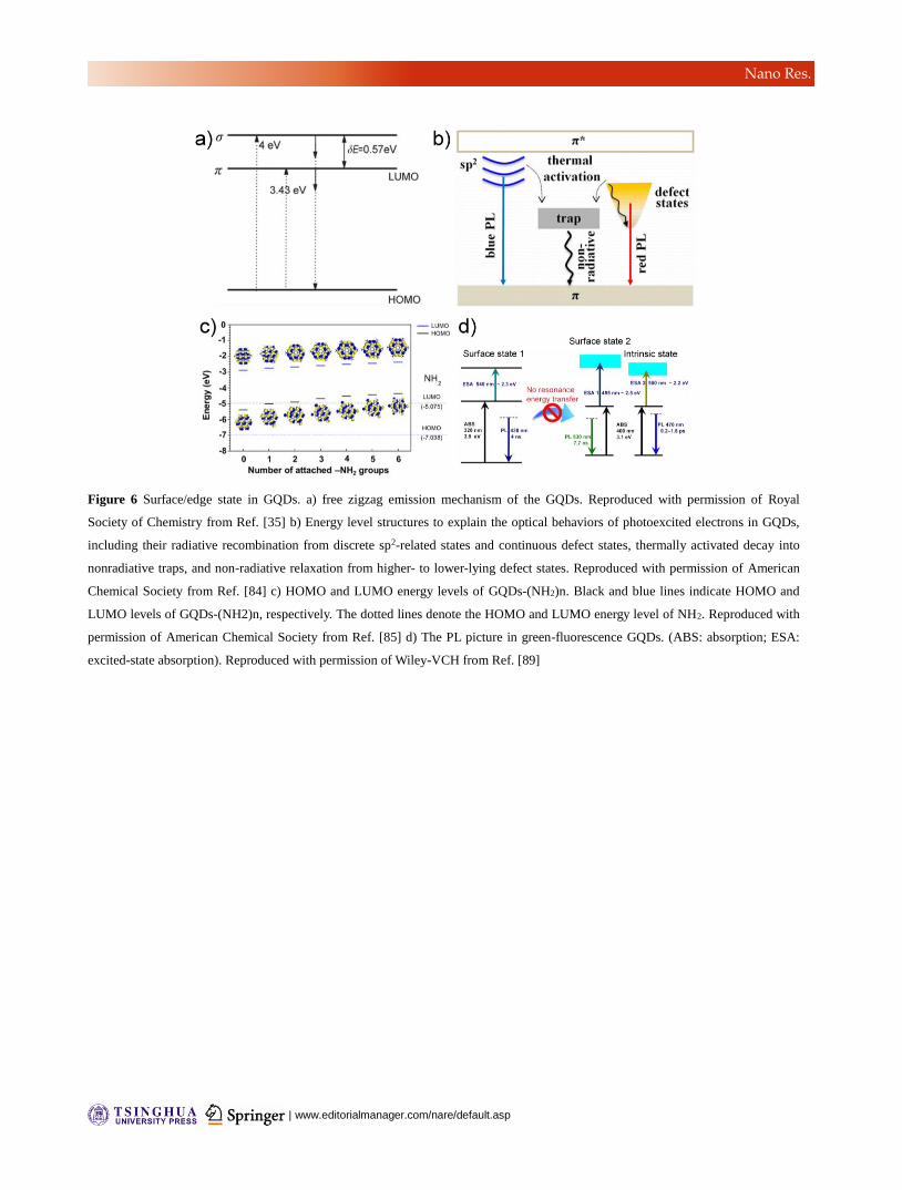
| www.editorialmanager.com/nare/default.asp
Nano Res.
Figure 6 Surface/edge state in GQDs. a) free zigzag emission mechanism of the GQDs. Reproduced with permission of Royal
Society of Chemistry from Ref. [35] b) Energy level structures to explain the optical behaviors of photoexcited electrons in GQDs,
including their radiative recombination from discrete sp2-related states and continuous defect states, thermally activated decay into
nonradiative traps, and non-radiative relaxation from higher- to lower-lying defect states. Reproduced with permission of American
Chemical Society from Ref. [84] c) HOMO and LUMO energy levels of GQDs-(NH2)n. Black and blue lines indicate HOMO and
LUMO levels of GQDs-(NH2)n, respectively. The dotted lines denote the HOMO and LUMO energy level of NH2. Reproduced with
permission of American Chemical Society from Ref. [85] d) The PL picture in green-fluorescence GQDs. (ABS: absorption; ESA:
excited-state absorption). Reproduced with permission of Wiley-VCH from Ref. [89]
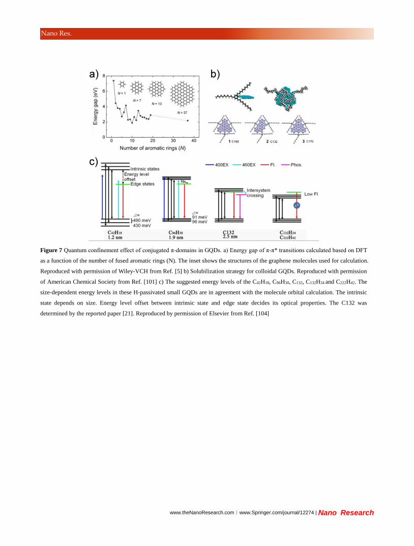
www.theNanoResearch.com∣www.Springer.com/journal/12274 | Nano Research
Nano Res.
Figure 7 Quantum confinement effect of conjugated π-domains in GQDs. a) Energy gap of π-π* transitions calculated based on DFT
as a function of the number of fused aromatic rings (N). The inset shows the structures of the graphene molecules used for calculation.
Reproduced with permission of Wiley-VCH from Ref. [5] b) Solubilization strategy for colloidal GQDs. Reproduced with permission
of American Chemical Society from Ref. [101] c) The suggested energy levels of the C42H18, C96H30, C132, C132H34 and C222H42. The
size-dependent energy levels in these H-passivated small GQDs are in agreement with the molecule orbital calculation. The intrinsic
state depends on size. Energy level offset between intrinsic state and edge state decides its optical properties. The C132 was
determined by the reported paper [21]. Reproduced by permission of Elsevier from Ref. [104]
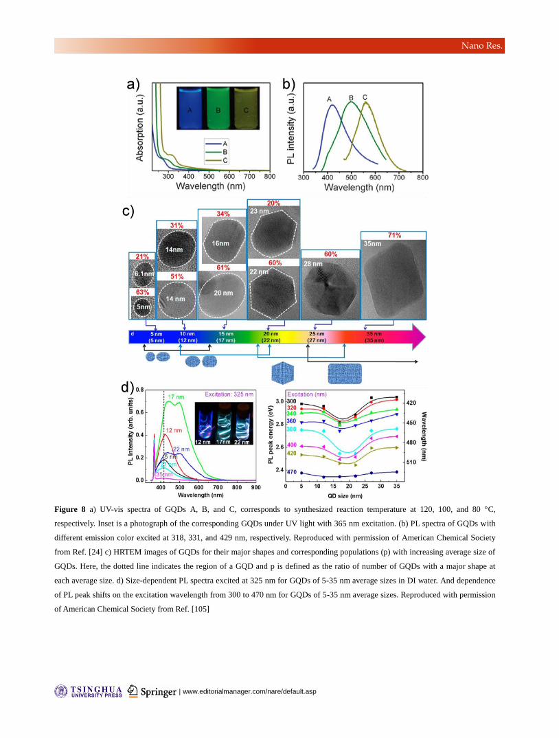
| www.editorialmanager.com/nare/default.asp
Nano Res.
Figure 8 a) UV-vis spectra of GQDs A, B, and C, corresponds to synthesized reaction temperature at 120, 100, and 80 °C,
respectively. Inset is a photograph of the corresponding GQDs under UV light with 365 nm excitation. (b) PL spectra of GQDs with
different emission color excited at 318, 331, and 429 nm, respectively. Reproduced with permission of American Chemical Society
from Ref. [24] c) HRTEM images of GQDs for their major shapes and corresponding populations (p) with increasing average size of
GQDs. Here, the dotted line indicates the region of a GQD and p is defined as the ratio of number of GQDs with a major shape at
each average size. d) Size-dependent PL spectra excited at 325 nm for GQDs of 5-35 nm average sizes in DI water. And dependence
of PL peak shifts on the excitation wavelength from 300 to 470 nm for GQDs of 5-35 nm average sizes. Reproduced with permission
of American Chemical Society from Ref. [105]
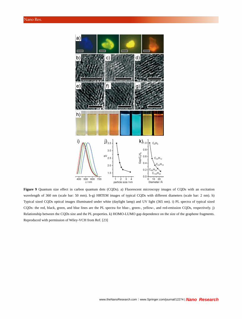
www.theNanoResearch.com∣www.Springer.com/journal/12274 | Nano Research
Nano Res.
Figure 9 Quantum size effect in carbon quantum dots (CQDs). a) Fluorescent microscopy images of CQDs with an excitation
wavelength of 360 nm (scale bar: 50 mm). b-g) HRTEM images of typical CQDs with different diameters (scale bar: 2 nm). h)
Typical sized CQDs optical images illuminated under white (daylight lamp) and UV light (365 nm). i) PL spectra of typical sized
CQDs: the red, black, green, and blue lines are the PL spectra for blue-, green-, yellow-, and red-emission CQDs, respectively. j)
Relationship between the CQDs size and the PL properties. k) HOMO-LUMO gap dependence on the size of the graphene fragments.
Reproduced with permission of Wiley-VCH from Ref. [23]
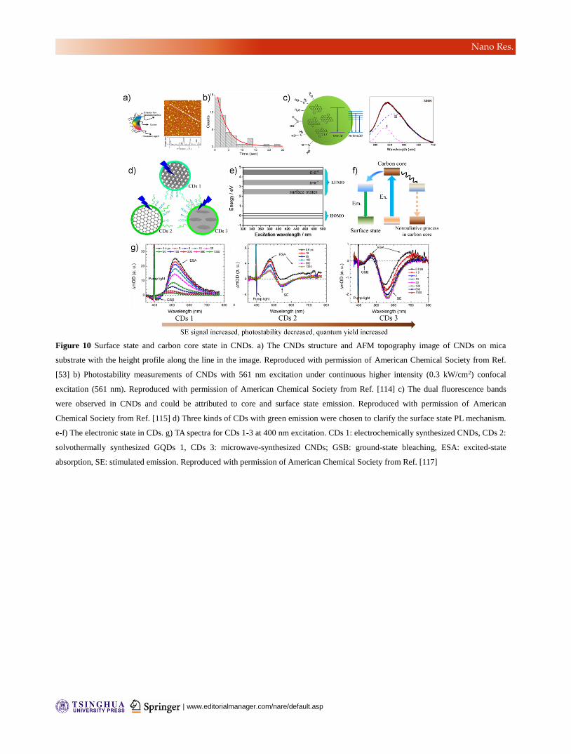
| www.editorialmanager.com/nare/default.asp
Nano Res.
Figure 10 Surface state and carbon core state in CNDs. a) The CNDs structure and AFM topography image of CNDs on mica
substrate with the height profile along the line in the image. Reproduced with permission of American Chemical Society from Ref.
[53] b) Photostability measurements of CNDs with 561 nm excitation under continuous higher intensity (0.3 kW/cm2) confocal
excitation (561 nm). Reproduced with permission of American Chemical Society from Ref. [114] c) The dual fluorescence bands
were observed in CNDs and could be attributed to core and surface state emission. Reproduced with permission of American
Chemical Society from Ref. [115] d) Three kinds of CDs with green emission were chosen to clarify the surface state PL mechanism.
e-f) The electronic state in CDs. g) TA spectra for CDs 1-3 at 400 nm excitation. CDs 1: electrochemically synthesized CNDs, CDs 2:
solvothermally synthesized GQDs 1, CDs 3: microwave-synthesized CNDs; GSB: ground-state bleaching, ESA: excited-state
absorption, SE: stimulated emission. Reproduced with permission of American Chemical Society from Ref. [117]
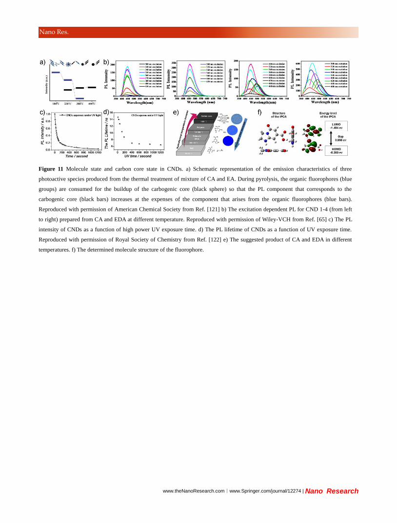
www.theNanoResearch.com∣www.Springer.com/journal/12274 | Nano Research
Nano Res.
Figure 11 Molecule state and carbon core state in CNDs. a) Schematic representation of the emission characteristics of three
photoactive species produced from the thermal treatment of mixture of CA and EA. During pyrolysis, the organic fluorophores (blue
groups) are consumed for the buildup of the carbogenic core (black sphere) so that the PL component that corresponds to the
carbogenic core (black bars) increases at the expenses of the component that arises from the organic fluorophores (blue bars).
Reproduced with permission of American Chemical Society from Ref. [121] b) The excitation dependent PL for CND 1-4 (from left
to right) prepared from CA and EDA at different temperature. Reproduced with permission of Wiley-VCH from Ref. [65] c) The PL
intensity of CNDs as a function of high power UV exposure time. d) The PL lifetime of CNDs as a function of UV exposure time.
Reproduced with permission of Royal Society of Chemistry from Ref. [122] e) The suggested product of CA and EDA in different
temperatures. f) The determined molecule structure of the fluorophore.
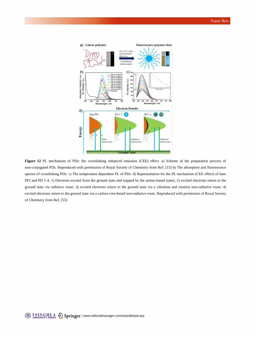
| www.editorialmanager.com/nare/default.asp
Nano Res.
Figure 12 PL mechanism of PDs: the crosslinking enhanced emission (CEE) effect. a) Scheme of the preparation process of
non-conjugated PDs. Reproduced with permission of Royal Society of Chemistry from Ref. [15] b) The absorption and fluorescence
spectra of crosslinking PDs. c) The temperature dependent PL of PDs. d) Representation for the PL mechanism (CEE effect) of bare
PEI and PD 1-4. 1) Electrons excited from the ground state and trapped by the amino-based states; 2) excited electrons return to the
ground state via radiative route; 3) excited electrons return to the ground state via a vibration and rotation non-radiative route; 4)
excited electrons return to the ground state via a carbon core-based non-radiative route. Reproduced with permission of Royal Society
of Chemistry from Ref. [55]