the op biological chemistry vol. 261, no. pp. …the jowrnai.op biological chemistry d 1986 by the...
TRANSCRIPT
THE JOWRNAI. OP BIOLOGICAL CHEMISTRY D 1986 by The American Society of Biological Chemists, Inc.
Vol. 261, No. 27. Issue of September 25, pp. 12604-12609 1986 Printed in 6.S.A.
Sphingosine Inhibition of Protein Kinase C Activity and of Phorbol Dibutyrate Binding in Vitro and in Human Platelets*
(Received for publication, February 25,1986)
Yusuf A. HannunSP, Carson R. Loomisll, Alfred H. Merrill, Jr.11, and Robert M. Bell7 From the Departments of $Medicine and liBwchemistry, Duke University Medical Center, Durham, North Carolina 27710 and the Department of [I Biochemist y, Emory University School of Medicine, A thntu, Georgia 30322
Sphingosine inhibited protein kinase C activity and phorbol dibutyrate binding. When the mechanism of inhibition of activity and phorboi dibutyrate binding was investigated in vitro using Triton X-100 mixed micellar methods, sphingosine inhibition was subject to surface dilution; 50% inhibition occurred when sphingosine was equimolar with sn- 1,2-dioleoylgIy- cero1 (diCle:,) or 40% of the ph~phatidylserine (PS) present. Sphingosine inhibition was modulated by Cas+ and by the mole percent of diCls:l and PS present. Sphingosine was a competitive inhibitor with respect to diClml, phorbol dibutyrate, and Ca2+. Increasing levels of PS markedly reduced inhibition by sphingo- sine. Since protein kinase C activity shows a coopera- tive dependence on PS, the kinetic analysis of compet- itive inhibition was only suggestive. Sphingosine in- hibited phorbol dibutyrate binding to protein kinase C but did not cause protein kinase C to dissociate from the mixed micelle surface. Sphingosine addition to hu- man platelets bloeked thrombin and sn- 1,2-diocta- noylglycerol-dependent phosphorylation of the 40- kDa (47 kDa) dalton protein. Moreover, sphingosine was subject to surface dilution in platelets. The mech- anism of sphingosine inhibition is discussed in relation to a previously proposed model of protein kinase C activation. The possible physiological role of sphingo- sine as a negative effector of protein kinase C is sug- gested and a plausible cycle for its generation is pre- sented. The potential physiological significance of sphingosine inhibition o f protein kinase C is further established in accompanying papers on HL-60 cells (Merrill, A. H., Jr., Sereni, A. M., Stevens, V. L., Hannun, Y. A., Bell, R. M., Kinkade, J. M., Jr. (1986) J. Biol. Chem. 261,12610-12615) and human neutro- phils (Wilson, E., Olcott, M. C., Bell, R. M., Merrill, A. H., Jr., and Lambeth, J. D. (1986) J. Biol. Chem. 261, 12616-12623). These results also suggest that sphin- gosine will be a useful inhibitor for investigating the function of protein kinase C in vitro and in living cells.
The central function of the phospholipid, Ca2+, and sn-1,Z- diacylglycerol-dependent protein kinase (protein kinase C ) in transducing intracellularly extracellular signals has recently been recognized (1). These extracellular agents which include neurotransmitters, hormones, and growth factors (1, 2 ) are
* This work was supported by Grant AM 20205 from the National Institutes of Health and Grant BC-511 from the American Cancer Society. The costs of publication of this article were defrayed in part by the payment of page charges. This article must therefore be hereby marked “advertisement” in accordance with 18 U.S.C. Section 1734 solely to indicate this fact.
$Recipient of National ~ n s t ~ t u ~ s of Health Physician Scientist Award 1Dll-ES-00155.
bound by specific cell surface receptors and elicit by trans- membrane signaling the generation of two second messengers by stimulating the degradation of phosphatidylinositols (1, 3, 4). Inositol 1,4,5-trisphosphate mobilizes intrace~lular calcium (4), whereas, sn-l,Z-diacylglycerol (DAG’) activates protein kinase C by a mechanism whereby the enzyme undergoes translocation from the cytosol to the plasma membrane (1, 5). The membrane contains phosphatidylserine which is re- quired for activation (6). Protein kinase C has been demon- strated to be the intracellular receptor of the tumor promoting phorbol esters which activate the enzyme by interaction at the sn-1,2-diacylglycerol site (7-10). The pleiotropic effects of protein kinase C activation and the recognition that it functions as a transducer of DAG and calcium second mes- sengers make detailed understanding of its mechanism of regulation desirable. Our laboratory has undertaken detailed structure-function analysis of diacylglycerol-protein kinase C interactions and studies on the mechanism of regulation by phosphatidylserine, calcium, sn-1,Z-diacylglycerols, and lipids in Triton X-100 mixed micelles (11-13).
The discovery or development of inhibitors of protein ki- nase C has the potential to further the understanding of specific functions of the enzyme in cells and animals. Several inhibitors of protein kinase C have been reported. These include calmodulin antagonists (14-16), H7 (17), adriamycin (15), alkyllysophospholipid (18), a nonsteroidal anti-estrogen, tamoxifen, (19), anti-neoplastic lipoidal amine (20), amiloride (Zl), verapamil (14), bilirubin (221, and palmitoylcarnitine (12). Herein, we report on the discovery that sphingosine is an inhibitor of protein kinase C .
In this article, we report on the inhibition of protein kinase C by sphingosine and related compounds, the phosphatidyl- serine, diacylglycerol (phorboI diester), and Ca2+ modulation of this inhibition, and the inhibition of phorbol dibutyrate binding, Furthermore, studies in human platelets demonstrate sphingosine inhibition of thrombin and ~C~-dependent acti- vation of protein kinase C and inhibition of phorbol ester binding. Sphingosine inhibition of protein kinase C may have physiological significance in that a regulated metabolic cycle may exist whereby sphingosine functions as a negative effec- tor of protein kinase C . In two accompanying manuscripts (23,241, the effects of sphingosine on neutrophil function and on HL-60 cell activities are documented.
EXPERIMENTAL PROCEDURES
Materials Charles River CD female rats were used for the source of protein
kinase C. Ultrogel AcA 44 and Ultrogel AcA 202 were from LKB. [“ZP]Orthophosphate, Aquasol 11, [yS2P1ATP, and [3HlPDBu (12.5
The abbreviations used are: DAG, sn-1,2-diacylglyceroi; ps, &os- phatidylserine; diC,., sn-1,2-dioleoylglycerol; dica, sn-l,f-diOCta- noyIgIycerol; PA, phosphatidic acid; PDBu, phorboi dibutyrate.
12604

Sphingosine Inhibition of Protein Kinase C 12605
Ci/nmol) were from New England Nuclear. Calf thymus histone type 111-S, phospholipase C, thrombin, phenylmethylsulfonyl fluoride, stearylamine, bovine serum albumin, phorbol dibutyrate, il-sphingan- ine, threosphingosine, and dihydrosphingosine were from Sigma. Leu- peptin was from the Peptide Institute (Osaka, Japan). 1,2-Dioleoyl- sn-glycerol-3-phosphoserine, 1,2-dioctanoylglycerl, and 1,2-dioleoyl- sn-glycero-3-phosphocholine were from Avanti Polar Lipids. sn-1,2- Diol~ylglycerol was synthesized from dioleoylphosphatidylcholine as previously described (25). Triton X-100 was from Research Products International Corp. Octylamine was from Aldrich. Swainsonine was a gift from Harry Broquist (Department of Biochemistry, Vanderbilt University), ceramide was a gift from Jim Walsh (Department of Biochemistry, Duke University), ~-acetylsphingosine was a gift from Barry Ganong (Department of Biochemistry, Duke University), 1,3- dihydroxy-2-amino-3-phenylpropane was a gift from Dennis Liotta (Department of Chemistry, Emory University). Phorboll2-myristate 13-acetate was from Pharmacia P-L Biochemicals.
Methods
Partial Purification of Protein Kinase C-Protein kinase C was partially purified from rat brain as previously described (13).
Mixed Micellar Assay for Protein Kinase C Actiuity-Protein kinase C was assayed with Triton X-100 mixed micelles as previously de- scribed (13). Sphingosine was dried down with the lipid cofactors.
Mixed Micellar Assay for PHJPDBu Binding to Protein Kinase C- [3HfPDBu binding was performed as previously described (26).
from freshly drawn blood essentially as described by Siess et al. (27). Preparation of Human Platelets-Human platelets were prepared
They were then suspended in modified Tyrode's buffer to a concen- tration of 2.5 X 10' plateletslml.
PHJPDBu Binding to Humn Platelets-Human platelets, pre- pared as described above, were suspended at a concentration of 2.5 X IO8 platelet/ml. 50 pi of the platelets were then incubated for 5 min with the indicated concentration of sphingosine in Eppendorf micro- fuge tubes. Sphingosine was prepared in 50% ethanol a t a concentra- tion 100-fold the final Concentration so that ethanol was kept at 0.5%. [3H]PBDu was added to 10 nM and incubated with the platelets at 37 "C for 10 min. The samples were then filtered on Whatman GF/C filters pre-washed with 5 mi of modified Tyrode's buffer con-
buffer, dried, and counted in 10 ml of Aquasol I1 in an LKB /3 counter. taining 0.1% bovine serum albumin, washed with 10 ml of the same
Nonspecific binding was determined in the presence of 1 ~ L M unlabeled PDBu and subtracted from the total counts to yield the specific binding.
40-kDa P h s p h v l a t w n in Human Platelet~-'~Pi at 0.2 mCi/ml was added to the piatelet suspension and labeling was allowed to proceed for 75 min at 37 "C, after which the platelets were pelleted at 600 X g for 10 min and resuspended in Tyrode's buffer to the same concentration. They were then afiquoted in Eppendorf microfuge tubes and pre-incubated at 37 "C for 5 min with the varying concen- trations of sphingosine. Platelets were then stimulated with either 5 p~ dioctanoylglycerol (added as an ethanol solution with the final concentration of ethanol 0.5%), 10 p~ phorbol 12-myristate 13- acetate, or 1 unitlml thrombin. The reactions were stopped after 30 s by the addition of an equal volume of 2 X sample buffer, and the samples were then boiled for 3 min. 0.1 ml were then loaded on 10% sodium dodecyl sulfate-polyacrylamide gels and electrophoresis was performed according to the method of Laemmli (28). Gels were subsequently fixed in water/methanol/acetic acid ( ~ 3 0 1 0 ) , dried, and autoradiographed.
whole platelets by the method of Bligh and Dyer (29). Phosphatidyl- Phospholipid Qmntitation-Phospholipids were extracted from
serine was purified by two-dimensional thin layer chromatography on Silica Gel H plates developed in chlorofo~/meth~o~/acetic acid (652510, v/v) in the first dimension and in chloroform/methanol/ 88% formic acid (65:25:10, v/v) in the second dimension. Phospho- lipids were quantitated by measuring phosphates according to the method of Ames (30).
The data shown is representative of a t least three sets of experi- ments.
RESULTS AND DISCUSSION
When the effect of sphingosine on protein kinase C activity was tested using the Tri ton X-100 mixed micelle assay (13) containing 6 mol % of PS and 2 mol % of diCle:,, sphingosine proved to be a potent inhibitor (Fig. 1). Under these condi- tions, the bulk concentration of PS was 260 y~ and diCls,.
OO 3 200 400 600 800 SPHINGOSINE L,uM)
FIG. 1. Inhibition of protein kinase C activity by sphingo- sine, Mixed micelles were formed with 3% (w/v) Triton X-100 containing PS at 6 mol %, diC1~, at 2 mol %, and sphingosine at 10- fold the indicated concentrations. The mixed micelles were then diluted 1: lO into the assay mixture. 1 mol % of sphingosine is e ~ i v a i e n t to 43 p ~ . Effect of sphingosine on protein kinase C (0) and on protein kinase M (0): Identical results were obtained with protein kinase M when sphingosine was added in 0.3% Triton X-100 solution without PS and diCIkI.
SPMNWSINE I . u W
FIG. 2. Effect of mixed micelle concentration on potency of sphingosine inhibition. A, mixed micelles were formed with Triton X-100 at 12% (w/v, U), 6% (A), 3% (A), and 1.5% (D) containing 7 mol % of PS, 1 mol % of diCls,l, and sphingosine at 10-fold the indicated concentrations. The mixed micelles were then diluted 1:lO into the assay mixture. B, mixed micelles were formed with 3% Triton X-100, 7 mol % of PS, 1 mol % of and 2-5 mol % of sphingosine. These were then diluted 1:20 (D), 1:lO (A), 1:5 (A), and 2 5 (U) into the assay mixture. In these experiments, 1 mol % of sphingosine corresponds to 21.5,43,86, and 172 p ~ , respectively.
was 86 y ~ . Therefore, 50% inhibition (100 p M ) occurred on a molar basis equivalent to [diC18:1] or 0.4 [PSI. The potency of sphingosine inhibition was markedly affected by the number of Triton X-100 mixed micelles containing 7 mol % of PS and 1 mol % of diC1B:l present in the assay (Fig. 2A). Thus, the effect of sphingosine was subject to surface dilution. When the data are expressed as mole percent (sphingosine:Triton X-loo), sphingosine inhibition at four different levels of Tri- ton X-100 mixed micelles (containing PS and diC 18.1 . ) was identical (Fig. 2B) implying that it i s the number of sphin- gosine molecules present in each mixed micelle that deter- mines the potency and not the absolute concentration. For surface active amphipathic molecules, the expression of inhib- itor potencies must be relative to the amount of surface (micelles in this case) as bulk concentrations are misleading. These results also imply that sphingosine interacts with the surface-bound protein kinase C probably by interfering with the function of its regulatory domain. To test this hypothesis, the catalytic domain (protein kinase M) was generated by proteolysis of protein kinase C 2 The activity of this catalytic
'The catalytic domain was generated by trypsin treatment of purified Protein kinase C (31) and purified by Ultrogel AcA 44 molecular sieve chromatography. The fractions showing pmtein ki- nase activity independent of ea2+, PS, and DAG were pooled and used for the above experiments.
12606 Sphingosine Inhibition of Protein Kinase C
domain which is independent of Ca", phospholipid, and DAG/phorbol esters (31) was not inhibited by sphingosine (Fig. 1). Thus, sphingosine does not appear to inhibit by interaction with the active site.
To further investigate the mechanism of sphingosine inhi- bition, the ~n-1 ,2 -d iC ,~~ dependency of protein kinase C ac- tivation was investigated at fixed PS (6 mol %) and Ca2+ (50 PM) and at several levels of sphingosine. Importantly, the inhibition by sphingosine was modulated strongly by diCla1 Fig. 3A). Double reciprocal plots (Fig. 3B) indicated essen- tially a competitive type of inhibition with respect to diClR1. Similarly, inhibition by sphingosine was overcome by increas- ing concentration of phorbol dibutyrate (Fig. 4A), and double reciprocal plots (Fig. 4B) showed competitive inhibition.
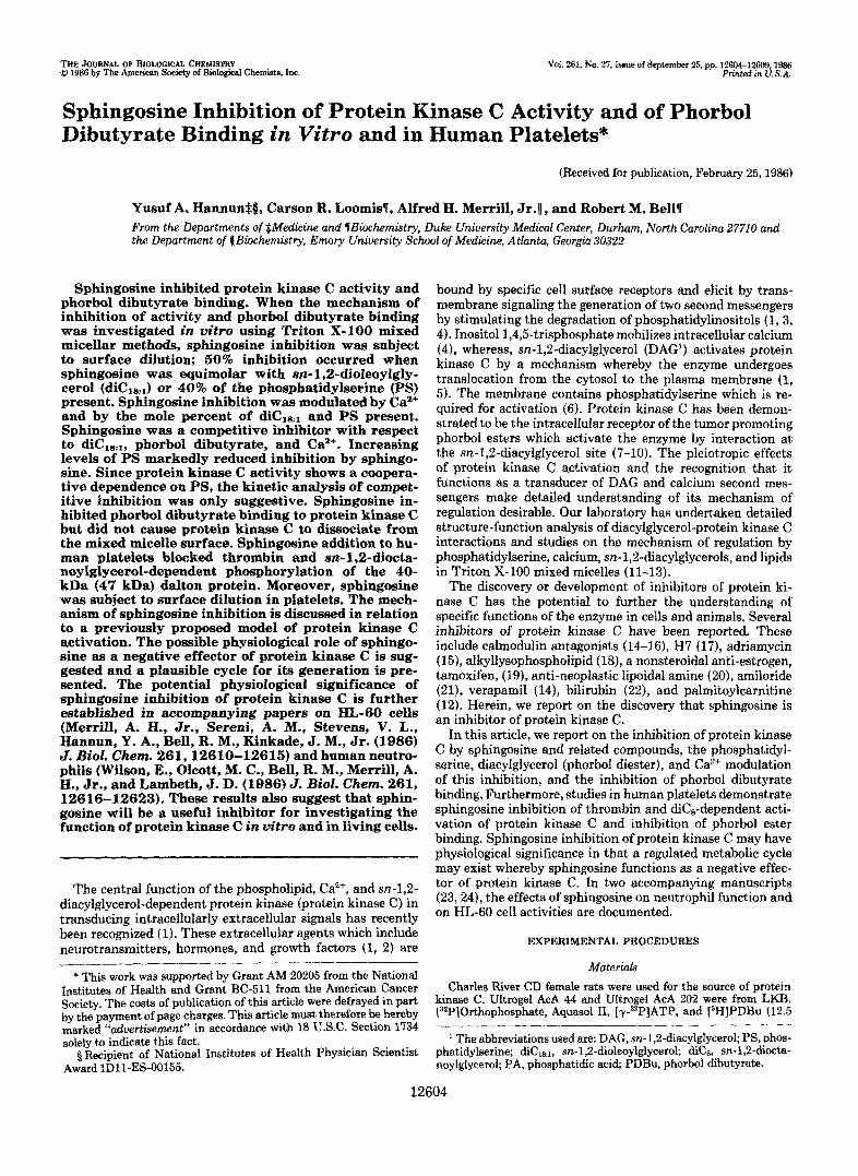
When the concentration of sphingosine was varied at 5, 6, and 7 mol % of PS and fixed diCls,l (2 mol %), the potency was modulated markedly (Fig. 5). This was especially true when PS was not saturating at 5 mol %. Shifting from 6 to 7
1 i
DIOLEOYLGLYCEAOL lrrml XI (OIOLEOYLGLYCEROL mal %I"
FIG. 3. Interaction of sphingosine with dioleoylglycerol. Mixed micelles were formed at 3% Triton X-100,6 mol % of PS, 0- 10 mol % of diC1,,, and sphingosine at 0 mol % (A), 2 mol % (A), 3 mol % (0), and 4 mol % (m). A, protein kinase C activity assayed in the presence of 50 p~ CaC12. B, double reciprocal plots with diClkl concentration in mole percent.
FIG. 4. Interaction of sphingosine with phorbol dibutyrate. Mixed micelles were formed with 10 mol % of PS and 0 mol % (A), 6 mol % (A), and 8 mol % (0) of sphingosine. PDBu was added as an aqueous solution. A, protein kinase C activity assayed in the presence of 10 p~ CaC12. B, double reciprocal plots of data in A.
mol % of PS, a level at which PS becomes saturating (Fig. 6), revealed that the curves were simply not displaced on a mole for mole basis. When PS was in excess, higher concentrations of sphingosine were required for inhibition (Fig. 5).
Next, the effect of 2 and 4 mol % of sphingosine on the PS dependence of protein kinase C activation was investigated (Fig. 6) . Interestingly, sphingosine caused a displacement of the PS dependence to higher levels which remained strongly cooperative in the presence or absence of inhibitor. When the PS dependencies were then plotted according to Hill, Hill numbers of 5.4, 6.9, and 8.8 were obtained for the curves generated at 0, 2, and 4 mol % of sphingosine, respectively. Double reciprocal plots were not constructed because of the cooperativity observed with PS and the sphingosine-depend- ent change in Hill numbers. In that additional PS completely overcame the inhibition by sphingosine, a competitive form of inhibition appears likely.
To further explore the mechanism of sphingosine inhibi- tion, the effect of the level of Ca2+ employed was examined at fixed PS and diC1,,, and at 0, 3, and 4 mol % of sphingosine. Double reciprocal plots of the calcium dependencies were linear and appeared competitive (Fig. 7).
The effect of sphingosine on phorbol ester binding was examined to further substantiate that sphingosine inhibition occurs by interfering with the regulatory domain of protein kinase C . Sphingosine inhibited phorbol ester binding to Triton X-100 mixed micelles containing 16 and 20 mol % of PS (Fig. 8). The concentration dependence of sphingosine displacement of phorbol dibutyrate, paralleled its ability to inhibit protein kinase C activation by PDBu (Fig. 4A). Sphin- gosine also inhibited phorbol binding when phosphatidic acid was used as the lipid cofactor (PA supports only 50% of the binding measured with PS).
The possibility that sphingosine causes inhibition of activ- ity and phorbol binding by preventing protein kinase C inter- action with and binding to mixed micelles was tested (Fig. 9A) by molecular sieve chromatography of the appropriate complexes (13). As seen in Fig. 9B, protein kinase C remained bound to Triton X-100 mixed micelles containing PS in the presence of sphingosine; sphingosine caused displacement of phorbol dibutyrate without breaking the association of protein kinase C with Ca*+ and the Triton X-lOO/PS mixed micelles.
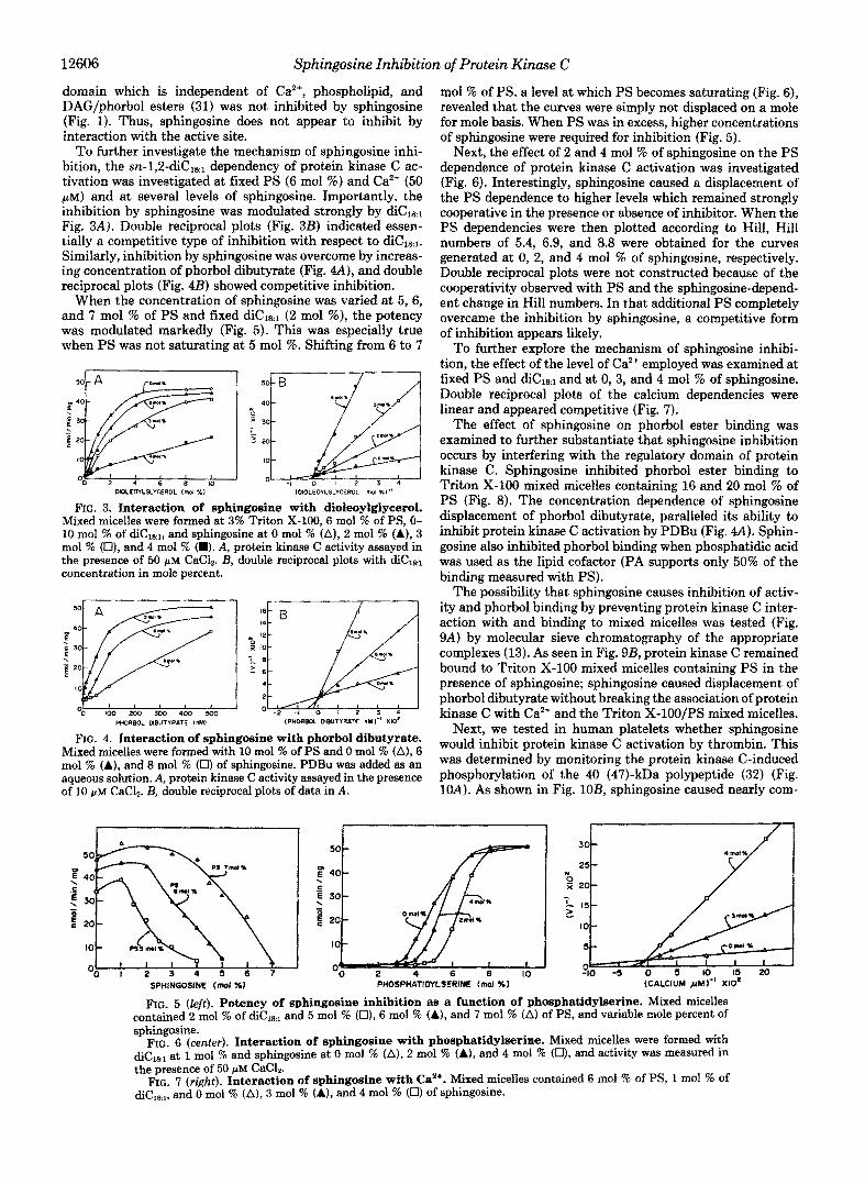
Next, we tested in human platelets whether sphingosine would inhibit protein kinase C activation by thrombin. This was determined by monitoring the protein kinase C-induced phosphorylation of the 40 (47)-kDa polypeptide (32) (Fig. 1OA). As shown in Fig. 1023, sphingosine caused nearly com-
I /
so -5 0 5 IO 1s 20 (CALCIUM JAM)" XIO'
FIG. 5 (left). Potency of sphingosine inhibition 88 a function of phosphatidylserine. Mixed micelles contained 2 mol % of diCl~,1 and 5 mol % (U), 6 mol % (A), and 7 mol % (A) of PS, and variable mole percent of sphingosine.
FIG. 6 (center). Interaction of sphingosine with phosphatidylserine. Mixed micelles were formed with diCtk1 a t 1 mol % and sphingosine at 0 mol % (A), 2 mol % (A), and 4 mol % (0), and activity was measured in the presence of 50 pM CaC12.
FIG. 7 (right). Interaction of sphingosine with Ca2+. Mixed micelles contained 6 mol % of PS, 1 mol % of diCikl, and 0 mol % (A), 3 mol % (A), and 4 mol % (0) of sphingosine.

Sphingosine lnhibition of Protein Kinase C 12607
i 1 4 a 12 16 20
SPHINGOSINE (mol %) FIG. 8. Sphingosine inhibition of phorbol dibutyrate bind-
ing. Mixed micelles contained PS at 16 mol % (U) and 20 mol % (A) or PA at 20 mol % (A) and 0-20 mol % of sphingosine. Binding studies were performed as described under "Methods." The data are plotted as percent of maximal binding under each condition. With 16 mol % of PS there was 82% of the [3H]PDBu binding seen with 20 mol % of PS. At 20 mol % of PA there was only 51% of the binding seen with 20 mol % of PS.
plete inhibition of 40-kDa phosphorylation at a concentration of 25 PM. Partial inhibition was seen with concentrations as low as 10 PM. Sphingosine also inhibited phorbol12-myristate 13-acetate and diC8-induced IO-kDa phosphorylation (data not shown).
To further analyze the mechanism by which sphingosine inhibits protein kinase C activation, PDBu binding to whole human platelets was studied (Fig. 11). Saturable and displace- able binding of [3HJPDBu to platelets was demonstrated (data not shown). Sphingosine was able to inhibit this binding in concentrations similar to those required to inhibit 40-kDa phosphorylation?
Since sphingosine is an amphiphilic molecule and would be expected to partition into a bilayer or micelle, a direct com- parison of its biologic effects to its in vitro potency in inhib- iting protein kinase C requires that its bulk concentration be expressed as mole percent of sphingosine to phospholipids. To accomplish this, the amount of PS and total phospholipids in platelet membranes were quantitated. Human platelets were found to have 20% of their total phospholipids as PS and the absolute PS concentration under the assay conditions was 200 PM. Therefore, the sphingosine concentrations (ex- pressed as mob percent of sphing0sine:phospholipids) re- quired to inhibit platelet 40-kDa phosphorylation were similar to those required for in uitro inhibition of protein kinase C: 3-5 mol % of sphingosine required for complete inhibition (or 15-25% of PS). In addition, this inhibition demonstrated the same reversibility observed in the in uitro system in that increasing the concentration of diC8 or of platelets (ie. PS) overcomes the inhibition (data not shown).
With these data in hand, we investigated the specificity of protein kinase C inhibition by sphingosine using a number of related molecules. As seen in Fig. 12, octylamine did not inhibit at the concentrations tested whereas stearylamine was nearly as effective as sphingosine. Swainsonine, structurally related to sphingosine (33), was not an inhibitor. N-Acetyl- sphingosine, ceramide, and 1,3-dihydroxy-2-amino-3-phenyl- propane analogue were without effect. Fatty acids, and cetyl- triethylammonium bromide were also inactive. However, 3- ketosphinganine, erythro-, and reo-sphingan~ne were all
Sphingosine Concentrations that produce 50% inhibition of phor- bo1 binding and 40-kDa phosphorylation are similar (Figs. 1OB and 11). However, inhibition of phorbol binding in platelets shows more complex kinetics, the significance of which is unknown at present.
"i 700 ;; Y
600 = 500
400 D;
300 2 200 I2.I
T
io0
Y
FRACTJON NUMEER
FK. 9. Interaction of PDBu and protein kinase C (PKC) with mixed micelles containing sphingosine on molecular sieves. An 80-ml (1.5 X 45-cm) Sephacryl S-200 column was equili- brated with 20 mM Tris-HC1, pH 7.5,10% sucrose, 50 mM 2-mercap- toethanol, 0.02% Triton X-100, and 200 GM CaCl,. 1 ml of protein kinase C concentrated 10-fold by Amicon (YM 10) ultrafi~tration was incubated for 5 min in the presence of 200 PM CaC12, 100 nM ['HI PDBu, and 1.0% (w/v) Triton X-100 mixed micelles containing 16 mol % of PS with or without 12 mol % of sphingosine. The sample was then chromatographed and the eluate was assayed for protein kinase C activity (A), and for [3H]PDBu (A, note that the ordinate on the left for bound [3H]PDB~ is magnified with respect to free ['HI PDBu whose ordinate is shown on the right). A, mixed micelles contained 16 mol % of PS. A fraction of [3H]PDBu (A) co-elutes with protein kinase C (A) at an tW, = 200,000. B, mixed miceiies contained 16 mol ?4 of PS and 12 mol % of sphingosine. Most of the bound ['HI PDBu was displaced. Arrows indicate elution position of blue dextran (A) , amylase @I, alcohol dehyrogenase (C) , Triton X-lOO/PS mixed micelles (Dl , protein kinase C when it is not bound to mixed micelles (El (and see Ref. 131, and bovine serum albumin (F).
inhibitors. Adriamycin, an inhibitor of protein kinase C {15), and sphingosine have similar structural features.
CONCLUDING REMARKS
A number of compounds that inhibit protein kinase C activity have been described, but little data is available on the mechanisms by which such inhibition occurs, leading to the assumption that they "perturb" the lipid bilayer making it unsuitable for protein kinase C activation. The mixed micelle assay for protein kinase C (11,131 has provided the necessary tool to examine the interaction of amphipathic and hydro- phobic molecules with protein kinase C. We have previously shown that protein kinase C interacts with a DAG. 4PS - Ca2+ complex (12,131. By competitively interacting with PS, CaZ+, and DAGlphorbol ester, sphingosine prevents the formation of an active Iipid-enzyme complex by displacement of the activator (DAG or phorbol ester) from the complex while maintaining the association of the enzyme to the surface (Fig.
12608 Sphingosine Inhibition of Protein Kinase C
FIG. 10. Sphingosine inhibition of platelet protein phoshorylation. A, the effect of increasing sphingosine con- centration on phosphorylation of the 40- kDa protein induced by thrombin. Lane I , control platelets; Lane 2, thrombin (1 unit/ml); Lane 3, 10 p~ sphingosine; Lane 4, 25 p M sphingosine; Lane 5, 50 pM; Lane 6, 100 pM; hne 7, 200 pM. B, the 40-kDa band was cut out and 32P was counted in Aquasol 11. The percent of maximal phosphorylation induced by thrombin is plotted as a function of sphingosine concentration.
1 2 3 4 5 6 7 . , “ . .- . ., _,. A
SPHINGOSINE O.LM) FIG. 11. Sphingosine inhibition of PDBu binding to plate-
lets. [3H]PDBu was at 25 nM and platelets at 2.5 X lo8 platelet/ml.
4 4 0 K D a
2 0 K D a
6 8 IO CONCENTRATION (mol % I
FIG. 12. Effects of stearylamine and octylamine on protein kinase C. Mixed micelles contained 5.5 mol % of PS, 1.0 mol % of diCle1 and sphingosine (O), octylamine (A), or stearylamine (A).
9). The results presented above show that sphingosine is a potent and reversible inhibitor of protein kinase C. This suggests that sphingosine may be a useful inhibitor of protein kinase C in different cell systems. Our results with platelets and the accompanying work in HL-60 cells (23) and neutro- phils (24) further attest to the usefulness of sphingosine as a protein kinase C inhibitor.
Sphingosine differs from the other known inhibitors of protein kinase C in that it is a natural component of cells comprising a critical component of ceramide, the building block of sphingomyelin and the glycosphingolipids (Fig. 13). Sphingosine and other naturally occurring long-chain (sphin- goid) bases are synthesized by serine palmitoyltransferase (34). Sphingosine could also be generated by the action of ceramidases (N-acylsphingosine amidohydrolases). These
” 0 25 50 75 160 125 1 %
SPHINGOSINE ( & M I
metabolic pathways raise the possibility that the generation of sphingosine intracellularly may serve as a regulated nega- tive effector of protein kinase C activity. Sphingosine levels may be regulated in response to either intra- or extracellular signals. In fact, sphingomyelin was observed to undergo rapid deacylation and N-acylation when L-929 fibroblasts were stimulated with specific antibody (35). The deacylation of sphingomyelin leads to the generation of sphingosylphos- phorylcholine (lysosphingomyelin). This molecule may lead to the generation of sphingosine through hydrolysis of the phosphorylcholine head group. Therefore, these catabolic pathways for the generation of sphingosine or one of its analogs may play a physiologic role in modulating the activity of protein kinase C. It is unlikely that the biosynthetic path- ways of sphingolipids will have such a physiologic function since previous studies have shown the levels of sphingosine and sphinganine to be exceedingly low (36). The potential for a physiologic inhibition of protein kinase C by sphingosine is also substantiated by the finding that sphingosine reversibly inhibits protein kinase C activity in human platelets. Also, the observed cytotoxicity of sphingosine in cells (37) is con- sistent with sphingosine acting as a potent inhibitor of protein kinase C. The activity of protein kinase C would then be expected to be a function of the concentration of PS (phos- pholipid), DAG, Ca2+, and the negative effector, sphingosine! Thus, a function of sphingolipids in regulation of transmem- brane signaling may emerge.
While such speculation clearly exceeds the available data, the discovery that sphingosine inhibits protein kinase C in vitro and in platelets, opens the door to critical tests of the hypothesis. The inhibition of protein kinase C by sphingosine in HL-60 cells (23), and in human neutrophils (24), docu- mented in the accompanying papers (23, 24) represents a beginning in understanding a possible new biologic role for sphingosine in cells. Furthermore, the demonstration that HL-60 cells contain elevated levels of sphingosine bases (23) is consistent with “developmental trapping” occurring by negative regulation of protein kinase C. In this speculative hypothesis, altered metabolism of sphingosine could represent the molecular basis of this form of leukemia.
‘This suggests an explanation for the different concentration dependencies of protein kinase C activation by the cell permeable diacylglycerol, diCs, observed in different cell types. In platelets (38), neutrophils (39), and A431 cells (40), micromolar amounts of dic8 was effective; whereas, in tracheal 2C5 cells (41), pituitary cells (42), and HL-60 cells (lo), 10-100 p~ amounts were required. Perhaps the higher dic8 concentrations required reflects the presence of an anti- signal (negative effector) in these cell types, such as sphingosine.

Sphingosine Inhibition of Protein Kinase C 12609
DIACYLGLYCEROL - PAL- COA + SERINE
GLYCEROL-P PIPԦ
GLYCEROLIPIDS
SPHINGOSINE NEGATIVE EFFECTOR FUNCTIONS
1 . INHIBITS PHOSPHORYLATION AND PHORBOL-BINDING
2. lNHlBlTS GROWTH. DIFFERENTIATION AND DEVELOPMENT
3. ANTI-TUMOR AGENT AND ANTI -TUMOR PROMOTER \ LYSOSPHINGOMYELIN SPHINGOMYELIN 7-
FA
FIG. 13. Positive and negative lipid effectors of protein kinase C. An overall view of the metabolism of complex lipids producing diacylglycerol “second messengers” and sphingosine “negative protein kinase C effectors” is shown. PIP’S stand for the phosphatidylinositol phosphates. We recognize that numerous routes could underlie the arrows and that more than one enzyme may lie under a given arrow. The question of whether sphingosine arises directly from palmitoyl-CoA and serine or via dihydroceramide is not illustrated. Sph~gomyelin synthesis occurs by the reaction, phosphatidylcholine + ceramide -+ sphingomyelin + DAG. Evidence that this enzyme resides in the plasma membrane (43) provides another potential route for DAG formation. The figure also points out how the negative effector (and positive effector) functions of sphingosine may be involved in biochemistry, cell biology, and pathology. Consideration of sphingosine providing a functional regulatory link for sphingolipids generally should not be ignored. These are supported fully by the accompan~ng manuscripts (23,24).
A c k ~ w ~ n ~ - W e thank Myung Ho Lee for preparing protein kinase M and Dave Lambeth for critically reading the manuscript.
REFERENCES 1. Nishizuka, Y. (1984) Nature 308,693-698 2. Nishizuka, Y. (1983) Philos. Trans. R. Soc. Lond. B. Biol. Sci.
3. Michell, R. H. (1979) Trends Biochem. Sci. 4, 128-131 4. Berridge, M. J. (1984) Biochem. J. 220,345-360 5. Wolf, M., Levine, H., 111, May, S., Cuatrecasas, P., and Sahyoun,
6. Kaibuchi. K.. Takai. Y.. and ~ ~ h i z u k a . Y. (1981) J. BioL Chem.
302,101-112
N. (1985) Nature 317,546-549
256,7146-7149 . . . . .
7. Castagna, M., Takai, Y., Kaibuchi, K., Sano, K., Kikkawa, U., and Nishizuka, Y. (1982) J. Bwl. Chem. 267.7847-7851
8. Niedel, J. E., Kuhn, L., and Vandenbark, G. R. (1983) Proc. Natl. A d . Sei. U. S. A. 80,36-40
9. Sharkey, N. A., and Blumberg, P. M. (1985) Biochem. Biophys. Res. Commun. 133,1051-1056
10. Ebeling, J. G., Vandenbark, G. R., Kuhn, L., Ganong, B., Bell, R. M., and Niedel, J. (1985) Ptvc. NatL Acad. Sci. U. S. A. 82,
11. Hannun, Y. A., Loomis, C. R., and Bell, R. M. (1986) J. BioL Chem. 261,7184-7190
12. Ganong, B. R., Loomis, C. R., Hannun, Y. A., and Bell, R. M. (1986) Proe. NatL Acad. Sei. U. S. A. 83,1184-1188
13. Hannun, Y. A., Loomis, C. R., and Bell, R. M. (1985) J. Biol. Chem. 260,10039-10043
14. Mori, T., Takai, Y., Minakuchi R., Yu, B., and Nishizuka, Y. (1980) J. Eiol. Chem. 266,8378-8380
15. Wise, B. C., Glass, D. B., Jen Chou, C. H., Raynor, R. L., Katoh, N., Schatzman, R. C., Turner, R. S., Kibler, R. F., and Kuo, J. F. (1982) J. BioL Chem. 267,8489-8495
16. Robinson, J. M., Badwey, J. A., Karnovsky, M. L., and Karnov- sky, M. J. (1985) J. CeU Bwl. 101,1052-1058
17. Hidaka, H., Inagaki, M., Kawamoto, S., and Sasaki, Y. (1984) Biochemistry 23,5036-5041
18. Helfman, D. M., Barnes, K. C., Kinkade, J. M., Jr., Volger, W. R., Shoji, M., and Kuo, J. F. (1983) Cancer Res. 43,2955-2961
19. O’Brian, C. A., Liskamp, R. M., Solomon, D. H., and Weinstein, I. B. (1985) Cancer Res. 46,2462-2465
20. Shoji, M., Vogler, W. R., and Kuo, J. F. (1985) Biochem. Biophys. Res. Commun. 127,590-595
21. Besteman, J. M., May, W. S., Jr., Levine, H., 111, Gragoe, E. J.,
815-819
Jr., and Cuatrecasas, P. (1985) J. BWL Chem. 260, 1155-1159 22. Sano, K., Nakamura, H., and Matsuo, T. (1985) Pediutr. Res. 19,
587-590 23. Merrill, A. H., Sereni, A., Stevens, W., Bell, R. M., Hannun, Y.,
and Kinkade, J. (1986) J. Biol. Chem. 261,12610-12615 24. Wilson, E., Olcott, M. C., Bell, R. M. Merrill, A. H., and Lambeth,
25. Mavis, R. D., Bell, R. M., and Vagelos, P. R. (1972) J. Biol. Chem.
26. Hannun, Y. A., and Bell, R. M. (1986) J. BioL Chem. 261,9341-
J. D. (1986) J. Bid. Chem 261,12616-12623
247,2835-2841
9347 27. Siess, W., Siegel, F. L., and Lapetina, E. G. (1983) J. BioL Chem,.
268,11236-11242 28. Laemmli, U. K. (1970) Nature 227,680-685 29. Bligh, E. G., and Dyer, W. J. (1959) Can. J. Biochem. Physiol.
37,911-917 30. Ames, B. N., and Dubin, D. T. (1960) J. Biol. Chem. 236, 769-
775 31. Inoue, M., Kishimoto, A., Takai, Y., and Nishizuka, Y. (1977) J.
BioL Chem. 262,7610-7616 32. Kawahara, Y., Takai, Y., Minakuchi, R, Sano, K., and Nishizuka,
Y. (1980) Biochem. Biophys. Res. C o m m u ~ 97,309-317 33. Colegate, S. M., Dorling, P. R., and Huxtable, C. R. (1979) Am.
34. Williams, R. D., Wang, E., and Merrill, A. H., Jr. (1984) Arch.
35. Ulrich, R. G., and Shearer, W. T. (1984) Biochem. Biophys. Res.
36. Wang, E., and Merrill, A. H., Jr. (1986) J. Biol. Chem. 261,
37. Merrill, A. H. (1983) Biochim. Biophys. Acta 764,284-291 38. Lapetina, E. G., Reep, B., Ganong, B. R., and Bell, R. M. (1985)
J. Bwl. Chem. 260, 1358-1361 39. Cox, C., Dougherty, R., Ganong, B., Bell, R., Niedel, J., and
Snyderman, R. (1986) J. Immunology 136,4611-4616 40. Davis, R. J., Ganong, B. R., Bell, R. M., and Czech, M. P. (1985)
J. BioL Chem. 260,1562-1566 41. Jetten, A. M., Ganong, B. R., Vandenbark, G. R., Shirley, J. E.,
andBe11, R. M. (1985) Proc. NatL Acad. Sci. U. S. A. 82,1941- 1945
42. Conn, P. M., Ganong, B. R., Ebeling, J., Staley, D., Niedel, J. E., and Bell, R. M. (1985) Biochem. Biophys. Res. Commun. 126, 532-539
43. Voelker, D., and Kennedy, E. P. (1983) Methods Entymof. 98, 596-598
J. Chem 32,2257-2264
Biochem. Biophys. 228,282-291
Commun. 121,605-611
3764-3769