the nine amino-terminal residues of 8 … keng et al. plg669z bamhi digest fill in ends with klenow...
TRANSCRIPT

MOLECULAR AND CELLULAR BIOLOGY, Feb. 1986, p. 355-364 Vol. 6, No. 20270-7306/86/020355-10$02.00/0Copyright C 1986, American Society for Microbiology
The Nine Amino-Terminal Residues of 8-Aminolevulinate SynthaseDirect 1-Galactosidase into the Mitochondrial Matrix
TERESA KENG, ERIC ALANI,t AND LEONARD GUARENTE*Department of Biology, Massachusetts Institute of Technology, Cambridge, Massachusetts 02139
Received 5 August 1985/Accepted 11 November 1985
8-Aminolevulinate synthase, the first enzyme in the heme biosynthetic pathway, is encoded by the nucleargene HEMI. The enzyme is synthesized as a precursor in the cytoplasm and imported into the matrix of themitochondria, where it is processed to its mature form. Fusions of 0-galactosidase to various lengths ofamino-terminal fragments of 8-aminolevulinate synthase were constructed and transformed into yeast cells.The subcelluar location of the fusion proteins was determined by organelle fractionation. Fusion proteins werefound to be associated with the mitochondria. Protease protection experiments involving the use of intactmitochondria or mitoplasts localized the fusion proteins to the mitochondrial matrix. This observation wasconfirmed by fractionation of the mitochondrial compartments and specific activity measurements of0-galactosidase activity. The shortest fusion protein contains nine amino acid residues of 8-aminolevulinatesynthase, indicating that nine amino-terminal residues are sufficient to localize (8-galactosidase to themitochondrial matrix. The amino acid sequence deduced from the DNA sequence of HEMI showed that theamino-terminal region of 8-aminolevulinate synthase was largely hydrophobic, with a few basic residuesinterspersed.
The vast majority of mitochondrial proteins are encodedby nuclear genes (12). Most of these proteins are made aslarger precursors in the cytoplasm before they are trans-ported to the mitochondria (21, 35, 43); the precursors arethen cleaved into their mature forms by a mitochondrialprotease (5). Mitochondrial precursor proteins can be trans-ported posttranslationally into mitochondria both in vitroand in vivo (13, 14, 38). Although import into the mitochon-drial matrix requires an electrochemical potential across theinner membrane of the mitochondrion (13, 45), an associa-tion of precursors with the outer surface of the mitochondri-on is found in the absence of the potential in several cases(22, 39). The first step of any posttranslational importprocess is probably binding of some targeting sequencepresent in the precursor to a receptor protein on the outersurface of the mitochondrion; for cytochrome c in Neuros-pora crassa, kinetics of the protein-mitochondrion associa-tion are consistent with the receptor hypothesis (22, 53). Forseveral imported proteins, amino-terminal peptide signalshave proved sufficient to direct heterologous proteins to amitochondrial location in vivo (11, 20, 25-28).To begin a genetic analysis of mitochondrial protein
import, we have initiated studies on the enzyme 8-aminolevulinate (8-ALA) synthase in Saccharomyces cere-visiae. This enzyme catalyzes the first step in the hemebiosynthetic pathway (48). It is found in the mitochondrialmatrix and is encoded by a nuclear gene, HEM] (16). Theenzyme is made as a precursor of 61,000 daltons and isprocessed to a mature form of 58,000 daltons (M. Douglas,personal communication). By complementation of the hemeauxotrophy caused by a mutation in the HEMI gene, wehave obtained a clone of this gene.To study the b-ALA synthase amino acid sequences that
are needed to direct the protein to the inner matrix of themitochondria, we have chosen the approach of fusion of the
* Corresponding author.t Present address: Department of Biochemistry and Molecular
Biology, Harvard University, Cambridge, MA 02138.
protein to P-galactosidase. ,-Galactosidase is an Escherichiacoli protein that can be expressed as a cytoplasmic protein inyeast cells (11, 20). By fusing various amino-terminal por-tions of b-ALA synthase to a lac repressor-,3-galactosidasemoiety and examining the final location of each of thesefusion proteins in the cell, sequences responsible for target-ing b-ALA synthase to the mitochondria can be identified.We report here that nine amino acid residues from the aminoterminus of this protein are sufficient to direct a foreignprotein, ,B-galactosidase, to the mitochondrial matrix.
MATERIALS AND METHODS
Strains. S. cerevisiae BWG1-7a (MATa leu2-3 leui2-112his4-519 adel-100 ura3-52) was used in this study. Strain 8D(MATca ura3-52 met13 gal80 [oliR]) was obtained from BrianOsborne, Massachusetts Institute ofTechnology. Strain TM2(ura3-52 heml) was obtained from Tom Mason, University ofMassachusetts, Amherst. E. coli YMC9 (AlacU169 hsd-hsm+) was used for plasmid isolation and propagation.Chemicals and media. Synthetic selective and YEP media
were as described by Sherman et al. (46). Galactose, lactate,and glucose, when added, were used at 2%. Buffered syn-thetic media with 5-bromo-4-chloro-3-indoyl-,-D-galactoside(XG) were as described previously (17). All nutritionalsupplements were added at 4 mg/ml. Heme was made up in0.1 M NaOH and added to a final concentration of 50 jig/ml.B-Aminolevulinic acid was added to 50 ,ug/ml.Complementation of heme auxotrophy. S. cerevisiae TM2,
a heml mutant, was transformed by the spheroplast method(23), with 1 ,ug each of RB161, RB112, or RB113 DNA. TheDNAs represent banks of partial Sau3A digests of yeastchromosomal DNA ligated into a YEp24 vector and wereobtained from D. Botstein, Massachusetts Institute of Tech-nology (8). The transformants were plated on syntheticglucose plates supplemented with heme and were selectedfor uracil prototrophy. Approximately 103 to 104 transform-ants were obtained per ,ug of DNA. Approximately 0.1 to 1%of transformants had become heme prototrophs. In each
355
at MA
SS
INS
T O
F T
EC
HN
OLO
GY
on June 4, 2009 m
cb.asm.org
Dow
nloaded from

356 KENG ET AL.
PLG669Z
BamHI digestFill in ends with KlenowInsert SmaI 8-mer linker
MP ~~~p
pHL 2 HEM A
SmaI dige
Sal I digest
Bal 31 treatment
Sma I Pv.;-'!.
,St
Pmp
HEMI A
loc Z
^ S,eB
XhoI digestBal 31 treotr
case, loss of the plasmid, detected by loss of uracilprototrophy, was accompanied by loss of heme prototrophy.The plasmids which could complement the heme auxotrophywere used to transform other heml mutants; each plasmidcould complement more than one hem! mutant. Plasmidswhich were able to complement the heml mutants weremapped and were found to contain a 4.6-kilobase (kb) EcoRIfragment in common and were later determined to representthe same locus as that isolated by Bard and Ingolia (2) and byM. Douglas. The 4.6-kb EcoRI fragment was subcloned intothe unique EcoRI site of the vector P72 to give the plasmidpLB6. A restriction map of pLB6 was derived.A noncomplementing 1.6-kb HindlIl fragment from these
plasmids, when introduced into the heml mutant strain TM2on an autonomously replicating plasmid, generated hemeprototrophic recombinants at a high frequency. Thus, weconclude that we have cloned the HEM! gene.DNA extractions and restriction site mapping. DNA was
isolated from yeast cells as described previously (46) andused to transform bacterial strain YMC9 (33). Plasmid DNAwas recovered from bacterial cells by the rapid boilingmethod (24). DNA used in plasmid constructions was iso-lated from cesium chloride gradients (15). All conditions forligations and restriction endonuclease digests were as rec-ommended by the suppliers (New England BioLabs, Boehr-inger Mannheim Biochemicals, or International Biotech-nologies, Inc.).
Construction of HEMJ-lacZ fusions. The first step toward'U constructing a HEMl-lacZ fusion was to construct a plasmid
which contained both HEMI and lacZ sequences. Thevector which provides the lacZ portion of the fusion isplasmid pLG669Z, which contains a fusion of upstreamCYC! sequences from S. cerevisiae, including the initiationcodon, to lacZ (18). The fusion junction is marked by aBamHI site. The 4.6-kb EcoRI fragment containing HEM!was isolated, and the ends were made flush with Klenowfragment. This DNA fragment was ligated into the SmaI siteof pLG669Z such that both HEM! and lacZ sequences weretranscribed in the same direction. (The direction of HEM!transcription was kindly provided by E. Skekly and M.Douglas [personal communication].) A SmaI octanucleotide
ment linker was then introduced into the BamHI site of thisvector. This intermediate plasmid, pHL2, was used to gen-erate HEMl-lacZ fusions (Fig. 1).
N pHL2 was cut at the unique Sall site internal to the HEM!coding region, and BAL 31 was added to digest the DNA inboth directions from the Sall site. The DNA was then cut atthe unique SmaI site adjacent to the lacZ coding region onthe plasmid to remove CYC! DNA. The DNA was subse-quently ligated. The addition, of the SmaI linker at theBamHI site next to lacZ obviates the need to add a linkerafter BAL 31 treatment and regenerates a BamHI site at thefusion junction. The BamHI junction faciliates DNA se-quencing of the fusions. Plasmids pHZ328, pHZ329,
pHZ 4-16pHZ 4LpHZ 4-15PHZ 4FPHZ 2FpHZ 2CpHZ IC
pHZC 18- 12pHZC 18-32pHZC 18-28pHZC 18-5
pHZC 18-14pHZC 18- 4pHZC 18 - 15
FIG. 1. Construction of HEMI-lacZ fusions. A 4.6-kb EcoRIfragment from pLB6 which contains HEM1 was ligated to SmaI-digested pLG669Z in the proper orientation. A SmaI linker wasinserted at the BamHI site of the resulting plasmitd to yield pHL2.To generate HEM!-iacZ fusions, pHL2 was cut at the unique Sallsite in HEM!, digested with BAL 31 nuclease, and restricted at theunique SmaI site before religation. Ligated plasmids were scored for
P-galactosidase activity as described in Materials and Methods. Thisprocedure yielded fusion plasmids pHZ328, pHZ329, pHZ233,pHZ4-16, pHZ4L, pHZ4-15, pHZ4F, pHZ2F, pHZ2C, and pHZ1C.To generate longer fusions, a Smal-PvuII fragment was inserted atthe SmaI site of pHL2. The resultant plasmid was cut at the uniqueXhol site, treated with BAL 31 nuclease, restricted at the SmaI site,and religated. This procedure resulted in plasmids pHZC18-12,pHZC18-32, pHZC18-28, pHZC18-5, pHZC18-15, pHZC18-4, andpHZC18-15. Restriction sites: R, EcoRI; P, PstI; A, Sall; X, XhoI;S, SmaI; and B, BamHI.
p L 86
MOL. CELL. BIOL.
at MA
SS
INS
T O
F T
EC
HN
OLO
GY
on June 4, 2009 m
cb.asm.org
Dow
nloaded from

MITOCHONDRIAL IMPORT OF HEMI-lacZ FUSION PROTEINS
pHZ233, pHZ4-16, pHZ4L, pHZ4-15, pHZ4F, pHZ2F,pHZ2C, and pHZlC were made in this manner.To generate fusions of the C18 series, which fuses longer
HEM] coding regions to lacZ, the plasmid pHL2 was cut atthe SmaI site adjacent to the lacZ coding region. A 2-kbPvuII-SmaI fragment of DNA from a ferrochelatase-encoding plasmid was inserted as a buffer region at this SmaIsite of pHL2 such that a SmaI site is regenerated adjacent tolacZ sequences. This intermediate plasmid was cut at theunique XhoI site, treated with BAL 31, and recut at the SmaIsite before ligation. This removed all non-HEMI codingsequences introduced. Plasmids pHZC18-12, pHZC18-32,pHZC18-28, pHZC18-5, pHZC18-4, pHZC18-14, andpHZC18-15 were constructed in this manner.
Construction of galactose-inducible HEMl-lacZ fusions. Tomake galactose-inducible HEMI-lacZ fusions, plasmidpLGSD5 was used. This plasmid contains the UASGALsegment between GALI and GAL1O. The segment is placed5' to a CYCI-lacZ region that is equivalent to that found inthe plasmid pLG669Z (19). A BamHI site also marks thefusion junction between CYCI and lacZ. pLGSD5 was cut atthe XhoI site adjacent to UASGAL, and the ends were madeflush with deoxynucleoside triphosphates and Klenow frag-ment. The plasmid DNA was then cut with BamHI, remov-ing all CYCI DNA. (Fig. 2). All the HEMI-lacZ fusionsconstructed in the original vector were cut at the KpnI siteupstream of the HEM] coding region, and the ends weremade flush with T4 DNA polymerase. The plasmids werethen cut again at the BamHI site at the fusion junction, andthe fragments coding for parts of HEM] were isolated andligated into the prepared pLGSD5 vector.
Screening of HEMI-lacZ fusions. All the fusions con-structed were put into E. coli YMC9. Because of the natureof the construction, two-thirds of the constructs would beexpected to generate fusions in which the translationalframes would be discontinuous across HEM] and lacZ. Toobtain fusions in which the translational frames were unin-terrupted, the transformed E. coli cells were plated onto LBplates with XG-ampicillin. Transformants that expressedP-galactosidase formed blue colonies, and their plasmidDNAs were isolated. Yeast transformants carrying theseplasmids were selected as Ura+ colonies on synthetic glu-cose plates. Transformants were picked and streaked on toXG plates, and after 2 days at 30°C, blue colonies weregrown in synthetic medium to an optical density at 600 nm of1.0 and assayed for the level of 3-galactosidase activity afterpermeabilization with sodium dodecyl sulfate and chloro-form (17, 18).DNA sequencing. DNA sequencing was performed by the
Sanger chain termination method (42) as modified by Bigginet al. (4); the M13 phages mplO and mpll were used togenerate single-stranded templates. pHZ4-16 was cut at theBamHI site and an upstream AluI site, and the sequence ofthe AluI-BamHI fragment was determined on both strands.Immunoblotting procedures. The immunoblotting proce-
dure was based on a procedure described by Burnette (7).Crude protein extracts from exponentially growing yeastcells were prepared as described by Yaffe and Schatz (51).The protein concentration of the extracts were determinedby the Bio-Rad protein assay (Bio-Rad Laboratories) (6).Approximately 4 ,ug of protein was run on each lane of a 5%sodium dodecyl sulfate-polyacrylamide gel (31). Proteinswere electrophoretically transferred onto a nitrocellulosefilter. The filter was washed and probed with anti-n-galactosidase antibodies (Cappel Laboratories) and with125I-labeled, affinity-purified protein A (Amersham Corp.).
pHZ plasmids
K
amp HEMI
;BKpnIl digestT4 DNA polymeraseBamHI digest
LigateBlue color
pLGSD5
UASGAL
1 B
samp X|
loc Z
XhoI digestFill in ends with KlenowBamHI digest
nies on XG camp plates
GAL
amp HEMI
pHZG plasmids
FIG. 2. Construction of galactose-inducible HEMI-lacZ fusions.KpnI-BamHl fragments containing different lengths of the HEM]coding region was inserted into pLGSD5 cut at XhoI and BamnHI.The resultant plasmids placed expression of HEMI-lacZ fusionproteins under control by UASGAL. Restriction sites: K, KpnI; B,BamHI; and X, Xhol.
The filters were then washed, and an autoradiogram of thefilter was made at -80°C with Cronex Lightning Plus screenonto Kodak SB5 film. Protein standards transferred onto thenitrocellulose filter were visualized by using a 0.1% (vol/vol)India ink solution in 10 mM Tris hydrochloride (pH 7.4)-150mM NaCl-0.05% Nonidet P-40.
Subcellular fractionation. Transformants of yeast strainBWG1-7a, carrying various galactose-inducible HEMI-lacZfusion plasmids, were grown in a rich lactate medium asdescribed by Daum et al. (9). Spheroplasts were preparedfrom late-log-phase cells and homogenized in buffer A' (0.6M sorbitol, 20 mM Tricine-KOH [pH 7.4; Sigma ChemicalCo.], 10 mM KCI, 1 mM dithiothreitol). The homogenatewas fractionated into a mitochondrial pellet and apostmitochondrial supernatant by the method of Reizman etal. (40). The mitochondrial pellet was washed once withbuffer A' and repelleted by centrifugation at 17,000 x g for10 min. The postmitochondrial fraction was centrifuged at17,000 x g to remove residual mitochondria. To monitormitochondria during the fractionation procedure, markerenzyme assays were performed. Density gradient analysesof washed mitochondria were performed on linear 20 to 70%sucrose gradients containing 50 mM Tris hydrochloride (pH7.4) and 0.5 mM phenylmethylsulfonyl fluoride. The gradi-ents were spun for 16 h at 20,000 rpm in a Beckman SW27.1rotor (11).
Mitoplasts were prepared by selective rupture of the outermembranes of the mitochondria (20). Crude mitochondriawere suspended in buffer A' at a concentration of 3 to 5mg/ml and diluted 10-fold with 20 mM Tricine-KOH (pH7.4)-10 mM KCI-1 mM dithiothreitol and kept on ice for 30min. The shocked mitochondria were collected by centrifu-
VOL. 6, 1986 357
at MA
SS
INS
T O
F T
EC
HN
OLO
GY
on June 4, 2009 m
cb.asm.org
Dow
nloaded from

358 KENG ET AL.
gation at 27,000 x g for 20 min. The protein concentration ofthe fractions was determined by the Bio-Rad dye bindingmethod (6). To ensure that the mitoplasts were free of intactmitochondria, assays for cytochrome b2, an intermembranespace enzyme, were performed (1). All mitoplast prepara-tions were free of this enzyme activity, which was quantita-tively released into the supernatant.The compartments of the mitochondria were fractionated
by the procedure of Daum et al. (9). To ensure that eachsubmitochondrial compartment was not contaminated withother compartments, assays for cytochrome b2, anintermembrane space enzyme, cytochrome c oxidase, aninner-membrane enzyme, and fumarase, a matrix enzyme,were performed on each fraction (1, 34, 37). Specific activitymeasurements of the enzymes indicated that the fractionatedcompartments were not significantly cross-contaminated.Protease protection experiments. Mitochondria or
mitoplasts were diluted to a concentration of 0.5 to 2 mg/mlin buffer A' (21). Proteinase K and Triton X-100, whenadded, were used at concentrations of 100 to 400 ,ug/ml and0.5%, respectively. After incubation at 30°C for 30 min, theprotease was inactivated by addition of phenylmethyl-sulfonyl fluoride to a final concentration of 1 mM. Thefractions were then assayed for fumarase and P-galacto-sidase activities.Enzyme assays. Fumarase, a mitochondrial matrix en-
zyme, and L-lactate ferricyanide reductase (cytochrome b2),an intermembrane space enzyme, were assayed as describedby Racker (37) and Appleby and Morton (1). Cytochrome coxidase, an inner-membrane enzyme, was assayed by themethod of Mason et al. (34). The cytoplasmic enzymeglyceraldehyde 3-phosphate dehydrogenase was assayed asdescribed by Krebs (30).
RESULTSConstruction of galactose-inducible HEMJ-lacZ fusions.
Our aim was to identify the sequences in HEMI which areneeded to direct the protein to the mitochondria. This wasaccomplished by constructing plasmids containing variouslengths of HEM] coding region fused to lacI'-lacZ andlooking at the subcellular location of these hybrid proteins,as described in Materials and Methods (Fig. 1 and 2). Thefragment of P-galactosidase used in these studies is normallyfound in the cytoplasm of yeast cells (11, 20).The initial constructs contained the HEM] promoter and
upstream sequences, such that expression of ,B-galactosidaseactivity depended on the HEM] regulatory sequences.These constructs were introduced into the yeast strainBWG1-7a, and the transformants were streaked onto XGplates and assayed for the level of ,-galactosidase activitypresent. Transformants which gave white colonies on XGplates were assumed to be out-of-frame fusions and were notstudied further. Blue colonies on XG plates represented cellswith plasmids bearing in-frame fusions and gave rise tolevels of ,-galactosidase activity ranging from 2 to 25 U (Fig.3).The positions of the fusion junctions for all the fusion
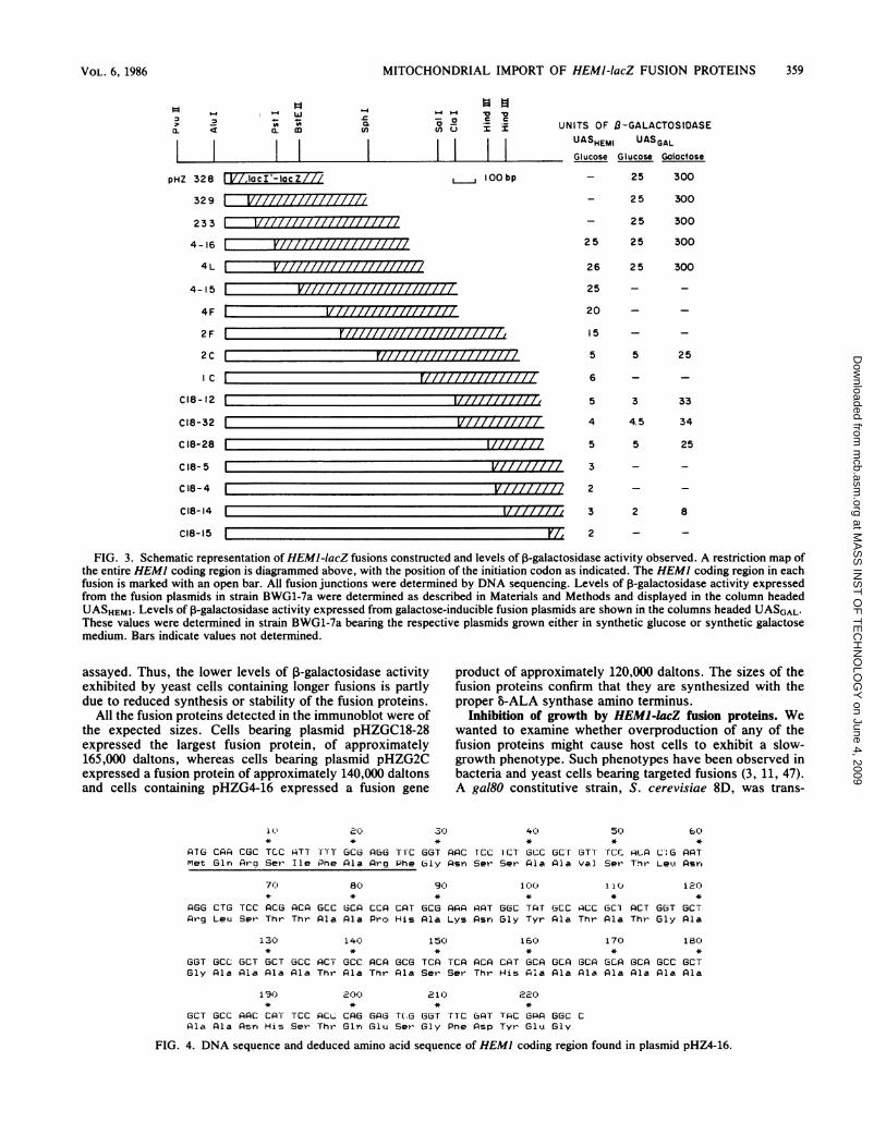
plasmids are shown in Fig. 3. The fusion endpoints of theseplasmids were determined by DNA sequencing. The fusionplasmids pHZ328, pHZ329, pHZ233, pHZ4-16, pHZ2C, andpHZC18-28 were found to contain 9, 35, 47, 75, 232, and 404codons of HEM], respectively. None of these fusion plas-mids could complement a HEM] mutation, indicating thatnone of them contains an intact HEMI gene and that none ofthe fusion proteins has B-ALA synthase activity in additionto ,B-galactosidase activity.
To obtain levels of expression of the fusion proteins whichcould be more easily detected in subcellular fractions, frag-ments containing the HEMI portion of some of the plasmidswere inserted into another vector, pLGSD5 (Fig. 2). Thisvector contains a lacZ portion that is identical to the one onthe original fusion plasmids. In addition, this vector containsa UASGAL positioned upstream of the lacZ region (19).Insertion of the HEM] fragments into this vector renderedthe HEMJ-IacZ fusions galactose inducible. Since HEM]upstream DNA in the constructs extends out to position-448, we might also expect the HEMI promoter to remainfunctional. These plasmids were used to transform S. cere-visiae BWG1-7a. The transformants displayed ,-galacto-sidase activity levels of 2 to 25 U in the absence of galactose,indicating the presence of a functional HEMI promoter, and25 to 300 U in the presence of galactose (Fig. 3).
Partial DNA sequence of HEM) coding region. The DNAsequence of the HEM] coding region of pHZ4-16 is shown inFig. 4. Because the lacZ translational frame is known, theHEM] coding sequence can be determined by counting backfrom the fusion junction to an AUG codon which is precededby two in-frame UAA stop codons 9 and 11 codons up-stream. pHZ4-16 was deduced to contain 75 amino acidresidues of HEMI. Translation of the DNA sequence intothe amino acid sequence revealed that, like other importedmitochondrial proteins, the amino-terminal 35 residues ofthis protein mostly consist of uncharged residues with 5basic residues; there are no acidic residues (32, 41, 49, 50).One striking feature of this sequence is an alanine-rich regionbetween residues 26 and 62, with 21 of 37 residues consistingof alanine, including a run of nine consecutive alanineresidues between residues 54 and 62.
Characterization of levels of HEM)-lacZ fusion gene prod-ucts. The fusions can be divided into two categories, basedon the levels of P-galactosidase activity expressed in yeastcells. When expression is from the HEM] upstream region,fusions pHZ328, pHZ329, pHZ233, pHZ4-16, pHZ4L,pHZ4-15, pHZ4F, and pHZ2F, containing 175 codons orfewer of HEM], all expressed approximately 20 to 30 U of3-galactosidase activity, whereas fusions pHZ2C, pHZ1C,pHZC18-12, pHZC18-32, pHZC18-28, pHZlC8-5, pHZC18-4, pHZC18-14, and pHZC18-15, containing at least 232codons of HEM], all exhibited approximately 5 U of activ-ity. The decreased level of expression of these longer fusiongenes is not due to a shift in the translational frame, as theDNA sequences of junctions of these fusions have estab-lished their reading frame to be correct (T. Keng and L.Guarente, unpublished data). The lower levels of activity ofthe longer fusion gene products were also observed in theconstructs, which placed them under UASGAL control.The difference in expression of P-galactosidase activity
could be due to a difference in the stability of the proteinssuch that the longer fusion proteins are more readily de-graded, or to a difference in the structure or localization ofthe fusion proteins. In the latter case, levels of the long andshort fusion proteins may be comparable, but the activity ofthe long fusion proteins may be reduced because of a failureto oligomerize their ,-galactosidase moieties. To distinguishbetween these two possibilities, crude extracts of strainscontaining some of the plasmids with galactose-inducibleHEMJ-lacZ fusions were run on sodium dodecyl sulfate-polyacrylamide gels, and the fusion proteins were detectedby immune blotting with anti-p-galactosidase antibodies (7).The immunoblot (Fig. 5) shows that the polypeptide levels offusion proteins detected in the extracts approximately cor-relate with the relative levels of ,-galactosidase activity
MOL. CELL. BIOL.
at MA
SS
INS
T O
F T
EC
HN
OLO
GY
on June 4, 2009 m
cb.asm.org
Dow
nloaded from
MITOCHONDRIAL IMPORT OF HEMJ-lacZ FUSION PROTEINS
_ 0 40< a. a)
iipHZ 328 tVl,lact -lacZlll
IO .c .I
enQI II
, lOObp
UNITS OF l3-GALACTOSIDASEUASHEMI UASGAL
Glucose Glucose Galactose
- 25 300
- 25 300
- 225 300
2 5 25 300
26 25 300
25 - -
20 - -
15 - -
5 5 25
6 - -
5 3 33
4 4.5 34
5 5 25
3 - -
2 - -
3 2 8
2 - -
FIG. 3. Schematic representation of HEMI-lacZ fusions constructed and levels of ,B-galactosidase activity observed. A restriction map ofthe entire HEMI coding region is diagrammed above, with the position of the initiation codon as indicated. The HEMI coding region in eachfusion is marked with an open bar. All fusion junctions were determined by DNA sequencing. Levels of 3-galactosidase activity expressedfrom the fusion plasmids in strain BWG1-7a were determined as described in Materials and Methods and displayed in the column headedUASHEM1. Levels of 1-galactosidase activity expressed from galactose-inducible fusion plasmids are shown in the columns headed UASGAL.These values were determined in strain BWG1-7a bearing the respective plasmids grown either in synthetic glucose or synthetic galactosemedium. Bars indicate values not determined.
assayed. Thus, the lower levels of ,-galactosidase activityexhibited by yeast cells containing longer fusions is partlydue to reduced synthesis or stability of the fusion proteins.
All the fusion proteins detected in the immunoblot were ofthe expected sizes. Cells bearing plasmid pHZGC18-28expressed the largest fusion protein, of approximately165,000 daltons, whereas cells bearing plasmid pHZG2Cexpressed a fusion protein of approximately 140,000 daltonsand cells containing pHZG4-16 expressed a fusion gene
product of approximately 120,000 daltons. The sizes of thefusion proteins confirm that they are synthesized with theproper b-ALA synthase amino terminus.
Inhibition of growth by HEMI-lacZ fusion proteins. Wewanted to examine whether overproduction of any of thefusion proteins might cause host cells to exhibit a slow-growth phenotype. Such phenotypes have been observed inbacteria and yeast cells bearing targeted fusions (3, 11, 47).A gal80 constitutive strain, S. cerevisiae 8D, was trans-
1 )I
PTG CPA CGC TCC PTTMet Gin Pro Set Ile
PO
TITT GCOPne Pla
*
APG6 Tl-C GGT APC rcc
Pro Phe ly Asn Set
40 5, 6C,
CT GCC GCT GTT rCc AcP C: G PATSer Aia Pia Val Ser Thr Let Asr,
70 80)* *
AGG CTG TCC AC6 PCP GCC 1CPAr-g Leu Ser Thr Thr APa APa
130
*
GGT GCC GCT GCT GCCGly Pla Pia Ala AlP
190
GCT GCC PAC CA-r TCCAla Ala Asn His Ser
140*
PCT GCCThr Pia
*
PCL CAGThr Gln
90)
*
CCA CAT GCG PAP APTPro His APa Lys Asr,
ACA GCG TCP TCA ACPThr Aia Ser- Ser- Thr
210
GGC TA'T GCCGly Tyr Pla
16()*
CAT 6CA GCAHis Ala Ala
1 0t
ACC GC1 ACTThr Ala Thr
12c)*
G6T GCTGly Ala
170 180* *
GCP GCA GCP GCC GCTPla Ala Ala Ala Ala
*
GAG T(.G GGT TTC GAT TAC GPA GGC CGlu Set, Gly Pne Psp Tyr GiU GlY
FIG. 4. DNA sequence and deduced amino acid sequence of HEM] coding region found in plasmid pHZ4-16.
329 1
233
4-16
4L
4-I5
4F
2F
2C
C
C18- 12
Cl8-32
C 18-28
C 18-5
C 18-4
Cl8-14
C18-15
VJIIIIIIIIIIIIIII11177VIIIIIIIIIIIIII777
F71illIlIlIllIlL177777777777.,V///7777777
17777777
V77777777177,777777
--FT,
359VOL. 6, 1986
L
at MA
SS
INS
T O
F T
EC
HN
OLO
GY
on June 4, 2009 m
cb.asm.org
Dow
nloaded from

360 KENG ET AL.
A B C D E F G H
200,030-
N
116,250j
92,500-
FIG. 5. Immunoblot of crude extracts prepared from strainBWG1-7a transformed with galactose-inducible fusion plasmids.The fusion proteins were detected with anti-,-galactosidase antibod-ies as described in Materials and Methods. Lanes: A and B,pLGSD5; C and D, pHZG4-16; E and F, pHZG2C; G and H,pHZGC18-28; A, C, E, and G, cells grown in lactate in the absenceof galactose; B, D, F, and H, cells grown with galactose in themedium. Lane D is underloaded.
formed with plasmids containing the galactose-induciblefusions pHZG4-16, pHZG4L, pHZG2C, pHZGC18-28, andthe control plasmid pLGSD5. The transformants werestreaked onto synthetic glucose, synthetic galactose, andsynthetic lactate plates. The plates were incubated at 30°C,and after 2 days the growth phenotypes of the differenttransformants were scored. In lactate medium, the UASGALis fully derepressed in a gal80 strain (52), whereas glucoserepresses the UASGAL (10, 36, 52).
After 2 days at 30°C, strain 8D transformed with the twolonger fusion plasmids pHZG2C and pHZGC18-28 showed agalactose-sensitive phenotype. The colonies formed on thesynthetic galactose plates were smaller than those formed bythe same strain transformed with pHZG4-16, pHZG4L, orthe control plasmid pLGSD5 (Fig. 6). On synthetic lactateplates, strain 8D transformed with all four galactose-inducible fusion plasmids, pHZG4-16, pHZG4L, pHZG2C,and pHZGC18-28, exhibited slow growth compared withcells bearing the control plasmid pLGSD5 (Fig. 6). Nogrowth inhibition was observed with cells bearing any of thefusion plasmids on synthetic plates containing glucose.
Cellular location of the HEM1-LacZ fusion gene product.Subcellular fractionation was performed to localize the fu-sion proteins; ,-galactosidase enzyme activity was used tolocate the fusion proteins.
TABLE 1. Localization of fusion proteins in subcellularfractionsa
% ,-galactosidase activity in:Fusion plasmid Postmitochondrial
Mitochondria supernatant
pLGSD5 0.3 99.7pHZG328 15.1 84.9pHZG329 31.6 68.4pHZG233 18.7 81.3pHZG4-16 24.0 76.0pHZG2C 76.0 24.0pHZGC18-28 74.1 25.9
a Cells were grown in a rich lactate medium. The cells were converted tospheroplasts and fractionated as described (9). Mitochondria prepared in thismanner contain <0.5% of whole-cell glyceraldehyde 3-phosphate dehydroge-nase activity and >99%o of whole-cell cytochrome b2 activity.
BWG1-7a transformants bearing plasmids with galactose-inducible HEMI-lacZ fusions and pLGSD5 were grownseparately in a rich lactate medium (9). Galactose was addedto induce high levels of synthesis of the ,B-galactosidasefusion proteins, and the cells were harvested and fraction-ated into a cytoplasmic supernatant and a mitochondrialpellet (40). The fractions were assayed for the level of3-galactosidase activity. The results are summarized in
Table 1. With the cytoplasmic ,3-galactosidase controlpLGSD5, only 0.3% of the enzyme activity was associatedwith the mitochondrial pellet, whereas 99.7% of the activitywas found in the cytoplasmic fraction. Cells bearing fusionplasmid pHZG328, pHZG329, pHZG233, or pHZG4-16, with9, 35, 47 and 75 amino acid residues of the amino terminus ofB-ALA synthase attached to ,B-galactosidase, respectively,had approximately 15 to 30% of the enzyme activity associ-ated with the mitochondria, whereas cells bearing the twolonger fusion genes pHZG2C (232 amino acid residues) andpHZG18-28 (404 amino acid residues) had approximately75% of the ,-galactosidase activity associated with themitochondria. Assays for glyceraldehyde 3-phosphate dehy-drogenase, a cytoplasmic marker enzyme, indicated that inthe subcellular fractionation of cells bearing the differentplasmids, not more than 0.2% of the cytoplasmic enzymeactivity was associated with the mitochondrial pellet. There-fore, in all cases examined the ,B-galactosidase activityassociated with the mitochondrial pellet was not due tocontamination of the pellet by cytoplasmic activity.To rule out the possibility that the cofractionation of the3-galactosidase activity with the mitochondria was due to
the formation of large aggregates of fusion protein, the
FIG. 6. Inhibition of growth by HEMJ-lacZ fusion proteins. S. cerevisiae 8D cells bearing galactose-inducible fusion plasmids werestreaked onto synthetic plates with glucose (A), galactose (B), or lactate (C) as the carbon source. Cells were incubated for 2 days at 30°Cand photographed. Segments: 1, pLGSD5; 2, pHZG4-16; 3, pHZG4L; 4, pHZG2C; 5, pHZGC18-28.
MOL. CELL. BIOL.
at MA
SS
INS
T O
F T
EC
HN
OLO
GY
on June 4, 2009 m
cb.asm.org
Dow
nloaded from
MITOCHONDRIAL IMPORT OF HEMJ-IacZ FUSION PROTEINS
mitochondrial pellets isolated from fusion-bearing strainswere further analyzed on sucrose density gradients (11).Mitochondria do not pellet in such gradients. Under theseconditions, the 3-galactosidase activity expressed from thefusion plasmid pHZG4-16 was found to comigrate with theactivity of the mitochondrial marker enzyme fumarase (Fig.7A and C). Identical results were obtained with mito-chondria prepared from strain BWG1-7a bearing fusionplasmids pHZG328, pHZG329, pHZ;G2C, and pHZGC18-28(data not shown). When mitochondria isolated from a strainbearing plasmid pLGSD5, coding for a cytoplasmic fusionprotein, were examined on these sucrose gradients, >90% ofthe low level of 1-galactosidase activity was found on top ofthe gradient (Fig. 7B and D). These results demonstrate aspecific association of the fusioq proteins with themitochondria. Results of immunofluorescence studies withanti-,B-galactosidase antibodies also indicate specific local-ization of the fusion proteins to the mitochondria (data notshown).Thus, in all cases examined, the HEM] encoding se-
quences present in the fusion plasmids were sufficient tQdirect the 13-galactosidase moiety to the mitochondria.
Localization of the HEMJ-lacZ gene product to within themitochondria. b-ALA synthase is located in the matrix of themitochondria (48). We wished to determine whether thefusion proteins encoded by plasmids pHZG328, pHZG329,pHZG4-16, pHZG2C, and pHZGC18-28 were also located inthe matrix. Hase et al. (20) and Schleyer et al. (44) showedthat any protein inside the mitochondria would be protectedfrom protease digestion by the inner and outer membranes ofthe organelle. We performed a similar experiment with themitochondrial pellets and found that in all cases, over 80% ofthe 1-galactosidase activity was protected from added pro-
-GALACTOSIDASE ACTIVITY
155 10FRACTION
a
5 10 15FRACT ION
FUMARASE ACTIVITY
5 10FRACTION
15 5 10FRACT ION
FIG. 7. ,-galactosidase activity encoded by HEMI-lacZ fusionplasmid cofractionating with fumarase activity on a sucrose gradi-ent. Mitochondria were prepared from BWG1-7a bearing fusionplasmids as described previously (40). Mitochondria (2.5 'mg[pHZG4-16J and 3.8 mg [pLGSD5]) were layered onto a 20 to 70%osucrose gradient and centrifuged. Fifteen fractions (approximately 1ml each) were collected from each gradient and assayed for theindicated enzymes. The bottoms of the gradients are to the left. (Aand C) pHZG4-16; (B and D) pLGSD5.
TABLE 2. Protection of ,B-galactosidase activity in mitochondriaand mitoplasts from proteinase K digestion
% P-Galactosidase activityProteinase K Triton X-100 remainingb in:Plsmda added added -.
Mitochondria Mitoplasts
pHZG328 - - 100 NDc+ - 128 ND- + 224 ND+ + 0.1 ND
pHZG329 - - 100 ND+ - 139 ND- + 180 ND+ + 0.1 ND
pHZG233 - - 100 100+ - 104 127- + 137 135+ + 0.1 0.1
pHZG4-16 - - 100 100+ - 87.4 71.6- + 136 111+ + 0.7 7.5
pHZG2C - - 100 100+ - 78.4 88.8- + 128 87.5+ + 0.2 0.9
pHZGC18-28 - - 100 100+ - 100 72.3- + 135 84.6+ + 0.4 9.8
a Host strain is S. cerevisiae BWG1-7a.bAll values are expressed as percentages of 0-galactosidase activity mea-
sured in the absence of Triton X-100 or proteinase K.c ND, Not determined.
teinase K (Table 2). However, over 95% of the activity ineach case was inactivated by proteinase K in the presence of0.5% Triton X-100, which solubilizes the mitochondrialmembranes; addition of 0.5% Triton X-100 alone in eachcase did not result in any net change of ,B-galactosidaseactivity. Identical results were observed in each case whenlevels of activity of fumarase, a matrix enzyme, were mea-sured (data not shown). These results showed that the fusiongene products in cells bearing plasmid pHZG328, pHZG329,pHZG233, pHGZG4-16, pHZG2C, or pHZGC18-28 wereassociated with the mitochondria in such a way as to beprotected from protease digestion by at least one membranebarrier.To determine whether the hybrid proteins were within the
inner mitochondrial membrane, mitoplasts were preparedfrom strain BlWG1-7a containing either plasmid pHZG233,pHZG4-16, pHZG2C, or pHZGC18-28. The mitoplasts con-sist of mitochondrial matrix surrounded by the inner mito-chondrial membrane. Outer membranes were removed byosmotic lysis in 0.06 M sorbitol (20). This treatment releasedthe intermembrane space enzyme cytochrome b2. In allcases examined, the 3-galactosidase activity was quantita-tively recovered with the mitoplasts. Moreover, as with theintact mitochondria, over 90% of the ,B-galactosidase activityin each case was protected from externally added proteinaseK (Table 2). Addition of 0.5% Triton X-100 rendered the0-galactosidase activity protease sensitive. The fate of thematrix enzyme fumarase paralleled that of the 1-
AZ 12
a-
8
01)
ui60 4
0
Z 2
z
2
(/) O
E 810
4
2
C
-
361VOL. 6, 1986
at MA
SS
INS
T O
F T
EC
HN
OLO
GY
on June 4, 2009 m
cb.asm.org
Dow
nloaded from

362 KENG ET AL.
i, 1
070OT|ZZ 8
Z E
0 aZ
40
2
pHZG 329
A B C D
(nWT
0
07 3zz
z 7t 2
-z
0
A B C D A B C D
FIG. 8. Distribution of ,-galactosidase activity in subfractions ofthe mitochondria. Yeast mitochondria from strain BWG1-7a bearingthe indicated plasmids were subfractionated as described previously(9). The ordinates mark the specific activities of ,-galactosidase. (A)cytoplasm; (B) membranes; (C) intermembrane space; (D) matrix.The membranes fraction represent 44 to 58% of mitochondrialproteins, the intermembrane space represents 13 to 24%, and thematrix represents 24 to 34%.
galactosidase activity (data not shown). The results demon-strate that the fusion proteins in all the cases examined wereprotected from protease inactivation by the inner mem-branes of the mitochondria. Thus, the fusions appear toreside in the matrix.To verify the matrix location of the fusion proteins, the
mnitochondria were fractionated into their various compart-ments: membranes, intermembrane space, and matrix (9).Measurements of specific activities of cytochrome c oxidase(inner membrane), cytochrome b2 (intermembrane space),and fumarase (matrix) indicated that there was no substantialcontamination of one compartment with any other. Specific-activity measurements of 3-galactosidase showed that instrains bearing plasmids pHZG328, pHZG329, and pHZG4-16, the fusion proteins are located in the matrix, whereas instrains bearing plasmids pHZG2C and pHZGC18-28, thefusion proteins are localized to both the membranes andmatrix compartments (Fig. 8).
DISCUSSIONAmino-terminal fragments containing as few as nine resi-
dues of the HEM] gene product B-ALA synthase direct,-galactosidase into the mitochondrial matrix. The locationof the fusion proteins was determined by using the method ofsubcellular fractionation. The fusion proteins were found tobe specifically associated with the mitochondria in sucrosedensity gradients. They were protected from protease diges-tion both in whole mitochondria and in mitoplasts, in whichthe outer membranes of the mitochondria were disrupted,and subfractionation of the mitochondria clearly showed thatthe fusion proteins were located in the matrix. Interestingly;the two longest fusion proteins tested (containing 232 and404 HEM] codons) also showed a substantial fraction of
their activity located in the membrane fractions. The prote-ase-resistant property of these fusion proteins in mitoplastpreparations suggests that they are associated with the insideof the inner membrane. The possibility that b-ALA synthaseitself displays an association with the inside of the innermembrane is currently being tested.Gene fusion as a method to study import has been suc-
cessfully used by Douglas et al. (11), Horwich et al. (25), andHurt et al. (26-28). Of particular interest is the finding that asfew as 12 amino-terminal residues of cytochrome oxidasesubunit IV will direct mouse dihydrofolate reductase, acytosolic protein, to the mitochondrial matrix (28). It wouldtherefore appear from our data and those of Hurt et al. thatmitochondrial signal sequences are, in general, very short. Acomparison of the first 9 residues of b-ALA synthase withthe first 12 residues of cytochrome oxidase subunit IVindicates that both sequences are largely hydrophobic, witha few basic residues interspersed (28) (Fig. 4). Precisehomology between these signals is not apparent.The amino-terminal 35 residues of 5-ALA synthase consist
largely of uncharged amino acids with a few basic residues(Fig. 4). This region is followed by an alanine-rich stretchhighlighted by a run of nine consecutive alanine residuesbetween positions 54 and 62. A similar run of 10 consecutivealanines is found in the cytochrome c peroxidase pre-piece(29). For b-ALA synthase, the alanine-rich region appearsnot to be required to direct import.Because of the low level of activity expressed from the
original fusion plasmids, we fused the HEMJ-lacZ fusions toa UASGAL and induced 10-fold-higher levels of synthesis offusion proteins with galactose. Douglas et al. (11) reportedthat an inability of the host cells to grow on a nonfermentablecarbon source is associated with the production of highlevels of Fl-ATPase-,-subunit-i-galactosidase fusion geneproduct. Similarly, a partial inhibition of growth is caused byall of our fusions when induced cells are grown in lactatemedia. The longer fusion proteins also inhibit growth ongalactose media. We are currently attempting to exploit thisphenotype to obtain mutations which define both cis- andtrans-acting components mediating import.
In several instances in which a ,-galactosidase moiety isfused to signals directing secretion through a bacterial mem-brane, the hybrid protein becomes "stuck" in the membrane(3, 47). It is unclear whether P-galactosidase contains se-quences which block the secretion machinery or whichprevent passage through the lipid bilayers in these cases. Wehave shown that fusions of 5-ALA synthase sequences toP-galactosidase via a 90-amino-acid "bridge" of the lacrepressor can result in matrix localization. The ability of themitochondrial machinery to import 3-galactosidase throughthe outer and inner membranes of the organelle may reflectunique aspects of the import process.
ACKNOWLEDGMENTSWe thank M. Douglas for communicating information prior to
publication, J. Masucci for constructing plasmids pHZG328,pHZG329, and pHZG233, R. Rosenberg and W.-K. Wong forinitiating studies on HEM], T. Bestor for help with the photographyand printing, and A. Mitchell, T. Bestor, P. Drain, N. Clarke, and P.Schimmel for critical reading of the manuscript.
This work was supported by a W. R. Grace grant to L.G. T.K.was a postdoctoral fellow of the Damon Runyon-Walter WinchellCancer Fund.
LITERATURE CITED1. Appleby, C. A., and R. K. Morton. 1959. Lactic dehydrogenase
and cytochrome b2 of Baker's yeast-purification and crystalli-
MOL. CELL. BIOL.
at MA
SS
INS
T O
F T
EC
HN
OLO
GY
on June 4, 2009 m
cb.asm.org
Dow
nloaded from

MITOCHONDRIAL IMPORT OF HEMJ-lacZ FUSION PROTEINS
zation. Biochem J. 71:492-499.2. Bard, M., and T. D. Ingolia. 1984. Plasmid-mediated comple-
mentation of a &-aminolevulinic-acid-requiring Saccharomycescerevisiae mutant. Gene 28:195-199.
3. Bassford, P. J., Jr., T. J. Silhavy, and J. R. Beckwith. 1979. Useof gene fusion to study secretion of maltose-binding protein inEscherichia coli periplasm. J. Bacteriol. 139:19-31.
4. Biggin, M. D., T. J. Gibson, and G. F. Hong. 1983. Buffergradient gels and 35S label as an aid to rapid DNA sequencedetermination. Proc. Natl. Acad. Sci. USA 80:3963-3965.
5. Bohni, P. C., G. Daum, and G. Schatz. 1983. Import of proteinsinto mitochondria-partial purification of a matrix-located pro-tease involved in cleavage of mitochondrial precursorpolypeptides. J. Biol. Chem. 258:4937-4943.
6. Bradford, M. M. 1976. A rapid and sensitive method for thequantitation of microgram quantities of protein utilizing theprinciple of protein-dye binding. Anal. Biochem. 72:248-254.
7. Burnette, W. N. 1981. "Western blotting": electrophoretictransfer of proteins from sodium dodecyl sulfate-polyacryl-amide gels to unmodified nitrocellulose and radiographic detec-tion with antibody and radioiodinated protein A. Anal.Biochem. 112:195-203.
8. Carlson, M., and D. Botstein. 1982. Two differentially regulatedmRNAs with different 5' ends encode secreted and intracellularforms of yeast invertase. Cell 28:145-154.
9. Daum, G., P. C. Bohni, and G. Schatz. 1982. Import of proteinsinto mitochondria-cytochrome b2 and cytochrome c peroxi-dase are located in the intermembrane space of yeastmitochondria. J. Biol. Chem. 257:13028-13033.
10. Douglas, H., and D. Hawthorne. 1966. Regulation of genescontrolling synthesis of the galactose pathway enzymes inyeast. Genetics 54:911-916.
11. Douglas, M. G., B. L. Gelier, and S. D. Emr. 1984. Intracellulartargeting and import of an F1-ATPase P-subunit-p-galactosidasehybrid protien into yeast mitochondria. Proc. Natl. Acad. Sci.USA 81:3983-3987.
12. Dujon, B. 1981. Mitochondrial genetics and functions, p.505-635. In J. N. Strathern, E. W. Jones, and J. R. Broach(ed.), The molecular biology of the yeast Saccharomyces - lifecycle and inheritance. Cold Spring Harbor Laboratory, ColdSpring Harbor, N.Y.
13. Gasser, S. M., G. Daum, and G. Schatz. 1982. Import of proteinsinto mitochondria - energy-dependent uptake of precursors byisolated mitochondria. J. Biol. Chem. 257:13034-13041.
14. Gasser, S. M., A. Ohashi, G. Daum, P. C. Bohni, J. Gibson,G. A. Reid, T. Yonetani, and G. Schatz. 1982. Imported mito-chondrial proteins cytochrome b2 and cytochrome cl are proc-essed in two steps. Proc. Natl. Acad. Sci. USA 79:267-271.
15. Godson, G. N., and D. Vapnek. 1973. A simple method ofpreparing large amounts of 4X174 supercoiled DNA. Biochim.Biophys. Acta 299:516-520.
16. Gollub, E. G., K.-P. Liu, J. Dayan, M. Adlersberg, and D. B.Sprinson. 1977. Yeast mutants deficient in heme biosynthesisand a heme mutant additionally blocked in cyclization of 2,3-oxidosqualene. J. Biol. Chem. 252:2846-2854.
17. Guarente, L. 1983. Yeast promoters and lacZ fusion designed tostudy expression of cloned genes in yeast. Methods Enzymol.101:181-191.
18. Guarente, L., and M. Ptashne. 1981. Fusion of Escherichia colilacZ to the cytochrome c gene of Saccharomyces cerevisiae.Proc. Natl. Acad. Sci. USA 78:2199-2203.
19. Guarente, L., R. R. Yocum, and P. Gifford. 1982. A GALJO-CYCI hybrid yeast promoter identifies the GAL4 regulatoryregion as an upstream site. Proc. Natl. Acad. Sci. USA79:7410-7414.
20. Hase, T., U. Miller, H. Reizman, and G. Schatz. 1984. A 70-kdprotein of the yeast mitochondrial outer membrane is targetedand anchored via its extreme amino terminus. EMBO J.3:3157-3164.
21. Hay, R., P. Bohni, and S. Gasser. 1984. How mitochondriaimport proteins. Biochim. Biophys. Acta 779:65-87.
22. Hennig, B., and W. Neupert. 1981. Assembly of cytochrome c.Apocytochrome c is bound to specific sites on mitochondria
before its conversion to holocytochrome c. Eur. J. Biochem.121:203-212.
23. Hinnen, A., J. B. Hicks, and G. R. Fink. 1978. Transformation ofyeast. Proc. Natl. Acad. Sci. USA 75:1929-1933.
24. Holmes, D. S., and M. Quigley. 1981. A rapid boiling method forthe preparation of bacterial plasmids. Anal. Biochem. 114:193-197.
25. Horwich, A. L., F. Kalousek, I. Meliman, and L. E. Rosenberg.1985. A leader peptide is sufficient to direct mitochondrialimport of a chimeric protein. EMBO J. 4:1129-1135.
26. Hurt, E. C., B. Pesold-Hurt, and G. Schatz. 1984. The amino-terminal region of an imported mitochondrial precursor poly-peptide can direct cytoplasmic dihydrofolate reductase into themitochondrial matrix. EMBO J. 3:3149-3156.
27. Hurt, E. C., B. Pesold-Hurt, and G. Schatz. 1984. The cleavableprepiece of an imported mitochondrial protein is sufficient todirect cytosolic dihydrofolate reductase into the mitochondrialmatrix. FEBS Lett. 178:306-310.
28. Hurt, E. C., B. Pesold-Hurt, K. Suda, W. Oppliger, and G.Schatz. 1985. The first twelve amino acids (less than half of thepre-sequence) of an imported mitochondrial protein can directmouse cytosolic dihydrofolate reductase into the yeast mito-chondrial matrix. EMBO J. 4:2061-2068.
29. Kaput, J., S. Goltz, and G. Blobel. 1982. Nucleotide sequence ofthe yeast nuclear gene for cytochrome c peroxidase precursor.J. Biol. Chem. 257:15054-15058.
30. Krebs, E. G. 1955. Glyceraldehyde-3-phosphate dehydrogenasefrom yeast. Methods Enzymol. 1:407-411.
31. Laemmli, U. K. 1970. Cleavage of structural proteins during theassembly of the head of bacteriophage T4. Nature (London)227:680-685.
32. Maarse, A. C., A. P. G. M. Van Loon, H. Reizman, I. Gregor, G.Schatz, and L. A. Grivell. 1984. Subunit IV of yeast cytochromec oxidase: cloning and nucleotide sequencing of the gene andpartial amino acid sequencing of the mature protein. EMBO J.3:2831-2837.
33. Mandel, M., and A. Higa. 1970. Calcium dependent bacterio-phage DNA infection. J. Mol. Biol. 53:159-162.
34. Mason, T. L., R. 0. Poyton, D. C. Wharton, and G. Schatz.1973. Cytochrome c oxidase from baker's yeast. J. Biol. Chem.248:1346-1354.
35. Neupert, W., and G. Schatz. 1981. How proteins are transportedinto mitochondria. Trends Biochem. Sci. 6:1-4.
36. Oshima, Y. 1982. Regulatory circuits for gene expression: themetabolism of galactose and phosphate, p. 159-180. In J.Strathern, E. Jones, and J. Broach (ed.), The molecular biologyof the yeast Saccharomyces - metabolism and gene expres-sion. Cold Spring Harbor Laboratory, Cold Spring Harbor,N.Y.
37. Racker, E. 1950. Spectrophotometric measurements of theenzymatic formation of fumaric acid and cis-aconitic acids.Biochim. Biophys. Acta 4:211-214.
38. Reid, G. A., and G. Schatz. 1982. Import of proteins intomitochondria- extramitochondrial pools and post-translationalimport of mitochondrial protein precursors in vivo. J. Biol.Chem. 257:13062-13067.
39. Reizman, H. 1982. Binding of precursors of cytoplasmically-synthesized mitochondrial proteins to isolated outer membranesof yeast mitochondria. In G. Akoyunoglou, A. E. Evangel-opoulos, J. Georgatsos, G. Palaiologos, A. Trakatellis, andC. P. Tsiganos (ed.), Cell function and differentiation, part B:biogenesis of energy transducing membranes and membraneand protein energetics. Alan R. Liss Inc., New York.
40. Reizman, H., R. Hay, S. Gasser, G. Daum, G. Schneider, C.Witte, and G. Schatz. 1983. The outer membrane of yeastmitochondria: isolation of outside-out sealed vesicles. EMBO J.2:1105-1111.
41. Sadler, I., K. Suda, G. Schatz, F. Kauderwitz, and A. Haid.1984. Sequencing of the nuclear gene for the yeast cytochromecl precursor reveals an unusually complex amino terminalpresequence. EMBO J. 3:2137-2143.
42. Sanger, F., S. Nicklen, and A. R. Coulson. 1977. DNA sequenc-ing with chain-terminating inhibitors. Proc. Natl. Acad. Sci.
VOL. 6, 1986 363
at MA
SS
INS
T O
F T
EC
HN
OLO
GY
on June 4, 2009 m
cb.asm.org
Dow
nloaded from

MOL. CELL. BIOL.
USA 74:5463-5467.43. Sthaitz, G., and R. A. gutow. 1983. How are proteins imported
into mitochondria? Cell 32:316-318.44. Schleyer, M., and W. Neupert. 1984. Transport of ADP/ATP
carrier intb mitochondria precursor imported in vitro ac-quires functional properties of the mature protein. J. Biol.Chem. 259:3487-3491.
45. Schleyer, M.+ B. Schmidt, and W. Neupert. 1982. Requirementof a membrane potential for the posttranslational transfer ofproteins into mitochondria. Eur. J. Biochem. 125:109-116.
46. Sherman, F., G. R. Fink, and J. B. Hicks. 1983. Methods inyeast genetics. Cold Spring Harbor Laboratory, Cold SpringHarbor, N.Y.
47. Silhavy, T., H. A. Shuman, J. Beckwith, and M. Schwartz. 1977.Use of gene fusions to study outer membrane protein localiza-tion in Escherichia coli. Proc. Natl. Acad. Sci. USA74:5411-5415.
48. Tait, G. H. 1978. The biosynthesis and degradation of heme, p.
1-48. In F. DeMatteis and W. N. Aldridge (ed.), Heme andhemoproteins. Springer-Verlagi New York.
49. Watson, M. E. E. 1984. Compilation of published signal se-quences. Nucleic Acids Res. 12:5145-5164.
50. Wright, R. M., C. Ko, M. G. Chmsky, and R. 0. Poyton. 1984.Isolation and sequence of the structural gene for cytochrome coxidase subunit VI from Saccharomyces cerevisiae. J. Biol.Chem. 259:15401-15407.
51. Yaffe, M. P., and G. Schatz. 1984. Two nuclear mutations thatblock mitochondrial protein import in yeast. Proc. Natl. Acad.Sci. USA 81:4819-4823.
52. Yocums, R. R., S. Hanley, R. West, Jr., and M. Ptashne. 1984.Use of lacZ fusions to delimit regulatory elements of theinducible divergent GALJ-GALIO promoter in Saccharomycescerevisiae. Mol. Cell. Biol. 4:1985-1998.
53. Zimmerman, R., B. Hennig, and W. Neupert. 1981. Differenttransport pathways of individual precursor proteins inmitochondria. Eur. J. Biochem. 116:455-460.
364 KENG ET AL.
at MA
SS
INS
T O
F T
EC
HN
OLO
GY
on June 4, 2009 m
cb.asm.org
Dow
nloaded from