the neurophysiological ankle-foot orthosis · odevelopmental techniques ... the neurophysiological...
TRANSCRIPT
The Neurophysiological Ankle-Foot Orthosis by Cyndi Ford, P.T.
Robert C. Grotz, M.D. Joanne Klope Shamp, C.P.O.
Since the late 1960's when Yates 1 and Lehneis 2 wrote the first articles pertaining to the use of plastics, orthotic practice has been revolutionized by the design possibilities afforded by total contact devices. However, prescription of lower extremity orthoses for neuro-logically involved patients has traditionally depended solely upon biomechanical principles even as neurophysiological approaches to treatment gained recognition and acceptance. Neur-odevelopmental Techniques (NDT) were developed as a theory of Karl and Berta Bobath and evolved to " a sensorimotor approach to control motor output and in doing so change sensory input ." 3 Handling techniques which counteract patterns of abnormal tonic reflex activity reduce spasticity and allow facilitation (activation) of normal postural reactions through stimulation of key points of control, which include points on the foot and ankle. Recent advances incorporating neurophysiological principles of inhibition and facilitation into the design of ankle-foot orthoses make possible tone-reducing devices with specific areas of pressure or contact to inhibit abnormal hypertonicity.
Eberle, Jeffries, and Zachazewski 4 recently reported success with an inhibitive AFO, a concept that was not feasible with metal orthotics. Their report stated that "the technique of fabrication used for the construction of a molded polypropylene AFO allows for all of the tone-inhibiting characteristics of casting . . . to be built into the A F O . "
Although tone-reducing AFO's inhibit abnormal hypertonicity in the affected lower extremity, the disadvantages inherent in traditional AFO's persist. Limited ankle dorsi-flexion and plantar flexion, create a negative influence upon independent knee and hip function. Floor reaction forces intended to prevent the typical hemiplegic knee recurvatum during stance phase also contribute to increased effort and decreased smoothness in gait. Tonic foot reflexes elicited by contact on the plantar surface of the foot as a means to facilitate normal movement are disregarded.
In an effort to address these gait concerns, an orthosis was designed based upon the neurode-velopmental concepts as described by Bobath 5
and Utley 6, and the foot reflexes as described by Duncan and Mott 7 with the following considerations in mind:
1. A design configuration intended to utilize both biomechanical principles to limit calcaneal varus and neurophysiological principles (of facilitation and inhibition) to obtain dynamic ankle dorsiflexion and plantar flexion.
2. Selection of a material with adequate flexibility, durability, and shape retention under conditions of continual deformation during ambulation.
3. Ease of donning for the one-handed patient.
DESIGN RATIONALE The Neurophysiological Ankle-Foot Orthosis
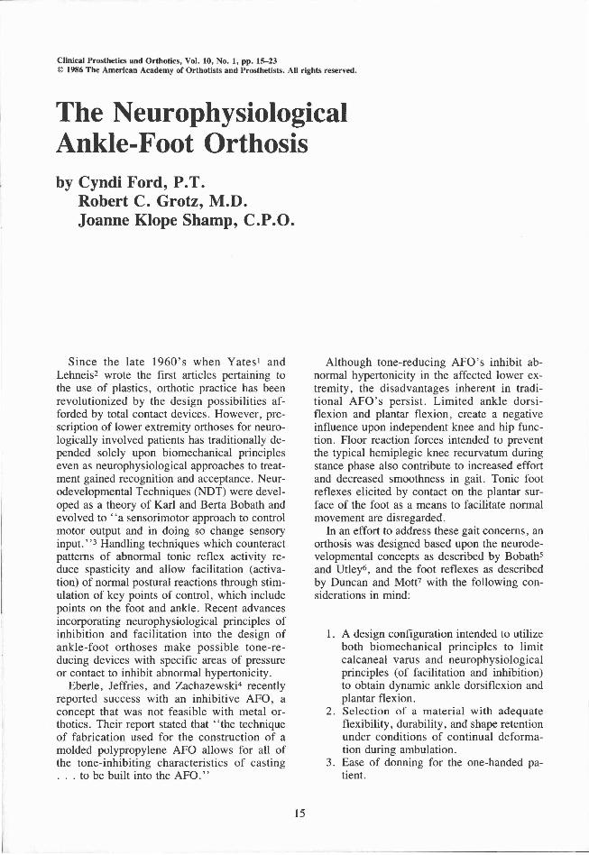
(NP-AFO) is a custom polypropylene device, vacuum-formed over a plaster model of the patient's affected lower extremity (Figure 1). Within the total contact design are incorporated the following forces:
1. A three-point pressure system to biome-chanically control calcaneal varus (Figure 2).
2. A biomechanical force medial to the achilles tendon to counterbalance and prevent excessive pronation and rotation of the orthosis in the shoe (Figure 3).
3. A neurophysiological force on the medial aspect of the calcaneus, extending to the plantar surface of the longitudinal arch without creating pressure under the navicular itself (Figure 3). This facilitates straight plane dorsiflexion.
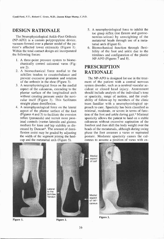
4. A neurophysiological force on the lateral aspect of the plantar surface of the foot (Figures 4 and 5) to facilitate the eversion reflex (peroneals) and recruit more proximal controls (vastus lateralis and gluteus medius) for knee and hip stability as discussed by Duncan 8 . The amount of dorsiflexion assist may be graded by adjusting the width of the segment joining the heel-cup and the metatarsal arch (Figure 5).
5. A neurophysiological force to inhibit the toe grasp reflex (toe flexors and gastroc-nemius-soleus) by unweighting of the metatarsal heads through use of a metatarsal arch (Figure 6).
6. Biomechanical function through flexibility of the foot and ankle due to the trimlines and configuration of the plastic NP-AFO (Figures 7 and 8).
PRESCRIPTION RATIONALE
The NP-AFO is designed for use in the treatment of the patient with a central nervous system disorder, such as a cerebral vascular accident or closed head injury. Assessment should include analysis of the individual's tone or spasticity, range of motion, and the availability of follow-up by members of the clinic team familiar with a neurophysiological approach to care. Spasticity has been classified as minimal, moderate, or severe in terms of function of the foot and ankle during gait. 9 Minimal spasticity allows the patient to land on a stable calcaneus without excessive supination of the forefoot and then shift the body weight over the heads of the metatarsals, although during swing phase the foot assumes a varus or supinated posture. Moderate spasticity causes the calcaneus to assume a position of varus with ex-
Figure 1. Figure 2. Figure 3.
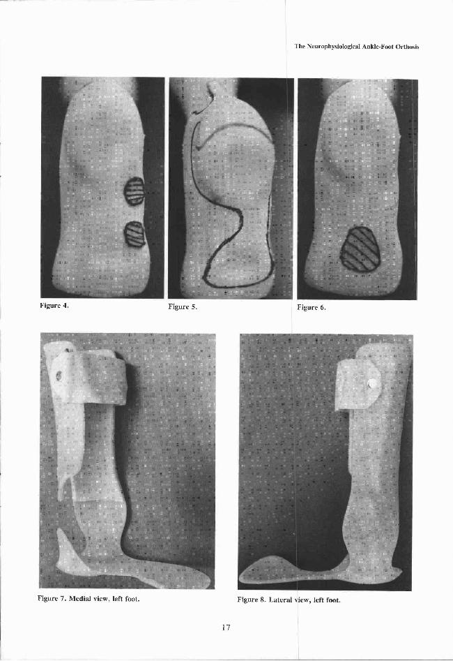
Figure 4. Figure 5. Figure 6.
Figure 7. Medial view, left foot. Figure 8. Lateral view, left foot.

cessive supination at initial contact; however, during midstance some pronation occurs and the body weight can again be transferred normally across the forefoot. Severe spasticity is characterized by the foot and ankle being held rigidly in a position of equinovarus throughout stance so that the body weight remains on the lateral aspect of the forefoot with little or no weightbearing through the heel or medial metatarsal heads. This varus position persists throughout swing phase also.
Patients exhibiting minimal or moderate spasticity are excellent candidates for the NP-AFO. Patients with severe spasticity are candidates only if their tone can be modified through handling techniques and/or inhibitive casting. The use of toe separators (Figures 9,10,11) as an adjunct treatment is also effective in patients with a separate toe grasp reflex to inhibit excess tone and reduce pain. 6 In order for the NP-AFO to function appropriately, the patient must have at least 15 degrees of passive dorsiflexion with the knee in flexion.
Follow-up by a clinic team familiar with the device is important to monitor the continued fit and function. With most AFO's the major concern may be skin breakdown. However, with the NP-AFO the change in fit due to edema, weight loss, or tone variations may require modifications to maintain the critical areas of contact.
Contraindications for this device are severe spasticity which cannot be modified through inhibitive casting or handling techniques, and early excessive pronation or calcaneal valgus with the foot pronated at initial contact of stance.
CLINICAL EXPERIENCE The NP-AFO has been prescribed for 35 pa
tients with the following diagnoses: 29 Cerebral Vascular Accidents (CVA), 4 Closed Head Injuries (CHI), 1 Cauda Equina Injury, and 1 undiagnosed Demyelinating Disease. Although three patients were lost to follow-up, the NP-AFO has continued to be worn by the remaining 32 with overwhelming acceptance which seems to be attributed to the comfort and function of the device. Of the four patients converted from traditionally designed orthoses (2 metal, 2 plastic AFO's ) , three have im-
Figure 9. Toe separa tors fabricated from Plastazote® with a Moleskin® cover and toe extension.
Figure 10. Toe separa tors in place under the toes.
Figure 11 . Super ior view showing tabs to hold in place under sock.

proved gait patterns and prefer the NP-AFO to their previous device. The fourth has rejected orthotic care due to refusal to adapt footwear from inappropriate styles with 2 1/2" heels. Four pat ients became independent ambulators without the use of any orthotic device.
FABRICATION Polypropylene was chosen as the thermo
plastic currently exhibiting the best conformance to the desired qualities, when used in the fabrication process described.
CASTING PROCEDURE The casting technique is similar to that de
scribed in Lower Limb Orthotics, A Manual10
and is a procedure commonly used by certified orthotists. The cast must be taken in a position of maximal dorsiflexion, preferably 20 degrees. The calcaneus, midfoot, and forefoot should be in a neutral position. It has been our experience that tone-reducing handling activi-
. ties performed by a physical therapist just prior to casting will help assure an optimal position. These activities include forefoot, midfoot, and hindfoot mobilizations as taught by Jan Utley. 6
The cast is removed upon hardening and filled with plaster to create a positive model for
use in vacuum-forming of the orthosis. The positive model is now ready for modifications to create the necessary biomechanical and neurophysiological forces.
MODIFICATION OF THE POSITIVE MODEL
As the key to function of the orthosis is selective inhibitive and facultative forces, accurate cast modification is essential. Plaster removal is performed in the following areas to a depth of 0.5 to 1 cm. depending upon the compressibility of the patient's extremity. These modifications must be sufficient to provide a very firm force to the skin as designated.
1. Medial and lateral to the achilles tendon using a Scarpa's knife to deeply groove the modification (Figure 12).
2. Medial aspect of the calcaneus extending to the plantar surface of the longitudinal arch without creating pressure under the navicular itself that would stimulate mid and forefoot supination (Figure 13).
3. Along the lateral plantar surface of the mid- and forefoot, excluding the base and head of the fifth metatarsal (Figure 14).
Figure 12.
Figure 13.
Figure 14.

4. Create a metatarsal arch 6mm. proximal of the metatarsal heads for the inhibitive function of unweighting the metatarsal heads and thereby reduce tone (Figure 6).
5. Smooth entire cast.
If an accurate negative cast and positive model were created, no further modifications are necessary.
VACUUM-FORMING PROCESS
Leather, nylon, or rope cording is applied to the cast (Figures 15 ,16 ,17) to create strengthening corrugations in the orthosis after molding.
A separating agent or material is used between the positive model and the hot plastic to create adequate vacuum and to leave a smooth inner surface. For our drape-forming process one layer of perlon with one layer of ladies' nylon knee-high stockings are applied and smoothed with talc. Stress-relieved 3/16" polypropylene is then drape-formed under vacuum to the positive model and allowed to cool for 24 hours.
TRIMLINES The orthosis is removed from the positive
model using a cast cutter and is sanded to finish according to the following trimlines:
1. Overall height of the orthosis is equal to the distance from the plantar surface of the calcaneus to the flare of the achilles tendon as it meets the gastrocnemius-soleus g roup , mul t ip l ied by 2. An average overall length for a 175cm. (5'9") adult is 25.5cm. (10").
2. Length of the plantar extension is terminated 6mm. proximal to the metatarsal heads for comfort.
3. The lateral trimlines (Figure 18) come as far anterior as possible and still allow passage of the leg into the orthosis. The posterior trimline (Figures 18 and 19) approaches the lateral margin of the achilles tendon, but may require modification to prevent a bowstring effect by the heel counter of the shoe against the NP-AFO.
Note that flexibility is enhanced by the narrowing anteriorly and posteriorly as the lateral side meets the heelcup.
4. The achilles tendon is left exposed to the point of flare with the gastronemius-soleus (Figure 19).
Figure 15. Figure 16.
Figure 17.
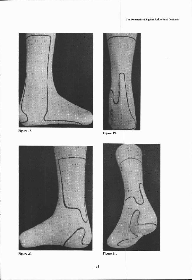
Figure 18. Figure 19.
Figure 20. Figure 2 1 .

5. The medial margin is trimmed so as to provide the appropriate forces and yet avoid contact on the medial malleolus and under the navicular. The open area provides for lack of resistance to dorsiflexion and plantar flexion (Figures 20 and 21).
6. The plantar extension (Figure 21) may be varied in width depending upon the size of the patient and flexibility desired, but as it serves only to join the metatarsal arch to the heelcup, it should remain as flexible as possible. The distal aspect, including the metatarsal pad, should span the distance between the shaft of the first metatarsal and the extreme lateral margin of the foot to allow maximum facilitation of the eversion reflex.
A full 1/8" Plastazote® liner is glued to the inner surface of the orthosis, with the exception of the areas contained by the patient's shoe to allow ease of donning the same size shoe previously worn by the patient. A Velcro® strap of 2" width is applied to the proximal anterior calf. A lace-tied or Velcro®-closed shoe is recommended to maintain the critical fit of the NP-AFO.
DISCUSSION The movement allowed by the NP-AFO en
courages dynamic control of the entire lower extremity . When si t t ing, normal weight-bearing attitude can occur with the foot rem a i n i n g in full con t ac t with the f loor throughout a full range of knee flexion. Analysis of the normal movements of the ankle during elevation from a chair has revealed to us that the ankle begins in dorsiflexion and continues to dorsiflex during the initial phase of the elevation before plantar flexing to a relatively neutral position. Devices which eliminate this normal range of dorsiflexion necessarily require a patient to work over an abnormal base and make difficult active weight-bearing during elevation. The ability to assume a normal weight-bearing surface in a position of power as allowed by the NP-AFO encourages weight-bearing on the affected extremity throughout all activities of daily living.
Further, dynamic control of the pelvis and knees are encouraged during ambulation by eliminating floor reaction forces inherent in
other AFO's. Without these abnormal forces, the patient experiences the normal movement of the pelvis and knee over the foot, allowing development of a propulsive toe-off with the NP-AFO.
Progressing from use of the NP-AFO to being independent of assistive devices is more feasible, as the patient has the opportunity to gain control of muscles through the normal range of movement.
SUMMARY The adequacy of traditional AFO's to pro
vide a safe, functional gait pattern is irrefutable. However, experience with patients who sustained a CVA five to fifteen years ago and received a traditional metal or plastic AFO reveals they now present problems related to overuse of the sound side: the pathomechanics resulting from a rigid ankle and/or increasing hypertonicity from abnormal weightbearing patterns. As more patients have increased lifespans following a CVA, treatments and orthotic care which assure prolonged quality of life become increasingly important. Neurophysiological treatment attempts to do this through emphasis upon normal movement patterns and integration of the affected and unaffected sides.
The NP-AFO is a biomechanically and neu-rophysiologically effective ankle-foot orthosis that is appropriate for creating a functional gait in the patient with a central nervous system disorder. The design allows for independent motion at the ankle, knee, and hip joints in a lightweight and cosmetic custom-made orthosis. The NP-AFO joins the inhibitive cast and other neurophysiological armamentarium in new approaches to the rehabilitation of the spastic or hypertonic patient.
REFERENCES 1 Yates, G., "A Method for Provision of Lightweight
Aesthetic Orthopaedic Appliances," Orthopaedics: Oxford, 1:2, pp 153-162 , 1968
2 Lehneis, H.R., "New Concepts in Lower Extremity Orthotics," Medical Clinics of North America, 53:3:3, pp. 585 -592 , 1969.
3 Bobath, K., "The Problem of Spasticity in the Treatment of Patients With Lesions of the Upper Motor Neurone," The Western Cerebral Palsy Centre, London, England.
4 Eberle, E.D.; Jeffries, M.; and Zachazewski, J.E.,
"Effect of Tone-Inhibiting Casts and Orthoses on Gait: A Case Report," Physical Therapy, 62:4 pp. 4 5 3 - 4 5 5 , 1982.
5 Bobath, B. and Bobath, K., Motor Development in Different Types of Cerebral Palsy, Heinman, London, 1975.
6 Utley, J., NDT Adult Hemiplegia and Closed Head Injury Certification Course, Columbus, Ohio, July, 1982.
7 Duncan, W. and Mott, D . , "Foot Reflexes and the Use of the Inhibitive Cast," Foot and Ankle, p. 145, 1983.
8 Duncan, W., "Tonic Reflexes of the Foot," Journal of Bone and Joint Surgery, July, 1960.
9 Sarno, J.E. and Lehneis , H .R. , "Be low-Knee Orthoses: A System for Prescription," Archives of Physical Medicine and Rehabilitation, Vol. 54, p. 548, December, 1975.
1 0 Rehabilitation Engineering Center, Moss Rehabilitation Hospital. Lower Limb Orthotics: A Manual, First Edition, Philadelphia, 1978.
ADDITIONAL READING Bobath, B. "The Application of Physiological Principles
to Stroke Rehabilitation—A Special Report," The Practitioner, December, 1979, Vol. 223, 7 9 3 - 4 .
Ibid, "The Treatment of Neuromuscular Disorders by Improving Patterns of Coordination," Psychotherapy.
Bobath, B. and Bobath, K., "The Importance of Memory Traces of Motor Efferent Discharges for Learning Skilled Movement," Developmental Medicine and Child Neurology, 1974, p. 16, pp. 8 3 7 - 8 .
Cherry, D.B. , "Review of Physical Therapy Alternatives for Reducing Muscle Contracture," Physical Therapy, Volume 60, Number 7, p. 877, July, 1980.
Effgen, S., "Integration of the Plantar Grasp Reflex as an Indicator of Ambulation Potential in Developmentally Disabled Infants," Physical Therapy, Volume 62, Number 4, pp. 4 3 3 - 3 5 , April, 1982.
Freedman and Herman, "Inhibition of EMG Activity in Human Triceps Surae Muscles During Sinusoidal Rotation of the Foot," Journal of Neurology, Neurosurgery and Psychiatry, 1975:38, pp. 3 3 6 - 4 5 .
Knutsson, E. et al., "Different Types of Disturbed Motor Control in the Gait of Hemiparetic Patients," Brain, 1979:102, p. 405.
Lehmann, J.F., "Biomechanics of Ankle-Foot Orthoses: Prescription and Design," Archives of Physical Medicine and Rehabilitation, Volume 60, May, 1979, p. 200.
Ibid, "Plastic Ankle-Foot Orthoses: Evaluation of Function", Archives of Physical Medicine and Rehabilitation, p. 402.
Ibid, "A Biomechanical Evaluation of Knee Stability in Below-Knee Braces," Archives of Physical Medicine and Rehabilitation, p. 688, December, 1970.
Manfredi, Sacco and Sideri, "The Tonic Ambulatory Foot Response," Brain, 1975: 98, pp. 167-80 .
Perry, et al., "Determinates of Muscle Action in Hemiparetic Lower Extremity," Clinical Orthopaedics and Related Research: p. 131, March-April, 1978.
Walters, R.L., "The Enigma of 'Carry Over'," International Rehabilitation Medicine, 1984:6, pp. 9 - 1 2 .
Watemabe, I. and Obubo, J., "The Role of Plantar Me-chanoreceptor in Equilibrium Control," Ann-NY-ACAD-Science, 1981: 374, pp. 8 5 5 - 6 4 .
Weiz, S., "Studies in Equilibrium Reaction," Journal of Nervous and Mental Disorders: 88, 1938, p. 150.
AUTHORS Cyndi Ford, P.T., is with the Edwin Shaw Hospital in
Akron, Ohio. Robert C. Grotz, M.D. , is Medical Director for Edwin
Shaw Hospital in Akron, Ohio. Joanne Klope Shamp, C.P.O., is with the Shamp Pros-
thetic-Orthotic Center in Norton, Ohio.