the neural integration of actions and their auditory ... · the neural integration of actions and...
TRANSCRIPT

Faculty of Social and Behavioural Sciences
Cognitive Psychology Unit
The Neural Integration of Actions and their Auditory Effects:
An fMRI-study
Henk van Steenbergen
Thesis submitted for the degree of M.Phil.
June 2007
Supervisor: Prof.dr. Bernhard Hommel
Co-reader: Dr. Sander Nieuwenhuis

2
Contents Preface............................................................................................................................................................3 Abstract ..........................................................................................................................................................4 1 Introduction ............................................................................................................................................5
1.1 The ideo-motor principle ................................................................................................................... 5 1.2 A two-stage model of voluntary action control ..................................................................................6 1.3 The neural correlates of action-effect integration.............................................................................. 8 1.4 The present study ........................................................................................................................... 10
2 Methods.................................................................................................................................................12 2.1 Participants .....................................................................................................................................12 2.2 Stimuli .............................................................................................................................................12 2.3 Task ................................................................................................................................................12 2.4 Image acquisition ............................................................................................................................13 2.5 Data analysis...................................................................................................................................14
2.5.1 Statistical analyses of behavioral data.....................................................................................14 2.5.2 Preprocessing of fMRI data .....................................................................................................14 2.5.3 Statistical analyses of fMRI data..............................................................................................14
3 Results ..................................................................................................................................................16 3.1 Behavioral data ............................................................................................................................... 16 3.2 Imaging data ................................................................................................................................... 16
3.2.1 Listening to neutral tones......................................................................................................... 16 3.2.2 Acquisition phase..................................................................................................................... 16 3.2.3 Test phase ............................................................................................................................... 17
4 Discussion ............................................................................................................................................23 4.1 Neural structures underlying action-effect integration.....................................................................23 4.2 Activity in the test phase ................................................................................................................. 24 4.3 Conclusion ......................................................................................................................................25
References ...................................................................................................................................................26

3
Preface This thesis is one of the fruits of my labor at the Leiden Institute for Brain and Cognition (LIBC) over the
past year. During the internship that was part of my research master studies I gained hands-on experience
in using the high-tech machinery and software needed to carry out a functional MRI study. Although nobody
can become an fMRI expert in such a short period of time, it is no exaggeration to state that my personal
learning curve, which grew like a logarithmic function, has reached a considerable level. Especially during
the steep slope of this learning curve it was very exciting to work on this project. When arrived at the more
flat gradient of the function, I learned to take care not to come under – as mathematicians call it – the “curse
of dimensionality.” In my own reading, this curse results in an urge to carry out new analyses, in order to
look at the high-dimensioned data at still another way, over and over again “ad infinitum”, though you will
never get what you are looking for. Still, I found that the threat of this curse can have positive outcomes
because you gradually become aware of the well-known fact that we are temporal creatures, and – possibly
only for that one simple reason – should enjoy the good of life… It was a delight that I had the time to dig up
the literature again at the end of the series of fMRI analyses. Rereading William James was inspiring, as
always. It was also a great joy to read about James’ philosophical ancestors and scientific successors. It
goes without saying that it nonetheless took some pains to eventually round off this thesis.
I am grateful to all those people from both the FSW and the LUMC building that were involved in
this study. In particular, I would like to thank prof. Bernhard Hommel for giving me the opportunity to take
part in this MRI project. I thank André Keizer and Serge Rombouts for always being willing to help me when
confronted with problems during the FSL analyses. I would also like to thank Lorenza Colzato and André for
spending several weekends to collect the brain imaging data for this study. I am grateful to Michiel Spapé
for contributing his expertise in audio processing. Last but not least I would like to thank my friends, fellow
students and parents for their moral support.
I hope you will enjoy reading this thesis.
Leiden, June 2007 Henk van Steenbergen

4
Abstract Elsner and Hommel’s two-stage model of voluntary action control assumes that the integration of actions
and their consequences is necessary to perform actions on purpose. The current event-related fMRI study
investigates which brain structures underlie this process of binding actions with their effects. In an
acquisition phase, subjects first performed a free-choice task. Contingent on left or right hand responses,
different tones were presented as the effect of their action. In the test phase, subjects had to listen to a
randomly presented auditory stream including these two tones and a third neutral tone that was not
presented earlier in the context of an action. Based on previous findings, we expected the premotor cortex
to be active during the action-effect tone presentation, but we could not replicate this effect. The acquisition
phase data, however, showed a systematic pattern of brain activation. Besides motor and auditory cortex
activation, we found the parahippocampal cortex active bilaterally which indicates memory encoding during
action-effect integration. Moreover, the contralateral posterior insula was active when subjects pressed the
key that triggered the presentation of a particular tone, suggesting a role of this structure in binding actions
with their auditory consequences.

5
1 Introduction Human beings can behave in a deliberate, goal-directed way. One of the biggest challenges cognitive
neuroscience faces is to explain how people learn to act on purpose. As will be discussed below, the ideo-
motor principle proposes that the processing of the consequences of one’s behavior can help in achieving
the desired outcomes. But, how can actions and their effects become integrated with each other, and how is
this process accomplished in the human brain?
1.1 The ideo-motor principle
The idea that people are not helpless automata, but agents who can use knowledge about the outcomes of
their actions in order to learn to act on purpose, had already been recognized by German philosophers in
the early 19th century. Although the term “ideo-motor behavior” was yet to be coined later on by the British
physiologist William B. Carpenter, the German scholar Johann F. Herbart had already formulated the main
elements of the ideo-motor principle. Taking a developmental stance, Herbart (1825) asked how the human
being can become a goal-directed creature throughout his lifetime. In other words, how can we explain that
a newborn baby, helpless and in need of parental care to survive, can eventually grow into an adult capable
of realizing goals they set on their own? Herbart proposed that a developmental process is involved, in
which two steps can be identified. First the mind (or soul, as he called it) is thought to observe from the
body which movements are accompanied by which sensory consequences. Subsequently, knowledge
about the relationship between actions and their effects can be used to act voluntarily. Action effects can
now serve as deliberate goals - ideas in the mind - that can cause the corresponding actions - motor
patterns - to occur. Hence, we can refer to goal-directed behavior as “ideo-motor behavior”, since actions
are thought to be initiated by just thinking of the sensory consequences of that action (Stock & Stock, 2004).
Although Herbart’s insights were elaborated by his successors, in particular Lotze and Harless,
they did not subject them to empirical test. However, a parallel British line of research, represented by
Carpenter (1852) and Laycock (1876), did use psychological experiments. Unfortunately, these researchers
restricted the ideo-motor theory to the domain of para-psychological phenomena. They used the term “ideo-
motor behavior” to refer to behavior that could be explained by “ideas” rather than by spiritual influences or
supernatural sources (Stock & Stock, 2004). It was only at the end of the 19th century that William James
(1890), the founding father of American psychology, brought both research roots together in “The Principles
of Psychology”. James applied the ideo-motor principle to explain everyday life behavior. Again, two stages
are thought to be implied. Because voluntary action assumes that you must be able to foresee the effects of
your action, James (1890, p.487) states that “no creature not endowed with divinatory power can perform
an act voluntarily for the first time”. Therefore, if people learn all their possibilities by the way of experience,
it follows that a voluntary movement is only possible after we have been informed about its consequences
earlier in our life. He went on to propose that in a second stage, “when a particular movement, having once
occurred in a random, reflex, or involuntary way, [and] has left an image of itself in the memory, (…) the
movement can be desired again, proposed as an end, and deliberately willed”.
William James not only cited anecdotal evidence for his ideo-motor theory, he even speculated
about the neural processes involved in this mechanism. Nevertheless, it was only from the 1960s on that
his theoretical account was picked up. This twilight of the ideo-motor principle was in particular due to the
focus on stimulus-driven behavior in the behaviorist era, where effects of one’s action were only thought to

6
signal hedonic value (e.g. Walker, 1969). It was during the cognitive revolution that theorists again started
reflecting on the theoretical relevance of information provided by action effects (cf. Greenwald, 1970). Yet, it
took until the end of the 20th century before the ideo-motor framework inspired systematic empirical
investigation. Significant insights have been provided by the work of Wolfgang Prinz (1990) and his
research group on the “common coding” principle. In particular the studies following Elsner & Hommel’s
(2001) publication on the two-stage model of voluntary action control have developed into an interesting line
of research.
1.2 A two-stage model of voluntary action control
Elsner and Hommel (2001) devised a model of voluntary action control, which allows explaining and
predicting empirical data. Inspired by the ideo-motor principle, it assumes two stages in obtaining voluntary
action control. In the first stage the contingencies between movements and effects are learned, whereas the
subsequent stage refers to the selection of goal-directed movements. Suppose, an infant still not able to
perform voluntary actions, produces a random movement that leads to a particular effect in the environment
which is in turn perceived. Elsner and Hommel’s model assumes that such a randomly generated motor
pattern (R) coincides with the activation in the cognitive system (E) associated with the registration of this
specific perceivable change in the environment (Figure 1 Left). Simply because of this temporal overlap in
activation of the motor and sensory pattern, the corresponding codes R and E are assumed to become
integrated, in such a way that activating one pattern on a later occasion will lead to activating the other one,
too. This R-E integration now makes it possible to select goal-directed movements in a second stage
(Figure 1 Right). In this stage, a movement R is thought to be recruited by just activating the perceptual
code E that belongs to the desired goal. In other words, a bidirectional association between response and
effect (R ↔ E) makes it possible to select a movement by simply mentally anticipating the representation of
the known effects of this movement. Though action selection might be influenced by extra intentional
processes, the spreading of activation from effect to response codes is claimed to happen automatically.
Elsner and Hommel (2001) mentioned four additional assumptions not formulated earlier by ideo-
motor theorists. First, actions are thought to be represented in a cognitively distributed fashion, that is, in
terms of their features. Accordingly, the more action-effect features are activated, the greater the activation
of the respective motor part will be. Second, movement effects from different sensory modalities are not
thought to be qualitatively distinct. This assumption makes the model applicable across different modalities.
Moreover, both distal movement effects (i.e. changes in the world caused by the body) and proximal effects
(i.e. changes of and in the body) are thought to become bound with actions. The fourth assumption, which
Figure 1. The two-stage model of the emergence of action control. From Elsner & Hommel (2001).

7
was crucial to test the model empirically, states that effect representations not only become activated by the
deliberate mental anticipation of certain action effects, but are also triggered when stimuli are perceived that
share features with the respective action effect.
Not only results of earlier studies, that for example have shown that the actor’s action goal can
influence the performance in Simon tasks (Hommel, 1993; Hommel, 1996), could be explained well by
Elsner and Hommel’s framework. A series of new behavioral studies were also shown to fit nicely with the
predicted patterns of results. In these studies, usually a paradigm was employed that comprised two phases
that reflect the proposed stages of the model. First, in an acquisition phase, stimuli are associated with a
preceding response. Participants for example perform a free two-choice response task, pressing either a
left (R1) or right key (R2). Immediately following each response, a matching tone is presented; for example,
a low tone is presented after the left key press (response contingent action effect E1) and a high tone is
presented after the right key press (response contingent action effect E2). Participants are instructed that
these tones are completely irrelevant for the task and should therefore be ignored. In order to test whether
responses and effects have become associated in a bidirectional way (R1 ↔ E1; R2 ↔ E2), a second
experimental phase is introduced. In this test phase, participants have for example to do a forced-choice
task in which the action effect now is presented as imperative stimulus requiring a left or right key press. In
this phase, performance during consistent and inconsistent tone-key mappings can be compared. For
example, one group of participants is required to respond to the stimulus with a tone-key mapping
consistent with that of the acquisition phase (E1 should be followed by R1, E2 should be followed by R2),
whereas another group is asked to use an inconsistent tone-key mapping (i.e. E1 should be followed by R2,
E2 should be followed by R1). Crucially, if it is true that bidirectional links between responses and contingent
effects are formed, we would expect faster finger responses for the consisted mapping group in comparison
to the inconsistent mapping group. Indeed, exactly this finding has been reported over and over again in
studies using (slightly adapted versions of) this typical paradigm (cf. e.g. Elsner & Hommel, 2001; Beckers,
De Houwer, & Eelen, 2002; Kunde, Koch, & Hoffmann, 2004). We could therefore speak of a “backward
conditioning” effect, where perceiving effects, earlier caused by behavior, are shown to influence selecting a
response later on. Other studies have found additional support for the assumption that events are coded in
terms of their features. Here, it was found that stimuli semantically related to action effects, that is, stimuli
with feature overlap, also prime actions earlier being associated with it (Hommel, Alonso, & Fuentes 2003;
cf. Alonso, Fuentes, & Hommel, 2006). Recent research indicates that multimodal effects can become
integrated with actions too (Dutzi & Hommel, 2006; Verschoor, 2006).
One central assumption of the two-stage model is that the formation of bidirectional links between
actions and their perceivable consequences is a very basic process. Temporal overlap is thought to be
sufficient to form these links. Action-effect binding is, for example, assumed already to emerge in the
exploration behavior of infants, who therefore eventually can become able to perform actions on purpose.
Indeed, developmental studies have found that action-effect relations can already become acquired by
human infants from their first birthday on (Elsner & Aschersleben, 2003; Elsner, 2007; Hauf, Elsner, &
Aschersleben, 2004; cf. Eenshuistra, Weidema, & Hommel, 2004; Kray, Eenshuistra, Kerstner, Weidema, &
Hommel, 2006). Furthermore, animal learning studies also have found evidence for the formation of
bidirectional R-E associations in rats (Maes, 2006; Meck, 1985; Trapold, 1970), cats (Brogden, 1962) and
pigeons (Urcuioli & De Marse, 1996), corroborating the idea that action-effect learning does not need
elaborated processing. That this process does not primarily depend on attention being focused on the
action-effect relationship or on an explicit intention to learn about it (Kunde, 2004), of course does not

8
exclude the possibility that stimulus saliency can moderate this integration process (cf. Dutzi & Hommel,
2006).
A recent study (Elsner & Hommel, 2004) also has shown that action-effect learning appears to be
influenced by the same factors as those that are known to affect instrumental learning. Action-effect
integration was found to depend on temporal contiguity. That is, priming effects in the test phase were most
pronounced if actions and effects were separated by less than 2 seconds in the training phase. Moreover,
probabilistic contingency also seems to influence action-effect learning because strong priming effects were
found only when the effect occurred rarely in the absence of the action, although the Delta-p rule (Rescorla,
1967) could not account for the found contingency relationship. For these reasons, the authors concluded
that probably a very simple Hebbian mechanism (Hebb, 1949) underlies the integration of an action and its
effects, which is unlikely to be overly complex or to put high demands on memory capacities.
1.3 The neural correlates of action-effect integration
The two-stage model of voluntary action control postulates an integration process that creates a
bidirectional pathway between codes of actions and their action effects. Analogous to the proposal by
Kahneman and Treisman (Kahneman, Treisman, & Gibbs, 1992), who have suggested that representations
of different perceptual features that are distributed over the brain but belong to the same object, can
become integrated in an “object file”, it has been proposed that codes of different features belonging to one
action can become integrated in an “action file” (cf. Stoet & Hommel, 1999; Hommel, Müsseler,
Aschersleben, & Prinz, 2001). Events, in turn, that include both perceptual context and action goals are
thought to become integrated in so-called “event files” (Hommel, 1998, 2004). This Theory of Event Coding
was found to be able to explain a number of results from behavioral studies (cf. Hommel et al., 2001). It
does however not specify how this binding of events is accomplished in the brain.
Fortunately, the object file literature has given some clues about this neural integration mechanism
(cf. Hommel, 2004). Some researchers have suggested, for example, that a mechanism of synchronization
of neural units representing features of the same event is involved in this integration process. Indeed a cell-
recording study of the cat has recently shown that perceptual and motor codes can become integrated
through neural synchronization (Roelfsema, Engel, Köning, & Singer, 1997). Research has also suggested
a role of working memory in indexing different features that belong together. Most relevant for the current
approach, other research has suggested that information may converge in dedicated parts of the brain.
The idea that perception and action may be represented by a common neural substrate was
supported by the boost of studies that followed the discovery of mirror- and canonical neurons. Particular
neurons in the ventral prefrontal cortex of the macaque brain called “mirror neurons”, which are activated
when a particular action is performed, were also found to be active when the animal was just observing the
same action carried out by another animal (Di Pellegrino, Fadiga, Fogassi, Gallese, & Rizzolatti, 1992;
Rizzolatti & Craighero, 2004). Interestingly, recent studies have shown that mirror neurons are not confined
to the visual domain of action observation, but can also become activated whilst perceiving auditory stimuli
related to certain actions (cf. Kohler, Keysers, Umilta, Fogassi, Gallese, & Rizzolatti, 2002). Canonical
neurons, on the other hand, have been found to be active both when an animal is grasping a particular
object and when this object is just observed (Gallese, Murata, Kaseda, Niki, & Sakata, 1994). Recent
studies have shown that both types of neurons can be observed in homologue areas of the human brain as
well (cf. Grezes, Armony, Rowe, & Passingham, 2003). Imaging studies revealed that especially the

9
premotor (PM) cortex including the supplementary motor area (SMA) is such a sensorimotor integration site
(Grezes & Decety, 2001; Jackson & Decety, 2004; cf. Schubotz & Von Cramon, 2003).
Rather than investigating the neural integration of actions and their natural, and usually proximal,
effects, a series of recent studies have focussed on action effects which were already highly overlearned in
one group but not in another group. Using this approach it was not only found that certain auditory effects
can interfere piano playing behavior only in pianists and not in novices (Drost, Rieger, Brass, Gunter, &
Prinz, 2005), but also that only in the professional pianist group a common network was activated both in
generating actions without hearing the effects and in perceiving action effects without performing the action.
This common network was found to comprise both a more action-related strip including PM, dorsolateral
and inferior frontal cortex areas, and a more perception-related strip covering the superior temporal gyrus
and supramarginal gyrus (Bangert et al., 2006; cf. Bangert & Altenmüller, 2003). Similar results have been
obtained in other musician studies (cf. e.g. Lotze, Scheler, Tan, Braun, & Birbaumer, 2003; Meister et al.,
2004). In the visual domain, the perception of highly overlearned action effects also has been shown to lead
to PM activity (cf. Calvo-Merino, Glaser, Grezes, Passingham, & Haggard, 2005; Tai, Scherfler, Brooks,
Sawamoto, & Castiello, 2004).
Although these between-subject comparisons provide strong evidence for the intimate neural
coupling of actions and their effects in the PM cortex, an experiment using a within-subject experimental
manipulation that is capable to find differential activity for stimuli either having been consistently associated
with, or not associated with a particular action, would be even more convincing. Lahav and colleagues
(Lahav, Saltzman, & Schlaug, 2007) used this method very recently, through letting nonmusicians first learn
some particular piece of music on a piano. Then, in a subsequent test phase, these participants were to
monitor several kinds of tone sequences, including parts of the trained piece. Indeed PM and parietal
cortices were found to be more active during the presentation of the sequences of tones that had been
trained before, in comparison to other tones. Another study has shown recently that PM areas can even be
reactivated specifically to the effector used earlier (Gazzola, Aziz-Zadeh, & Keysers, 2006). Here it was
shown that perceiving sounds which were associated as effects of particular actions earlier on, activates
ventral or dorsal PM regions depending on whether mouth or hand movements had been associated with it.
Although the just mentioned researchers do not seem to be aware of our theoretical framework described
above, their results are nicely in concord with this approach. According to our two-stage model of voluntary
action, we indeed would have predicted that perceiving stimuli having feature overlap with action effects,
should lead to the recruitment of those motor-related areas of the brain that represent the planning of this
particular action.
A recent PET (positron emission tomography) study by Elsner and colleagues (2002), explicitly
addressing the ideo-motor theory of voluntary action control, even found support for a bidirectional link
being formed between actions and their effects when simple key presses and tones were used. Similar to
the acquisition phase used in the earlier described behavioral experiments, subjects first made self-initiated
left or right key presses that were, for example, followed by a low or high tone respectively. Next, they were
to monitor a stream of tones without using their hands during test trials in a PET scanner. Using a
parametrical block design, the authors were able to find specific neural activation for tones earlier
associated (low and high tones) versus those not associated with a finger response (neutral tones).
Replicating previous findings, PM cortex (caudal SMA) was found to be active during the perception of
these action-effect tones. Right hippocampus activity was also found to be significantly correlated with the
amount of response-related tones presented. This observation, thus, not only provides additional evidence

10
for the idea that the presentation of action-effects leads to response preparation in the brain, but also shows
that this process may be accompanied by memory retrieval.
Elsner et al.’s findings were not totally satisfying however, because no reactivation specific to the
particular action linked to the action effect could be found; during the presentation of low tones we would
have expected left hand preparation processes, whereas the presentation of high tones should have led to
right hand motor preparation. That response selection could not found to result in a lateralized PM
activation for left and right hand separately was probably due to the design used not being powerful enough
to reveal such differences. Because behavioral experiments usually find action-effect bindings that are
specific for left and right hand responses, more research is needed to resolve this point.
1.4 The present study
The abundance of studies discussed earlier indicates that the central assumption of Elsner & Hommel’s
(2001) two-stage model of action control, namely that effects associated with particular actions can trigger
response selection processes when they are presented later on, is supported by both behavioral and
neuroimaging data. Nonetheless, knowledge about the neural processes underlying the binding of actions
and their effects is still very limited. Although several studies, using a conjunction analysis, identified the PM
cortex being involved in both generating actions and perceiving their effects, only a few studies have
investigated how the brain creates new bidirectional links between actions and their effects. Even the Elsner
et al. (2002) study apt to test our theoretical framework, only investigated neural activation during the test
phase and not during acquisition trials. Another limitation of previous research is that most imaging studies
did not systematically investigate the role of different effectors involved in action-effect integration. Although
one study (Gazzola et al., 2006) has found a dorsal-ventral distinction for reactivations of mouth and hand
movements, studies usually let subjects just perform either right-handed or bimanual actions. The only
imaging study that has systematically investigated reactivation of left and right hand actions separately
(Elsner et al., 2002), failed to find a lateralization effect in PM reactivation, possibly due to power limitations.
The goal of the present study was to overcome these shortcomings. Using an functional magnetic
resonance imaging (fMRI) design reflecting the basic structure of the Elsner et al. (2002) study, we aimed to
investigate the neural processes involved in the integration of actions and their auditory effects, as well as
the brain structures involved in perceiving these effects in a latter stage. In the acquisition phase, subjects
first had to make voluntarily left- and right-hand key presses which were always consistently followed by a
tone (called left-response effect tones and right-response effect tones). Because actions and their effects
were assumed to become bound together during the acquisition phase, we expected not only neural
activation of auditory cortex and regions associated with action generation (especially cerebellum, PM and
primary motor cortex), but also hypothesized to find some neural correlates of this integration process.
Because action-effect integration is thought to involve an associative learning mechanism we expected the
hippocampus to become active too, since this region has been found to be involved in implicit associative
learning tasks (Degonda et al., 2005).
In the subsequent test phase, both left-response and right-response effect tones, as well as neutral
tones not earlier associated with an action, were presented to the subject who was not allowed to respond
to these tones. It was hypothesized that the presentation of action-effect tones would lead to PM activation
contralateral to the hand with which the tone had been associated. Neutral tones were expected not to
trigger PM activation. Because we used an event-related fMRI design, rather than a parametric block-

11
design as used by Elsner and colleagues (2002), we were not only able to compare differences between
hemodynamic responses to tones associated versus to tones not associated with actions, but could also
clarify the neural activation involved in these events per se, that is, when compared with baseline activity.
On the basis of Elsner et al.’s findings, it was also expected that retrieving action-effect bindings in the test
phase is accompanied by hippocampal activity during the monitoring of action-effect tones rather than
during the presentation of neutral tones.

12
2 Methods
2.1 Participants
Thirteen right-handed volunteers (4 males and 9 females), all students from Leiden University, aged 19 to
29 years, participated in this fMRI study. Before taking part in our study, they were screened for a history of
psychiatric illness. After subjects were informed about the scanning procedure and received general
instruction, they gave written informed consent and performed the task in an MRI scanner. This study was
approved by the Medical Ethical Committee of Leiden University Medical Centre. Participants received a
financial compensation of € 30 for participating in this study and another subsequent study, which took
about an hour in total. Because of recording problems, one participant was excluded from our analysis.
2.2 Stimuli
The auditory action-effect stimuli used in this experiment consisted of spoken words that were played in
reverse order leading to three easily distinguishable sounds. After recording the Dutch words “rood” (red),
“groen” (green) and “paars” (purple) spoken by different male actors, the following steps of audio
adjustments were performed using Adobe Audition 2.0. All stimuli, which were 300 ms long, were treated
with 10 ms ramp up/down to avoid starting-click noises and were reversed so that no word was
recognizable. A fast Fourier transform filter was applied to them, enhancing around each half kHz since a
recording of the EPI (echoplanar imaging) scanner noise had previously revealed maximum amplitudes in
every 1000 Hz. The dynamics were adjusted by expanding 2.17 : 1 below -79.9 dB as a noise-gate,
compressing 1.96 : 1 between -10.4 dB and -4.9 dB and delimiting peaks by compressing 5.44 : 1 above -
4.9 dB. The resultant waveforms, that were much less dynamic due to the compression, were normalized to
0 dB so that they differed neither in digital (peak) nor in perceived (RMS) loudness as much anymore.
As a fixation tone an 80-ms square wave of 1.5 kHz, including a rise and fall time of 10 ms, was
used. The claxon sound used in the test phase was a 2 kHz square wave with the same duration and
envelope but with harmonics at 2.5, 4.5, 6.5 and 8.5 kHz. Serving as a cue to open their eyes, a standard
Windows ME Notify sound repeating three times (total duration of 4000 ms) was presented to the subjects.
2.3 Task
After being given general information about the scanning procedure, participants were placed in the MRI
scanner. A pad was placed under their knees to provide a comfortable supine position. Sounds were
presented in both ears through the standard scanner-compatible headphones. The scan session started
after the subject’s index fingers were brought to a key fixed at their legs.
The experiment programmed in E-prime (Version 1.2; Psychology Software Tools, Inc.) consisted
of three phases. Each phase started with an instruction projected at a screen outside the scanner, which
was viewed via a mirror. After subjects had worked through these instruction screens, they were to close
their eyes during the task. A very distinctive notification sound indicated the end of a block, after which
participants were expected to open their eyes again and either had to read new instructions or had to take a
break. During the task, the screen shows a message asking the subject to close their eyes. The bottom line
of this screen was used to inform the experimenter about subjects’ performance.

13
As a first phase (lasting about 4 minutes), subjects were instructed to listen to a series of sounds
without using their hands. One particular 300-ms sound, called the neutral tone, was presented 50 times,
using a fixed semi-random sequence of intervals between trials (mean = 4340 ms; min = 2300 ms; max =
16700 ms) that was optimized for current fMRI design using Optseq software (Dale, 1999). This phase
serves merely to avoid surprise effects produced by the presentation of this tone in the test phase, later on.
Next, subjects were instructed to voluntarily press one of the two keys after they heard a fixation
tone. They were instructed to use both hands about equally often without using a particular response
pattern in this acquisition phase. Immediately (35 ms) following the left or right key press, a certain tone was
presented. Participants were instructed that these tones were not important for the task and only indicated
the registration of the key presses. The sound consistently following the left response is called the “left-
response effect tone” whereas the tone immediately presented following the right response is called the
“right-response effect tone” in the remainder of this thesis. The assignment of the three sounds to the left-
response effect, right-response effect, and neutral tones was balanced across subjects. After 50 trials
(about 4 minutes) subjects received a short break and were informed about the proportion of left and right
responses obtained. After the break, this phase lasted until at least a total of 50 trials for each response
were acquired. Inter-trial intervals (mean = 3990 ms; min = 2000 ms; max = 14400 ms) were optimized
using Optseq. Response times lasting longer than 3 seconds were omitted.
In the final test phase, subjects were instructed to monitor a stream of tones without using their
hands. All three different kinds of tones (the two action-effect tones and the neutral tone) were presented 50
times in an optimized semi-random sequence. In order to maintain the participant’s attention, these 150
tones were intermitted by 10 other attention trials across this phase. In these attention trials, the program
waited until the subject responded to a particular claxon sound by pressing both the left and right key
simultaneously. A random number of between 9 and 15 normal trials always preceded these attention trials.
After 80 trials (about 6 minutes), subjects received a break after which they completed this phase.
Optimized intervals between trials had an average duration of 4200 ms (min = 2000 ms; max = 17600 ms).
2.4 Image acquisition
Images were recorded with a Philips Achieva 3-T MR scanner (Philips Medical Systems, Best, The
Netherlands). Functional images were acquired using a SENSE parallel imaging Gradient Echo EPI
sequence of 38 axial slices (resolution = 2.75 mm3 isotropic; repetition time [TR] = 2211 ms; echo time [TE]
= 30 ms; flip angle = 80°; field of view = 220 mm; matrix = 80 x 80). For each experimental phase a
scanning session was started after the subject had read the instructions for that phase. Image acquisition
was stopped when the subject finished that particular phase, resulting in an average number of 115, 246
and 375 volumes for the listening-, acquisition- and test phase, respectively. A T1-weighted structural image
(MPRAGE; 1.2 mm3 isotropic) and a high-resolution EPI scan (2 mm3 isotropic) were obtained for
registration purposes.

14
2.5 Data analysis
2.5.1 Statistical analyses of behavioral data
Descriptive statistics were calculated for reaction times (RT) and number of key presses, for both left and
right responses in the acquisition phase, and for attention-trial responses in the test phase. Paired t-tests
were used to compare the number of responses and RTs for left- and right hand responses.
2.5.2 Preprocessing of fMRI data
The pre-processing of the images as well as the statistical analyses were done using FSL (FMRIB’s
Software Library, Oxford U.K.; FSL version 3.2 [Smith et al., 2004]). Volumes that were acquired from the
moment on at which subjects heard the sound indicating this phase was finished, were cut from the
functional data. Next, the following preprocessing statistics were applied to the functional data: motion
correction (Jenkinson, Bannister, Brady, & Smith, 2002), slice-timing correction, spatial realignment to the
first volume of that run, Gaussian smoothing with a kernel of 8 mm³ FWHM and removal of non-brain tissue
(Smith, 2002). The realigned images were first spatially normalized to the high-resolution EPI and then to
the MNI template brain (re-sampled voxel size: 2 mm³) before smoothing. For the experiment a 100-s
temporal high-pass filter was applied to the data and model, and temporal autocorrelation was estimated
(and corrected for) using a first-order autoregressive function.
2.5.3 Statistical analyses of fMRI data
For the listening phase, regressors of the 300-ms neutral tone presentation convoluted with a canonical
HRF were created. Acquisition phase regressors were defined for the first 50 responses of the left- and right
hand separately, ranging from the onset of a response until the offset of the action-effect stimulus (duration:
0.335 sec). The fixation tones, extra responses following the first 50 ones and the breaks were modeled
separately. The test phase model included regressors for each of the three tone types. The breaks as well
as the ten attention trials were also added as regressors. In addition, all three models include the temporal
derivatives of all the regressors.
For the listening phase, we calculated the contrast that modeled the activation during the neutral
tone presentation, in comparison to baseline. For the acquisition phase we were interested in the contrast
that modeled the activity of the first 50 trials for each hand separately, compared with baseline activity. A
difference contrast between left-response effect tone and right-response effect tone activation was also
calculated. The test phase model comprised the contrasts of the activity of the left-response effect tone,
right-response effect tone and neutral tone (vs. baseline activity). Because neither contrast that calculates
differences between (combinations of) these three conditions of the test phase shows any supra-threshold
activity (p < 0.001; uncorrected for multiple comparisons), no other contrasts for this phase are described
here. All used contrasts include both the respective regressor and its temporal derivative. We report only
activity and local maxima that are above a threshold corresponding to p < 0.001 uncorrected for multiple
comparisons.
Talairach Daemon software (http://ric.uthscsa.edu/resources/) and the mni2tal tool
(http://imaging.mrc-cbu.cam.ac.uk/imaging/MniTalairach) were used respectively to automatically define
Talairach atlas labels and to convert coordinates from MNI brain to equivalent Talairach coordinates using a
non-linear transformation. If coordinates of local maxima did not result in an atlas label, the name of a close

15
neighboring area was looked up with the help of an Online Talairach Space Utility (http://wwwneuro03.uni-
muenster.de/ger/t2tconv/conv3d.html). Tables of local maxima do not show clusters that have volumes
equal to or smaller than 10 voxels. Statistical maps were projected onto a T1-weighted MRI template
background, being an average over 27 scans (Colin, Montreal Neurological Institute).

16
3 Results
3.1 Behavioral data
All twelve subjects appeared to have understood the instructions. No responses were given during the first
phase in which subjects were only to listen to the neutral tones. In the acquisition phase only a few (0 - 4)
extra trials were needed to obtain the minimum of 50 trials for both the left and right hand response. No
hand preference was found (0.25 ± 0.179 [mean ± SE] extra left responses vs. 0.83 ± 0.405 extra right
responses; t[11] = -1.20, N.S.) A paired t-test also did not reveal differences in RTs for left and right hand
trials (404.3 ± 20.1 ms respectively 387.9 ± 26.5 ms; t[11] = 1.83, N.S.). It took subjects an average of 541 ±
60 ms to respond to the claxon sound with both index fingers in the ten attention trials of the test phase.
3.2 Imaging data
3.2.1 Listening to neutral tones
Our first listening phase required subjects just to listen to one particular tone presented to them for 50
times. Not surprisingly, bilateral primary and secondary auditory cortex activation was found to be
significantly correlated with the presentation of the neutral tones. See Figure 2 and Table 1.
3.2.2 Acquisition phase
The voluntary left- and right key presses followed by an auditory effect were accompanied by a highly
distributed pattern of brain activity (see Figure 3A and B, and Table 2). Again primary and secondary
auditory cortices were found to be active bilaterally. In the medial temporal lobe, bilateral parahippocampal
cortex for both left and right key presses was found to be involved too. Moreover, a number of motor-related
areas were observed to become active, including cerebellum, basal ganglia, bilateral M1 (primary motor
cortex), and PM cortex including the SMA (cf. Table 2). Primary (S1) and secondary (S2) somatosensory
areas were active too. Anterior cingulate cortex and other frontal regions were also found to be active.
X = 58 Y = 22 Z = 4
Figure 2. Activity of the auditory cortex in the first phase where subjects are listening to neutral tones. Activation
of p < 0.001 (uncorrected for multiple comparisons) is shown. Radiological convention is used (left side of the
picture shows right side of the brain, and vice versa). The saggital slice shows activity in the left hemisphere.

17
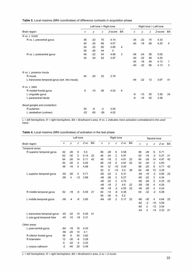
As Figure 3C shows (cf. Table 3), several significant differences were found when comparing the
activation of left-response + effect tone integration and right-response + effect tone integration contrasts.
S1M1 was observed to be more active at the contralateral side of the hand used, whereas the cerebellum
shows an ipsilateral mapping. The SMA shows a contralateral pattern too. Most intriguingly, the activity in
the posterior insula was also found to depend on which hand was used. Although this method is not
adequate to resolve this point, visual inspection (cf. Figure 3) seems to suggest that the found right
posterior insular region is more or less only active in the trials where a left-hand response was given, while
this holds vice versa for the left posterior insular region.
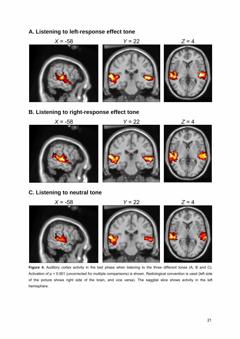
3.2.3 Test phase
In the test phase, participants listened to a stream of tones including left-response effect tones, right-
responses effect tones and neutral tones. Figure 4 shows that the presentation of these tones always
resulted in a significant activation of the auditory cortex (cf. Table 4). However, we could not find evidence
for activation in motor-related areas in the brain. No significant differences were found when contrasting
these conditions with each other.
Table 1. Local maxima (MNI coordinates) of activation during the listening phase
Brain region x y z Z-score BA
Temporal areas R superior temporal gyrus 64 -20 6 5.03 62 -14 -2 4.95 66 -28 8 4.83 64 -26 2 4.82 68 -10 -2 4.64 L superior temporal gyrus -60 -22 4 5.11 -62 -18 4 5.05 -64 -30 8 4.8 42 -58 -32 8 4.5 R middle temporal gyrus 62 -6 -6 4.83 L middle temporal gyrus -62 -10 -4 4.88 -56 -34 4 4.46 22 Other areas R midbrain 12 -26 -12 3.49 L = left hemisphere, R = right hemisphere, BA = Brodmann’s area

18
A. Left-response + effect tone integration Z = -26 Z = 16 Z = 50 Y = 22
B. Right-response + effect tone integration
Z = -26 Z = 16 Z = 50 Y = 22
C. Difference between Left- and Right-response + effect tone Z = -20 Z = 16 Z = 52 Y = 34
Figure 3. Activation during the acquisition phase for activation of left (A) and right (B) response + following
action-effect tone compared to baseline. C shows the differences between contrasts A and B (red and blue colors
indicate positive respectively negative activation). In the first column, cerebellum activity is shown. The second
column shows auditory and insular cortex activation. S1M1 and SMA activity are shown in the third column.
Coronal slides show active S1M1, S2, auditory cortex, parahippocampal cortex and insulae. Activations of p <
.001 (uncorrected for multiple comparisons) are shown. Images use radiological convention (left side of the
picture is right side of the brain and vice versa).

19
Table 2. Local maxima (MNI coordinates) of left and right-response tone during the acquisition phase
Activation Left response + tone Activation Right response + tone
Brain region x y z Z-score BA x y z Z-score BA
Temporal areas R superior temporal gyrus 66 -16 8 5.51 64 -26 10 5.12 66 -24 6 5.17 62 -22 4 4.99 64 -20 4 5.16 48 -22 6 4.96 64 -8 4 5.01 22 66 -26 14 4.88 42 60 -10 4 4.86 L superior temporal gyrus -58 -22 8 5.62 -54 -26 8 5.12 -66 -28 12 5.3 42 -50 -18 4 4.98 -56 -14 2 4.84 -52 -16 4 4.8 -50 -20 2 4.69 R transverse temporal gyrus 50 -22 10 5.09 L transverse temporal gyrus -58 -22 12 5.61 41 R middle temporal gyrus 60 -22 -6 4.8 R parahippocampal gyrus* 14 -32 -6 3.92 * 14 -32 -6 3.71 * L parahippocampal gyrus -16 -32 -8 3.74 -2 -40 0 3.74 -14 -30 -4 3.7 Motor-related areas R postcentral gyrus 42 -24 54 4.92 3 58 -22 50 3.96 2 44 -28 52 3.57 58 -12 50 3.21 L postcentral gyrus -52 -20 50 5.04 R medial frontal gyrus 4 -12 56 4.53 6 L paracentral lobule -8 -20 46 4.11 -2 -26 52 4.11 R putamen 30 -6 -2 3.31 12 2 6 3.53 * 30 -10 4 3.31 16 16 -4 3.32 * 16 14 -8 3.27 L medial globus pallidus -10 2 2 3.86 -10 -4 0 3.44 * R basal ganglia 12 -6 4 3.5 * L caudate head -4 0 2 3.62 * R (anterior) cingulate gyrus 4 -4 58 4.48 6 6 12 36 4.26 24 6 12 36 4.23 24 4 6 42 4.11 32 L cingulate gyrus 0 -8 46 4.26 32 -2 -8 48 4.52 -4 16 34 4.08 -6 16 32 4.15 24 -2 6 44 4.09 32 R cerebellum 42 -60 -28 3.94 4 -80 -18 3.86 36 -64 -26 3.77 12 -86 -24 4.15 48 -68 -26 3.67 52 -66 -26 3.64 56 -62 -22 3.63 * 30 -86 -24 3.58 * 34 -90 -24 3.21 * L cerebellum 0 -78 -18 4.14 * -34 -78 -24 3.74 -24 -78 -24 3.72 -28 -70 -24 3.61 -24 -62 -24 3.5 -28 -52 -26 3.38 -36 -64 -24 3.28

20
Table 2 (continued). Local maxima (MNI coord.) of left and right-response tone during the acquisition phase
Activation Left response + tone Activation Right response + tone
Brain region x y z Z-score BA x y z Z-score BA
Parietal areas R precuneus 4 -74 34 3.74 7 4 -74 38 3.52 L precuneus -2 -76 40 3.96 7 -4 -70 36 3.77 -4 -72 36 3.95 7 0 -70 36 3.7 0 -72 36 3.84 -32 -76 36 3.33 -34 -76 36 3.5 -28 -80 36 3.26 R inferior parietal lobe -56 -46 22 4.51 40 52 -30 48 3.42 40 -36 -44 44 3.64 -36 -48 50 3.56 40 L postcentral gyrus -54 -22 16 5.06 Frontal areas R middle frontal gyrus 40 44 24 3.48 54 4 46 4.14 6 38 48 24 3.47 54 4 40 4.13 9 60 0 44 3.7 54 16 42 3.51 54 16 36 3.39 36 40 24 3.37 10 L middle frontal gyrus -34 38 34 3.58 -36 36 30 3.5 -38 42 24 3.49 -44 38 30 3.42 -40 40 24 3.34 10 -36 44 18 3.24 -40 44 16 3.24 R inferior frontal gyrus 54 34 -2 3.5 47 54 32 -4 3.53 L inferior frontal gyrus -46 42 16 3.35 46 -40 38 12 3.4 R superior frontal gyrus 32 48 26 3.22 R precentral gyrus 60 0 40 3.52 Other areas R thalamus (medial dorsal nucleus) 6 -20 4 4.13 6 -18 6 3.72 L thalamus -16 -30 2 3.78 -10 -10 2 3.37 R lingual gyrus 8 -88 -22 4.23 18 2 -86 -6 4.12 18 2 -86 -8 4.14 18 6 -90 -18 3.97 18 L lingual gyrus -2 -74 -14 4.04 * -10 -98 -6 3.85 17 R cuneus 4 -78 32 3.73 4 -80 32 3.57 19 2 -78 20 3.3 L cuneus -4 -76 32 3.67 7 R precuneus 2 -68 28 3.57 31 R inferior occipital gyrus 24 -96 -16 3.9 L superior occipital gyrus -30 -82 32 3.17 L = left hemisphere, R = right hemisphere, BA = Brodmann’s area
21
A. Listening to left-response effect tone X = -58 Y = 22 Z = 4
B. Listening to right-response effect tone X = -58 Y = 22 Z = 4
C. Listening to neutral tone X = -58 Y = 22 Z = 4
Figure 4. Auditory cortex activity in the test phase when listening to the three different tones (A, B and C).
Activation of p < 0.001 (uncorrected for multiple comparisons) is shown. Radiological convention is used (left side
of the picture shows right side of the brain, and vice versa). The saggital slice shows activity in the left
hemisphere.
22
Table 3. Local maxima (MNI coordinates) of difference contrasts in acquisition phase
Left tone > Right tone Right tone > Left tone
Brain region x y z Z-score BA x y z Z-score BA
R vs. L S1M1 R vs. L precentral gyrus 36 -22 70 4.14 -34 -22 70 4.33 40 -20 66 4.07 -40 -18 58 4.25 4 34 -32 60 3.85 4 36 -26 44 4 R vs. L postcentral gyrus 46 -22 54 4.68 3 -46 -24 58 5.05 34 -34 52 3.97 -40 -32 48 4.25 -54 -18 48 4.13 1 -40 -32 66 4.13 3 R vs. L posterior insula R insula 44 -20 22 3.79 L transverse temporal gyrus (ext. into insula) -44 -22 12 3.67 41 R vs. L SMA R medial frontal gyrus 6 -10 56 4.03 6 L cingulate gyrus -8 -10 50 3.39 24 L paracentral lobule -6 -16 50 3.38 Basal ganglia and cerebellum R putamen 30 -8 -2 3.54 L cerebellum (culmen) -22 -50 -26 4.02 L = left hemisphere, R = right hemisphere, BA = Brodmann’s area. R vs. L indicates more activation contralateral to the used hand.
Table 4. Local maxima (MNI coordinates) of activation in the test phase
Left tone Right tone Neutral tone
Brain region x y z Z-sc BA x y z Z-sc BA x y z Z-sc BA
Temporal areas R superior temporal gyrus 62 -26 8 5.5 66 -28 6 5.58 66 -28 6 5.71 62 -30 2 5.14 22 46 -24 2 4.97 62 -18 -2 5.27 21 64 -26 14 5.11 42 48 -18 2 4.91 22 64 -28 14 4.97 42 50 -30 4 5.05 68 -18 6 4.87 42 52 -30 2 4.83 48 -16 0 4.68 54 12 -10 3.45 68 -20 6 4.71 42 52 6 -14 3.4 38 64 -46 10 3.29 L superior temporal gyrus -50 -26 4 5.71 -60 -22 2 5.31 -46 -18 -2 4.65 22 -56 4 -12 3.88 -48 -26 2 5.27 -60 -22 2 4.34 -46 -22 2 4.75 -60 -26 0 4.33 22 -48 -18 2 4.6 22 -58 -38 4 4.26 -46 -18 -2 4.55 22 -46 -28 4 4.24 R middle temporal gyrus 62 -18 -6 5.05 21 64 -14 -6 5.38 64 -32 -2 5.08 56 -30 -4 5.2 L middle temporal gyrus -58 -4 -8 3.85 -64 -30 2 5.17 22 -66 -30 4 4.64 22 -62 -2 -10 3.59 -60 2 -12 3.54 -54 4 -14 3.33 21 L transverse temporal gyrus -40 -32 10 5.05 41 L sub-gyral temporal lobe -40 -10 -18 3.31 Other areas L post-central gyrus -64 -18 16 4.43 -56 -20 14 4.1 R inferior frontal gyrus 36 8 30 3.62 R brainstem 12 -30 -8 3.28 6 -32 -4 3.23 L corpus callosum -2 -40 20 3.49 L = left hemisphere, R = right hemisphere, BA = Brodmann’s area, Z-sc = Z-score.

23
4 Discussion The purpose of our study was to investigate the neural processes underlying action-effect integration. In the
acquisition phase, subjects first performed a free-choice task. Contingent on their left or right responses an
auditory stimulus was presented as an effect of that action. In the following test phase, subjects had to
listen to these two left and right response effect tones and the neutral tone which was not presented earlier
in the context of an action. Auditory stimulus presentation was always found to result in a bilateral activity of
both primary and secondary auditory cortex, which indicates that the auditory stimuli were processed
despite the noisy background of EPI sequences in our MRI environment. We could not find evidence for the
predicted increase in PM (premotor) activity for the action-effect tones in the test-phase. The neuroimaging
data from the acquisition phase, however, provides significant insights. It suggests that especially the
posterior insular cortex is important for integrating one’s actions with its auditory effects. The following
sections elaborate on the findings of the acquisition phase and test phase and their implications.
4.1 Neural structures underlying action-effect integration
As expected, the generation of a voluntary key press was associated with the activation of brain areas
known to be involved in motor tasks, including lateral PM cortex, SMA, S1M1, cerebellum and basal
ganglia. As usually found, the BOLD (blood oxygen level-dependent) response in S1M1 was greater during
contralateral response, whereas cerebellar activation shows an ipsilateral pattern (see Figure 3C). Further,
the SMA shows activation greater for the contralateral hand too, a finding reported earlier by Beltramelio
and colleagues (1998). Frontal areas were also active during the acquisition phase trials, probably
indicating the involvement of some control processes in this task.
The bilateral activation of the parahippocampal cortex probably indicates the involvement of
memory encoding processes during the binding of actions and their effects. This result is in line with earlier
studies that have found hippocampus being activated during implicit associative learning tasks (e.g.
Degonda et al., 2005). Admittedly, we have only found the parahippocampal cortex to be active, rather than
the hippocampus proper. Since the parahippocampal area provides most of the input to the hippocampus
proper, this area usually shows a larger BOLD response (cf. Brewer & Moghekar, 2002). This probably is
the reason why this region is dominating our statistical map
Our most intriguing observation regards the contralateral activation of the posterior insular cortex
during the acquisition of the left- and right-response effect tone contingencies. We suggest that this
posterior insular region is involved in the process of integrating actions with their auditory effects. Earlier
research has attributed several functions to the insular region. Some studies have suggested, for example,
that the insulae, including the secondary somatosensory cortices at its lateral boundaries, are part of a
somesthetic network that is active in both normal and painful finger stimulation (Deuchert et al., 2002;
Farrell, Laird, & Egan, 2005) as well as in passive finger movement (Blakemore, Oakley, & Frith, 2003).
Other researchers have proposed that the insula is part of a brain network that underlies a body schema
serving corporal awareness (Chaminade, Meltzoff, & Decety, 2005; Berlucchi & Aglioti, 1997). The insular
cortex has been found to be involved in the processing of auditory information, too (Bamiou, Musiek, &
Luxon, 2003). Recent imaging studies also suggest that this region integrates information from multiple
senses (Calvert, 2001; Hadjikhani, & Roland, 1998). Crucially, studies on post-stroke recovery of motor
function in patients (cf. Augustine, 1996) as well as stimulation studies in animals (Showers & Lauer, 1961;
Sugar, Chusid, & French, 1948) have shown that the insular cortex may also function as an additional motor

24
part of the brain. That this region can become active even without a reafferent signal was recently
demonstrated in a study that reveals insular cortex to be active whilst hemiplegic patients attempt to lift their
feet (Nowak, Holm, Biering-Sorensen, Secher, & Friberg, 2005; cf. Williamson, Fadel, & Mitchell, 2006).
Previous studies thus have attributed both sensory and motor functions to the insular cortex.
Anatomical evidence is also consistent with the idea that the insular cortex is a multi-faced sensory and
motor area (Augustine, 1996). The insula not only shares reciprocal connections with several parietal and
temporal areas, but also receives a number of fibers from the PM cortex, SMA and primary and secondary
somatosensory areas (Augustine, 1996). Even direct efferent fibers to the spinal cord have been found for
this region (Keizer & Kuypers, 1989). Taken together, these findings suggest that the insular cortex may
serve as a convergence zone of both sensory and motor information (Jantzen, Steinberg, & Kelsoa, 2005).
This characteristic makes the insula an excellent candidate for a neural site that mediates the integration of
actions and their perceived effects.
Consistent with this idea, Farrer and colleagues (Farrer & Frith, 2002; Farrer, Franck, Georfieff,
Frith, Decety, & Jeannerod, 2003) recently have found that the sense of agency is depended on activity of
the insular cortex. In their PET study (Farrer et al., 2003), subjects had to make movements with a joystick
whilst they receive feedback about these movements on a screen showing a virtual hand holding a joystick.
Subject’s degree of control of the movements on the screen was manipulated through varying the amount
of discrepancy between the actual joystick movement and the feedback on the screen. The activity of the
right posterior insular cortex was found to co-vary with the amount of concordance between what the
subject did and what they saw. Given that the insula integrates sensory information of multiple senses, the
authors concluded that the insular cortex might mediate the integration of the many sensory signals, both
proximal and distal, that are associated with an action (Farrer & Frith, 2002; Farrer et al., 2003).
Given these findings, our observation of contralateral insular activation for left and right hand
actions seems nicely in concord with an interpretation that treats the insular cortex as a brain region where
actions and their effects become integrated together. As discussed, the ideo-motor principle assumes that
action-effect learning is needed in order to become a goal-directed actor. Surprisingly, a lesion mapping
study of 27 stroke patients with and without anasognosia symptoms has recently revealed that damage to
exactly the (right) posterior insular cortex is associated with a disturbed sense of agency (Karnath, Baier, &
Nagele, 2005). Because prior research also has shown that the insula is involved in auditory processing, we
tentatively conclude that not only proximal somatosensory but also the distal auditory action effects in our
study have become integrated with the action via the insulae. Our results suggest that the insular region
contralateral to the side of the used effector is primarily involved in this process. An alternative interpretation
of insular activity merely reflecting motor-related activity cannot be ruled out given the fact that our study did
not use a baseline condition of trials including only responses and no subsequent auditory stimuli. This
interpretation, however, is rather unlikely because the insula is not an area that is typically activated during
motor tasks in brain imaging experiments (Karnath et al., 2005).
4.2 Activity in the test phase
Listening to the effect tones, presented earlier with or without the context of an action, leads to auditory
cortex activation. We could however not find evidence for SMA or PM activation triggered by the action-
effect tones. This failure to replicate the finding of Elsner and colleagues (2002) could be due to the
neuroimaging technique we adopted in this study. First, in contrast to a PET environment, the background

25
noise of the MRI scanner may have overshadowed the processing of the auditory stimuli probably leading
to less salient differences in processing the three tones. Furthermore, because we used an event-related
design rather than a (parametric) block design, current imaging technique probably was not powerful
enough to find the expected effect in neural structures underlying action planning (cf. Liu, Frank, Wong, &
Buxton, 2001). Another difference with the Elsner et al. study is that our event-related design did not allow
rapid stimulus presentation in the test phase. Moreover, only one acquisition session preceding the test
phase was used in our study, whereas the earlier study used extra training blocks in between the test
sessions. We suppose that a future fMRI study using an adapted and more powerful design should be able
to find differential PM activity induced by action-effect tones. Moreover, given the strong effects found in
behavioral studies, using a test phase that allows responses to action effects may even result in much
stronger PM activation differences.
4.3 Conclusion
Although the chosen experimental design did not allow us to find premotor activity triggered by the
presentation of action-effect tones, the acquisition phase data of our fMRI study provides new insights.
Besides motor and auditory cortex activity, we found bilateral parahippocampal cortex and contralateral
insular cortex being involved in this action-effect integration process. This finding not only supports the
assumption that action-effect learning involves memory processes as has been found in other imaging
studies using implicit associative learning tasks, but also shows that dedicated areas of the brain seems to
be involved in integrating perception and action. Our study is the first that suggests that the contralateral
posterior insular cortex may play a key role in the integration of ones actions and their auditory
consequences.

26
References Alonso, D., Fuentes, L.J., & Hommel, B. (2006). Unconscious symmetrical inferences: a role of
consciousness in event integration. Consciousness and Cognition,15, 386-396.
Augustine, J.R. (1996). Circuitry and functional aspects of the insular lobe in primates including humans.
Brain Research Reviews, 22, 229-244.
Bamiou, D.-E., Musiek, F.E., & Luxon, L.M. (2003). The insula (Island of Reil) and its role in auditory
processing: literature review. Brain Research Reviews, 42, 143-154.
Bangert, M., & Altenmüller, E.O. (2003). Mapping perception to action in piano practice: a longitudinal DC-
EEG study. BMC Neuroscience, 4, 26.
Bangert, M., Peschel, T., Schlaug, G., Rotte, M., Drescher, D., Hinrichs, H., Heinze, H., & Altenmüller, E.
(2006). Shared networks for auditory and motor processing in professional pianists: evidence from
fMRI conjunction. NeuroImage, 30, 917-926.
Beckers, T., De Houwer, J., & Eelen, P. (2002). Automatic integration of non-perceptual action effect
features: the case of the associative affective Simon effect. Psychological Research, 66, 166-173.
Beltramelio, A., Cerini, R., Puppini, G., EI-Dalati, G., Viola, S., Martone, E., Cordopatri, D., Manfredi, M.,
Aglioti, S., & Tassinari, G. (1998). Motor representation of the hand in the human cortex: an f-MRI
study with a conventional 1.5 T clinical unit. Italian Journal of Neurological Sciences, 19, 277-284.
Berlucchi, G., & Aglioti, S. (1997). The body in the brain: neural bases of corporeal awareness. Trends in
Neurosciences, 20, 560-564.
Blakemore, S.J., Oakley, D.A., & Frith, C.D. (2003). Delusions of alien control in the normal brain.
Neuropsychologia, 41, 1058-1067.
Brewer, J.B., & Moghekar, A. (2002). Imaging the medial temporal lobe: exploring new dimensions. Trends
in Cognitive Sciences, 6, 217-223.
Brogden, W. J. (1962). Contiguous conditioning. Journal of Experimental Psychology, 64, 172-76.
Calvert, G.A. (2001). Crossmodal processing in the human brain: insights from functional neuroimaging
studies. Cerebral Cortex, 11, 1110-1123.
Calvo-Merino, B., Glaser, D.E., Grèzes, J., Passingham, R.E., & Haggard, P. (2005). Action observation
and acquired motor skills: an fMRI study with expert dancers. Cerebral Cortex, 15, 1243-1249.
Carpenter, W. B. (1852). On the influence of suggestion in modifying and directing muscular movement,
independently of volition. Proceedings of the Royal Institution, 147–154.
Chaminade, T., Meltzoff, A.N., & Decety, J. (2005). An fMRI study of imitation: action representation and
body schema. Neuropsychologia, 43, 115-127.
Dale, A.M. (1999). Optimal experimental design for event-related fMRI. Human Brain Mapping, 8, 109-114.
Degonda, N., Mondadori, C.R.A., Bosshardt, S., Schmidt, C.F., Boesiger, P., Nitsch, R.M., Hock, C., &
Henke, K. (2005). Implicit associative learning engages the hippocampus and interacts with explicit
associative learning. Neuron, 46, 505-520.
Deuchert, M., Ruben, J., Schwiemann, J., Meyer, R., Thees, S., Krause, T., Blankenburg, F., Villringer, K.,
Kurth, R., Curio, G., & Villringer, A. (2002). Event-related fMRI of the somatosensory system using
electrical finger stimulation. NeuroReport, 13, 365-369.
Di Pellegrino G., Fadiga L., Fogassi V., Gallese V., & Rizzolatti G. (1992). Understanding motor events: a
neurophysiological study. Experimental Brain Research, 91, 176-180.

27
Drost, U.C., Rieger, M., Brass, M., Gunter, T.C., & Prinz, W. (2005). Action-effect coupling in pianists.
Psychological Research, 69, 233–241.
Dutzi, I.B. & Hommel, B. (2006). Spontaneous but goal-dependent binding of actions and their effects.
Manuscript submitted for publication.
Eenshuistra, R.M., Weidema, M.A., & Hommel, B. (2004). Development of the acquisition and control of
action–effect associations. Acta Psychologica, 115, 185-209.
Elsner, B. (2007). Infants’ imitation of goal-directed actions: the role of movements and action effects. Acta
Psychologica, 124, 44-59
Elsner, B. & Aschersleben, G. (2003). Do I get what you get? Learning about the effects of self-performed
and observed actions in infancy. Consciousness & Cognition, 12, 732-751.
Elsner, B. & Hommel, B. (2001). Effect anticipation and action control. Journal of Experimental Psychology:
Human Perception and Performance, 27, 229-240.
Elsner, B., & Hommel, B. (2004). Contiguity and contingency in action-effect learning. Psychological
Research, 68, 138-154.
Elsner, B., Hommel, B., Mentschel, C., Drzezga, A., Prinz, W., Conrad, B., & Siebner. H. (2002). Linking
actions and their perceivable consequences in the human brain. NeuroImage, 17, 364-372.
Farrell, M.J., Laird, A.R., & Egan, G.F. (2005). Brain activity associated with painfully hot stimuli applied to
the upper limb: a meta-analysis. Human Brain Mapping, 25, 129-139.
Farrer, C., & Franck, N., Georgieff, N., Frith, C.D., Decety, J., Jeannerod, M. (2003). Modulating the
experience of agency: a positron emission tomography study. NeuroImage, 18, 324-333.
Farrer, C., & Frith, C.D. (2002) Experiencing oneself vs another person as being the cause of an action: the
neural correlates of the experience of agency. NeuroImage, 15, 596-603.
Gallese, V., & Lakoff, G. (2005). The brain’s concepts: The role of the sensory-motor system in conceptual
knowledge. Cognitive Neuropsychology, 22, 455-479.
Gallese, V., Murata, A., Kaseda, M., Niki, N., & Sakata, H. (1994). Deficit of hand preshaping after
muscimol injection in monkey parietal cortex. NeuroReport, 5, 1525–1529.
Gazzola, V., Aziz-Zadeh, L., & Keysers, C. (2006). Empathy and the somatotopic auditory mirror system in
humans. Current Biology, 16, 1824-1829.
Greenwald, A.G. (1970). Sensory feedback mechanisms in performance control: with special reference to
the ideo-motor mechanism. Psychological Review, 77, 73–99.
Grèzes, J., Armony, J.L., Rowe, J., & Passingham, E. (2003). Activations related to “mirror” and “canonical”
neurones in the human brain: an fMRI study. NeuroImage, 18, 928-937.
Grèzes, J., & Decety, J. (2001). Functional anatomy of execution, mental simulation, observation, and verb
generation of actions: a meta-analysis. Human Brain Mapping, 12, 1-19.
Hadjikhani, N., & Roland, P.E. (1998). Cross-modal transfer of information between the tactile and the
visual representations in the human brain: a positron emission tomographic study. The Journal of
Neuroscience, 18, 1072-1084.
Hauf, P., Elsner, B., & Aschersleben, G. (2004). The role of action effects in infants’ action control.
Psychological Research, 68, 115-125.
Hebb, D.O. (1949). The Organization of Behavior. New York: Wiley.
Herbart, J. F. (1825). Psychologie als Wissenschaft neu gegründet auf Erfahrung, Metaphysik und
Mathematik. Zweiter, analytischer Teil. Königsberg, Germany: Unzer.

28
Hommel, B. (1993). Inverting the Simon effect by intention: determinants of direction and extent of effects of
irrelevant spatial information. Psychological Research, 55, 270-279.
Hommel, B. (1996). The cognitive representation of action: automatic integration of perceived action effects.
Psychological Research, 59, 176-186.
Hommel, B. (1998). Event files: evidence for automatic integration of stimulus-response episodes. Visual
Cognition, 5, 183-216.
Hommel, B. (2004). Event files: feature binding in and across perception and action. Trends in Cognitive
Sciences, 8, 494-500.
Hommel, B., Alonso, D., & Fuentes, L.J. (2003). Acquisition and generalization of action effects. Visual
Cognition, 10, 965-986.
Hommel, B., Müsseler, J., Aschersleben, G., & Prinz, W. (2001). The Theory of Event Coding (TEC): a
framework for perception and action planning. Behavioral and Brain Sciences, 24, 849-937.
Jackson, Ph.L., & Decety, J. (2004). Motor cognition: a new paradigm to study self–other interactions.
Current Opinion in Neurobiology, 14, 259-263.
James, W. (1890). The principles of psychology, Vols. I and II. Cambridge, MA: Harvard University Press.
Jantzen, K.J., Steinberg, F.L., & Kelsoa, J.A.S. (2005). Functional MRI reveals the existence of modality
and coordination-dependent timing networks. NeuroImage, 25, 1031-1042.
Jenkinson, M., Bannister, P.R., Brady, M., & Smith, S.M. (2002). Improved optimization for the robust and
accurate linear registration and motion correction of brain images. NeuroImage, 17, 825-841.
Kahneman, D., Treisman, A.., & Gibbs, B.J. (1992). The reviewing of object files: object-specific integration
of information. Cognitive Psychology, 24, 175-219.
Karnath, H.-O., Baier, B., & Nagele, T. (2005). Awareness of the functioning of one’s own limbs mediated
by the insular cortex? The Journal of Neuroscience, 25, 7134-7138.
Keizer, K., & Kuypers, H.G.J.M. (1989). Distribution of corticospinal neurons with collaterals to the lower
brain stem reticular formation in monkey (Macaca fascicularis). Experimental Brain Research, 74,
311-318.
Kohler, E., Keysers, C., Umilta, M.A., Fogassi, L., Gallese, V., & Rizzolatti, G. (2002). Hearing sounds,
understanding actions: Action representation in mirror neurons. Science, 297, 846-848.
Kray, J., Eenshuistra, R., Kerstner, H., Weidema, M., & Hommel, B. (2006). Language and action control:
the acquisition of action goals in early childhood. Psychological Science, 17, 737-741.
Kunde, W. (2004). Response priming by supraliminal and subliminal action effects. Psychological
Research, 68, 91-96.
Kunde, W., Koch, I., & Hoffmann, J. (2004). Anticipated action effects affect the selection, initiation, and
execution of actions. The Quarterly Journal of Experimental Psychology, 57A, 87-106.
Kray, J., Eenshuistra, R.M., Kerstner, H., Weidema, M.A., Hommel, B. (2006). Language and action control:
the acquisition of action goals in early childhood. Psychological Science, 17, 737-741.
Lahav, A., Saltzman, E., & Schlaug, G. (2007). Action representation of sound: audiomotor recognition
network while listening to newly acquired actions. The Journal of Neuroscience, 27, 308-314.
Laycock, T. (1876). Reflex, automatic, and unconscious cerebration: a history and a criticism. The Journal
of Mental Science, 21, 477-498.
Liu, T.T., Frank, L.R., Wong, E.C., Buxton, R.B. (2001). Detection power, estimation efficiency, and
predictability in event-related fMRI. NeuroImage, 13, 759-773.
29
Lotze, M., Scheler, G., Tan, H.R., Braun, C., Birbaumer, N. (2003). The musician’s brain: functional imaging
of amateurs and professionals during performance and imagery. NeuroImage, 20, 1817-1829.
Maes, J.H.R. (2006). Response bias induced in rats by response effects. The Quarterly Journal of
Experimental Psychology, 59, 1346-1356.
Meck, W. H. (1985). Postreinforcement signal processing. Journal of Experimental Psychology: Animal
Behavior Processes, 11, 52-70.
Meister, I.G., Krings, T., Foltys, H., Boroojerdi, B., Muller, M., Topper, R., & Thron, A., 2004. Playing piano
in the mind: An fMRI study on music imagery and performance in pianists. Cognitive Brain Research,
19, 219-228.
Nowak, M., Holm, S., Biering-Sorensen, F., Secher, N.H., & Friberg, L. (2005). “Central command” and
insular activation during attempted foot lifting in paraplegic humans. Human Brain Mapping, 25, 259-
265.
Prinz, W. (1990). A common coding approach to perception and action. In: O. Neumann & W. Prinz (ed.),
Relationships between perception and action: Current approaches. Springer.
Rescorla, R.A. (1967). Pavlovian conditioning and its proper control procedures. Psychological Review, 74,
71-80.
Rizzolatti, G., & Craighero, L. (2004). The mirror-neuron system. Annual Review of Neuroscience, 27, 169-
192.
Roelfsema, P.R., Engel, A.K., König, P., & Singer, W. (1997). Visuomotor integration is associated with zero
time-lag synchronization among cortical areas. Nature, 385, 157-161.
Schubotz, R.I., & Von Cramon, D.Y. (2003). Functional–anatomical concepts of human premotor cortex:
evidence from fMRI and PET studies. Neuroimage, 20, 120-131.
Showers, M.J., & Lauer, E.W. (1961). Somatovisceral motor patterns in the insula. J Comp Neurol, 117,
107-115.
Smith, S.M. (2002). Fast robust automated brain extraction. Human Brain Mapping, 17, 143-155.
Smith, S.M., Jenkinson, M., Woolrich, M.W., Beckmann, C.F., Behrens, T.E., Johansen-Berg, H., Bannister,
P.R., De Luca, M., Drobnjak, I., Flitney, D.E., Niazy, R.K., Saunders, J., Vickers, J., Zhang, Y., De
Stefano, N., Brady, J.M., & Matthews, P.M. (2004). Advances in functional and structural MR image
analysis and implementation as FSL. NeuroImage, 23 (Suppl 1), 825-841.
Stock, A. & Stock, C. (2004). A short history of ideo-motor action. Psychological Research, 68, 176-188.
Stoet, G., & Hommel, B. (1999). Action planning and the temporal binding of response codes. Journal of
Experimental Psychology: Human Perception and Performance, 25, 1625-1640.
Sugar, O., Chusid, J.G., & French, J.D. (1948). A second motor cortex in the monkey (Macaca mulatta). J
Neuropathol Exp Neurol, 7, 182-189.
Tai, Y.F., Scherfler, C., Brooks, D.J., Sawamoto, N., & Castiello, U. (2004). The human premotor cortex is
‘mirror’ only for biological actions. Current Biology, 14, 117-120.
Trapold, M.A. (1970). Are expectancies based upon different reinforcement events discriminably different?
Learning and Motivation, 1, 129-140.
Urcuioli, P. J., & De Marse, T. (1996). Associative processes in differential outcome discriminations. Journal
of Experimental Psychology: Animal Behavior Processes, 22, 192-204.
Verschoor, S.A. (2006). The Role of Attention and Language in Action-Effect Binding. Unpublished
manuscript.

30
Walker, E.L. (1969). Reinforcement: “the one ring.” In: J. T. Tapp (Ed.), Reinforcement and behavior. New
York: Academic Press.
Williamson, J.W., Fadel, P.J., & Mitchell, J.H. (2006). New insights into central cardiovascular control during
exercise in humans: a central command update. Experimental Physiology, 91, 51-58.