the muscular dystrophies - wesleyan...
TRANSCRIPT

For personal use. Only reproduce with permission from The Lancet Publishing Group.
SEMINAR
THE LANCET • Vol 359 • February 23, 2002 • www.thelancet.com 687
The commonest form of these inherited disorders—Duchenne muscular dystrophy—was originally described byEdward Meryon, an English doctor. At a meeting of theRoyal Medical and Chirurgical Society in 1851,1 and laterpublished in the transactions of the society,2 he described indetail the clinical presentation of this disorder, beginning inearly childhood with progressive muscle wasting andweakness and leading to death in late adolescence. Heshowed that the disease was familial and only affected boys;most importantly, he demonstrated that the spinal cord atnecropsy was normal. Therefore, this was a disease ofmuscle (myogenic) and was not secondary to anterior-horncell degeneration. Furthermore, his detailed histologicalstudies led him to conclude that the muscle membrane orsarcolemma was broken down and destroyed. Thisobservation was singularly important, because we nowknow that the primary defect resides in the sarcolemma.However, Meryon’s observations were neglected for manyyears for various reasons,3 and the disorder becameeponymously associated with Duchenne in Paris, whodetailed the clinical and muscle histology some years later.4,5
Over the following years, investigators gradually realisedthat, although Duchenne muscular dystrophy was by far thecommonest and one of the most serious forms of thedisease, muscular dystrophy was in fact a group of inheriteddisorders, all characterised by variable degrees anddistribution of muscle wasting and weakness.
Clinically defined types of muscular dystrophyOn the basis of distribution of predominant muscleweakness, six major forms can be delineated (figure 1), withthe addition of congenital dystrophy, in which muscleweakness is more generalised (panel 1).
Congenital muscular dystrophyChildren with this heterogeneous group of autosomalrecessively inherited disorders present with hypotonia andweakness at birth or within the first few months of life.Several different forms have been recognised, some withand some without significant mental retardation.6 Twoforms without mental deficiency are caused by an absence
Lancet 2002; 359: 687–95
Peninsula Medical School, Department of Neurology, Royal Devonand Exeter Hospital, Exeter EX2 5DW, UK (Prof A E H Emery FRCP)(e-mail: [email protected])
of merosin (laminin �2, a muscle extracellular protein)7 orvery occasionally integrin �7.8 Deficiency of merosin can beshown by western-blot analysis or muscle immuno-histochemistry, and also with chorionic villus material forprenatal diagnosis.9 Although children with merosin-deficient congenital muscular dystrophy are not mentallyretarded, magnetic resonance imaging of the braininvariably shows white-matter changes. Most affectedchildren might eventually be able to stand with somesupport, but few learn to walk. Muscle weakness is usuallynon-progressive, but many joint contractures develop withimmobility. Although cardiac function is normal, the long-term outlook is not good because many patients developserious feeding and respiratory problems.
The incidence of Fukuyama congenital musculardystrophy in Japan is second only to Duchenne musculardystrophy, but is rare elsewhere. This disorder is namedafter Yukio Fukuyama from Tokyo, who first described thecondition in 1960.10 Onset is in infancy with hypotonia andmuscle weakness. Affected children are rarely able to walk.Most are mentally retarded and many have epilepsy. Theprotein product of the responsible gene has been namedfukutin,11 but its function is not clear. Of the rarer variantsof congenital muscular dystrophy, the responsible genes andtheir gene products have only been identified for rigid spinesyndrome and muscle-eye-brain disease.
Duchenne and Becker muscular dystrophy The clinical features of these X-linked disorders have beendescribed in detail.12 In Duchenne muscular dystrophy,onset is in early childhood, with difficulties in running and,later, climbing stairs. Weakness of the knee and hipextensors results in Gower’s manoeuvre: a child climbs uphis thighs, pushing down on them, to extend the hips andtrunk. Some degree of mental impairment is usual, about20% of affected boys have an IQ of less than 70. Mostpatients have enlarged calves, hence a previous term for thedisorder was pseudohypertrophic muscular dystrophy;however, calf hypertrophy not only is seen in Duchennemuscular dystrophy but also is present in other dystrophies,such as Becker dystrophy. Weakness is mainly proximal and
The muscular dystrophies
Alan E H Emery
Seminar
The muscular dystrophies are inherited myogenic disorders characterised by progressive muscle wasting and weakness ofvariable distribution and severity. They can be subdivided into several groups, including congenital forms, in accordancewith the distribution of predominant muscle weakness: Duchenne and Becker; Emery-Dreifuss; distal; facioscapulo-humeral; oculopharyngeal; and limb-girdle which is the most heterogeneous group. In several dystrophies the heart can beseriously affected, sometimes in the absence of clinically significant weakness. The genes and their protein products thatcause most of these disorders have now been identified. This information is essential to establish an accurate diagnosisand for reliable genetic counselling and prenatal diagnosis. There is, as yet, no way of greatly affecting the long-termcourse of any of these diseases. However, advances in gene manipulation and stem-cell therapy suggest cautiousoptimism for finding an effective treatment in the not-too-distant future.
Search strategy and selection criteria
The material covered in this review was obtained entirely frompersonal knowledge, the reference sources listed, andPubMed literature searches.

For personal use. Only reproduce with permission from The Lancet Publishing Group.
progressive. Ultimately, a wheelchair becomes necessary,in most cases by age 12. Pneumonia compounded bycardiac involvement is the most frequent cause of death,which happens in the late teens or early 20s. However,with more attention to respiratory care and various formsof assisted ventilation, even including tracheostomy, manyaffected individuals now survive into their late 20s andbeyond.13
In Becker muscular dystrophy,14,15 the distribution ofmuscle wasting and weakness is closely similar to that inDuchenne muscular dystrophy, but the course of thedisease is more benign, with age of onset around 12 years;some patients have no symptoms until much later in life.
Loss of ambulation also varies fromadolescence onwards, with deathusually in the fourth or fifth decade. Aproportion of cases, as in Duchennemuscular dystrophy, have some degreeof mental impairment.
In both Duchenne and Beckermuscular dystrophies, about 5–10% offemale carriers show some degree ofmuscle weakness, and frequently haveenlarged calves—so-called manifestingcarriers. Such weakness is oftenasymmetric, and it can develop inchildhood or not become evident untiladult life, and could be slowlyprogressive or remain static. Becauseweakness is essentially proximal,differentiation from limb-girdlemuscular dystrophy is essential forgenetic counselling. Most importantly,female carriers might develop dilatedcardiomyopathy, which can arise evenwithout apparent weakness.16
The Duchenne gene is located atXp21,17,18 which affects the sarcolemmalprotein dystrophin,19 and is allelic withBecker muscular dystrophy. Dystrophinis usually absent in patients withDuchenne muscular dystrophy, but isreduced in amount or abnormal in sizein people with Becker musculardystrophy.20 However, in rare cases ofDuchenne muscular dystrophy,dystrophin can be detected, or isoccasionally undetectable in mild casesof this disorder.21
Emery-Dreifuss muscular dystrophy This disorder is characterised by atriad22,23 of manifestations. First, thereare early contractures—before there isany clinically significant weakness—ofthe Achilles tendons, elbows, andposterior cervical muscles, with initiallimitation of neck flexion; later, forwardflexion of the entire spine becomesrestricted. Second, slowly progressivemuscle wasting and weakness with ahumeroperoneal distribution—ie, prox-imal in the upper limbs and distal in thelower limbs—happens early in thecourse of the disease. Later, theproximal limb-girdle musculaturebecomes affected. Third, cardio-myopathy arises, which usually presentsas cardiac conduction defects, ranging
from sinus bradycardia, prolongation of PR interval, tocomplete heart block. Atrial paralysis with absent P waveson electrocardiography should always prompt exclusion ofthis muscular dystrophy. Cardiac involvement generallybecomes evident as muscle weakness progresses. Evidenceof cardiac disease is usually present by age 30 years.
The X-linked form of Emery-Dreifuss musculardystrophy is caused by mutations of the STA gene atXq28, which encodes the nuclear membrane proteinemerin.24 In almost all cases of this dystrophy, there iscomplete absence of emerin in muscle. Immuno-histochemistry of skin fibroblasts or exfoliative buccal cellsshows a mosaic pattern of expression of this protein in
SEMINAR
688 THE LANCET • Vol 359 • February 23, 2002 • www.thelancet.com
A B C
D E F
Figure 1: Distribution of predominant muscle weakness in different types of dystrophyA, Duchenne-type and Becker-type; B, Emery-Dreifuss; C, limb-girdle; D, facioscapulohumeral; E, distal, F, oculopharyngeal. Shaded=affected areas. (Reproduced from BMJ 1998; 317: 991–95by permission of the BMJ Publishing Group).

For personal use. Only reproduce with permission from The Lancet Publishing Group.
female carriers, and thus provides a valuable test foridentification of such individuals.
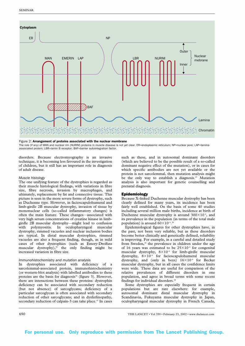
The autosomal dominant type of this disorder is clinicallyvery similar to the X-linked forms, but is caused bymutations of the LMNA gene at 1q21. This gene encodeslamins A and C,25 which make up part of the nuclearlamina—a fibrous layer on the nucleoplasmic side of theinner nuclear membrane. These lamins interact with chro-matin and other proteins of the inner nuclear membrane(lamina-associated proteins) and emerin (figure 2).Diagnosis of autosomal dominant Emery-Dreifuss musculardystrophy can only be verified by mutation analysis, and notby muscle protein studies. There is a rarer more seriousautosomal recessive form of this dystrophy, which is alsocaused by mutations of the LMNA gene.26
Emery-Dreifuss muscular dystrophy with serious cardiacmanifestations can arise in the absence of any muscleweakness.27 For this reason, the disorder could contribute toone of the causes of sudden death in apparently healthyyoung adults.
Distal muscular dystrophyIn this muscular dystrophy, weakness is mainly distal. Thedisorder can be divided into two main groups: late onset(over 40 years of age) with autosomal dominant inheritance,
including Welander’s disease; and early onset (less than 30years of age) with autosomal recessive inheritance.However, apart from one recessive form (Miyoshi type),which is associated with deficiency of the sarcolemmal-associated protein dysferlin, the underlying cause of thesedystrophies is unknown.28 Since variations in fibre size andrimmed vacuoles—rather than dystrophic changes—areseen on muscle histology, these disorders might better beregarded as myopathies rather than dystrophies.
Facioscapulohumeral muscular dystrophy This dystrophy derives its name from the muscle groupsthat are mainly affected first: facial and shoulder girdle.Later, foot extensors and pelvic-girdle muscles becomeinvolved. The heart is not implicated in most cases, thougharrhythmias and conduction defects have been described.29
Mental impairment is not a feature, but retinal vasculardisease and hearing loss can arise. This autosomal dominantdisorder is associated with subtelomeric deletion ofchromosome 4q, with loss of 3·3 kb tandem-repeat units.Loss of ten or fewer repeats causes the disorder, and ingeneral, the lower the number of repeats the more clinicallyserious the disorder. However the function of the particulargene (or genes) that causes the disorder is not clear.
Oculopharyngeal muscular dystrophyThis disorder has mainly been studied in French Canada,where the disease can be traced back to immigrants fromFrance in 1634. However, although most frequent inCanada, the disorder also occurs in other parts of NorthAmerica and Europe. Onset is around the third decade oflife, affecting the extraocular muscles (though frank diplopiais rare), and upper facial muscles with ptosis, and there isweakness of the neck and proximal upper (and even lower)limb musculature. Dysphagia is a serious feature. Afrequent presentation is ptosis and dysphagia.30
The gene locus (at 14q) codes for the poly-(A)-bindingprotein; in the first exon there is usually a (GCG)6
triplet expansion. In affected individuals, this expansion has a further two to seven repeats.31 The resultant expansionhampers normal transport of mRNA from the nucleus.
Limb-girdle muscular dystrophyIn this disorder, weakness affects mainly the proximal limb-girdle musculature. So far, 15 genetically different typeshave been identified, which show great clinical and geneticheterogeneity. Autosomal dominant forms are very rare andgenerally less severe than recessive types; they have beenreviewed in detail elsewhere.32 Several of these disorders areassociated with clinically significant cardiac involvement(types 1B, 1D, 2C, 2E, and 2F), and affected individualsshould therefore be carefully monitored for signs of cardiacdisease.
Laboratory diagnosisSerum creatine kinaseMeasurement of serum concentration of creatine kinase is asimple and inexpensive diagnostic test for severe forms ofdystrophy known to be associated with high concentrations.In Duchenne muscular dystrophy, serum creatine kinasevalues are raised from birth, and testing in neonates for earlydiagnosis could reduce the possibility of further affectedboys in a family.33 There is still a worrying delay in diagnosisof the disorder in early childhood.34
ElectromyographyThis method is important for establishment of themyopathic nature of dystrophy and for exclusion ofneurogenic causes of weakness, including peripheral nerve
SEMINAR
THE LANCET • Vol 359 • February 23, 2002 • www.thelancet.com 689
Panel 1: Gene loci and protein defects in thecommonest forms of muscular dystrophy
Disorder Gene locus Protein defectCongenital (AR) 6q Laminin �2 (merosin)
12q Laminin receptor (�7 integrin)
9q Fukutin (Fukuyama dystrophy)
1p Selenoprotein N1 (rigid spine syndrome)
1p Glycosyltransferase (muscle-eye-braindisease)
Duchenne and Becker (XR) Xp21 DystrophinEmery-Dreifuss (XR) Xq28 Emerin Emery-Dreifuss (AD/AR) 1q Lamin A/CDistal (AD) 14q, 2q ?Distal (AR) 2p DysferlinFacioscapulohumeral (AD) 4q ?Oculopharyngeal (AD) 14q Poly(A)-binding protein
2 (PAB 2)Limb-girdle (AD)
1A 5q Myotilin1B 1q Lamin A/C1C 3p Caveolin 31D 6q ?1E 7q ?1F 2q ?
Limb-girdle (AR)2A 15q Calpain-3 2B 2p Dysferlin2C 13q �-sarcoglycan2D 17q �-sarcoglycan (adhalin)2E 4q �-sarcoglycan2F 5q �-sarcoglycan2G 17q Telethonin2H 9q ?2I 19q Fukutin-related
Modes of inheritance: AR=autosomal recessive; AD=autosomaldominant; XR=X-linked recessive. ?=unknown.
For personal use. Only reproduce with permission from The Lancet Publishing Group.
disorders. Because electromyography is an invasivetechnique, it is becoming less favoured in the investigationof children, but it still has an important role in diagnosisof adult disease.
Muscle histologyThe one unifying feature of the dystrophies is regarded astheir muscle histological findings, with variations in fibresize, fibre necrosis, invasion by macrophages, andultimately, replacement by fat and connective tissue. Thispicture is seen in the more severe forms of dystrophy, suchas Duchenne type. However, in facioscapulohumeral andlimb-girdle 2B muscular dystrophy, invasion of tissue bymononuclear cells (so-called inflammatory changes) isoften the main feature. These changes—associated withvery high serum concentrations of creatine kinase in limb-girdle 2B muscular dystrophy—might lead to confusionwith polymyositis. In oculopharyngeal musculardystrophy, rimmed vacuoles and nuclear inclusion bodiesare typical. In distal muscular dystrophies, rimmedvacuoles are also a frequent finding, though, as in mildcases of other dystrophies (such as Emery-Dreifussmuscular dystrophy),35 the only finding might beincreased variation in fibre size.
Immunohistochemistry and mutation analysisIn dystrophies associated with deficiency of asarcolemmal-associated protein, immunohistochemistry(or western-blot analysis) with labelled antibodies to theseproteins are the basis for diagnosis36 (figure 3). However,there are interactions between these proteins: dystrophindeficiency can be associated with secondary reduction(but not absence) of sarcoglycans; deficiency of aparticular sarcoglycan is often associated with secondaryreduction of other sarcoglycans; and in dysferlinopathy,secondary reduction of calpain-3 can take place.37 In cases
such as these, and in autosomal dominant disorders(which are believed to be the possible result of a so-calleddominant negative effect of the mutation), or in cases forwhich specific antibodies are not yet available or theprotein is not sarcolemmal, then mutation analysis mightbe the only way to establish a diagnosis.37 Mutationanalysis is also important for genetic counselling andprenatal diagnosis.
EpidemiologyBecause X-linked Duchenne muscular dystrophy has beenclearly defined for many years, its incidence has beenfairly well established. On the basis of some 40 studiesincluding several million male births, incidence at birth ofDuchenne muscular dystrophy is around 300�10–6, andits prevalence in the population (in terms of the total malepopulation) is around 60�10–6.38
Epidemiological figures for other dystrophies have, inthe past, not been very reliable, but as these disordersbecome better clinically and genetically defined, reliabilityis improving. For example, in a careful and detailed studyfrom Sweden,39 the prevalence in children under the ageof 16 years was estimated to be 25�10–6 for congenitalmuscular dystrophy, 8�10–6 for limb-girdle musculardystrophy, 8�10–6 for facioscapulohumeral musculardystrophy, and (only in boys) 16�10–6 for Beckermuscular dystrophy, but in all cases the confidence limitswere wide. These data are useful for comparison of therelative prevalences of different disorders in onepopulation, and agree in broad terms with some recentfindings for individual disorders.38
Some dystrophies are especially frequent in certainpopulations but are rare elsewhere: for example,autosomal dominant distal muscular dystrophy inScandinavia, Fukuyama muscular dystrophy in Japan,oculopharyngeal muscular dystrophy in French Canada,
SEMINAR
690 THE LANCET • Vol 359 • February 23, 2002 • www.thelancet.com
Cytoplasm
NP
EMERIN LAPMAN
Lamina
Chromatin
LBR
BAF
Outer
Inner
NURIMNuclearmebrane
ER
Figure 2: Arrangement of proteins associated with the nuclear membraneThe role (if any) of MAN and nuclear rim (NURIM) proteins in muscle disease is not yet clear. ER=endoplasmic reticulum; NP=nuclear pore; LAP=lamina-associated protein; LBR=lamin B receptor; BAF=barrier autointegration factor.

For personal use. Only reproduce with permission from The Lancet Publishing Group.
and several autosomal recessive limb-girdle musculardystrophies in communities in Brazil, North America, andthe Middle East.
Broadening the definition of muscular dystrophyRecognition of the molecular basis of certain dystrophieshas led to further research, which has shown a broadeningof the associated phenotype. Most importantly, anassociation with cardiomyopathy has been recorded formany forms of dystrophy, which in some cases is associatedwith conduction defects. In several disorders—most notablysome cases of Becker and Emery-Dreifuss musculardystrophy—the associated cardiomyopathy might be thepresenting and main feature, rather than muscle weakness.This finding has implications for management (panel 2).
Intrafamilial and interfamilial variationThe accepted idea of one gene�one protein�one disease is,in many monogenic disorders, now proving to be anoversimplification. In the �-thalassaemias for example, theassociated phenotypes indicate not only the heterogeneity ofmutations in the �-globin gene but also the effects ofmodifier genes and environmental factors.40 This problem isnow clearly evident in the dystrophies. For example,different mutations of the LMNA gene can present not onlyas Emery-Dreifuss muscular dystrophy but also as dilatedcardiomyopathy associated with conduction defects but nomuscle involvement,41 limb-girdle 1B muscular dystrophy,42
and even Dunnigan partial lipodystrophy.43 These quitedifferent disorders are therefore allelic. There is as yet nosatisfactory explanation for this occurrence, thoughevidence is emerging that defective assembly of the nuclearlamina could be a shared feature of these disorders.44
However, the same specific mutation of the LMNA genecan result in different phenotypes in the same family: thefull syndrome of Emery-Dreifuss muscular dystrophy mightbe present in one branch of a family, but in another branchaffected individuals might only ever develop cardiacproblems.45 Families have also been reported in which an
identical mutation of the dysferlin gene can result in somefamily members developing limb-girdle 2B musculardystrophy whereas others develop a quite distinct (Miyoshi-type) distal muscular dystrophy.46 Identical mutations of theCAV3 gene can cause not only limb-girdle 1C musculardystrophy but also non-dystrophic disorders of hereditaryrippling muscle disease and idiopathic high creatine kinasein blood with no muscle weakness.47 The answer to these,and associated, occurrences might be found in the searchfor modifier genes,48 possibly retrotransposons,49 byexpression profiling,50 by proteomics and protein bindingand folding,51 or even the effects of certain infectiousagents.52
PathophysiologyAlthough much research is focused on future availability ofsome form of gene therapy, discovery of an effective drugtreatment is also possible. However, this finding woulddepend on a clear understanding of the pathophysiology ofthese disorders. When dystrophin was discovered to be theprotein defect in Duchenne muscular dystrophy,researchers naturally assumed that, since this protein is
SEMINAR
THE LANCET • Vol 359 • February 23, 2002 • www.thelancet.com 691
Figure 3: Frozen sections of muscle biopsy specimens stained with labelled antibodies Antibodies to dystrophin (A, D), �-sarcoglycan (B, E), and laminin �2 (merosin; C, F) from a control (A, B, C), a case of Duchenne muscular dystrophy (D),one of limb-girdle 2D muscular dystrophy (E), and one of congenital muscular dystrophy (F). Bar=50 �m. (Reproduced with permission of Caroline Sewry.)
Panel 2: Muscular dystrophies associated withdilated cardiomyopathy
Disorder Protein defect*Limb-girdle 1B muscular dystrophy Lamin A/C*AD Emery-Dreifuss muscular dystrophy Lamin A/C*Limb-girdle 1D muscular dystrophy ?Limb-girdle 2C muscular dystrophy �-sarcoglycanLimb-girdle 2E muscular dystrophy �-sarcoglycanLimb-girdle 2F muscular dystrophy �-sarcoglycan*XR Emery-Dreifuss muscular dystrophy EmerinXR Duchenne muscular dystrophy DystrophinXR Becker muscular dystrophy Dystrophin
*Conduction defects. AD=autosomal dominant; XR=X-linked recessive;?=unknown.

For personal use. Only reproduce with permission from The Lancet Publishing Group.
associated with the sarcolemma, deficiency of dystrophinwould result in breakdown of muscle membrane. Thisprocess would then lead to loss of muscle enzymes,including creatine kinase, and subsequent developmentof muscle weakness. However, for many reasons, thisstructural hypothesis is proving an oversimplification.53,54
Furthermore, although several forms of dystrophy areassociated with deficiencies of various proteins associatedwith the muscle membrane (figure 4), others—eg,Emery-Dreifuss, oculopharyngeal, and limb-girdle 1A,1B, 2A, and 2G muscular dystrophies—are not. Forexample, there is no satisfactory explanation about howdefects in nuclear membrane proteins can result inmuscle weakness and cardiac disease.
Binding relations of various sarcolemmal-associatedproteins are proving far more complex than waspreviously believed.52 One possibility is that interactionsbetween these various proteins might induceconformational changes in calcium channels resulting intheir enhanced activity, particularly through abnormalacetylcholine-receptor-cytoskeletal interactions.55 Thisprocess would then lead to mitochondrial dysfunction,and ultimately, to cell death. Increased intracellularcalcium has been known for some time to be animportant early finding in Duchenne musculardystrophy.56,57 Therefore, relations between sarcolemmal-associated proteins and calcium channels might be arelevant process in the pathophysiology of at least somedystrophies.
Genetic counselling and prenatal diagnosisPrevention by counselling and prenatal diagnosis is nowpossible for almost all muscular dystrophies. However,for prevention to be reliable a precise diagnosis isessential. Diagnosis is based mainly on careful clinical
examination to establish the main type of dystrophy,followed by relevant laboratory investigations, which inmany cases—and certainly when prenatal diagnosis is tobe considered—should include identification of thespecific mutation. These laboratory investigations areoften complex and need considerable expertise.58 Forthese reasons, referral to a centre specialising in suchdisorders is important. The European NeuromuscularCentre (now based in the Netherlands) has published acomprehensive listing of clinicians and scientists inEurope who are specialists in these disorders,59 withfurther information available on its websites(www.enmc.org and enmc.spc.ox.ac.uk).
Management and treatmentManagement of individuals with dystrophy depends verymuch on the type of dystrophy and its severity. Becauseof the severity and high frequency of Duchenne musculardystrophy, most concern has centred on management ofthis disorder.
SurgeryIn general, early surgery (eg, division of heel cords) is notrecommended: not only does it fail to improve musclestrength or walking ability but also there are anaestheticrisks (see later), and the period of bed-rest after suchsurgery might actually be detrimental. However, surgicalcorrection of contractures might be helpful in later stagesof disease when walking is becoming difficult, and is theaim then to prolong ambulation. Surgical correction ofscoliosis is now becoming widely accepted. After theoperation (usually the Luque technique) sitting becomesmuch easier and more comfortable. Surgery might helpto preserve lung function and possibly lengthen life for afew extra years. However, this procedure is a major
SEMINAR
692 THE LANCET • Vol 359 • February 23, 2002 • www.thelancet.com
Laminin �2
VI collagen(endomysium)
Nerve
NMJ
Myonucleus
Actin
Lamin A/Cand emerin
Utrophin
Rapsyn
Dystroglycans SarcoglycansLaminin receptor (integrin �7)
Dystrophin
SyntrophinsDystrobrevin
Dysferlin
Caveolin-3Sarcospan
pm
bm
AChR
�
�
�
N
N
�
Figure 4: Diagrammatic representation of various proteins implicated in different forms of muscular dystrophy bm=basement membrane; pm=plasma membrane; NMJ=neuromuscular junction; AChR=acetylcholine receptor. (Reproduced from Neuromuscul Disord 2000;10: 228–32 by permission of Elsevier Science.)

For personal use. Only reproduce with permission from The Lancet Publishing Group.
operation with significant complications. The indicationsfor surgery in Duchenne and other types of dystrophyhave been reviewed.60
Medical managementThere is no cure for any of the dystrophies: emphasis is onrespiratory care and treatment of cardiologicalcomplications. With respect to respiratory care, symptomsthat suggest nocturnal hypoventilation and that are oftenunder-recognised include disturbed sleep with nightmares,early morning headaches, and daytime drowsiness. Ifreduced respiratory function is suspected it should beconfirmed by measurements of vital capacity. Respiratoryinsufficiency can be treated by non-invasive intermittentpositive-pressure ventilation with some form of nasal mask,which has revolutionised care of such patients. Workershave concluded in a consensus report61 that ventilatorysupport should be available for all patients with symptoms.Furthermore, elective tracheostomy—ie, doing theprocedure to ensure adequate lung function for the futureand not in response to acute infection—is gainingacceptance.62 With a tracheostomy and assisted ventilation,boys with Duchenne muscular dystrophy for example, cannow survive into their third decade.
All individuals with muscular dystrophy are at risk ofchest infections and respiratory complications postop-eratively. Furthermore, patients with Duchenne andBecker muscular dystrophy are at high risk ofmyoglobinuria, and succinylcholine should be avoided.12
The anaesthetist needs to be informed of the diagnosis ofdystrophy before any operation needing generalanaesthesia is done.
Early detection of a cardiomyopathy in dystrophy isimportant, and methods of detection includeelectrocardiography and echocardiography. Treatment ofsymptoms includes conventional use of diuretics,angiotensin-converting-enzyme inhibitors, and digitalisglycosides. Early detection of cardiac-conduction defects(eg, in Emery-Dreifuss muscular dystrophy) is essential,since fitting of a pacemaker can be lifesaving.
Drug treatmentMany pharmacological agents have been tried inDuchenne muscular dystrophy,12 but none has provedeffective in arresting the course of the disease. However,there have been no less than 16 trials of glucocorticoids,beginning with the encouraging study of Drachman andcolleagues in 1974,63 which suggested a possible slowing ofthe disease process, at least in the short term. Yet there isno agreement on long-term effectiveness of these agents onthe course of the disease, because different steroids withdifferent regimens have been used in various centres withno universal agreement.64 Therefore, there is a need for aCochrane-style systematic review of published trials. Onthe basis of the findings of such a review, a large trial withagreed criteria could be set up.
Future prospects for treatmentAn effective pharmacological agent might be found whenmore is understood about the pathophysiology of thesedisorders. However, other approaches are also possible.These include some form of gene or stem-cell treatment.With respect to gene therapy,65 one possibility is use of aviral vector. There are undoubtedly many problems withthis approach.66 Nevertheless, some success has beenachieved, with an adeno-associated virus carrying a humanmini-dystrophin gene, in amelioration of musculardystrophy in the mdx mouse model.67 Encouraging resultshave also been reported with an adeno-associated virus
carrying a human �-sarcoglycan gene in the hamster modelof limb-girdle 2F muscular dystrophy.68 Similarly,adenovirus-mediated gene transfer prevents musculardystrophy in �-sarcoglycan-deficient mice.69 However, twodrawbacks to this approach in treatment of man will be toensure delivery of vector to all important muscle groups,including the heart, and to keep the host’s immunologicalresponse to the vector and the protein product of thetransferred gene to a minimum.
Other molecular approaches being studied include useof oligonucleotides to circumvent or repair a particularmutation70 or use of an aminoglycoside antibiotic (eg,gentamicin), which causes read-through of stop codons;71
however, these mutations make up a small proportion ofall cases in Duchenne muscular dystrophy, and gentamicincan have serious oto-nephrotoxic side-effects. Anotherantibiotic with the same molecular effect but that is lesstoxic might prove therapeutic (eg, negamicin).
Another approach could be to upregulate a protein thatcould compensate for a deficient protein, comparable with upregulation of fetal haemoglobin in certainhaematological conditions.72 In the mdx mouse there isgood evidence that upregulation of utrophin, a dystrophin-related protein, ameliorates the dystrophy.73,74 Further-more, in mice with congenital muscular dystrophy with adeficiency of laminin �2 (merosin) protein, upregulation ofagrin ameliorates the disorder.75 In man, the hope is thatpharmacological agents with the ability to upregulate theseproteins can be found, which might then be of therapeuticvalue.
Finally, the possibility of stem-cell therapy is beingexplored. Researchers have shown that a small proportionof bone marrow (haemopoietic) stem cells from normalmice can relocate in the muscle of mdx mice and producedystrophin.76 This approach is especially exciting andopens up the possibility of perhaps being able to replacedystrophin-deficient muscle cells in the heart andelsewhere in the body with stem cells derived from varioussources.77
Conflict of interest statementNone declared.
References1 Meryon E. On fatty degeneration of the voluntary muscles: report of
the Royal Medical and Chirurgical Society, Dec 9, 1851. Lancet 1851;2: 588–89.
2 Meryon E. On granular and fatty degeneration of the voluntarymuscles. Medico-Chirurgical Trans 1852; 35: 73–84.
3 Emery AEH, Emery MLH. The history of a genetic disease: Duchennemuscular dystrophy or Meryon’s disease. London: Royal Society ofMedicine Press, 1995.
4 Duchenne GBA. Case 68: Paraplégie cérébrale, congénitale,hypertrophique. In: L’Électrisation localisée et de son application a lapathologie et a la thérapeutique, 2nd edn. Paris: J-B Baillière et Fils,1861: 354–56.
5 Duchenne GBA. Recherches sur la paralysie musculaire pseudo-hypertrophique ou paralysie myo-sclérosique. Archives GénéralesMédecine 1868; 11: 5–25, 179–209, 305–21, 421–43, 552–88.
6 Muntoni F, Guicheney P. ENMC sponsored workshop on congenitalmuscular dystrophy held in Naarden, The Netherlands, Oct 27–28,2000. Neuromuscul Disord 2002; 12: 69–78.
7 Tomé FM, Evangelista T, Leclerc A, et al. Congenital musculardystrophy with merosin deficiency. C R Acad Sci III 1994; 317:351–57.
8 Hayashi YK, Chou FL, Engvall E, et al. Mutations in the integrin �-7 gene cause congenital myopathy. Nat Genet 1998; 19:94–97.
9 Muntoni F, Sewry C, Wilson L, et al. Prenatal diagnosis in congenitalmuscular dystrophy. Lancet 1995; 345: 591.
10 Fukuyama Y, Kawazura M, Haruna H. A peculiar form of congenitalprogressive muscular dystrophy: report of 15 cases. Pediatr Univ Tokyo1960; 4: 5–8.
SEMINAR
THE LANCET • Vol 359 • February 23, 2002 • www.thelancet.com 693

For personal use. Only reproduce with permission from The Lancet Publishing Group.
11 Kobayashi K, Nakahori Y, Miyake M, et al. An ancient retrotransposalinsertion causes Fukuyama-type congenital muscular dystrophy. Nature1998; 394: 388–92.
12 Emery AEH. Duchenne muscular dystrophy, 2nd edn. Oxford: OxfordUniversity Press, 1993.
13 Simonds AK, Muntoni F, Heather S, Fielding S. Impact of nasalventilation on survival in hypercapnic Duchenne muscular dystrophy.Thorax 1998; 53: 949–52.
14 Becker PE, Kiener F. Eine neue X-chromosomale muskeldystrophie.Arch Psychiatr Nervenkrankheiten 1955; 193: 427–48.
15 Becker PE. Two new families of benign sex-linked recessive musculardystrophy. Rev Can Biol 1962; 21: 551–66.
16 Grain L, Cortina-Borja M, Forfar C, et al. Cardiac abnormalities andskeletal muscle weakness in carriers of Duchenne and Becker musculardystrophies and controls. Neuromuscul Disord 2001; 11: 186–91.
17 Kunkel LM, Monaco AP, Middlesworth W, Ochs HD, Latt SA.Specific cloning of DNA fragments absent from the DNA of a malepatient with an X chromosome deletion. Proc Natl Acad Sci USA 1985;82: 4778–82.
18 Ray PN, Belfall B, Duff C, et al. Cloning of the breakpoint of an X;21translocation associated with Duchenne muscular dystrophy. Nature1985; 318: 672–75.
19 Hoffman EP, Brown RH, Kunkel LM. Dystrophin: the protein productof the Duchenne muscular dystrophy locus. Cell 1987; 51: 919–28.
20 Monaco AP, Bertelson CJ, Liechti-Gallati S, Moser H, Kunkel LM.An explanation for the phenotypic differences between patients bearingpartial deletions of the DMD locus. Genomics 1988; 2: 90–95.
21 Hattori N, Kaido M, Nishikagi T, et al. Undetectable dystrophin canstill result in a relatively benign phenotype of dystrophinopathy.Neuromuscul Disord 1999; 9: 220–36.
22 Emery AEH, Dreifuss FE. Unusual type of benign X-linked musculardystrophy. J Neurol Neurosurg Psychiatry 1966; 29: 338–42.
23 Emery AEH. Emery-Dreifuss syndrome. J Med Genet 1989; 26:637–41.
24 Bione S, Maestrini E, Rivella S, et al. Identification of a novel X-linked gene responsible for Emery-Dreifuss muscular dystrophy. Nat Genet 1994; 8: 323–27.
25 Bonne G, Di Barletta MR, Varnous S, et al. Mutations in the geneencoding lamin A/C cause autosomal dominant Emery-Dreifussmuscular dystrophy. Nat Genet 1999; 21: 285–88.
26 Di Barletta RM, Ricci E, Galluzzi G, et al. Different mutations in theLMNA gene cause autosomal dominant and autosomal recessiveEmery-Dreifuss muscular dystrophy. Am J Hum Genet 2000; 66:1407–12.
27 Yohanka S, Vytopil M, Bednarik J, et al. A mutation in the X-linkedEmery-Dreifuss muscular dystrophy gene in a patient affected with a conduction cardiomyopathy. Neuromuscul Disord 2001; 11:411–13.
28 Nonaka I. Distal myopathies. Curr Opin Neurol 1999; 12: 493–99.29 Laforêt P, Eymard B, Becane HM, et al. Cardiac involvement in
facioscapulohumeral muscular dystrophy: the experience of theSalpêtrière Hospital in 2001 (Abstract). Neuromuscul Disord 2001; 11:616–17.
30 Brais B, Rouleau GA, Bouchard J-P, Fardeau M, Tomé FMS.Oculopharyngeal muscular dystrophy. Semin Neurol 1999; 19:59–66.
31 Brais B, Bouchard J-P, Xie Y-G, et al. Short GCG expansions in thePABP2 gene cause oculopharyngeal muscular dystrophy. Nat Genet1998; 18: 164–67.
32 Bushby KMD. The limb-girdle muscular dystrophies. In: Emery AEH,ed. The muscular dystrophies. Oxford: Oxford University Press, 2001:109–36.
33 van Ommen GJB, Scheuerbrandt G. Workshop report: neonatalscreening for muscular dystrophy. Neuromuscul Disord 1993; 3: 231–39.
34 Bushby K, Hill A, Steele JG. Failure of early diagnosis in symptomaticDuchenne muscular dystrophy. Lancet 1999; 353: 557–58.
35 Sewry CA, Brown SC, Mercuri E, et al. Skeletal muscle pathology inautosomal dominant Emery-Dreifuss muscular dystrophy with laminA/C mutations. Neuropathol Appl Neurobiol 2001; 27: 281–90.
36 Sewry CA. Immunocytochemical analysis of human musculardystrophy. Microsc Res Tech 2000; 48: 142–54.
37 Pogue R, Anderson LVB, Pyle A, et al. Strategy for mutation analysisin the autosomal recessive limb-girdle muscular dystrophies.Neuromuscul Disord 2001; 11: 80–87.
38 Emery AEH. Population frequencies of inherited neuromusculardiseases - a world survey. Neuromuscul Disord 1991; 1: 19–29.
39 Darin N, Tulinius M. Neuromuscular disorders in childhood: adescriptive epidemiological study from western Sweden. NeuromusculDisord 2000; 10: 1–9.
40 Weatherall DJ. Phenotype-genotype relationships in monogenicdisease: lessons from the thalassaemias. Nat Rev 2001; 2: 245–55.
41 Fatkin D, MacRae C, Sasaki T, et al. Missense mutations in the rod
domain of the lamin A/C gene as causes of dilated cardiomyopathy andconduction system disease. New Engl J Med 1999; 341: 1715–24.
42 Muchir A, Bonne G, van der Kooi AJ, et al. Identification of mutationsin the gene encoding lamins A/C in autosomal dominant limb-girdlemuscular dystrophy with atrioventricular conduction disturbances(LGMD1B). Hum Mol Genet 2000; 9: 1453–59.
43 Shackleton S, Lloyd DJ, Jackson SNJ, et al. LMNA, encoding laminA/C, is mutated in partial lipodystrophy. Nat Genet 2000; 24: 153–56.
44 Morris GE. The role of the nuclear envelope in Emery-Dreifussmuscular dystrophy. Trends Mol Med 2001; 7: 572–77.
45 Bonne G, Mercuri E, Muchir A, et al. Clinical and molecular geneticspectrum of autosomal dominant Emery-Dreifuss muscular dystrophydue to mutations of the lamin A/C gene. Ann Neurol 2000; 48: 170–80.
46 Ueyama H, Kumamoto T, Nagao S, et al. A new dysferlin genemutation in two Japanese families with limb-girdle muscular dystrophy2B and Miyoshi myopathy. Neuromuscul Disord 2001; 11: 139–45.
47 Betz RC, Schoser BGH, Kasper D, et al. Mutations in CAV3 causemechanical hyper-irritability of skeletal muscle in rippling muscledisease. Nat Genet 2001; 28: 218–19.
48 Pennacchio LA, Rubin EM. Genomic strategies to identify mammalianregulatory sequences. Nat Rev 2001; 2: 100–09.
49 Whitelaw E, Martin DIK. Retrotransposons as epigenetic mediators ofphenotypic variation in mammals. Nat Genet 2001; 27: 361–65.
50 Chen Y-W, Zhao P, Borup R, Hoffman EP. Expression profiling in themuscular dystrophies: identification of novel aspects of molecularpathophysiology. J Cell Biol 2000; 151: 1321–36.
51 Norwood FLM, Sutherland-Smith AJ, Keep NH, Kendrick-Jones J.The structure of the N-terminal actin-binding domain of humandystrophin and how mutations in this domain may cause Duchenne orBecker muscular dystrophy. Structure 2000; 8: 481–91.
52 Emery AEH. Muscular dystrophy into the new Millennium.Neuromuscul Disord 2002; (in press).
53 Brown SC, Lucy JA, eds. Dystrophin: gene, protein and cell biology.Cambridge: Cambridge University Press, 1997.
54 Burton EA, Davies KE. The pathogenesis of Duchenne musculardystrophy. In: Mattson MP, ed. Pathogenesis of neurodegenerativedisorders. Totowa: Humana Press, 2001: 239–84.
55 Carlson CG. The dystrophinopathies: an alternative to the structuralhypothesis. Neurobiol Dis 1998; 5: 3–15.
56 Emery AEH, Burt D. Intracellular calcium and pathogenesis andantenatal diagnosis of Duchenne muscular dystrophy. BMJ 1980; 280:355–57.
57 Bertolini TE, Cornelio F, Bhattacharya SK, et al. Calcium andmagnesium content in fetuses at risk for Duchenne musculardystrophy. Neurology 1984; 34: 1436–40.
58 Bushby KMD, Anderson LVB, eds. Muscular dystrophy: methods andprotocols. Totowa: Humana Press, 2001.
59 ENMC. Who’s who in neuromuscular disorders in ENMC. Baarn:ENMC, 1999.
60 Merlini L, Forst J. Surgical management of muscular dystrophy. In:Emery AEH, ed. The muscular dystrophies. Oxford: Oxford UniversityPress, 2001: 284–96.
61 Consensus report. Clinical indications for noninvasive positive pressure ventilation in chronic respiratory failure due to restrictive lung disease, COPD, and nocturnal hypoventilation. Chest 1999;116: 521–34.
62 Bachy J. Pulmonary rehabilitation. New York: Hanley and Beifus,1996.
63 Drachman DB, Toyka KV, Myer E. Prednisone in Duchenne musculardystrophy. Lancet 1974; 142: 1409–12.
64 Dubowitz V. Report of the 75th ENMC international workshop:treatment of muscular dystrophy. Neuromuscul Disord 2000; 10:313–20.
65 Howell JM, Karpati G, Kakulas BA, eds. Gene therapy for Duchennemuscular dystrophy. Neuromuscul Disord 1997; 7: 273–368.
66 Howell JM. Is there a future for gene therapy? Neuromuscul Disord1999; 9: 102–07.
67 Wang B, Li J, Xiao X. Adeno-associated virus vector carrying humanminidystrophin genes effectively ameliorates muscular dystrophy inmdx mouse model. Proc Natl Acad Sci USA 2000; 97: 13714–19.
68 Xiao X, Li J, Tsao Y-P, et al. Full functional rescue of a completemuscle (TA) in dystrophic hamsters by adeno-associated virus vector-directed gene therapy. J Virol 2000; 74: 1436–42.
69 Allamand V, Donahue KM, Straub V, et al. Early adenovirus-mediatedgene transfer effectively prevents muscular dystrophy in alpha-sarcoglycan-deficient mice. Gene Ther 2000; 7: 1385–91.
70 Kapsa R, Quigley A, Lynch GS, et al. In vivo and in vitro correction ofthe mdx dystrophin gene nonsense mutation by short-fragmenthomologous replacement. Hum Gene Ther 2001; 12: 629–42.
71 Barton-Davis ER, Cordier L, Shoturma DI, et al. Aminoglycosideantibiotics restore dystrophin function to skeletal muscles of mdx mice.J Clin Invest 1999; 104: 375–81.
SEMINAR
694 THE LANCET • Vol 359 • February 23, 2002 • www.thelancet.com
For personal use. Only reproduce with permission from The Lancet Publishing Group.
72 Olivieri NF, Rees DC, Ginder GD, et al. Treatment of thalassaemiamajor with phenylbutyrate and hydroxyurea. Lancet 1997; 350:491–92.
73 Tinsley JM, Potter AC, Phelps SR, et al. Amelioration of thedystrophic phenotype of mdx mice using a truncated utrophintransgene. Nature 1996; 384: 349–53.
74 Rafael JA, Tinsley JM, Potter AC, et al. Skeletal muscle-specificexpression of a utrophin transgene rescues utrophin-dystrophindeficient mice. Nat Genet 1998; 19: 79–82.
75 Moll J, Barzaghi P, Lin S, et al. An agrin minigene rescues dystrophicsymptoms in a mouse model for congenital muscular dystrophy. Nature 2001; 413: 302–07.
76 Gussoni E, Soneoka Y, Strickland CD, et al. Dystrophin expression inthe mdx mouse restored by stem cell transplantation. Nature 1999; 401:390–94.
77 Torrente Y, D’Angelo MG, Li Z, et al. Transplacental injection ofsomite-derived cells in mdx mouse embryos for the correction ofdystrophin deficiency. Hum Mol Genet 2000; 9: 1843–52.
SEMINAR
THE LANCET • Vol 359 • February 23, 2002 • www.thelancet.com 695
A word of advice
Henry Gans
Uses of error
1. Niewiarowski S, Kowalski E. Un nouvel anticoagulant derive du fibinogene. Revue Hemat 1958; 13: 320.
522 Colorado Avenue, Stuart, FL 34994, USA (H Gans MD)
In research, chance observations are not uncommon. In1958, when studying the effect of endotoxin onhaemostasis in the dog, I happened to notice that in thepresence of fibrinolysis a decline in the fibrinogen didn’talways mean it was consumed or broken down. If I repeated the procedure on plasma samples that by theclotting technique contained little or no fibrinogen I would find, to my great surprise, that it was present if I used the salting-out technique instead. I was mystified. I pursued this strange finding for months. Since I was newto the field, I tried to learn all I could but couldn’t findanything on it. I talked to my advisor but he was involvedin other matters and since I had just started to work in hislab, ascribed my findings to errors in technique.Subsequently he raised other objections that took a lot oftime to refute. I had the feeling that he thought I wassuffering from some kind of delusion. I becamesidetracked and never got down to really study theproblem in earnest. However, after some time I collectedall the data and wrote them up. I called this phenomenoncryptofibrinogenaemia. My advisor barely looked at the
paper. It just didn't fit in with anything known at the time. The next spring, my advisor helped to organise a
continuation course in haematology for generalpractioners. He invited me to lunch at the CampusClub, where he introduced me to the two visitinghaematology professors and urged me to tell them aboutmy findings.
After a brief outline, one of the visitors exclaimedlaughing: “Crypto-fibrinogenaemia, my foot! You havediscovered a phenomenon that has been described bytwo Polish investigators. What you have demonstrated isthe anticoagulant or anti-thrombin effect of thefibrinogen breakdown products released duringfibrinolysis. Their presence interferes with the clotting offibrinogen!”
Sure enough, the phenomenon I had observed hadrecently been described and we had missed it because ithad been initially published in French.1 This taught meto think for myself. Had I done so from the start andpursued the problem singlemindedly, I might well havecome up with the right answer.