the mammalian circadian timing system: organization … · anrv404-ph72-26 ari 18 january 2010...
TRANSCRIPT

ANRV404-PH72-26 ARI 18 January 2010 10:13
The Mammalian CircadianTiming System: Organizationand Coordination of Centraland Peripheral ClocksCharna Dibner,1 Ueli Schibler,2 and Urs Albrecht3
1Division of Endocrinology, Diabetes and Nutrition, Geneva University Hospital (HUG),CH-1211 Geneva-14, Switzerland; email: [email protected] of Molecular Biology & NCCR Frontiers in Genetics, Sciences III,University of Geneva, CH-1211 Geneva-4, Switzerland; email: [email protected] of Medicine, Division of Biochemistry, University of Fribourg,CH-1700 Fribourg, Switzerland; email: [email protected]
Annu. Rev. Physiol. 2010. 72:517–49
The Annual Review of Physiology is online atphysiol.annualreviews.org
This article’s doi:10.1146/annurev-physiol-021909-135821
Copyright c© 2010 by Annual Reviews.All rights reserved
0066-4278/10/0315-0517$20.00
Key Words
SCN, robustness, circadian synchronization, FEO, MASCO, drugsand rhythms, reward system, circadian metabolism
AbstractMost physiology and behavior of mammalian organisms follow dailyoscillations. These rhythmic processes are governed by environmentalcues (e.g., fluctuations in light intensity and temperature), an internalcircadian timing system, and the interaction between this timekeepingsystem and environmental signals. In mammals, the circadian timekeep-ing system has a complex architecture, composed of a central pacemakerin the brain’s suprachiasmatic nuclei (SCN) and subsidiary clocks innearly every body cell. The central clock is synchronized to geophysicaltime mainly via photic cues perceived by the retina and transmitted byelectrical signals to SCN neurons. In turn, the SCN influences circa-dian physiology and behavior via neuronal and humoral cues and viathe synchronization of local oscillators that are operative in the cells ofmost organs and tissues. Thus, some of the SCN output pathways serveas input pathways for peripheral tissues. Here we discuss knowledge ac-quired during the past few years on the complex structure and functionof the mammalian circadian timing system.
517
Ann
u. R
ev. P
hysi
ol. 2
010.
72:5
17-5
49. D
ownl
oade
d fr
om a
rjou
rnal
s.an
nual
revi
ews.
org
by K
anto
n-un
d U
nive
rsita
tsbi
b. -
Unv
ersi
ty o
f Fr
ibou
rg o
n 02
/14/
10. F
or p
erso
nal u
se o
nly.

ANRV404-PH72-26 ARI 18 January 2010 10:13
SCN: suprachiasmaticnucleus
INTRODUCTION
Most light-sensitive organisms from cyanobac-teria to humans are equipped with time-measuring devices, known as circadian clocks,that allow them to anticipate daytime and henceto organize their physiology and behavior ina proactive rather than a responsive manner.As insinuated by their name, circadian clocks(“circa diem” means “approximately a day”)cannot measure 24 h with high precision buthave to be periodically synchronized to geo-physical time. The photoperiod is the mostdominant environmental Zeitgeber (time giver)for the phase entrainment of circadian oscil-lators in all investigated organisms, includingcyanobacteria, fungi, green plants, and meta-zoans. In mammals, the circadian timing sys-tem is composed of virtually as many clocksas there are cells, as most cells harbor self-sustained and cell-autonomous circadian oscil-lators. This raises the question of how lightcan establish phase coherence in cells of opaqueorganisms.
The answer lies in the hierarchical archi-tecture of the mammalian timing system. Spe-cialized suprachiasmatic nuclei (SCN) neuronsreceive photic information from the retina viasynaptic transmission by axons of the retinohy-pothalamic tract. This electrical information isconverted into chemical information that altersthe phase of clock gene expression in a subset ofSCN neurons. Because the oscillators of SCNneurons are tightly coupled (see Reference 1),the new phase is rapidly established in all SCNneurons. Owing to the paracrine and synapticcommunication of SCN cells, the SCN oscil-lators never desynchronize in animals deprivedof external timing cues. Moreover, the coupledSCN neurons maintain phase coherence dur-ing days to weeks in organotypic tissue explants(see Reference 1). The SCN then transmits itsrhythmic information to cells in other brainregions and peripheral organs via a variety ofoutputs. These include neuronal connections,endocrine signals, body temperature rhythms,and indirect cues, provoked by oscillatingbehavior. For example, rest-activity cycles
generate feeding-fasting rhythms, which ap-pear to be dominant Zeitgebers for manyperipheral organs, such as liver, pancreas, kid-ney, heart, and skeletal muscles. Although themolecular pathways through which feedingrhythms synchronize peripheral clocks are stillpoorly understood, it is tempting to speculatethat nutrient-sensing hormones or intracellu-lar metabolites may be involved. As revealed bystudies on laboratory rodents and cultured cells,many parallel signaling pathways can reset thephase in peripheral cell types, and this redun-dancy renders the molecular dissection of thesesynchronization pathways particularly daunt-ing. In this review, we discuss various routesby which the SCN may coordinate circadianphysiology in the brain and peripheral tissues.
CENTRAL CLOCKSAND THE BRAIN
As mentioned above, the circadian system ofmammals encompasses all organs, tissues, andcells. One of the hallmarks of this system is itsability to synchronize the individual circadianclocks at all levels. The brain, however, has asomewhat special position because it is sepa-rated by the blood-brain barrier from the restof the body. As a consequence, synchroniza-tion mechanisms that coordinate peripheral or-gans and tissues do not necessarily have thesame effect on the brain. Only nervous signalsand blood-borne lipophilic signaling moleculessuch as glucocorticoids, which can pass throughthe blood-brain barrier, can affect the brain.
Synchronization between central and pe-ripheral clocks and synchronization of cellularclocks within the brain impact circadian tim-ing, physiology, and behavior. In the followingsections we discuss different oscillators in thebrain and their function.
Suprachiasmatic Nuclei: Master Clockor Master Synchronizer?
Initially, circadian rhythms were seen as a dif-fuse time-measuring capacity of the organismas a whole, until Pittendrigh (2) developed the
518 Dibner · Schibler · Albrecht
Ann
u. R
ev. P
hysi
ol. 2
010.
72:5
17-5
49. D
ownl
oade
d fr
om a
rjou
rnal
s.an
nual
revi
ews.
org
by K
anto
n-un
d U
nive
rsita
tsbi
b. -
Unv
ersi
ty o
f Fr
ibou
rg o
n 02
/14/
10. F
or p
erso
nal u
se o
nly.

ANRV404-PH72-26 ARI 18 January 2010 10:13
idea of a distinct light-sensitive oscillator thatserves as a pacemaker for the organism. Fromthen on researchers were investigating brainstructures that could serve as pacemakers,mainly using wheel-running behavior of labo-ratory rodents as a readout. Lesion experimentsin the brain led to the identification of a pairedstructure in the hypothalamus located justabove the optic chiasma, the SCN (Figure 1).This structure appeared to be important forrhythmicity in corticosterone secretion and lo-comotor activity (3, 4). Transplantation of fetalSCN tissue into the third ventricle of previouslySCN-lesioned hamsters restored circadian
−−−−−−−−−−−−−−−−−−−−−−−−−−−−−−−−−−→Figure 1(a) Main afferent pathways to the SCN inrat. Orange arrows represent photic input, and bluearrows represent nonphotic input to the SCN. 5-HT,serotonin; DRN, dorsal raphe nucleus; IGL, inter-geniculate leaflet; GABA, gamma-aminobutyric acid;GHT, geniculohypothalamic tract; Glu, glutamate;MRN, median raphe nucleus; NPY, neuropeptideY; PACAP, pituitary adenylate cyclase–activatingpeptide; RHT, retinohypothalamic tract; SCN,suprachiasmatic nuclei. (b) Efferent pathways fromthe SCN (red ) to hypothalamic ( yellow) and thalamic( green) brain regions. AMY, amygdala; ARC, arcuatenucleus; BNST, bed nucleus of the stria terminalis;DMH, dorsomedial hypothalamus; HB, habenula;IGL, intergeniculate leaflet; LS, lateral septum;POA, preoptic area; PVN, paraventricular nucleus ofthe hypothalamus; PVT, paraventricular nucleus ofthe thalamus; SCN, suprachiasmatic nuclei; sPVZ,subparaventricular zone. (c) Pathways from the PVNto the adrenal gland. Three different output systemsare highlighted: the neuroendocrine neuronsof the PVN-controlling pituitary (Pit) hormones(blue arrows), parasympathetically projectingneurons in the PVN that target the dorsal motornucleus of the vagus (DMV) ( purple arrow), andsympathetically projecting neurons in the PVN thattarget the spinal cord preganglionic neurons locatedin the intermediolateral columns (IML) (red arrows).From the IML the pineal gland (Pin) is regulated viathe superior cervical ganglia (SCG). The neuronalmessage sets the sensitivity of the organs for thearrival of hormones. The neuronal and hormonalmessages from the organs feed back to the brain.ME, median eminence. ACTH, adrenocorticotropichormone; CRH, corticotropin-releasing hormone;AVP, arginine vasopressin.
locomotor activity (5). Furthermore, SCNtissue from Tau mutant hamsters displayingcircadian rhythmicity with a shortened periodlength restored rhythms in SCN-lesionedwild-type hamsters with a period length char-acteristic of the mutant donor (6). Similarly,SCN grafts from wild-type mice could restore
SCN
Retina
DRNMRN
SCN
Arousal/day length
Activity/exercise
RHT
GHT
Glu, PACAP
5-HT
5-HT
NPY, GABA
Light
PVN
ARC
DMH
POABNST
LS
IGL
HB
AMY
sPVZ
IML
DMV
PVN
Pit
Pin
Corticosterones
ACTHME
CRH, AVP
Otherperipheralclocks
SCG
Sympat
hetic
proje
ctio
nsPara
sym
pat
het
ic
pro
ject
ion
s
a
b
c
PVT
IGL
www.annualreviews.org • Central and Peripheral Clocks 519
Ann
u. R
ev. P
hysi
ol. 2
010.
72:5
17-5
49. D
ownl
oade
d fr
om a
rjou
rnal
s.an
nual
revi
ews.
org
by K
anto
n-un
d U
nive
rsita
tsbi
b. -
Unv
ersi
ty o
f Fr
ibou
rg o
n 02
/14/
10. F
or p
erso
nal u
se o
nly.

ANRV404-PH72-26 ARI 18 January 2010 10:13
RHT:retinohypothalamictract
GHT:geniculohypothalamictract
pRGC:photosensitive retinalganglion cells
VIP: vasoactiveintestinal polypeptide
PACAP: pituitaryadenylate cyclase–activating protein
circadian rhythmicity in genetically arrhythmicmice (7). These experiments demonstrateda prominent role of the SCN for circadianrhythmicity. When the SCN were isolatedas a hypothalamic “island” (8) or when SCNneurons were cultured in vitro, electricalactivity was circadian (9–11), even after threeweeks in culture (12). Furthermore, individualneurons showed circadian firing rhythms,demonstrating the cellular nature of circadianrhythms (13). Additionally, metabolic activityand glucose uptake in the SCN were dependenton time (14) as well as on expression of clockgenes (15, 16) and proteins (17), illustratingclock activity in the SCN at the physiologicaland molecular levels.
The above-mentioned observations led tothe view that the SCN are containing the clockof an organism and that other rhythms maybe simply driven by this master clock in thebrain, because secretion of vasopressin, one ofthe peptides transmitted by the SCN, followedthe SCN’s pattern of electrical and metabolicactivity. However, there was evidence that oxy-gen consumption in rat liver suspension cul-ture was circadian (18), indicating the pres-ence of self-sustaining clocks in the periphery.This notion has been substantiated, leading tothe view that a self-sustained clock not onlyis a property of the SCN and its neurons butis present in most, if not all, tissues, organs,and cells (see below). Therefore, the SCN isprobably best viewed as a conductor of an or-chestra of clocks that synchronizes all clocksand integrates information from the peripheryto generate coherent systemic rhythms in theorganism.
The Suprachiasmatic Nuclei: A Relayto Synchronize Physiology toEnvironmental Changes
To adapt an organism’s physiology to changingenvironmental parameters, information fromoutside has to reach body structures that canintegrate this information and send appropri-ate signals to various tissues. The SCN havethe capacity to serve as such a relay between
the external and internal worlds and the body(Figure 1).
Input to the SCN: information from the en-vironment. The SCN can be influenced viathree major input pathways: the retinohypotha-lamic tract (RHT), the geniculohypothalamictract (GHT), and serotonergic (5HT) inputfrom the dorsal raphe nucleus (DRN) and me-dian raphe nucleus (MRN) (Figure 1a). Fromthese three input pathways the RHT mediatesphotic information (Figure 1a), whereas theGHT and the raphe nuclei provide nonphoticinformation to the SCN (Figure 1a). The SCNvary in their temporal responsiveness to thesestimuli. For example, in nocturnal rodents, ex-posure to light causes shifts in the phase of theSCN clock primarily during subjective night(the activity period in rodents), whereas non-photic cues elicit these shifts mainly during thesubjective day (the resting period in rodents).
Mammals perceive light information mainlyvia the retina of the eye, where non-image-forming photoreceptors termed photosensitiveretinal ganglion cells (pRGC) (19–21), whichexpress the photopigment melanopsin [a ho-molog of a photoreceptor in amphibian skin(22–24)], send photic information directly tothe SCN via the RHT (25). The monosynap-tic RHT fibers end directly on neurons in theventrolateral part of the SCN that express va-soactive intestinal polypeptide (VIP) (26, 27).The molecules implicated in the photic signal-ing are the excitatory neurotransmitter gluta-mate (28) and the neuropeptide pituitary adeny-late cyclase–activating protein (PACAP) (29).Their release leads to the activation of severalsignaling pathways (reviewed in Reference 30)and evokes chromatin remodeling (31) and in-duction of clock genes (32–34) and immedi-ate early genes (35). One important signalingmechanism for transmitting light informationin an SCN neuron involves Ca2+ influx and in-tracellular Ca2+ levels modulated by the inositoltrisphosphate receptor (36) and the ryanodinereceptor (37). Regulation of the ryanodine re-ceptor appears to involve circadian clock pro-teins in vitro (38), providing evidence that the
520 Dibner · Schibler · Albrecht
Ann
u. R
ev. P
hysi
ol. 2
010.
72:5
17-5
49. D
ownl
oade
d fr
om a
rjou
rnal
s.an
nual
revi
ews.
org
by K
anto
n-un
d U
nive
rsita
tsbi
b. -
Unv
ersi
ty o
f Fr
ibou
rg o
n 02
/14/
10. F
or p
erso
nal u
se o
nly.

ANRV404-PH72-26 ARI 18 January 2010 10:13
clock may regulate components of its own in-put pathway. Recently, a role of inhibitor of DNAbinding (Id) genes in the regulation of photicentrainment of the mammalian clock has beenproposed (39). In particular, Id2 can inhibitCLOCK:BMAL1 transactivation of Per1 andAVP (arginine-vasopressin) promoters in vitro.
However, the most extensively studiedpathway is the extracellular signal–regulatedkinase (ERK) pathway. Activation of ERKculminates in the phosphorylation of cAMPresponse element–binding (CREB) protein(40–42). Phosphorylated CREB binds tocAMP response elements (CRE) located inthe promoters of target genes, activating theirtranscription (43). Per1 and Per2 genes containa CRE within their promoter regions; hence,these core clock genes can be activated inde-pendently of the CLOCK:BMAL1-controlledE-box activation (44, 45). Interestingly, thesegenes are inducible only when light hits theretina during the night (or subjective night,when animals are kept in constant darkness),which is during the time when mammals re-spond behaviorally with an advance or a delayof periodic activity. This adaptation is termedresetting and probably reflects the necessityof daily adaptation of the internal clock to thecurrent solar time. Per1 gene induction appearsto play an important role in the resettingprocess because antisense oligonucleotidesagainst Per1 inhibit phase delays (46) and phaseadvances (47). Similarly, photic induction ofthe Per2 gene may be involved in phase delays(48). Consistent with these observations is thefinding that light-dependent phase advances ordelays are impaired in mice mutant in the Per1or Per2 genes, respectively (49). However, itappears that behavioral responses to light arestrongly dependent on the light history that anorganism experienced (50, 51).
Interestingly, the RHT projects not only tothe SCN but also to the intergeniculate leaflets(IGL) (Figure 1a) (52, 53). From the IGL theGHT projects to the SCN and therefore can in-directly confer processed light information byreleasing neuropeptide Y (NPY) and gamma-aminobutyric acid (GABA) (52, 54). Hence, the
CREB protein:calcium/cAMPresponse element–binding protein
LD: light-dark
DD: dark-dark
SCN receive two different signals upon lightstimulation of the retina, one directly via theRHT and the other indirectly via the GHT.The delay between the RHT and the GHT sig-nals may provide additional information lead-ing to a more differentiated response of theSCN to light. This may partially explain theimportance of light history in molecular andbehavioral responses. Consistent with this vieware the observations that the IGL modulatesphotoperiod responsiveness in Siberian ham-sters (55) and that NPY-deficient mice show al-tered circadian response to simulated “natural”photoperiods (56). The IGL, however, is stimu-lated not only via the RHT from the retina butalso via a nonphotic pathway emanating fromthe DRN (57) (Figure 1a). This indicates thatthe pathway via the IGL allows an integrationof photic and nonphotic signals to entrain theSCN.
The third important afferent input to theSCN comes from the MRN in hamsters (57,58) and from both the MRN and DRN in rats(59) (Figure 1a). These fibers end within theVIP-immunoreactive cells of the SCN core re-gion (60, 61), to which the retinal afferentsalso project (62). When this 5HT path is in-fluenced by administration of agonists or an-tagonists of 5HT, locomotor activity in bothlight-dark (LD) and dark-dark (DD) is affected(63). Therefore, it is believed that the 5HT tractparticipates in nonphotic regulation of the SCNand entrainment of the circadian clock.
Output from the SCN: passing informationto the body. To function as a pacemaker andsynchronizer for other brain and peripheralclocks, the intrinsic timekeeping signal fromthe SCN has to be transmitted. In the brain,the connections of the SCN with other brainstructures have been elucidated by injectingantero- and retrograde tracers showing that theSCN efferents terminate in a range of brainsites (Figure 1b) (64). Within the hypothala-mus SCN efferents terminate most densely inthe subparaventricular zone (sPVZ) (65) androstrally in the preoptic area (POA), the bednucleus of the stria terminalis (BNST), and the
www.annualreviews.org • Central and Peripheral Clocks 521
Ann
u. R
ev. P
hysi
ol. 2
010.
72:5
17-5
49. D
ownl
oade
d fr
om a
rjou
rnal
s.an
nual
revi
ews.
org
by K
anto
n-un
d U
nive
rsita
tsbi
b. -
Unv
ersi
ty o
f Fr
ibou
rg o
n 02
/14/
10. F
or p
erso
nal u
se o
nly.

ANRV404-PH72-26 ARI 18 January 2010 10:13
DMH: dorsomedialhypothalamus
ARC: arcuate nucleus
PVN: paraventricularnucleus
HB: habenula
lateral septum (LS). Dorsally, they terminate inthe dorsomedial hypothalamus (DMH) and thearcuate nucleus (ARC). In the thalamus, axonsfrom the SCN innervate the paraventricular nu-cleus (PVN) and possibly the IGL. Other SCNprojections like those to the habenula (HB) andamygdala (AMY) have to be confirmed. Of note,specific subdivisions within the SCN project todesignated areas, and these patterns of projec-tions can differ between species (reviewed inReferences 66 and 67).
Neurochemicals transmit SCN informationvia the axonal connections mentioned above.Ultrastructural studies have shown that ∼30%of SCN axons contain both GABA and peptideneurotransmitters (68, 69). Electrophysiologi-cal studies have also shown that glutamate isan SCN transmitter (70) and conveys circadiansignals to hypothalamic target structures (71).Additionally, the following molecules have beendescribed as SCN output signals: AVP (72),cardiolipin-like cytokine (73), prokineticin 2(PK2) (74), VIP (75), and transforming growthfactor α (TGFα) (76, 77). For some of theseneurochemicals, rhythms of synthesis in theSCN have been observed. However, it is un-clear whether mRNA rhythms result in cyclicalrelease of these transmitters in the target sitesof SCN efferents.
The best studied of these candidate outputsis PK2. In the SCN its expression peaks inthe middle of subjective day, and this patternof expression is altered by light exposure (78).PK2 receptor mRNA expression is observed inmany brain areas known to receive SCN effer-ents including the DMH, the LS, and the PVN(Figure 1b). Exogenous application of PK2suppresses locomotor activity in rats during thenight but increases it during the day, indicat-ing that PK2 transmits circadian activity of theSCN (74, 79). However, mice lacking PK2 orthe PK2 receptor display dampened rhythms inactivity and thermoregulation, suggesting thatfactors other than PK2 can sustain circadian lo-comotor activity (80, 81).
The transplantation experiments of SCNtissue described above make evident that pro-jections from SCN neurons are not required
for the establishment of locomotor activityrhythms. Transplanted SCN tissue can be im-planted into many brain areas and restorerhythms, as can fetal SCN grafts encapsulatedin material preventing neurite outgrowth intothe host brain (82). Thus, it appears that an as-yet-unknown paracrine factor(s) released fromSCN tissue can act to coordinate the expres-sion of rhythms in wheel-running activity. Thesite of action of these factors is unknown, butlesion experiments suggest that the sPVZ is in-volved. Interestingly, SCN grafts do not restorerhythms in the neuroendocrine axis. This im-plicates that SCN efferents control hormonerhythms such as melatonin and corticosterone(5, 83).
Hence, the autonomic nervous system playsan important role as a hand of the SCN mas-ter pacemaker. In support of this view are datasuggesting that the SCN controls (indirectly;see Figure 1c) pineal as well as adrenal cor-tex function. Tracing studies and physiologi-cal experiments indicate that, apart from theclassical neuroendocrine control of the adrenalcortex by the PVN-CRH-ACTH (paraventric-ular nucleus–adrenocorticotropic hormone–corticotropin-releasing hormone) cascade, theautonomic projections of the SCN via the PVNto the IML determine daily changes in sensi-tivity of the adrenal gland to ACTH (71, 84,85). This is further highlighted by the findingthat light activates the murine adrenal gland,as observed via timing of gene expression andglucocorticoid release (86). Moreover, light af-fects gene expression via the autonomic ner-vous system in the liver (87). It appears that theSCN affect not only hormone secretion but alsomodulate the sensitivity of the target organs ofthese hormones by neuronal mechanisms. Thisnotion is supported by the finding that injec-tion of transneuronal tracers into various or-gans/tissues results in the labeling of neuronsin the SCN via sympathetic and parasympa-thetic branches of the autonomic nervous sys-tem (88–91). Because neurons in the SCN arelabeled via both the parasympathetic and thesympathetic systems, the SCN probably influ-ences both the rest and the activity periods of
522 Dibner · Schibler · Albrecht
Ann
u. R
ev. P
hysi
ol. 2
010.
72:5
17-5
49. D
ownl
oade
d fr
om a
rjou
rnal
s.an
nual
revi
ews.
org
by K
anto
n-un
d U
nive
rsita
tsbi
b. -
Unv
ersi
ty o
f Fr
ibou
rg o
n 02
/14/
10. F
or p
erso
nal u
se o
nly.

ANRV404-PH72-26 ARI 18 January 2010 10:13
circadian rhythms. Taken together, the data in-dicate that the SCN can transmit circadian in-formation to influence cyclic physiology in pe-ripheral organs not only by hormones but alsoby direct nervous control of these organs (re-viewed in Reference 92).
The findings described above have to betaken with a grain of salt. Although most mam-mals show circadian rest/activity patterns, someare nocturnal and others diurnal. Hence, hor-monal signaling as well as neuronal projectionsmay differ between such organisms. However,in the SCN there is no distinction betweennocturnal and diurnal animals with regard tothe phase of clock gene expression because thelight-dependent synchronization is similar inall investigated species (93, 94). Examinationof the mRNA profile of the putative outputfactor TGFα in the SCN showed that in thediurnal rodent Arvicanthis ansorgei the patternof expression does not match the behavioralrhythm, whereas it does in nocturnal animals(95). Furthermore, the phase of circadian geneexpression varies greatly in peripheral tissues ofdiurnal and nocturnal animals (96). Therefore,the signals emanating from the SCN must beinterpreted in an opposite manner in diurnalcompared with nocturnal mammals. Oneplausible explanation for these observations isthat the downstream actions of SCN outputfactors differ between nocturnal and diurnalanimals. Identification of such factors will beof great interest to the field of chronobiology.
Multiple Brain Clocks:Multiple Clock Mechanisms?
The first autonomous oscillator in mam-mals outside the SCN was identified in theretina (97). Cultured retina displayed circa-dian rhythms in melatonin synthesis that en-trained to light in vitro, continued oscillatingin constant darkness, and were temperaturecompensated, all hallmarks of an autonomousoscillator. Cultured retina from Tau mutanthamsters showed an accelerated circadian mela-tonin rhythm of 20 h, which paralleled the pe-riod of locomotor rhythms in these hamsters
OB: olfactory bulbs
(97, 98), indicating that the oscillations in theretina are generated by similar mechanisms asin the SCN. Interestingly, reports on clock geneexpression in the retina are contradictory withregard to the pattern and phase of oscillatinggene expression. This hints to variations in theclock mechanism that may depend on tissue-or species-specific factors. For example, a vari-ation in the clock mechanism is observed in theforebrain of mice, where NPAS2 acts as a func-tional substitute for CLOCK (99, 100). As re-vealed by loss-of-function genetics, NPAS2 andCLOCK can functionally replace each other inthe SCN. In contrast, CLOCK is indispens-able for circadian gene expression in peripheraltissues (101).
The discovery of clock genes enabled theidentification of brain areas that contain themolecular machinery necessary for the genera-tion of circadian rhythms. Thus, daily oscilla-tions in gene expression have been identifiedin a number of brain regions (102), includ-ing nuclei in the thalamus and hypothalamus,AMY, olfactory bulbs (OB), and cerebellum (seeFigure 2) (for a comprehensive list, seeReference 103). Transgenic rats expressing theluciferase gene under the murine Per1 promoterallowed visualization of intrinsic rhythmicity ofPer1 promoter activation in isolated brain re-gions (104). Twenty-seven different brain re-gions were cultured, and approximately halfwere rhythmic. The SCN displayed sustainedcircadian Per1 bioluminescence, whereas allother brain regions investigated showed damp-ened oscillations or were arrhythmic. This find-ing is in keeping with the observations that thecellular oscillators are coupled in the SCN butnot in other brain regions (or peripheral tis-sues; see Reference 1). From the extra-SCN os-cillators the OB (Figure 2) showed the mostrobust rhythms, and tissues with a neuroen-docrine function such as the ARC, PVN, andpituitary gland (Pit) displayed only slight damp-ening of bioluminescence over time. MaximalPer1 promoter activity was observed during theday in the SCN, but not in other brain regions.Thus, notable differences in phase and robust-ness as well as in the kinetics of light-induced
www.annualreviews.org • Central and Peripheral Clocks 523
Ann
u. R
ev. P
hysi
ol. 2
010.
72:5
17-5
49. D
ownl
oade
d fr
om a
rjou
rnal
s.an
nual
revi
ews.
org
by K
anto
n-un
d U
nive
rsita
tsbi
b. -
Unv
ersi
ty o
f Fr
ibou
rg o
n 02
/14/
10. F
or p
erso
nal u
se o
nly.

ANRV404-PH72-26 ARI 18 January 2010 10:13
OB
CB
CX
Pi
Hip
DGHB
DMH
ARCSCN
Ret
DRN
MRN
Pin
VTANAc
AMY
BNST
MEPit
RVLM
NTSOVLT
PVN
SON
PVT
VLPOLH
Figure 2Potential circadian oscillators in the mammalian brain. Self-sustained circadian oscillators are shown shadedin red, semiautonomous oscillators in blue, and slave oscillators in green. AMY, amygdala; ARC, arcuatenucleus; BNST, bed nucleus of the stria terminalis; CB, cerebellum; CX, cortex; DG, dentate gyrus;DMH, dorsomedial hypothalamus; DRN, dorsal raphe nucleus; HB, habenula; Hip, hippocampus;LH, lateral hypothalamus; ME, median eminence; MRN, median raphe nucleus; NAc, nucleus accumbens;NTS, nucleus of the solitary tract; OB, olfactory bulb; OVLT, vascular organ of the lamina terminalis;Pi, piriform cortex; Pin, pineal gland; Pit, pituitary gland; PVN, paraventricular nucleus of thehypothalamus; PVT, paraventricular nucleus of the thalamus; Ret, retina; RVLM, rostral ventrolateralmedulla; SCN, suprachiasmatic nuclei; SON, supraoptic nucleus; VLPO, ventrolateral preoptic area;VTA, ventral tegmental area.
phase resetting exist between different brainareas (104, 105). Attenuated oscillations inextra-SCN brain rhythms may be attributableto either a lower robustness of oscillators inindividual cells or a less efficient synchroniza-tion of individually oscillating cells. Fibroblastskept in culture display robust oscillations, butbecause individual cells have slightly differentperiods, they desynchronize over time. Hence,after a transient synchronization by a serumshock or a dexamethasone treatment, oscilla-tions of gene expression in the entire popula-tion dampen after a few days (106, 107). Simi-larly, in cultured OB, individual neurons displaycircadian oscillations in Per1:luciferase expres-sion that differ in phase and periods, and there-fore the compound rhythms of OB are lost overtime (108). In the intact animal, signals fromthe SCN appear to synchronize populations ofweakly coupled or noncoupled cells in the brain,rather than impose rhythmicity on otherwisearrhythmic cells in non-SCN brain regions.
The brain is composed of various cell types;neurons and glia are the most prominent ones.Neurons and glia are metabolically dependenton each other (109) and have complementaryfunctions. The degree of metabolic dependencyas well as the electophysiological communica-tion between these two cell types may vary indifferent brain regions, which may have conse-quences on the degree of synchronization be-tween these cells. Therefore, it is important toevaluate whether the circadian clock mecha-nism as well as the synchronization propertiesof these two cell types in the brain are identi-cal. A recent study evaluated the expression ofthe clock genes Per1 and Per2 using mice ex-pressing the Venus and DsRED proteins underthe control of the Per1 and Per2 promoters, re-spectively (110). Consistent with previous ob-servations (102), the expression of the Per1 andPer2 transgenes was only partially overlappingin various brain regions. Detailed cellular anal-ysis in the dentate gyrus (DG), striatum, and
524 Dibner · Schibler · Albrecht
Ann
u. R
ev. P
hysi
ol. 2
010.
72:5
17-5
49. D
ownl
oade
d fr
om a
rjou
rnal
s.an
nual
revi
ews.
org
by K
anto
n-un
d U
nive
rsita
tsbi
b. -
Unv
ersi
ty o
f Fr
ibou
rg o
n 02
/14/
10. F
or p
erso
nal u
se o
nly.

ANRV404-PH72-26 ARI 18 January 2010 10:13
cortex revealed that Per2:DsDRED is mainlyobserved in cells expressing glial fibrillary acidicprotein (GFAP), a marker for glial cells. In con-trast, Per1:Venus is expressed in neurons and tosome extent also in glial cells (110). These re-sults indicate cell type–specific segregation ofPer1 and Per2 promoter activation, suggestinga possible divergent role of Per1 and Per2 inmouse brain, as suggested previously on thebasis of microarray data (111). Furthermore,these results indicate that variations in the clockmechanism in neurons and glia may exist. Inline with this view is the observation that Per2mutant mice have a specific defect in astro-cytes (a glial cell type). In these animals theastrocyte-specific glutamate transporter Eaat1(Glast) is reduced in its expression, and as a con-sequence uptake of glutamate by astrocytes isreduced, leading to an inefficient clearance ofglutamate from the synaptic cleft (112). Futureexperiments will hopefully shed light on the im-portance of the segregation of Per1 and Per2gene activation in the two main cell types in thebrain and the impact such segregation may haveon intercellular coupling and cell-autonomousoscillations.
In the brain, the functional relevance of cir-cadian clock gene expression can be measuredby evaluating the firing rate of neurons. Inthe SCN, similar periods of clock gene expres-sion and firing rates are observed. In particular,Per1 transcription positively correlates with fir-ing rate in a linear fashion in Per1:GFP (greenfluorescent protein) mice (113). In vasoac-tive intestinal peptide receptor 2 (VPAC2)receptor–deficient mice, Per1, Per2, and Cry1gene expression is attenuated in the SCN (114),and similar blunted rhythms are observed in thecellular excitability in these animals (115–117).However, it is unclear whether all extra-SCNbrain regions translate clock gene expressioncycles into physiologically relevant firing raterhythms. Cell firing activities outside the SCNwere evaluated in several studies (118), but onlya few of the brain areas, including the OB andthe lateral HB, maintained electrical rhythmsand oscillating clock gene expression in theabsence of the SCN (105, 108, 119).
An independent self-sustained circadian os-cillator in the OB may be important to regulatethe sensitivity level of olfactory neurons, thusallowing an animal to follow an odor concentra-tion gradient toward a food source in a daytime-dependent manner. Likewise, increased respon-siveness to stimulatory signals from potentialmates may be advantageous. The HB has beenproposed to contain at least a semiautonomousoscillator (119). Because the HB receives directnonvisual projections from the retina (120), andbecause together with the nucleus accumbens(NAc) (121) the HB projects to the pineal gland,the raphe nuclei, the substantia nigra, and theventral tegmental area (VTA) (122–125), theHB may be important in the integration of di-urnal signals to regulate reward-related behav-ior. SCN-independent oscillators in particularbrain regions may help an animal to temporallycoordinate its basic needs, such as feeding andmating.
Brain Clocks, Drugs,and the Reward System
Brain structures that regulate and control be-havior by inducing pleasurable effects make upthe reward system. These structures include,among others, the VTA, the NAc, the HB, andthe prefrontal cortex. The presentation of a re-ward more than once reinforces the intensity ofa given behavior. Primary rewards are elicitedby food, water, and sex, which are all neces-sary for survival of the individual and/or species.Secondary rewards derive their values from pri-mary rewards and, for humans, include music,pleasant touch, and money. Rewards modify be-havior, modulate learning capabilities, and in-fluence mood. For example, food, alcohol, andcocaine positively influence the reward system,improve subjective well-being, and encouragerepetitive ingestion, which will eventually leadto a compulsive behavior. Such reward-relatedbehavior exhibits a recurring pattern with aperiod of approximately 24 h (126–130), in-dicating an interaction between the circadianand the reward systems in the brain. Seasonalpatterns also appear to influence the use of
www.annualreviews.org • Central and Peripheral Clocks 525
Ann
u. R
ev. P
hysi
ol. 2
010.
72:5
17-5
49. D
ownl
oade
d fr
om a
rjou
rnal
s.an
nual
revi
ews.
org
by K
anto
n-un
d U
nive
rsita
tsbi
b. -
Unv
ersi
ty o
f Fr
ibou
rg o
n 02
/14/
10. F
or p
erso
nal u
se o
nly.

ANRV404-PH72-26 ARI 18 January 2010 10:13
addictive drugs such as alcohol, which is oftenaccompanied by depression (131). This seasonalincidence of affective illness is reflected in bio-chemical determinants (132), suggesting a cel-lular and molecular basis of such disorders. Insupport of this view are observations that hu-mans with genetically determined sleep disor-ders are more prone to addiction (133). Ratswith an abnormal circadian rhythm and micewith a mutation in the Per2 gene absorb ethanolat increased levels when simultaneously offeredpure drinking water and a dilute solution ofalcohol (112, 134).
Further evidence for the involvement ofclock components in drug-induced behaviorcomes from mice with disrupted Per1 or Per2genes. In these animals locomotor sensitiza-tion and conditioned preference for cocaineare abnormal (126). Furthermore, expression ofthese genes is induced by cocaine in the dor-sal striatum and the NAc, brain regions im-portant for cocaine-mediated behavioral effects(135, 136). Interestingly, cocaine differentiallyaffects expression of clock genes in the brain,depending on the treatment schedule (acuteor chronic) and the brain area (137). Cocaineand other drugs of abuse such as metham-phetamine (see below) influence the reward
HB
VTANAc
Ret
Figure 3A potential role of the habenula (HB) in the reward system. The ventraltegmental area (VTA)–nucleus accumbens (NAc) pathway plays a critical rolein reward. Dopaminergic projections innervate the NAc. How muchstimulation from the VTA reaches the NAc may be influenced by the HB,which signals to the VTA when no reward is expected. The NAc in turn feedsback to the HB, thereby establishing a regulatory circuit balancing dopaminelevels in the brain. Direct light input to the HB may influence dopaminebalance and adjust it to the diurnal cycle. Ret, retina.
system in part by modulating dopamine neuro-transmission in the mesolimbic dopamine re-ward circuit, including the VTA and NAc ofthe striatum (138, 139) (Figure 3). Several in-teractions between dopamine and the circadianclock have been reported. Dopamine neuronsin the retina regulate adaptation to light (140),and dopamine D2 receptor–null mice show im-paired light masking (141). Signaling via thedopamine D2 receptor also potentiates circa-dian transcriptional regulation in the retina(142). Mice with a point mutation in the geneClock display increased excitability of dopamineneurons, cocaine reward, and expression of ty-rosine hydroxylase, the rate-limiting enzyme indopamine synthesis (143). Furthermore, theseanimals display a mania-like phenotype sim-ilar to that observed in patients with bipo-lar disorder (144). A molecular link betweenthe circadian clock mechanism and dopaminemetabolism has just recently been established(145). In mice, the clock proteins BMAL1,NPAS2, and PER2 regulate expression levelsand activity of monoamine oxidase A (MaoA),an enzyme important in dopamine degradation.In this mechanism, PER2 acts as a coactivatorand modulates dopamine levels in the mesolim-bic dopaminergic system. This has an impact onmood-related behavior in mice, leading to theconclusion that the clock can influence mood.In support of this conclusion is that seasonalaffective disorder (SAD) in humans correlateswith the frequency of specific single-nucleotidepolymorphisms in BMAL1, NPAS2, and PER2(146).
Methamphetamine-sensitive circadian os-cillator (MASCO). The influence of drugsof abuse on circadian behavior was firststudied in rats (147) by using metham-phetamine [(2S)-N-methyl-1-phenylpropan-2-amine]. Methamphetamine affects locomotoractivity, the length of alpha (the duration be-tween onset and offset of daily activity), and theperiod length of the activity rhythm. Even inconstant darkness, these effects persist but dis-appear after withdrawal of methamphetamine.Interestingly, methamphetamine can induce
526 Dibner · Schibler · Albrecht
Ann
u. R
ev. P
hysi
ol. 2
010.
72:5
17-5
49. D
ownl
oade
d fr
om a
rjou
rnal
s.an
nual
revi
ews.
org
by K
anto
n-un
d U
nive
rsita
tsbi
b. -
Unv
ersi
ty o
f Fr
ibou
rg o
n 02
/14/
10. F
or p
erso
nal u
se o
nly.

ANRV404-PH72-26 ARI 18 January 2010 10:13
robust activity rhythms in SCN-lesioned rats(148) as well as in Clock and Cry1/Cry2 mu-tant mice that are arrhythmic under con-stant darkness conditions (149, 150), suggest-ing that an oscillator that is independent ofthe SCN and is sensitive to methamphetaminemust exist. All the behavioral responses ob-served in rats were confirmed in mice, andthese effects do not depend on rhythmicconsumption of methamphetamine, indicat-ing that a methamphetamine-sensitive circa-dian oscillator (MASCO) also exists in mice(151). Because methamphetamine acts primar-ily on dopaminergic cells in the brain, themesolimbic-dopaminergic system (VTA, NAc,striatum) (see Figure 3) is probably affectedby this drug. This would lead to alterations indopamine (or other catecholamine) levels in thebrain.
At the molecular level, repeated injectionsof methamphetamine cause a sensitized in-crease in Per1 gene expression specifically inthe mouse striatum without affecting Per1 orPer2 gene expression in the master clock ofthe SCN (152). Furthermore, acute injectionof methamphetamine increases the expressionof Per1, Bmal1, and Npas2 genes in the stria-tum. Additionally, chronic daytime metham-phetamine treatment shifts rhythmic Per1 andPer2 expression in the striatum from a noctur-nal to a diurnal rhythm without affecting therhythm in the SCN (153). Among genes alteredin expression by methamphetamine treatment,the clock gene Per2 was most affected (154).Interestingly, mutation of the Per2 gene leadsto a reduction of dopamine degradation (155),highlighting the mutual interaction of the cir-cadian clock with dopamine levels in the stria-tum. This suggests that known clock compo-nents either are part of the MASCO or linkthe MASCO to the regular circadian oscillator(RCO). In favor of the second possibility arethe findings that all clock gene mutants havingmethamphetamine in their drinking water candisplay rhythmic behavior with a period lengthof between 25 and 37 h, even when the SCNis surgically removed (156). Therefore, one canhypothesize that some clock components are
MASCO:methamphetamine-sensitive circadianoscillator
involved in coupling the long-period MASCOto the shorter-period RCO to bring the twointo resonance. The molecular makeup of theMASCO remains to be investigated.
Because it appears that the MASCO isan SCN-independent oscillator, the questionarises as to what the anatomical substrate forMASCO might be. A Mn2+-enhanced MRIstudy revealed neuronal dysfunction of a long-projecting multisynaptic pathway in responseto methamphetamine after injection of Mn2+ inthe HB (157). This pathway includes the VTA,striatum, and NAc, which as discussed abovemake up the mesolimbic dopaminergic sys-tem. Hence, it appears that methamphetamineaffects the mesolimbic dopaminergic system(Figure 3), in which dopamine degradation is atleast in part under circadian clock control (155).But what role does the HB play? A recent in-vestigation shows that neurons in the lateral HBsignal to dopamine neurons when no reward isexpected (158). This finding is of great impor-tance because the firing rate of dopamine neu-rons increases when a reward is given unexpect-edly and decreases when an expected reward isomitted. This firing has been proposed to re-flect reward prediction errors, a parameter thatreflects the difference between the expected asopposed to the actually obtained reward value(159). Thus, the lateral HB seems to be criticallyinvolved in predicting reward. Because the HBis directly innervated by the retina (120), it hasthe possibility, at least theoretically, of relatingreward with the day/night cycle and thus mod-ulating behavior toward maximal efficiency forobtaining a reward such as food. In a natural set-ting, this is of utmost importance for an animalto survive in a competitive environment.
Food-entrainable oscillator (FEO). Whenfood availability is limited to a few hours duringeach day, mammals quickly alter daily rhythmsof physiology and behavior, such as locomotoractivity, body temperature, and corticosteronesecretion, to correlate with the food availabilityrhythm (160–162). Increases in locomotor ac-tivity occur shortly before the time of daily foodpresentation and is called food-anticipatory
www.annualreviews.org • Central and Peripheral Clocks 527
Ann
u. R
ev. P
hysi
ol. 2
010.
72:5
17-5
49. D
ownl
oade
d fr
om a
rjou
rnal
s.an
nual
revi
ews.
org
by K
anto
n-un
d U
nive
rsita
tsbi
b. -
Unv
ersi
ty o
f Fr
ibou
rg o
n 02
/14/
10. F
or p
erso
nal u
se o
nly.

ANRV404-PH72-26 ARI 18 January 2010 10:13
FAA: food-anticipatory activity
FEO: food-entrainable oscillator
TTFL: transcrip-tional/translationalfeedback loop
activity (FAA). FAA appears in SCN-lesionedanimals, yet only under a 24-h feeding sched-ule, demonstrating the existence of an SCN-independent food-entrainable oscillator (FEO)that is not mimicked by a passive hourglassmechanism or by an associated memory pro-cess (161, 163). The anatomical substrate forthe FEO is still unknown. Lesion of candidateareas such as the hypothalamus or limbic systemhas failed to identify a principal site (164, 165),supporting the possibility that the anatomicalsubstrata for the FEO are dispersed in variousbrain regions or located in peripheral tissues(166). However, recent experiments suggestthat the integrity of the dorsomedial hypotha-lamus (DMH) is important, as lesions in thisregion can affect behavioral expression of FAAin rats (22; but see also Reference 167). Further-more, robust oscillations of Per1 and Per2 geneexpression are seen in the DMH only under re-stricted feeding conditions (168). Interestingly,Per2 but not Per1 mutant mice exhibit attenu-ated food anticipation in mice, as monitored bywheel-running activity, spontaneous locomotoractivity, and body temperature (169). Mice witha mutation in the Clock gene show normal FAA(170), but mutation of its analog Npas2 delaysFAA in mice (99). Similarly, Cry1/Cry2 double-knockout mice display delayed onset and re-duced stability of FAA (171). Mice deficient inthe Bmal1 gene show robust FAA (172, 173).Overall, it appears that FAA does not necessar-ily require the known clock genes. Blunted FAAin Per2 mutant animals, containing a Per2 genewith a small in-frame deletion (111), may bedue to an effect on FAA that is independent ofthe function of Per2 in the circadian clock. Thisfinding may highlight a critical link between theFEO and the RCO in which Per2 may have therole of a synchronizer comparable to its rolein linking the activating and inhibiting loops ofthe core oscillator (174).
Similarities between MASCO and FEO. Ina natural setting, animals seek food, and the re-warding properties of food strongly influencefood-seeking behavior. Similarly, drug addic-tion relies on the action of compounds like
cocaine or methamphetamine to stimulatebrain regions that convey pleasure, which pro-vides motivation and promotes drug-seekingbehavior. Food and drug-seeking behavior ap-pear to be linked via hormones, which regulatenot only feeding but also neuronal activity in themesolimbic dopaminergic pathway. In particu-lar, leptin reduces the firing rates of dopamin-ergic neurons (175), indicating that leptin ex-erts a direct influence on dopamine neuronsvia leptin receptors in the VTA (176). Interest-ingly, leptin-deficient mice show reduced levelsof dopamine in the NAc, and leptin treatmentincreases the synthesis and activity of tyrosinehydroxylase (TH) (176). Mice with a mutationin the Clock gene display elevated leptin levels(177) and TH activity with increased dopaminelevels in the VTA (143). Hence, feeding abnor-malities and abnormal behavior of Clock mutantmice in mood-related behavioral tests (144) ap-pear to be related. However, it remains to bedetermined whether the anatomical substratesof the MASCO and FEO are identical or over-lap to some degree.
PERIPHERAL CLOCKS
Circadian Oscillators Are Functionalin Most Body Cells
Shortly after the discovery of the first bona fideclock genes in mammals, it became obvious thatcircadian clocks are ticking not only in neuronsof SCN but also in most, if not all, peripheraltissues (106, 178). Moreover, peripheral clocksappear to have a similar molecular makeup asthe clocks operative in SCN neurons (179).Thus, in both SCN cells and peripheral cells,the rhythm-generating molecular circuitry isthought to be based on a delayed transcrip-tional/translational feedback loop (TTFL)involving essentially the same core clock com-ponents (see Reference 1). Intriguingly, cul-tured cells (106) and tissue explants from liver,lung, kidney, spleen, pancreas, heart, stomach,skeletal muscle, lung, cornea, thyroid gland,and adrenal gland exhibit robust circadian os-cillations in gene expression (178, 180–182). In
528 Dibner · Schibler · Albrecht
Ann
u. R
ev. P
hysi
ol. 2
010.
72:5
17-5
49. D
ownl
oade
d fr
om a
rjou
rnal
s.an
nual
revi
ews.
org
by K
anto
n-un
d U
nive
rsita
tsbi
b. -
Unv
ersi
ty o
f Fr
ibou
rg o
n 02
/14/
10. F
or p
erso
nal u
se o
nly.

ANRV404-PH72-26 ARI 18 January 2010 10:13
contrast, analysis of clock gene expression inmouse thymus and testis revealed somewhatconflicting results: One research group couldnot find cyclic mRNA accumulation, whereasanother described a 12-h cycle for clock geneexpression (183, 184). Circadian oscillatorsmay not function properly in the immatureand differentiating cells that make up a largeproportion of these two tissues. For example,embryonic stem cells, which can be regardedas the least-differentiated cells, do not appearto be capable of circadian rhythm genera-tion (E. Kowalska & S.A. Brown, personalcommunication).
In peripheral organs a large number of keyphysiological functions are subject to daily os-cillations. These include xenobiotic and endo-biotic detoxification by liver, kidney, and smallintestine (185); carbohydrate (186) and lipid(187) metabolism by liver, muscle, and adiposetissue; renal plasma flow and urine production;and parameters of the cardiovascular systemsuch as blood pressure and heart beat rates (re-viewed in Reference 188). Given the hierarchi-cal architecture of the circadian timing system,these peripheral functions can be coordinatedby systemic cues emanating from the SCN, suchas neuronal signals and circulating hormones ormetabolites, and/or by local peripheral oscilla-tors synchronized by the SCN (for reviews, seeReferences 188–190).
Genome-wide transcriptome profiling stud-ies performed on peripheral tissues such as liver,heart, and adrenal gland suggest that many cel-lular functions are subject to circadian regula-tion. Depending on the tissue and on the algo-rithms used for data mining, between 2% and10% of all detected genes are rhythmically ex-pressed. The liver is the most extensively stud-ied organ in terms of circadian transcription.Independent studies have revealed close to 1000circadian transcripts in the liver (191–200).
As expected, many of the rhythmically ex-pressed liver genes encode key enzymes in-volved in metabolic pathways, energy home-ostasis, food processing, and detoxification(185). It could be argued that economizingenergy expenditure is the major virtue of
temporally limited gene expression. However,cells are generally quite wasteful with their fuelresources (see Reference 201 and referencestherein), and it is thus unlikely that the minorenergy savings associated with the temporallygated activity of a few genes is the major pur-pose of circadian gene expression. Rather, thesequestration of chemically incompatible pro-cesses to different time windows and the tempo-ral limitation of potentially harmful processesto time spans during which they are absolutelyrequired may constitute the major tasks of cir-cadian peripheral clocks (see Reference 202 andreferences therein). For example, a simultane-ously high expression of glycogen synthase andglycogen phosphorylase would not be compati-ble with the conversion of glucose into glycogenand vice versa during the absorptive and postab-sorptive phases, respectively. Hence, the an-tiphasic expression of these two enzymes in livermakes physiological sense (203). Furthermore,cytochrome p450 mono-oxidases, which are in-volved in xenobiotic detoxification, can producegenotoxic reactive oxygen species from molecu-lar oxygen and thereby cause collateral damage.Therefore, the tight regulation of such enzymesby both hepatocyte oscillators and acute regula-tory mechanisms may be meaningful. We thushypothesize that the anticipation of metabolicpathways to optimize food processing, the lim-itation of metabolic processes with adverse sideeffects to time periods when they are needed,and the sequestration of chemically incompati-ble reactions to different time windows are thethree most important purposes of peripheralclocks, at least in metabolically active tissues(reviewed in Reference 202).
Intriguingly, Hogenesch and colleagues(193) have recently demonstrated that, in addi-tion to mRNAs with a 24-h accumulation cycle,two clusters of transcripts cycling with 12-h and8-h periods exist in liver. This original discov-ery further suggests the significance of oscillat-ing gene expression for the temporal separationof metabolic processes.
Studies published during the past fewyears provide evidence that posttranscriptionalmechanisms are also implicated in the fine
www.annualreviews.org • Central and Peripheral Clocks 529
Ann
u. R
ev. P
hysi
ol. 2
010.
72:5
17-5
49. D
ownl
oade
d fr
om a
rjou
rnal
s.an
nual
revi
ews.
org
by K
anto
n-un
d U
nive
rsita
tsbi
b. -
Unv
ersi
ty o
f Fr
ibou
rg o
n 02
/14/
10. F
or p
erso
nal u
se o
nly.

ANRV404-PH72-26 ARI 18 January 2010 10:13
tuning of circadian gene expression. Thus, inliver many proteins issued from constitutivelyaccumulating mRNAs oscillate in abundance,supposedly owing to their rhythmic translationand/or degradation rates (204). Moreover, thehepatocyte-specific microRNA miR122, whosetranscription is under the control of the clockcomponent REV-ERBα, affects the cyclic ex-pression of numerous regulators and enzymesimplicated in lipid and cholesterol metabolism(205).
Properties of Peripheral Oscillators
It was originally thought that only oscillatorsin SCN neurons are self-sustained, whereas theperipheral clocks dampen after several cycles.However, Yoo et al. (182) have shown that circa-dian timekeepers in liver and lung explants cangenerate up to 20 (or more) daily cycles of Per2-luciferase expression. In spite of the resem-blance between central and peripheral clocks,the terms of master and slave oscillators are stilljustified. In fact, the phases of peripheral oscil-lators exhibit large differences in SCN-lesionedanimals. Furthermore, Bittman and colleagues(206) demonstrated that a functional SCN isrequired to maintain phase coherence betweenhepatocytes of the same liver. Hence, periph-eral oscillators do not appear to be coupled viaparacrine communication signals (206). Thislatter report also suggests that the transplan-tation of an SCN into SCN-lesioned animalscan restore phase coherence of circadian geneexpression in liver, kidney, and skeletal muscle,but not in heart, spleen, or adrenal medulla.Perhaps the synchronization of circadian os-cillators in the latter three tissues requires in-puts from the peripheral nervous system, which
probably cannot be restored by an implantedSCN. In a separate study, Bittman and col-leagues (207) also performed elegant parabio-sis experiments demonstrating that diffusibleSCN-derived signals are sufficient for the phaseentrainment of liver and kidney.
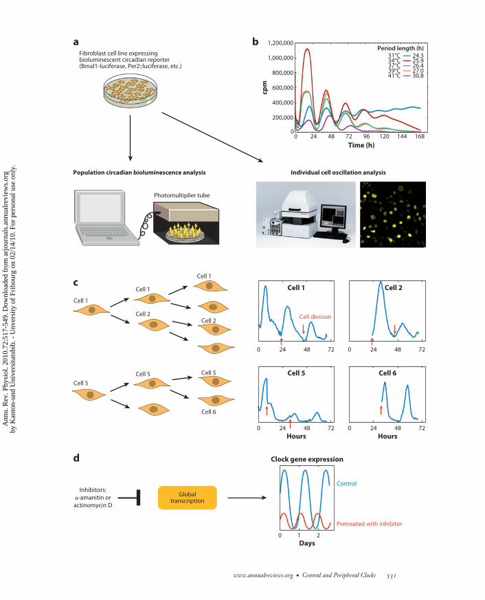
In 1998 Balsalobre et al. (106) showed thatimmortalized Rat-1 fibroblasts, which had beenpropagated in culture for many decades, possessrobust circadian oscillators. Since then, circa-dian gene expression has been observed in avariety of cultured cell lines and primary fibrob-lasts, including murine NIH-3T3 fibroblasts(208), mouse and human primary fibroblasts(209), and human U2OS osteosarkoma cells(193). The circadian oscillators of such culturedcells can be transiently synchronized by a puz-zling variety of signaling pathways involvingboth transmembrane and nuclear receptors(188). Single-cell recordings and mathematicalanalysis of bioluminescence cycles generatedby cell populations revealed that fibroblastsharbor self-sustained, cell-autonomous os-cillators (107, 210) (Figure 4), in keepingwith the observations made with peripheraltissue explants (182). The fibroblast clocksexhibit robust but desynchronized oscillationsunder normal continuous culture conditions.However, as outlined above, these clocks maybe transiently synchronized by the activationof several signal transduction pathways.
Peripheral clocks are resilient to large fluc-tuations in temperature and overall tran-scription rates. The studies with cultured fi-broblasts suggest that, when examined at thesingle-cell level, peripheral oscillators are atleast as robust as those operative in isolatedSCN neurons. Thus, oscillators in cultured
−−−−−−−−−−−−−−−−−−−−−−−−−−−−−−−−−−−−−−−−−−−−−−−−−−−−−−−−−−−−−−−−−−−−−−−−−−−−−−−−−−−−−−−−−→Figure 4Robustness of mammalian cellular clocks. (a) Experimental system for the analysis of circadian gene expression in cell populations orsingle cells (adapted with permission from References 107 and 215). (b) Temperature fluctuations within the physiological range do notdramatically alter period length. Surprisingly, the cellular circadian oscillators run slightly faster at lower temperatures (temperatureovercompensation). (c) During cell division the circadian oscillators keep ticking in daughter cells, and in spite of the sudden division ofthe cell’s contents during this process, the phases undergo only minor advances or delays (adapted from Reference 107). (d ) Inhibitionof RNA synthesis by α-amanitin (an inhibitor of RNA polymerase II–dependent transcription) or actinomycin D (an inhibitor of RNApolymerase I– and II–dependent transcription) does not abolish circadian oscillations.
530 Dibner · Schibler · Albrecht
Ann
u. R
ev. P
hysi
ol. 2
010.
72:5
17-5
49. D
ownl
oade
d fr
om a
rjou
rnal
s.an
nual
revi
ews.
org
by K
anto
n-un
d U
nive
rsita
tsbi
b. -
Unv
ersi
ty o
f Fr
ibou
rg o
n 02
/14/
10. F
or p
erso
nal u
se o
nly.
ANRV404-PH72-26 ARI 18 January 2010 10:13
Cell 1
Cell 5
Cell 1
Cell 1
Cell 2Cell 2
Cell 5 Cell 5
Cell 6
Fibroblast cell line expressingbioluminescent circadian reporter(Bmal1-luciferase, Per2::luciferase, etc.)
Population circadian bioluminescence analysis
Photomultiplier tube
Individual cell oscillation analysis
a b
c
d
0 724824 0 724824
0 724824 0 724824
Cell division
Cell 1
Cell 5 Cell 6
Cell 2
Globaltranscription
Inhibitors:
α-amanitin or
actinomycin D
Clock gene expression
cpm
0 24 48 72 96 120 144 168
Time (h)
0
200,000
400,000
600,000
800,000
1,000,000
1,200,000
31°C34°C37°C39°C41°C
24.325.926.427.030.8
Period length (h)
Hours Hours
Control
Pretreated with inhibitor
0 1 2
Days
www.annualreviews.org • Central and Peripheral Clocks 531
Ann
u. R
ev. P
hysi
ol. 2
010.
72:5
17-5
49. D
ownl
oade
d fr
om a
rjou
rnal
s.an
nual
revi
ews.
org
by K
anto
n-un
d U
nive
rsita
tsbi
b. -
Unv
ersi
ty o
f Fr
ibou
rg o
n 02
/14/
10. F
or p
erso
nal u
se o
nly.

ANRV404-PH72-26 ARI 18 January 2010 10:13
fibroblasts and SCN neurons keep tickingthroughout a cell’s lifetime. In fibroblasts, cir-cadian gene expression even persists duringcell division, and the phase of these oscilla-tions is passed on to daughter cells with onlyminor advances or delays. Another remarkableaspect of peripheral clock robustness is temper-ature compensation, a property described sev-eral decades ago for many organisms (211, 212).In contrast to most chemical and biochemicalprocesses, which are accelerated with increas-ing temperature, the period length of circadianoscillators remains nearly constant over a widerange of physiological temperatures (213, 214).This feature even applies to circadian oscilla-tors operative in cells of homeotherm organ-isms, such as cultured mammalian fibroblasts.In fact, the oscillators of these cells are eventemperature overcompensated, in that they tickslightly faster at lower temperatures (215–217)(Figure 4b). In Drosophila, the Per protein playsa key role in temperature compensation, andperL mutant flies lose temperature compensa-tion and exhibit longer periods at higher tem-peratures (211). We found that Per1-deficientprimary mouse tail fibroblasts, in contrast totheir wild-type counterparts, are not tempera-ture (over)compensated. Rather, they oscillatewith similar and even slightly shorter periods atelevated temperatures. This suggests that PERproteins play an important role in the temper-ature control of circadian oscillators in mam-malian cells as well (215).
Besides being temperature compensated,peripheral clocks also appear to be resilientagainst fluctuations in oscillator components.As mentioned above, cell division, during whichthe cellular contents are cut approximatelyin half, does not abrogate oscillator functionbut causes only relatively modest phase shifts(Figure 4c) (107). Fibroblast clocks also sup-port large variations in general transcriptionrates. Thus, circadian oscillator function per-sists in mouse fibroblasts exposed to sublethaldoses of the transcription inhibitors α-amanitinand actinomycin D, which lower general RNApolymerase II–dependent transcription by up tothreefold (Figure 4d ) (215). Breathtaking work
from Kondo and colleagues (218) recently re-vealed that in cyanobacteria circadian proteinphosphorylation can persist in the absence oftranscription and translation, at least at certaintemperatures. Moreover, this group succeededin reconstituting a circadian oscillator in the testtube with just three clock proteins (KaiA, KaiB,and KaiC) and ATP (219). However, the sameresearch team recently showed that circadianclock gene transcription is required for fullyoperative clock function in vivo (220). Hence,the coordinated interaction between posttrans-lational and transcriptional/translational regu-latory mechanisms appears to account for thegeneration of robust daily rhythms in cyanobac-teria. Work on mammalian and insect oscilla-tors resulted in similar conclusions for meta-zoan systems (221, 222), which may explainwhy absolute cellular concentrations of knownclock components do not appear to play pre-ponderant roles in keeping the clocks tick-ing (see above). The latter notion is also sup-ported by a recent report by Fan and colleagues(223), demonstrating that the overexpressionof CRY1, CRY2, and BMAL1, at least withincertain limits, does not abrogate oscillatorfunction.
The resilience of cellular clocks to changesin temperature and gene expression may beeven more important in peripheral cell typesthan in the SCN. In contrast to SCN oscillators,whose intercellular communication reinforcestheir resilience to perturbations (224), periph-eral clocks are rather autistic and must thusrely on cell-autonomous robustness. For exam-ple, cellular transcription rates can vary dra-matically (>20-fold) in different tissues (225).Moreover, even in homeotherm animals likemammals, the ambient temperature can fluctu-ate significantly between internal organs (e.g.,liver, kidney) and tissues exposed to outsidetemperature (e.g., skin, mucosa, testicles). Inaddition, fever and hypothermia can lead tolarge temperature changes, even in internal or-gans. Because small period-length alterationscan result in large phase changes, the robust-ness of peripheral clocks may be important toassure phase coherence.
532 Dibner · Schibler · Albrecht
Ann
u. R
ev. P
hysi
ol. 2
010.
72:5
17-5
49. D
ownl
oade
d fr
om a
rjou
rnal
s.an
nual
revi
ews.
org
by K
anto
n-un
d U
nive
rsita
tsbi
b. -
Unv
ersi
ty o
f Fr
ibou
rg o
n 02
/14/
10. F
or p
erso
nal u
se o
nly.

ANRV404-PH72-26 ARI 18 January 2010 10:13
An intriguing area of research concerns thebalance between different core clock compo-nents necessary for proper clockwork function.The single-cell analysis of fibroblasts from Per1knockout mice performed by us and others(224) revealed that the low-amplitude oscilla-tions exhibited by these cells at the populationlevel are the result of a small fraction of robustlycycling cells and a large fraction of arrhythmiccells. Therefore, some cells appear to be moresensitive to the absence of PER1 than do othercells, perhaps because PER2 production is up-regulated in these cells.
Fibroblast oscillators and behavior. Clockgenes are operative in most, if not all, skin celltypes and may play a role in several processessuch as cellular proliferation (226) and hair fol-licle cycling (227). Brown and colleagues (209)have compared the wheel-running behavior andthe period length of skin fibroblasts of vari-ous mice carrying mutations in different coreclock genes. Even though the cellular clocksoperative in fibroblasts did not perfectly reflectthe central clockwork, there was a qualitativecorrelation between locomotor activity and fi-broblast gene expression. Given that the assess-ment of the circadian period length in humanbeings is complicated and expensive, the pos-sibility of studying human circadian oscillatorproperties by using primary fibroblasts was awelcome opportunity. Thus, the skin fibrob-last system was also used successfully to char-acterize amplitude and phase shift properties inindividuals with “normal” period lengths butdifferent behavioral phenotypes. These studiessuggested that human chronotypes may be in-fluenced not only by the period length of theircircadian oscillators but also by cellular com-ponents that affect period length, amplitude,and phase (228). Moreover, this system maybe exploited for diagnostic purposes, conceiv-ably for subjects with circadian disorders. Forexample, Vanselow and coworkers (229) havefound that a mutation in an mPer2 phospho-acceptor site corresponding to the hPER2 mu-tation associated with human familial advancedsleep phase syndrome (FASPS) phenocopies the
short period and advanced phase in culturedfibroblasts.
Synchronization of Peripheral Clocks
As mentioned above, the SCN central pace-maker must establish phase coherence in thebody by synchronizing billions of individualcell clocks every day (202, 224). The SCN usesmany routes to establish phase coherence in theperiphery. Thus, feeding rhythms, driven byrest-activity rhythms, are strong Zeitgebers formany tissues (178, 230). Likewise, body tem-perature rhythms, influenced directly by theSCN and by activity cycles controlled by theSCN, appear to play a role in the resetting ofperipheral timekeepers (231). In addition, theSCN also uses more direct timing cues, suchas humoral and neuronal signals, to entrain thephases of peripheral clocks (87, 232) (Figure 5).Peripheral clocks continue to oscillate in SCN-lesioned mice, but their phases are no longercoordinated in these behaviorally arrhythmicanimals (182, 206).
Entrainment of peripheral clocks by in-direct cues imposed by oscillating behav-ior: feeding-fasting cycles and temperature.Daily feeding-fasting cycles appear to be thedominant Zeitgebers for several peripheral or-gans, including liver, kidney, pancreas, andheart. The timing of food intake influencesthe expression profile of many circadian genesin these organs. Normally, the feeding-fastingcycles are in phase with rest-activity cycles.Strikingly, daytime-restricted feeding of noc-turnal rodents inverts the phase of gene ex-pression in peripheral organs, thereby uncou-pling peripheral clocks from the SCN (230,233). The entrainment pathways from feeding-fasting cycles may include hormones secretedupon feeding and fasting [for example, chole-cystokinin, peptide YY, oxyntomodulin, ghre-lin, leptin (234)], food metabolites (for example,glucose, cholesterol, fatty acids, heme), post-prandial temperature elevations, and intracellu-lar redox state [NAD(P)H/NAD(P)+ ratio (235and references therein)].
www.annualreviews.org • Central and Peripheral Clocks 533
Ann
u. R
ev. P
hysi
ol. 2
010.
72:5
17-5
49. D
ownl
oade
d fr
om a
rjou
rnal
s.an
nual
revi
ews.
org
by K
anto
n-un
d U
nive
rsita
tsbi
b. -
Unv
ersi
ty o
f Fr
ibou
rg o
n 02
/14/
10. F
or p
erso
nal u
se o
nly.

ANRV404-PH72-26 ARI 18 January 2010 10:13
Rest-activitycycles
Feeding-fastingcycle
Feeding-relatedhormones
Metabolites(e.g., NAD(P)H/NAD(P)+)
Sympatheticnervous system
Humoralsignals
Bodytemperature
Directsignals
SCN(central clock)
Peripheral clocks(organs)
SCN
SCN
Figure 5Peripheral clock entrainment pathways (see text for explanations).
There is growing evidence for the inter-play between energy metabolism and the cir-cadian clock (236–238). First, the dominanceof feeding cycles as a Zeitgeber for peripheral
clocks implies that the circadian timing sys-tem plays an important role in nutrient pro-cessing and energy homeostasis. In addition, aswe mentioned above, transcriptome profiling
534 Dibner · Schibler · Albrecht
Ann
u. R
ev. P
hysi
ol. 2
010.
72:5
17-5
49. D
ownl
oade
d fr
om a
rjou
rnal
s.an
nual
revi
ews.
org
by K
anto
n-un
d U
nive
rsita
tsbi
b. -
Unv
ersi
ty o
f Fr
ibou
rg o
n 02
/14/
10. F
or p
erso
nal u
se o
nly.

ANRV404-PH72-26 ARI 18 January 2010 10:13
studies revealed that many genes involved inmetabolism are rhythmically expressed (191,192, 194, 199, 239, 240). Furthermore, at leastin vitro, the DNA-binding activity of BMAL1-CLOCK is strongly influenced by the ratio ofreduced to oxidized NAD cofactors, which areoften considered to be readouts of the cellularmetabolic state (241).
Mammalian SIRT1, an NAD+-dependentdeacetylase, has been recently identified as anovel regulator of circadian gene expression(242, 243). SIRT1, the mammalian orthologof yeast Sir2, is involved in transcriptional si-lencing, genome stability, and longevity (244,245). Asher and colleagues (242) demonstratedthat SIRT1 exhibits circadian accumulation inmouse hepatocytes and cultured fibroblasts andthat it is essential for high-magnitude circa-dian transcription of several core clock genes,including Bmal1, Rorα, Per2, and Cry1. More-over, SIRT1 binds CLOCK-BMAL1 in a cir-cadian manner and promotes the deacetylationand subsequent degradation of PER2 (242).
Temperature fluctuations represent an im-portant entrainment cue in Drosophila, Neu-rospora, and mammals (246 and referencestherein). Shallow temperature rhythms imitat-ing body temperature fluctuations can maintainpreviously induced rhythms in peripheral os-cillators, and can phase shift peripheral clocks,without affecting the phase of SCN (231). In ze-brafish kept in constant darkness, temperaturesteps of as little as 2◦C can entrain circadianrhythms in developing larvae (and in zebrafishcell lines) (247).
Using a novel screen dubbed differentialdisplay of DNA-binding proteins (DDDP)of mouse liver nuclear extracts, Reinke andcoworkers (248) have found a highly rhythmicactivity of heat-shock factor 1 (HSF1). HSF1drives the expression of heat-shock proteins atthe onset of the dark phase, when the animalsstart to be behaviorally active. The onset offeeding is followed by a postprandial rise inbody temperature, which makes it difficult todiscriminate between temperature-dependentand chemical HSF1 activation pathways. Nev-ertheless, recent studies with cultured cells
DDDP: differentialdisplay of DNA-binding proteins
HSF1: heat-shockfactor 1
indicate that HSF1 not only governs part of thecytoprotection system but also plays a role inthe synchronization of peripheral cells (C. Saini& U. Schibler, unpublished results).
Entrainment by hormonal and neuronal sig-nals emanating from the SCN. The SCNalso employs more direct signals, such as neuraland humoral outputs, to synchronize peripheralclocks.
Plasma glucocorticoid hormone levels ex-hibit robust daily oscillations in both labo-ratory rodents and humans, and these cyclesare driven by SCN via the hypothalamic-Pit-adrenal axis (249). Interestingly, the glucocor-ticoid nuclear hormone receptor is expressedin virtually all cell types, except in SCN neu-rons (see Reference 250 and references therein).In keeping with this expression pattern, dex-amethasone, a glucocorticoid receptor agonist,acts as a strong Zeitgeber in vivo. Dexametha-sone synchronizes rat-1 fibroblasts and causesphase shifts in peripheral oscillators but doesnot affect SCN rhythms (250). Thus, glu-cocorticoid hormones possess clock-resettingproperties and represent an important phase-entrainment signal from the SCN. Due to theredundancy of synchronization pathways, thesteady-state phase can be established in the ab-sence of glucocorticoid receptors in peripheraltissues such as the liver. Nonetheless, the in-volvement of glucocorticoid signaling can bedemonstrated by determining the kinetics offeeding-induced phase inversion. Thus, hepa-tocytes devoid of a functional glucocorticoid re-ceptor gene adapt the phase much more rapidlyto daytime feeding than do wild-type hepato-cytes (251).
The autonomic nervous system constitutesan additional direct synchronization pathwayemployed by the SCN. For example, by sur-gically disrupting liver innervation, Buijs andcoworkers (87) demonstrated that light mayaffect liver gene expression not only via thehormonal pathway but also via autonomic in-put. Hence, the autonomic nervous systemmay play a role in the resetting of periph-eral clocks after phase shift–inducing light
www.annualreviews.org • Central and Peripheral Clocks 535
Ann
u. R
ev. P
hysi
ol. 2
010.
72:5
17-5
49. D
ownl
oade
d fr
om a
rjou
rnal
s.an
nual
revi
ews.
org
by K
anto
n-un
d U
nive
rsita
tsbi
b. -
Unv
ersi
ty o
f Fr
ibou
rg o
n 02
/14/
10. F
or p
erso
nal u
se o
nly.
ANRV404-PH72-26 ARI 18 January 2010 10:13
exposure (87). In addition, the same group hasfound that plasma glucose and insulin concen-trations are affected by administering gluta-matergic agonists or GABAergic antagonists.The hyperglycemic effect of the GABA antag-onist is absent in SCN-ablated animals. Thecontrol of daily glucose metabolism in the liverindicates an important role for GABAergic/glutamatergic SCN inputs to the hypothala-mic preautonomic neurons that are connectedto the liver (252). Furthermore, an elegantstudy by Okamura and colleagues (86) revealedthat adrenal innervation is required for light-induced corticosterone secretion by the adrenalgland, thus representing a direct output fromthe SCN to the adrenal cortex.
Under certain circumstances, different tim-ing cues can be in conflict with each other.We mention above that food availability duringthe inactivity phase will override more directSCN-driven phase-resetting signals in the liver(194, 251). By contrast, in the submaxillarysalivary gland, temporal food restriction failsto entrain circadian gene expression. However,upon sympathetic denervation, the submaxil-lary gland oscillators rapidly adjust their phaseto the inverted feeding regimen. This findingfurther suggests that sympathetic inputs are im-portant for the phase entrainment of peripheraloscillators and that multiple synchronizationpathways can contribute to peripheral oscilla-tor synchronization in the same tissue (232).
A Few Selected Outputsof Peripheral OscillatorsHepatic clocks in glucose and lipidmetabolism. Rhythmically expressed livergenes are implicated in the metabolism offatty acids, cholesterol, bile acids, amino acids,and xenobiotics (185, 196, 199, 239). Thedisruption of circadian oscillator functioncauses alterations in metabolism (reviewedin Reference 253). For instance, mutationsin the essential clock genes Bmal1 and Clocklead to various metabolic disorders (254).Elegant work by Weitz and colleagues (186)suggests that functional liver clocks contributeto glucose homeostasis by driving a daily
rhythm of hepatic glucose export. Using theCre-loxP recombination strategy in mice,these authors inactivated Bmal1 either in allcells (Bmal1−/−) or specifically in hepatocytes(L-Bmal1−/−). Their experiments suggestthat BMAL1 function is important for theregulation of total body fat, glucose clear-ance, and insulin production. Animals witha liver-specific Bmal1 disruption suffer fromhypoglycemia during the inactivity phase. Yet,mice deficient in BMAL1 in all cells do notshow overt problems with their resting bloodsugar levels. Hence, in L-Bmal1−/− mice, thelack of liver rhythms—rather than just the lackof the BMAL1 transcription factor—must bethe cause of impaired glucose homeostasis.Of note, Weitz and colleagues (186) havedescribed a similar pattern of metabolic defectsin Per1−/−, Per2−/− double-mutant mice,which also have a disrupted clock. Moreover, agenetic linkage analysis in humans has recentlyshown that hPER2 activity may be associatedwith blood glucose levels as well (255).
Recent studies by Le Martelot and col-leagues (187) further underscore the tight linkbetween the circadian clock and metabolism inthe liver. These authors have demonstrated animportant role for REV-ERBα in the circadianregulation of cholesterol and bile acid synthe-sis (187). Genetic loss- and gain-of-functionexperiments suggest that REV-ERBα partici-pates in the circadian modulation of sterol reg-ulatory element–binding protein (SREBP) ac-tivity and thereby in the daily expression ofSREBP target genes involved in cholesteroland lipid metabolism (187). This regulationis accomplished via the cyclic transcription ofInsig2, which encodes a transmembrane pro-tein that sequesters SREBPs to the endoplasmicreticulum (ER) membranes and thereby inter-feres with the proteolytic activation of SREBPsin Golgi membranes. Probably through thismechanism, REV-ERBα also participates in theregulation of cyclic cholesterol-7α-hydroxylase(Cyp7a1) expression. Thus, the authors of thisreport speculate that the SREBP-dependentproduction of cholesterol is accompanied by thegeneration of oxysterols, which in turn activate
536 Dibner · Schibler · Albrecht
Ann
u. R
ev. P
hysi
ol. 2
010.
72:5
17-5
49. D
ownl
oade
d fr
om a
rjou
rnal
s.an
nual
revi
ews.
org
by K
anto
n-un
d U
nive
rsita
tsbi
b. -
Unv
ersi
ty o
f Fr
ibou
rg o
n 02
/14/
10. F
or p
erso
nal u
se o
nly.

ANRV404-PH72-26 ARI 18 January 2010 10:13
the nuclear receptor LXR and hence the LXRtarget gene Cyp7a1.
Pancreas clocks, melatonin signaling, anddiabetes. An additional connection betweencircadian oscillators and glucose homeostasishas emerged from recent studies on melatoninreceptors in the pancreas (256). Melatonin isa circulating neurohormone that is secretedpredominantly by the pineal gland at night.The hormone regulates circadian rhythms byassisting the translation of photoperiodic in-formation in the brain. Melatonin signaling ismainly mediated by two receptors, MT1 andMT2, encoded by the MTNR1A and MTNR1Bgenes, respectively. The strongest MT2 expres-sion levels are detected in the retina and in theSCN. MT2-mediated melatonin signaling mayindirectly regulate glucose level and insulin se-cretion through the SCN (257). In healthy in-dividuals insulin secretion follows a circadianrhythm. This rhythm, generated within theislets, may be induced by melatonin, which pro-vokes a phase shift in insulin secretion (256).Indeed, MTNR1A is highly expressed in pan-creatic islets and is, in general, more abun-dant than MTNR1B. The presence of MTNR1Bwas recently confirmed in both pancreatic isletsand sorted β-cells, suggesting a possible directrole of MT2 in the regulation of insulin se-cretion (258). Genetic studies in humans haverevealed a possible link between MTNR1B al-lele variants and hyperglycemia, impaired early-phase insulin secretion, and β-cell function(258, 259). Thus, MTNR1B, expressed in hu-man pancreatic β-cells, may represent an im-portant connection between circadian clocksand glucose homeostasis, and its detailed anal-ysis may open new avenues for the treat-ment of type 2 diabetes. Recent studies ontransgenic mice underscore the involvement ofmelatonin signaling in islet function. Analysisof circadian gene expression in pancreas (andliver) of melatonin receptor knockout mice re-vealed changes in both phase and amplitude.Moreover, an upregulation of insulin secre-tion was detected in isolated islets of MT1 andMT2 single-knockout mice and MT1/MT2
double-knockout animals, confirming the neg-ative action of melatonin signaling on insulinsecretion (260).
CONCLUSIONS ANDPERSPECTIVES
Remarkable progress has been made in our un-derstanding of the mammalian circadian timingsystem during the past few years. The molecularoscillator model has been considerably revisedand amended. In particular, a plethora of post-translational regulatory mechanisms have beenshown to contribute critically to proper clockfunction in addition to the canonical TTFL.Furthermore, the evidence for a tight couplingof circadian gene expression to metabolic cy-cles in mammalian cells, originally proposed byRutter and colleagues (241), is now stronger. Anew level of complexity has recently been addedto the mammalian circadian timing system byfindings that microRNAs participate in the con-trol of posttranscriptional clock output path-ways (205, 261). However, we must considerthat core clock proteins also perform noncir-cadian functions. That is, when we study or-ganisms with mutations in core clock genes,we must discriminate between clock gene phe-notypes and clock phenotypes. For example,the deficiencies of clock mutant mice in boneformation, tumor surveillance, and metabolicpathways may have no causal relationship withthe circadian clock. Indeed, the rigorous dis-crimination between clock function and clockgene function is not a trivial matter and to datehas been accomplished in only a few isolatedcases by so-called resonance studies (262–264).Now that period-length mutations are avail-able for laboratory mice (265 and referencestherein), such studies could be performed formammalian organisms. Even in the absence ofsuch evidence, the facts remain that hundreds ofgenes are expressed in a cyclic manner and thatmost physiology follows a daily rhythm. Alas,this knowledge is largely ignored in the clinic,and only a few pioneers are making efforts toexplore this temporal information in designingbetter therapies (266).
www.annualreviews.org • Central and Peripheral Clocks 537
Ann
u. R
ev. P
hysi
ol. 2
010.
72:5
17-5
49. D
ownl
oade
d fr
om a
rjou
rnal
s.an
nual
revi
ews.
org
by K
anto
n-un
d U
nive
rsita
tsbi
b. -
Unv
ersi
ty o
f Fr
ibou
rg o
n 02
/14/
10. F
or p
erso
nal u
se o
nly.

ANRV404-PH72-26 ARI 18 January 2010 10:13
The synchronization pathways of peripheralclocks by the SCN remain largely unknown.Many signaling pathways are expected to par-ticipate in the phase entrainment of periph-eral clocks. Hence, the elimination of a sin-gle pathway is unlikely to significantly affectthe steady-state phase of circadian clocks. Amore sensitive approach would be the recording
of phase-shifting kinetics. Ideally, such record-ing would be performed at a high tempo-ral resolution in living animals. It will thusbe of utmost importance to develop novelnoninvasive whole-body imaging techniquesallowing the recording of central and periph-eral gene expression patterns in freely movinganimals.
SUMMARY POINTS
1. The main circadian oscillator in the brain is located in the suprachiasmatic nuclei (SCN).Secondary oscillators exist in the brain.
2. Light adjusts the phase of the SCN oscillator to the environmental light-dark cycle.
3. Neural and humoral signals from the SCN can synchronize central and peripheral oscil-lators and vice versa.
4. In the periphery, circadian clocks are functional in most body cells.
5. Peripheral oscillators are self-sustained; cell-autonomous; and resilient to cell division,to acute stress, and to fluctuations in temperature and general transcription rate.
6. The combination of environmental inputs—such as daily fasting-feeding cycles, tem-perature cycles, the cellular redox state, and direct neural and humoral signals from theSCN—represents essential entrainment cues for the peripheral clocks.
DISCLOSURE STATEMENT
The authors are not aware of any affiliations, memberships, funding, or financial holdings thatmight be perceived as affecting the objectivity of this review.
LITERATURE CITED
1. Welsh DK, Takahashi JS, Kay SA. 2010. Suprachiasmatic nucleus: cell autonomy and network properties.Annu. Rev. Physiol. 72:551–77
2. Pittendrigh CS. 1960. Circadian rhythms and the circadian organization of living systems. Cold SpringHarbor Symp. Quant. Biol. 25:159–84
3. Moore RY, Eichler VB. 1972. Loss of a circadian adrenal corticosterone rhythm following suprachias-matic lesions in the rat. Brain Res. 42:201–6
4. Stephan FK, Zucker I. 1972. Circadian rhythms in drinking behavior and locomotor activity of rats areeliminated by hypothalamic lesions. Proc. Natl. Acad. Sci. USA 69:1583–86
5. Lehman MN, Silver R, Gladstone WR, Kahn RM, Gibson M, Bittman EL. 1987. Circadian rhythmicityrestored by neural transplant. Immunocytochemical characterization of the graft and its integration withthe host brain. J. Neurosci. 7:1626–38
6. Ralph MR, Foster RG, Davis FC, Menaker M. 1990. Transplanted suprachiasmatic nucleus determinescircadian period. Science 247:975–78
7. Sujino M, Masumoto KH, Yamaguchi S, van der Horst GT, Okamura H, Inouye ST. 2003. Suprachi-asmatic nucleus grafts restore circadian behavioral rhythms of genetically arrhythmic mice. Curr. Biol.13:664–68
538 Dibner · Schibler · Albrecht
Ann
u. R
ev. P
hysi
ol. 2
010.
72:5
17-5
49. D
ownl
oade
d fr
om a
rjou
rnal
s.an
nual
revi
ews.
org
by K
anto
n-un
d U
nive
rsita
tsbi
b. -
Unv
ersi
ty o
f Fr
ibou
rg o
n 02
/14/
10. F
or p
erso
nal u
se o
nly.

ANRV404-PH72-26 ARI 18 January 2010 10:13
8. Inouye ST, Kawamura H. 1979. Persistence of circadian rhythmicity in a mammalian hypothalamic“island” containing the suprachiasmatic nucleus. Proc. Natl. Acad. Sci. USA 76:5962–66
9. Green DJ, Gillette R. 1982. Circadian rhythm of firing rate recorded from single cells in the rat suprachi-asmatic brain slice. Brain Res. 245:198–200
10. Groos G, Hendriks J. 1982. Circadian rhythms in electrical discharge of rat suprachiasmatic neuronesrecorded in vitro. Neurosci. Lett. 34:283–88
11. Shibata S, Oomura Y, Kita H, Hattori K. 1982. Circadian rhythmic changes of neuronal activity in thesuprachiasmatic nucleus of the rat hypothalamic slice. Brain Res. 247:154–58
12. Bos NP, Mirmiran M. 1990. Circadian rhythms in spontaneous neuronal discharges of the culturedsuprachiasmatic nucleus. Brain Res. 511:158–62
13. Welsh DK, Logothetis DE, Meister M, Reppert SM. 1995. Individual neurons dissociated from ratsuprachiasmatic nucleus express independently phased circadian firing rhythms. Neuron 14:697–706
14. Schwartz WJ, Gainer H. 1977. Suprachiasmatic nucleus: use of 14C-labeled deoxyglucose uptake as afunctional marker. Science 197:1089–91
15. Sun ZS, Albrecht U, Zhuchenko O, Bailey J, Eichele G, Lee CC. 1997. RIGUI, a putative mammalianortholog of the Drosophila period gene. Cell 90:1003–11
16. Tei H, Okamura H, Shigeyoshi Y, Fukuhara C, Ozawa R, et al. 1997. Circadian oscillation of a mammalianhomologue of the Drosophila period gene. Nature 389:512–16
17. Hastings MH, Field MD, Maywood ES, Weaver DR, Reppert SM. 1999. Differential regulation ofmPER1 and mTIM proteins in the mouse suprachiasmatic nuclei: new insights into a core clock mech-anism. J. Neurosci. 19:RC11
18. Langner R, Rensing L. 1972. Circadian rhythm of oxygen consumption in rat liver suspension culture:changes of pattern. Z. Naturforsch. Teil B 27:1117–18
19. Berson DM, Dunn FA, Takao M. 2002. Phototransduction by retinal ganglion cells that set the circadianclock. Science 295:1070–73
20. Moore RY, Speh JC, Card JP. 1995. The retinohypothalamic tract originates from a distinct subset ofretinal ganglion cells. J. Comp. Neurol. 352:351–66
21. Provencio I, Jiang G, De Grip WJ, Hayes WP, Rollag MD. 1998. Melanopsin: an opsin in melanophores,brain, and eye. Proc. Natl. Acad. Sci. USA 95:340–45
22. Gooley JJ, Lu J, Chou TC, Scammell TE, Saper CB. 2001. Melanopsin in cells of origin of the retino-hypothalamic tract. Nat. Neurosci. 4:1165
23. Hattar S, Liao HW, Takao M, Berson DM, Yau KW. 2002. Melanopsin-containing retinal ganglioncells: architecture, projections, and intrinsic photosensitivity. Science 295:1065–70
24. Provencio I, Rodriguez IR, Jiang G, Hayes WP, Moreira EF, Rollag MD. 2000. A novel human opsinin the inner retina. J. Neurosci. 20:600–5
25. Moore RY, Lenn NJ. 1972. A retinohypothalamic projection in the rat. J. Comp. Neurol. 146:1–1426. Ibata Y, Takahashi Y, Okamura H, Kawakami F, Terubayashi H, et al. 1989. Vasoactive intestinal peptide
(VIP)-like immunoreactive neurons located in the rat suprachiasmatic nucleus receive a direct retinalprojection. Neurosci. Lett. 97:1–5
27. Tanaka M, Ichitani Y, Okamura H, Tanaka Y, Ibata Y. 1993. The direct retinal projection to VIP neuronalelements in the rat SCN. Brain Res. Bull. 31:637–40
28. Castel M, Belenky M, Cohen S, Ottersen OP, Storm-Mathisen J. 1993. Glutamate-like immunoreactivityin retinal terminals of the mouse suprachiasmatic nucleus. Eur. J. Neurosci. 5:368–81
29. Hannibal J, Ding JM, Chen D, Fahrenkrug J, Larsen PJ, et al. 1997. Pituitary adenylate cyclase-activatingpeptide (PACAP) in the retinohypothalamic tract: a potential daytime regulator of the biological clock.J. Neurosci. 17:2637–44
30. Hirota T, Fukada Y. 2004. Resetting mechanism of central and peripheral circadian clocks in mammals.Zool. Sci. 21:359–68
31. Crosio C, Cermakian N, Allis CD, Sassone-Corsi P. 2000. Light induces chromatin modification in cellsof the mammalian circadian clock. Nat. Neurosci. 3:1241–47
32. Albrecht U, Sun ZS, Eichele G, Lee CC. 1997. A differential response of two putative mammaliancircadian regulators, mper1 and mper2, to light. Cell 91:1055–64
www.annualreviews.org • Central and Peripheral Clocks 539
Ann
u. R
ev. P
hysi
ol. 2
010.
72:5
17-5
49. D
ownl
oade
d fr
om a
rjou
rnal
s.an
nual
revi
ews.
org
by K
anto
n-un
d U
nive
rsita
tsbi
b. -
Unv
ersi
ty o
f Fr
ibou
rg o
n 02
/14/
10. F
or p
erso
nal u
se o
nly.

ANRV404-PH72-26 ARI 18 January 2010 10:13
33. Shearman LP, Zylka MJ, Weaver DR, Kolakowski LF Jr, Reppert SM. 1997. Two period homologs:circadian expression and photic regulation in the suprachiasmatic nuclei. Neuron 19:1261–69
34. Shigeyoshi Y, Taguchi K, Yamamoto S, Takekida S, Yan L, et al. 1997. Light-induced resetting of amammalian circadian clock is associated with rapid induction of the mPer1 transcript. Cell 91:1043–53
35. Morris ME, Viswanathan N, Kuhlman S, Davis FC, Weitz CJ. 1998. A screen for genes induced in thesuprachiasmatic nucleus by light. Science 279:1544–47
36. Hamada T, Liou SY, Fukushima T, Maruyama T, Watanabe S, et al. 1999. The role of inositoltrisphosphate-induced Ca2+ release from IP3-receptor in the rat suprachiasmatic nucleus on circadianentrainment mechanism. Neurosci. Lett. 263:125–28
37. Ding JM, Buchanan GF, Tischkau SA, Chen D, Kuriashkina L, et al. 1998. A neuronal ryanodinereceptor mediates light-induced phase delays of the circadian clock. Nature 394:381–84
38. Pfeffer M, Muller CM, Mordel J, Meissl H, Ansari N, et al. 2009. The mammalian molecular clockworkcontrols rhythmic expression of its own input pathway components. J. Neurosci. 29:6114–23
39. Duffield GE, Watson NP, Mantani A, Peirson SN, Robles-Murguia M, et al. 2009. A role for Id2 inregulating photic entrainment of the mammalian circadian system. Curr. Biol. 19:297–304
40. Coogan AN, Piggins HD. 2003. Circadian and photic regulation of phosphorylation of ERK1/2 andElk-1 in the suprachiasmatic nuclei of the Syrian hamster. J. Neurosci. 23:3085–93
41. Kornhauser JM, Mayo KE, Takahashi JS. 1996. Light, immediate-early genes, and circadian rhythms.Behav. Genet. 26:221–40
42. Obrietan K, Impey S, Storm DR. 1998. Light and circadian rhythmicity regulate MAP kinase activationin the suprachiasmatic nuclei. Nat. Neurosci. 1:693–700
43. Grewal SS, York RD, Stork PJ. 1999. Extracellular-signal-regulated kinase signalling in neurons. Curr.Opin. Neurobiol. 9:544–53
44. Motzkus D, Maronde E, Grunenberg U, Lee CC, Forssmann W, Albrecht U. 2000. The human PER1gene is transcriptionally regulated by multiple signaling pathways. FEBS Lett. 486:315–19
45. Travnickova-Bendova Z, Cermakian N, Reppert SM, Sassone-Corsi P. 2002. Bimodal regulation ofmPeriod promoters by CREB-dependent signaling and CLOCK/BMAL1 activity. Proc. Natl. Acad. Sci.USA 99:7728–33
46. Akiyama M, Kouzu Y, Takahashi S, Wakamatsu H, Moriya T, et al. 1999. Inhibition of light- or glutamate-induced mPer1 expression represses the phase shifts into the mouse circadian locomotor and suprachi-asmatic firing rhythms. J. Neurosci. 19:1115–21
47. Tischkau SA, Mitchell JW, Tyan SH, Buchanan GF, Gillette MU. 2003. Ca2+/cAMP response element-binding protein (CREB)-dependent activation of Per1 is required for light-induced signaling in thesuprachiasmatic nucleus circadian clock. J. Biol. Chem. 278:718–23
48. Wakamatsu H, Takahashi S, Moriya T, Inouye ST, Okamura H, et al. 2001. Additive effect of mPer1and mPer2 antisense oligonucleotides on light-induced phase shift. NeuroReport 12:127–31
49. Albrecht U, Zheng B, Larkin D, Sun ZS, Lee CC. 2001. MPer1 and mper2 are essential for normalresetting of the circadian clock. J. Biol. Rhythms 16:100–4
50. Spoelstra K, Albrecht U, van der Horst GT, Brauer V, Daan S. 2004. Phase responses to light pulses inmice lacking functional per or cry genes. J. Biol. Rhythms 19:518–29
51. Spoelstra K, Daan S. 2008. Effects of constant light on circadian rhythmicity in mice lacking functionalcry genes: dissimilar from per mutants. J. Comp. Physiol. A 194:235–42
52. Moore RY, Card JP. 1994. Intergeniculate leaflet: an anatomically and functionally distinct subdivisionof the lateral geniculate complex. J. Comp. Neurol. 344:403–30
53. Pickard GE. 1985. Bifurcating axons of retinal ganglion cells terminate in the hypothalamic suprachias-matic nucleus and the intergeniculate leaflet of the thalamus. Neurosci. Lett. 55:211–17
54. Harrington ME, Nance DM, Rusak B. 1985. Neuropeptide Y immunoreactivity in the hamster geniculo-suprachiasmatic tract. Brain Res. Bull. 15:465–72
55. Freeman DA, Dhandapani KM, Goldman BD. 2004. The thalamic intergeniculate leaflet modulatesphotoperiod responsiveness in Siberian hamsters. Brain Res. 1028:31–38
56. Kim HJ, Harrington ME. 2008. Neuropeptide Y-deficient mice show altered circadian response tosimulated natural photoperiod. Brain Res. 1246:96–100
540 Dibner · Schibler · Albrecht
Ann
u. R
ev. P
hysi
ol. 2
010.
72:5
17-5
49. D
ownl
oade
d fr
om a
rjou
rnal
s.an
nual
revi
ews.
org
by K
anto
n-un
d U
nive
rsita
tsbi
b. -
Unv
ersi
ty o
f Fr
ibou
rg o
n 02
/14/
10. F
or p
erso
nal u
se o
nly.

ANRV404-PH72-26 ARI 18 January 2010 10:13
57. Meyer-Bernstein EL, Morin LP. 1996. Differential serotonergic innervation of the suprachiasmaticnucleus and the intergeniculate leaflet and its role in circadian rhythm modulation. J. Neurosci. 16:2097–111
58. Leander P, Vrang N, Møller M. 1998. Neuronal projections from the mesencephalic raphe nuclearcomplex to the suprachiasmatic nucleus and the deep pineal gland of the golden hamster (Mesocricetusauratus). J. Comp. Neurol. 399:73–93
59. Moga MM, Moore RY. 1997. Organization of neural inputs to the suprachiasmatic nucleus in the rat.J. Comp. Neurol. 389:508–34
60. Bosler O, Beaudet A. 1985. VIP neurons as prime synaptic targets for serotonin afferents in rat suprachi-asmatic nucleus: a combined radioautographic and immunocytochemical study. J. Neurocytol. 14:749–63
61. Kiss J, Leranth C, Halasz B. 1984. Serotoninergic endings on VIP-neurons in the suprachiasmatic nucleusand on ACTH-neurons in the arcuate nucleus of the rat hypothalamus. A combination of high resolutionautoradiography and electron microscopic immunocytochemistry. Neurosci. Lett. 44:119–24
62. van den Pol AN, Tsujimoto KL. 1985. Neurotransmitters of the hypothalamic suprachiasmatic nucleus:immunocytochemical analysis of 25 neuronal antigens. Neuroscience 15:1049–86
63. van Esseveldt KE, Lehman MN, Boer GJ. 2000. The suprachiasmatic nucleus and the circadian time-keeping system revisited. Brain Res. Brain Res. Rev. 33:34–77
64. Watts AG, Swanson LW. 1987. Efferent projections of the suprachiasmatic nucleus. II. Studies usingretrograde transport of fluorescent dyes and simultaneous peptide immunohistochemistry in the rat.J. Comp. Neurol. 258:230–52
65. Morin LP, Goodless-Sanchez N, Smale L, Moore RY. 1994. Projections of the suprachiasmatic nuclei,subparaventricular zone and retrochiasmatic area in the golden hamster. Neuroscience 61:391–410
66. Morin LP. 2007. SCN organization reconsidered. J. Biol. Rhythms 22:3–1367. Morin LP, Allen CN. 2006. The circadian visual system, 2005. Brain Res. Rev. 51:1–6068. Buijs RM, Wortel J, Hou YX. 1995. Colocalization of γ-aminobutyric acid with vasopressin, vasoactive
intestinal peptide, and somatostatin in the rat suprachiasmatic nucleus. J. Comp. Neurol. 358:343–5269. Castel M, Morris JF. 2000. Morphological heterogeneity of the GABAergic network in the suprachias-
matic nucleus, the brain’s circadian pacemaker. J. Anat. 196(Pt. 1):1–1370. Hermes ML, Coderre EM, Buijs RM, Renaud LP. 1996. GABA and glutamate mediate rapid neuro-
transmission from suprachiasmatic nucleus to hypothalamic paraventricular nucleus in rat. J. Physiol.496(Pt. 3):749–57
71. Kalsbeek A, van Heerikhuize JJ, Wortel J, Buijs RM. 1996. A diurnal rhythm of stimulatory input tothe hypothalamo-pituitary-adrenal system as revealed by timed intrahypothalamic administration of thevasopressin V1 antagonist. J. Neurosci. 16:5555–65
72. Kalsbeek A, Buijs RM, van Heerikhuize JJ, Arts M, van der Woude TP. 1992. Vasopressin-containingneurons of the suprachiasmatic nuclei inhibit corticosterone release. Brain Res. 580:62–67
73. Kraves S, Weitz CJ. 2006. A role for cardiotrophin-like cytokine in the circadian control of mammalianlocomotor activity. Nat. Neurosci. 9:212–19
74. Cheng MY, Bullock CM, Li C, Lee AG, Bermak JC, et al. 2002. Prokineticin 2 transmits the behaviouralcircadian rhythm of the suprachiasmatic nucleus. Nature 417:405–10
75. Kalsbeek A, Buijs RM. 1992. Peptidergic transmitters of the suprachiasmatic nuclei and the control ofcircadian rhythmicity. Prog. Brain Res. 92:321–33
76. Kramer A, Yang FC, Snodgrass P, Li X, Scammell TE, et al. 2001. Regulation of daily locomotor activityand sleep by hypothalamic EGF receptor signaling. Science 294:2511–15
77. Li X, Sankrithi N, Davis FC. 2002. Transforming growth factor-α is expressed in astrocytes of thesuprachiasmatic nucleus in hamster: role of glial cells in circadian clocks. NeuroReport 13:2143–47
78. Cheng MY, Bittman EL, Hattar S, Zhou QY. 2005. Regulation of prokineticin 2 expression by light andthe circadian clock. BMC Neurosci. 6:17
79. Cheng MY, Leslie FM, Zhou QY. 2006. Expression of prokineticins and their receptors in the adultmouse brain. J. Comp. Neurol. 498:796–809
80. Li JD, Hu WP, Boehmer L, Cheng MY, Lee AG, et al. 2006. Attenuated circadian rhythms in micelacking the prokineticin 2 gene. J. Neurosci. 26:11615–23
www.annualreviews.org • Central and Peripheral Clocks 541
Ann
u. R
ev. P
hysi
ol. 2
010.
72:5
17-5
49. D
ownl
oade
d fr
om a
rjou
rnal
s.an
nual
revi
ews.
org
by K
anto
n-un
d U
nive
rsita
tsbi
b. -
Unv
ersi
ty o
f Fr
ibou
rg o
n 02
/14/
10. F
or p
erso
nal u
se o
nly.

ANRV404-PH72-26 ARI 18 January 2010 10:13
81. Prosser HM, Bradley A, Chesham JE, Ebling FJ, Hastings MH, Maywood ES. 2007. Prokineticinreceptor 2 (Prokr2) is essential for the regulation of circadian behavior by the suprachiasmatic nuclei.Proc. Natl. Acad. Sci. USA 104:648–53
82. Silver R, LeSauter J, Tresco PA, Lehman MN. 1996. A diffusible coupling signal from the transplantedsuprachiasmatic nucleus controlling circadian locomotor rhythms. Nature 382:810–13
83. Meyer-Bernstein EL, Morin LP. 1999. Electrical stimulation of the median or dorsal raphe nuclei reduceslight-induced FOS protein in the suprachiasmatic nucleus and causes circadian activity rhythm phaseshifts. Neuroscience 92:267–79
84. Buijs RM, Wortel J, Van Heerikhuize JJ, Feenstra MG, Ter Horst GJ, et al. 1999. Anatomical andfunctional demonstration of a multisynaptic suprachiasmatic nucleus adrenal (cortex) pathway. Eur. J.Neurosci. 11:1535–44
85. Kaneko M, Hiroshige T, Shinsako J, Dallman MF. 1980. Diurnal changes in amplification of hormonerhythms in the adrenocortical system. Am. J. Physiol. 239:R309–16
86. Ishida A, Mutoh T, Ueyama T, Bando H, Masubuchi S, et al. 2005. Light activates the adrenal gland:timing of gene expression and glucocorticoid release. Cell Metab. 2:297–307
87. Cailotto C, Lei J, van der Vliet J, van Heijningen C, van Eden CG, et al. 2009. Effects of nocturnal lighton (clock) gene expression in peripheral organs: a role for the autonomic innervation of the liver. PLoSOne 4:e5650
88. Buijs RM, Chun SJ, Niijima A, Romijn HJ, Nagai K. 2001. Parasympathetic and sympathetic control ofthe pancreas: a role for the suprachiasmatic nucleus and other hypothalamic centers that are involved inthe regulation of food intake. J. Comp. Neurol. 431:405–23
89. Gerendai I, Toth IE, Boldogkoi Z, Medveczky I, Halasz B. 2000. CNS structures presumably involvedin vagal control of ovarian function. J. Auton. Nerv. Syst. 80:40–45
90. Kreier F, Fliers E, Voshol PJ, Van Eden CG, Havekes LM, et al. 2002. Selective parasympathetic inner-vation of subcutaneous and intra-abdominal fat—functional implications. J. Clin. Investig. 110:1243–50
91. la Fleur SE, Kalsbeek A, Wortel J, Buijs RM. 2000. Polysynaptic neural pathways between the hypotha-lamus, including the suprachiasmatic nucleus, and the liver. Brain Res. 871:50–56
92. Kalsbeek A, Palm IF, La Fleur SE, Scheer FA, Perreau-Lenz S, et al. 2006. SCN outputs and thehypothalamic balance of life. J. Biol. Rhythms 21:458–69
93. Caldelas I, Poirel VJ, Sicard B, Pevet P, Challet E. 2003. Circadian profile and photic regulation of clockgenes in the suprachiasmatic nucleus of a diurnal mammal Arvicanthis ansorgei. Neuroscience 116:583–91
94. Mrosovsky N, Edelstein K, Hastings MH, Maywood ES. 2001. Cycle of period gene expression in adiurnal mammal (Spermophilus tridecemlineatus): implications for nonphotic phase shifting. J. Biol. Rhythms16:471–78
95. Tournier BB, Dardente H, Vuillez P, Pevet P, Challet E. 2007. Expression of TGFα in the suprachias-matic nuclei of nocturnal and diurnal rodents. Neuroscience 145:1138–43
96. Vosko AM, Hagenauer MH, Hummer DL, Lee TM. 2009. Period gene expression in the diurnal degu(Octodon degus) differs from the nocturnal laboratory rat (Rattus norvegicus). Am. J. Physiol. Regul. Integr.Comp. Physiol. 296:R353–61
97. Tosini G, Menaker M. 1996. Circadian rhythms in cultured mammalian retina. Science 272:419–2198. Tosini G, Menaker M. 1998. The tau mutation affects temperature compensation of hamster retinal
circadian oscillators. NeuroReport 9:1001–599. Dudley CA, Erbel-Sieler C, Estill SJ, Reick M, Franken P, et al. 2003. Altered patterns of sleep and
behavioral adaptability in NPAS2-deficient mice. Science 301:379–83100. Reick M, Garcia JA, Dudley C, McKnight SL. 2001. NPAS2: an analog of clock operative in the mam-
malian forebrain. Science 293:506–9101. DeBruyne JP, Weaver DR, Reppert SM. 2007. CLOCK and NPAS2 have overlapping roles in the
suprachiasmatic circadian clock. Nat. Neurosci. 10:543–45102. Feillet CA, Mendoza J, Albrecht U, Pevet P, Challet E. 2008. Forebrain oscillators ticking with different
clock hands. Mol. Cell Neurosci. 37:209–21103. Guilding C, Piggins HD. 2007. Challenging the omnipotence of the suprachiasmatic timekeeper: Are
circadian oscillators present throughout the mammalian brain? Eur. J. Neurosci. 25:3195–216
542 Dibner · Schibler · Albrecht
Ann
u. R
ev. P
hysi
ol. 2
010.
72:5
17-5
49. D
ownl
oade
d fr
om a
rjou
rnal
s.an
nual
revi
ews.
org
by K
anto
n-un
d U
nive
rsita
tsbi
b. -
Unv
ersi
ty o
f Fr
ibou
rg o
n 02
/14/
10. F
or p
erso
nal u
se o
nly.

ANRV404-PH72-26 ARI 18 January 2010 10:13
104. Abe M, Herzog ED, Yamazaki S, Straume M, Tei H, et al. 2002. Circadian rhythms in isolated brainregions. J. Neurosci. 22:350–56
105. Abraham U, Prior JL, Granados-Fuentes D, Piwnica-Worms DR, Herzog ED. 2005. Independentcircadian oscillations of Period1 in specific brain areas in vivo and in vitro. J. Neurosci. 25:8620–26
106. Balsalobre A, Damiola F, Schibler U. 1998. A serum shock induces circadian gene expression in mam-malian tissue culture cells. Cell 93:929–37
107. Nagoshi E, Saini C, Bauer C, Laroche T, Naef F, Schibler U. 2004. Circadian gene expression inindividual fibroblasts: cell-autonomous and self-sustained oscillators pass time to daughter cells. Cell119:693–705
108. Granados-Fuentes D, Prolo LM, Abraham U, Herzog ED. 2004. The suprachiasmatic nucleus entrains,but does not sustain, circadian rhythmicity in the olfactory bulb. J. Neurosci. 24:615–19
109. Magistretti PJ, Sorg O, Naichen Y, Pellerin L, de Rham S, Martin JL. 1994. Regulation of astrocyteenergy metabolism by neurotransmitters. Ren. Physiol. Biochem. 17:168–71
110. Cheng HY, Alvarez-Saavedra M, Dziema H, Choi YS, Li A, Obrietan K. 2009. Segregation of expressionof mPeriod gene homologs in neurons and glia: possible divergent roles of mPeriod1 and mPeriod2 inthe brain. Hum. Mol. Genet. 18:3110–24
111. Zheng B, Albrecht U, Kaasik K, Sage M, Lu W, et al. 2001. Nonredundant roles of the mPer1 andmPer2 genes in the mammalian circadian clock. Cell 105:683–94
112. Spanagel R, Pendyala G, Abarca C, Zghoul T, Sanchis-Segura C, et al. 2005. The clock gene Per2influences the glutamatergic system and modulates alcohol consumption. Nat. Med. 11:35–42
113. Quintero JE, Kuhlman SJ, McMahon DG. 2003. The biological clock nucleus: a multiphasic oscillatornetwork regulated by light. J. Neurosci. 23:8070–76
114. Maywood ES, Reddy AB, Wong GK, O’Neill JS, O’Brien JA, et al. 2006. Synchronization and mainte-nance of timekeeping in suprachiasmatic circadian clock cells by neuropeptidergic signaling. Curr. Biol.16:599–605
115. Brown TM, Colwell CS, Waschek JA, Piggins HD. 2007. Disrupted neuronal activity rhythms in thesuprachiasmatic nuclei of vasoactive intestinal polypeptide-deficient mice. J. Neurophysiol. 97:2553–58
116. Cutler DJ, Haraura M, Reed HE, Shen S, Sheward WJ, et al. 2003. The mouse VPAC2 receptor conferssuprachiasmatic nuclei cellular rhythmicity and responsiveness to vasoactive intestinal polypeptide invitro. Eur. J. Neurosci. 17:197–204
117. Pakhotin P, Harmar AJ, Verkhratsky A, Piggins H. 2006. VIP receptors control excitability of suprachi-asmatic nuclei neurones. Pflugers Arch. 452:7–15
118. Yamazaki S, Kerbeshian MC, Hocker CG, Block GD, Menaker M. 1998. Rhythmic properties of thehamster suprachiasmatic nucleus in vivo. J. Neurosci. 18:10709–23
119. Zhao H, Rusak B. 2005. Circadian firing-rate rhythms and light responses of rat habenular nucleusneurons in vivo and in vitro. Neuroscience 132:519–28
120. Qu T, Dong K, Sugioka K, Yamadori T. 1996. Demonstration of direct input from the retina to thelateral habenular nucleus in the albino rat. Brain Res. 709:251–58
121. Parent A, Gravel S, Boucher R. 1981. The origin of forebrain afferents to the habenula in rat, cat andmonkey. Brain Res. Bull. 6:23–38
122. Amat J, Sparks PD, Matus-Amat P, Griggs J, Watkins LR, Maier SF. 2001. The role of the habenularcomplex in the elevation of dorsal raphe nucleus serotonin and the changes in the behavioral responsesproduced by uncontrollable stress. Brain Res. 917:118–26
123. Herkenham M, Nauta WJ. 1979. Efferent connections of the habenular nuclei in the rat. J. Comp. Neurol.187:19–47
124. Rønnekleiv OK, Møller M. 1979. Brain-pineal nervous connections in the rat: an ultrastructure studyfollowing habenular lesion. Exp. Brain Res. 37:551–62
125. Tavakoli-Nezhad M, Schwartz WJ. 2006. Hamsters running on time: Is the lateral habenula a part ofthe clock? Chronobiol. Int. 23:217–24
126. Abarca C, Albrecht U, Spanagel R. 2002. Cocaine sensitization and reward are under the influence ofcircadian genes and rhythm. Proc. Natl. Acad. Sci. USA 99:9026–30
127. Baird TJ, Gauvin D. 2000. Characterization of cocaine self-administration and pharmacokinetics as afunction of time of day in the rat. Pharmacol. Biochem. Behav. 65:289–99
www.annualreviews.org • Central and Peripheral Clocks 543
Ann
u. R
ev. P
hysi
ol. 2
010.
72:5
17-5
49. D
ownl
oade
d fr
om a
rjou
rnal
s.an
nual
revi
ews.
org
by K
anto
n-un
d U
nive
rsita
tsbi
b. -
Unv
ersi
ty o
f Fr
ibou
rg o
n 02
/14/
10. F
or p
erso
nal u
se o
nly.

ANRV404-PH72-26 ARI 18 January 2010 10:13
128. Jones EM, Knutson D, Haines D. 2003. Common problems in patients recovering from chemical de-pendency. Am. Fam. Physician. 68:1971–78
129. Raymond RC, Warren M, Morris RW, Leikin JB. 1992. Periodicity of presentations of drugs of abuseand overdose in an emergency department. J. Toxicol. Clin. Toxicol. 30:467–78
130. Wasielewski JA, Holloway FA. 2001. Alcohol’s interactions with circadian rhythms. A focus on bodytemperature. Alcohol. Res. Health 25:94–100
131. McGrath RE, Yahia M. 1993. Preliminary data on seasonally related alcohol dependence. J. Clin. Psy-chiatry 54:260–62
132. Wirz-Justice A, Richter R. 1979. Seasonality in biochemical determinations: a source of variance and aclue to the temporal incidence of affective illness. Psychiatry Res. 1:53–60
133. Shibley HL, Malcolm RJ, Veatch LM. 2008. Adolescents with insomnia and substance abuse: conse-quences and comorbidities. J. Psychiatr. Pract. 14:146–53
134. Rosenwasser AM, Fecteau ME, Logan RW. 2005. Effects of ethanol intake and ethanol withdrawal onfree-running circadian activity rhythms in rats. Physiol. Behav. 84:537–42
135. Sanchis-Segura C, Spanagel R. 2006. Behavioural assessment of drug reinforcement and addictive fea-tures in rodents: an overview. Addict. Biol. 11:2–38
136. Yuferov V, Kroslak T, Laforge KS, Zhou Y, Ho A, Kreek MJ. 2003. Differential gene expression in therat caudate putamen after “binge” cocaine administration: advantage of triplicate microarray analysis.Synapse 48:157–69
137. Uz T, Ahmed R, Akhisaroglu M, Kurtuncu M, Imbesi M, et al. 2005. Effect of fluoxetine and cocaineon the expression of clock genes in the mouse hippocampus and striatum. Neuroscience 134:1309–16
138. Luscher C. 2007. Drugs of abuse. In Basic and Clinical Pharmacology, ed. BG Katzung, pp. 511–25. NewYork: McGraw-Hill
139. Nestler EJ, Carlezon WA Jr. 2006. The mesolimbic dopamine reward circuit in depression. Biol. Psychiatry59:1151–59
140. Witkovsky P. 2004. Dopamine and retinal function. Doc. Ophthalmol. 108:17–40141. Doi M, Yujnovsky I, Hirayama J, Malerba M, Tirotta E, et al. 2006. Impaired light masking in dopamine
D2 receptor-null mice. Nat. Neurosci. 9:732–34142. Yujnovsky I, Hirayama J, Doi M, Borrelli E, Sassone-Corsi P. 2006. Signaling mediated by the dopamine
D2 receptor potentiates circadian regulation by CLOCK:BMAL1. Proc. Natl. Acad. Sci. USA 103:6386–91
143. McClung CA, Sidiropoulou K, Vitaterna M, Takahashi JS, White FJ, et al. 2005. Regulation of dopamin-ergic transmission and cocaine reward by the Clock gene. Proc. Natl. Acad. Sci. USA 102:9377–81
144. Roybal K, Theobold D, Graham A, DiNieri JA, Russo SJ, et al. 2007. Mania-like behavior induced bydisruption of CLOCK. Proc. Natl. Acad. Sci. USA 104:6406–11
145. Hampp G, Ripperger JA, Houben T, Schmutz I, Blex C, et al. 2008. Regulation of monoamine oxidaseA by circadian-clock components implies clock influence on mood. Curr. Biol. 18:678–83
146. Partonen T, Treutlein J, Alpman A, Frank J, Johansson C, et al. 2007. Three circadian clock genes Per2,Arntl, and Npas2 contribute to winter depression. Ann. Med. 39:229–38
147. Honma K, Honma S, Hiroshige T. 1986. Disorganization of the rat activity rhythm by chronic treatmentwith methamphetamine. Physiol. Behav. 38:687–95
148. Honma K, Honma S, Hiroshige T. 1987. Activity rhythms in the circadian domain appear in suprachi-asmatic nuclei lesioned rats given methamphetamine. Physiol. Behav. 40:767–74
149. Honma S, Yasuda T, Yasui A, van der Horst GT, Honma K. 2008. Circadian behavioral rhythms inCry1/Cry2 double-deficient mice induced by methamphetamine. J. Biol. Rhythms 23:91–94
150. Masubuchi S, Honma S, Abe H, Nakamura W, Honma K. 2001. Circadian activity rhythm inmethamphetamine-treated Clock mutant mice. Eur. J. Neurosci. 14:1177–80
151. Tataroglu O, Davidson AJ, Benvenuto LJ, Menaker M. 2006. The methamphetamine-sensitive circadianoscillator (MASCO) in mice. J. Biol. Rhythms 21:185–94
152. Nikaido T, Akiyama M, Moriya T, Shibata S. 2001. Sensitized increase of period gene expression in themouse caudate/putamen caused by repeated injection of methamphetamine. Mol. Pharmacol. 59:894–900
544 Dibner · Schibler · Albrecht
Ann
u. R
ev. P
hysi
ol. 2
010.
72:5
17-5
49. D
ownl
oade
d fr
om a
rjou
rnal
s.an
nual
revi
ews.
org
by K
anto
n-un
d U
nive
rsita
tsbi
b. -
Unv
ersi
ty o
f Fr
ibou
rg o
n 02
/14/
10. F
or p
erso
nal u
se o
nly.

ANRV404-PH72-26 ARI 18 January 2010 10:13
153. Iijima M, Nikaido T, Akiyama M, Moriya T, Shibata S. 2002. Methamphetamine-induced, suprachias-matic nucleus-independent circadian rhythms of activity and mPer gene expression in the striatum ofthe mouse. Eur. J. Neurosci. 16:921–29
154. Yamamoto H, Imai K, Takamatsu Y, Kamegaya E, Kishida M, et al. 2005. Methamphetamine modulationof gene expression in the brain: analysis using customized cDNA microarray system with the mousehomologues of KIAA genes. Brain Res. Mol. Brain Res. 137:40–46
155. Hampp G, Albrecht U. 2008. The circadian clock and mood-related behavior. Commun. Integr. Biol.1:1–3
156. Mohawk JA, Baer ML, Menaker M. 2009. The methamphetamine-sensitive circadian oscillator does notemploy canonical clock genes. Proc. Natl. Acad. Sci. USA 106:3519–24
157. Hsu YH, Chen CC, Zechariah A, Yen CC, Yang LC, Chang C. 2008. Neuronal dysfunction of a longprojecting multisynaptic pathway in response to methamphetamine using manganese-enhanced MRI.Psychopharmacology 196:543–53
158. Matsumoto M, Hikosaka O. 2007. Lateral habenula as a source of negative reward signals in dopamineneurons. Nature 447:1111–15
159. Schultz W. 2002. Getting formal with dopamine and reward. Neuron 36:241–63160. Boulos Z, Terman M. 1980. Food availability and daily biological rhythms. Neurosci. Biobehav. Rev. 4:119–
31161. Mistlberger RE. 1994. Circadian food-anticipatory activity: formal models and physiological mecha-
nisms. Neurosci. Biobehav. Rev. 18:171–95162. Stephan FK. 2002. The “other” circadian system: food as a Zeitgeber. J. Biol. Rhythms 17:284–92163. Stephan FK, Swann JM, Sisk CL. 1979. Entrainment of circadian rhythms by feeding schedules in rats
with suprachiasmatic lesions. Behav. Neural Biol. 25:545–54164. Davidson AJ, Cappendijk SL, Stephan FK. 2000. Feeding-entrained circadian rhythms are attenuated
by lesions of the parabrachial region in rats. Am. J. Physiol. Regul. Integr. Comp. Physiol. 278:R1296–304165. Mistlberger RE, Mumby DG. 1992. The limbic system and food-anticipatory circadian rhythms in the
rat: ablation and dopamine blocking studies. Behav. Brain Res. 47:159–68166. LeSauter J, Hoque N, Weintraub M, Pfaff DW, Silver R. 2009. Stomach ghrelin-secreting cells as
food-entrainable circadian clocks. Proc. Natl. Acad. Sci. USA 106:13582–87167. Landry GJ, Simon MM, Webb IC, Mistlberger RE. 2006. Persistence of a behavioral food-anticipatory
circadian rhythm following dorsomedial hypothalamic ablation in rats. Am. J. Physiol. Regul. Integr. Comp.Physiol. 290:R1527–34
168. Mieda M, Williams SC, Richardson JA, Tanaka K, Yanagisawa M. 2006. The dorsomedial hypothalamicnucleus as a putative food-entrainable circadian pacemaker. Proc. Natl. Acad. Sci. USA 103:12150–55
169. Feillet CA, Ripperger JA, Magnone MC, Dulloo A, Albrecht U, Challet E. 2006. Lack of food anticipationin Per2 mutant mice. Curr. Biol. 16:2016–22
170. Pitts S, Perone E, Silver R. 2003. Food-entrained circadian rhythms are sustained in arrhythmic Clk/Clkmutant mice. Am. J. Physiol. Regul. Integr. Comp. Physiol. 285:R57–67
171. Iijima M, Yamaguchi S, van dr Horst GT, Bonnefont X, Okamura H, Shibata S. 2005. Altered food-anticipatory activity rhythm in Cryptochrome-deficient mice. Neurosci. Res. 52:166–73
172. Pendergast JS, Nakamura W, Friday RC, Hatanaka F, Takumi T, Yamazaki S. 2009. Robust food antic-ipatory activity in BMAL1-deficient mice. PLoS One 4:e4860
173. Storch KF, Weitz CJ. 2009. Daily rhythms of food-anticipatory behavioral activity do not require theknown circadian clock. Proc. Natl. Acad. Sci. USA 106:6808–13
174. Shearman LP, Sriram S, Weaver DR, Maywood ES, Chaves I, et al. 2000. Interacting molecular loopsin the mammalian circadian clock. Science 288:1013–19
175. Hommel JD, Trinko R, Sears RM, Georgescu D, Liu ZW, et al. 2006. Leptin receptor signaling inmidbrain dopamine neurons regulates feeding. Neuron 51:801–10
176. Fulton S, Pissios P, Manchon RP, Stiles L, Frank L, et al. 2006. Leptin regulation of the mesoaccumbensdopamine pathway. Neuron 51:811–22
177. Turek FW, Joshu C, Kohsaka A, Lin E, Ivanova G, et al. 2005. Obesity and metabolic syndrome incircadian Clock mutant mice. Science 308:1043–45
www.annualreviews.org • Central and Peripheral Clocks 545
Ann
u. R
ev. P
hysi
ol. 2
010.
72:5
17-5
49. D
ownl
oade
d fr
om a
rjou
rnal
s.an
nual
revi
ews.
org
by K
anto
n-un
d U
nive
rsita
tsbi
b. -
Unv
ersi
ty o
f Fr
ibou
rg o
n 02
/14/
10. F
or p
erso
nal u
se o
nly.

ANRV404-PH72-26 ARI 18 January 2010 10:13
178. Yamazaki S, Numano R, Abe M, Hida A, Takahashi R, et al. 2000. Resetting central and peripheralcircadian oscillators in transgenic rats. Science 288:682–85
179. Yagita K, Tamanini F, van der Horst GT, Okamura H. 2001. Molecular mechanisms of the biologicalclock in cultured fibroblasts. Science 292:278–81
180. Yamamoto T, Nakahata Y, Soma H, Akashi M, Mamine T, Takumi T. 2004. Transcriptional oscillationof canonical clock genes in mouse peripheral tissues. BMC Mol. Biol. 5:18
181. Yamazaki S, Yoshikawa T, Biscoe EW, Numano R, Gallaspy LM, et al. 2009. Ontogeny of circadianorganization in the rat. J. Biol. Rhythms 24:55–63
182. Yoo SH, Yamazaki S, Lowrey PL, Shimomura K, Ko CH, et al. 2004. PERIOD2::LUCIFERASE real-time reporting of circadian dynamics reveals persistent circadian oscillations in mouse peripheral tissues.Proc. Natl. Acad. Sci. USA 101:5339–46
183. Alvarez JD, Sehgal A. 2005. The thymus is similar to the testis in its pattern of circadian clock geneexpression. J. Biol. Rhythms 20:111–21
184. Liu S, Cai Y, Sothern RB, Guan Y, Chan P. 2007. Chronobiological analysis of circadian patterns intranscription of seven key clock genes in six peripheral tissues in mice. Chronobiol. Int. 24:793–820
185. Gachon F, Olela FF, Schaad O, Descombes P, Schibler U. 2006. The circadian PAR-domain basic leucinezipper transcription factors DBP, TEF, and HLF modulate basal and inducible xenobiotic detoxification.Cell Metab. 4:25–36
186. Lamia KA, Storch KF, Weitz CJ. 2008. Physiological significance of a peripheral tissue circadian clock.Proc. Natl. Acad. Sci. USA 105:15172–77
187. Le Martelot G, Claudel T, Gatfield D, Schaad O, Kornmann B, et al. 2009. REV-ERBα participates incircadian SREBP signaling and bile acid homeostasis. PLoS Biol. 7:e100018
188. Gachon F, Nagoshi E, Brown SA, Ripperger J, Schibler U. 2004. The mammalian circadian timingsystem: from gene expression to physiology. Chromosoma 113:103–12
189. Kornmann B, Schaad O, Reinke H, Saini C, Schibler U. 2007. Regulation of circadian gene expressionin liver by systemic signals and hepatocyte oscillators. Cold Spring Harbor Symp. Quant. Biol. 72:319–30
190. Stratmann M, Schibler U. 2006. Properties, entrainment, and physiological functions of mammalianperipheral oscillators. J. Biol. Rhythms 21:494–506
191. Akhtar RA, Reddy AB, Maywood ES, Clayton JD, King VM, et al. 2002. Circadian cycling of the mouseliver transcriptome, as revealed by cDNA microarray, is driven by the suprachiasmatic nucleus. Curr.Biol. 12:540–50
192. Duffield GE, Best JD, Meurers BH, Bittner A, Loros JJ, Dunlap JC. 2002. Circadian programs oftranscriptional activation, signaling, and protein turnover revealed by microarray analysis of mammaliancells. Curr. Biol. 12:551–57
193. Hughes ME, DiTacchio L, Hayes KR, Vollmers C, Pulivarthy S, et al. 2009. Harmonics of circadiangene transcription in mammals. PLoS Genet. 5:e1000442
194. Kornmann B, Schaad O, Bujard H, Takahashi JS, Schibler U. 2007. System-driven and oscillator-dependent circadian transcription in mice with a conditionally active liver clock. PLoS Biol. 5:e34
195. McCarthy JJ, Andrews JL, McDearmon EL, Campbell KS, Barber BK, et al. 2007. Identification of thecircadian transcriptome in adult mouse skeletal muscle. Physiol. Genomics 31:86–95
196. Miller BH, McDearmon EL, Panda S, Hayes KR, Zhang J, et al. 2007. Circadian and CLOCK-controlledregulation of the mouse transcriptome and cell proliferation. Proc. Natl. Acad. Sci. USA 104:3342–47
197. Oishi K, Amagai N, Shirai H, Kadota K, Ohkura N, Ishida N. 2005. Genome-wide expression analysisreveals 100 adrenal gland-dependent circadian genes in the mouse liver. DNA Res. 12:191–202
198. Oishi K, Miyazaki K, Kadota K, Kikuno R, Nagase T, et al. 2003. Genome-wide expression analysis ofmouse liver reveals CLOCK-regulated circadian output genes. J. Biol. Chem. 278:41519–27
199. Panda S, Hogenesch JB, Kay SA. 2002. Circadian rhythms from flies to human. Nature 417:329–35200. Ueda HR, Chen W, Adachi A, Wakamatsu H, Hayashi S, et al. 2002. A transcription factor response
element for gene expression during circadian night. Nature 418:534–39201. Vander Heiden MG, Cantley LC, Thompson CB. 2009. Understanding the Warburg effect: the
metabolic requirements of cell proliferation. Science 324:1029–33202. Schibler U. 2007. The daily timing of gene expression and physiology in mammals. Dialog. Clin. Neurosci.
9:257–72
546 Dibner · Schibler · Albrecht
Ann
u. R
ev. P
hysi
ol. 2
010.
72:5
17-5
49. D
ownl
oade
d fr
om a
rjou
rnal
s.an
nual
revi
ews.
org
by K
anto
n-un
d U
nive
rsita
tsbi
b. -
Unv
ersi
ty o
f Fr
ibou
rg o
n 02
/14/
10. F
or p
erso
nal u
se o
nly.

ANRV404-PH72-26 ARI 18 January 2010 10:13
203. Ishikawa K, Shimazu T. 1980. Circadian rhythm of liver glycogen metabolism in rats: effects of hypotha-lamic lesions. Am. J. Physiol. 238:E21–25
204. Reddy AB, Karp NA, Maywood ES, Sage EA, Deery M, et al. 2006. Circadian orchestration of thehepatic proteome. Curr. Biol. 16:1107–15
205. Gatfield D, Le Martelot G, Vejnar CE, Gerlach D, Schaad O, et al. 2009. Integration of microRNAmiR-122 in hepatic circadian gene expression. Genes Dev. 23:1313–26
206. Guo H, Brewer JM, Lehman MN, Bittman EL. 2006. Suprachiasmatic regulation of circadian rhythmsof gene expression in hamster peripheral organs: effects of transplanting the pacemaker. J. Neurosci.26:6406–12
207. Guo H, Brewer JM, Champhekar A, Harris RB, Bittman EL. 2005. Differential control of peripheralcircadian rhythms by suprachiasmatic-dependent neural signals. Proc. Natl. Acad. Sci. USA 102:3111–16
208. Akashi M, Nishida E. 2000. Involvement of the MAP kinase cascade in resetting of the mammaliancircadian clock. Genes Dev. 14:645–49
209. Brown SA, Fleury-Olela F, Nagoshi E, Hauser C, Juge C, et al. 2005. The period length of fibroblastcircadian gene expression varies widely among human individuals. PLoS Biol. 3:e338
210. Welsh DK, Yoo SH, Liu AC, Takahashi JS, Kay SA. 2004. Bioluminescence imaging of individualfibroblasts reveals persistent, independently phased circadian rhythms of clock gene expression. Curr.Biol. 14:2289–95
211. Konopka RJ, Pittendrigh C, Orr D. 1989. Reciprocal behaviour associated with altered homeostasis andphotosensitivity of Drosophila clock mutants. J. Neurogenet. 6:1–10
212. Pittendrigh CS. 1954. On temperature independence in the clock system controlling emergence time inDrosophila. Proc. Natl. Acad. Sci. USA 40:1018–29
213. Kaushik R, Nawathean P, Busza A, Murad A, Emery P, Rosbash M. 2007. PER-TIM interactions with thephotoreceptor cryptochrome mediate circadian temperature responses in Drosophila. PLoS Biol. 5:e146
214. Takeuchi T, Hinohara T, Kurosawa G, Uchida K. 2007. A temperature-compensated model for circadianrhythms that can be entrained by temperature cycles. J. Theor. Biol. 246:195–204
215. Dibner C, Sage D, Unser M, Bauer C, d’Eysmond T, et al. 2009. Circadian gene expression is resilientto large fluctuations in overall transcription rates. EMBO J. 28:123–34
216. Izumo M, Johnson CH, Yamazaki S. 2003. Circadian gene expression in mammalian fibroblasts revealedby real-time luminescence reporting: temperature compensation and damping. Proc. Natl. Acad. Sci. USA100:16089–94
217. Tsuchiya Y, Akashi M, Nishida E. 2003. Temperature compensation and temperature resetting of circa-dian rhythms in mammalian cultured fibroblasts. Genes Cells 8:713–20
218. Tomita J, Nakajima M, Kondo T, Iwasaki H. 2005. No transcription-translation feedback in circadianrhythm of KaiC phosphorylation. Science 307:251–54
219. Nakajima M, Imai K, Ito H, Nishiwaki T, Murayama Y, et al. 2005. Reconstitution of circadian oscillationof cyanobacterial KaiC phosphorylation in vitro. Science 308:414–15
220. Kitayama Y, Nishiwaki T, Terauchi K, Kondo T. 2008. Dual KaiC-based oscillations constitute thecircadian system of cyanobacteria. Genes Dev. 22:1513–21
221. Gallego M, Virshup DM. 2007. Post-translational modifications regulate the ticking of the circadianclock. Nat. Rev. Mol. Cell Biol. 8:139–48
222. Lakin-Thomas PL. 2006. Transcriptional feedback oscillators: maybe, maybe not. J. Biol. Rhythms 21:83–92
223. Fan Y, Hida A, Anderson DA, Izumo M, Johnson CH. 2007. Cycling of CRYPTOCHROME proteinsis not necessary for circadian-clock function in mammalian fibroblasts. Curr. Biol. 17:1091–100
224. Liu AC, Welsh DK, Ko CH, Tran HG, Zhang EE, et al. 2007. Intercellular coupling confers robustnessagainst mutations in the SCN circadian clock network. Cell 129:605–16
225. Schmidt EE, Schibler U. 1995. Cell size regulation, a mechanism that controls cellular RNA accu-mulation: consequences on regulation of the ubiquitous transcription factors Oct1 and NF-Y and theliver-enriched transcription factor DBP. J. Cell Biol. 128:467–83
226. Geyfman M, Andersen B. 2009. How the skin can tell time. J. Investig. Dermatol. 129:1063–66227. Lin KK, Kumar V, Geyfman M, Chudova D, Ihler AT, et al. 2009. Circadian clock genes contribute to
the regulation of hair follicle cycling. PLoS Genet. 5:e1000573
www.annualreviews.org • Central and Peripheral Clocks 547
Ann
u. R
ev. P
hysi
ol. 2
010.
72:5
17-5
49. D
ownl
oade
d fr
om a
rjou
rnal
s.an
nual
revi
ews.
org
by K
anto
n-un
d U
nive
rsita
tsbi
b. -
Unv
ersi
ty o
f Fr
ibou
rg o
n 02
/14/
10. F
or p
erso
nal u
se o
nly.

ANRV404-PH72-26 ARI 18 January 2010 10:13
228. Brown SA, Kunz D, Dumas A, Westermark PO, Vanselow K, et al. 2008. Molecular insights into humandaily behavior. Proc. Natl. Acad. Sci. USA 105:1602–7
229. Vanselow K, Vanselow JT, Westermark PO, Reischl S, Maier B, et al. 2006. Differential effects of PER2phosphorylation: molecular basis for the human familial advanced sleep phase syndrome (FASPS). GenesDev. 20:2660–72
230. Damiola F, Le Minh N, Preitner N, Kornmann B, Fleury-Olela F, Schibler U. 2000. Restricted feedinguncouples circadian oscillators in peripheral tissues from the central pacemaker in the suprachiasmaticnucleus. Genes Dev. 14:2950–61
231. Brown SA, Zumbrunn G, Fleury-Olela F, Preitner N, Schibler U. 2002. Rhythms of mammalian bodytemperature can sustain peripheral circadian clocks. Curr. Biol. 12:1574–83
232. Vujovic N, Davidson AJ, Menaker M. 2008. Sympathetic input modulates, but does not determine, phaseof peripheral circadian oscillators. Am. J. Physiol. Regul. Integr. Comp. Physiol. 295:R355–60
233. Stokkan KA, Yamazaki S, Tei H, Sakaki Y, Menaker M. 2001. Entrainment of the circadian clock in theliver by feeding. Science 291:490–93
234. Konturek SJ, Konturek JW, Pawlik T, Brzozowski T. 2004. Brain-gut axis and its role in the control offood intake. J. Physiol. Pharmacol. 55:137–54
235. Schibler U. 2009. The 2008 Pittendrigh/Aschoff lecture: peripheral phase coordination in the mam-malian circadian timing system. J. Biol. Rhythms 24:3–15
236. Kaasik K, Lee CC. 2004. Reciprocal regulation of haem biosynthesis and the circadian clock in mammals.Nature 430:467–71
237. Rutter J, Reick M, McKnight SL. 2002. Metabolism and the control of circadian rhythms. Annu. Rev.Biochem. 71:307–31
238. Tu BP, McKnight SL. 2006. Metabolic cycles as an underlying basis of biological oscillations. Nat. Rev.Mol. Cell Biol. 7:696–701
239. Storch KF, Lipan O, Leykin I, Viswanathan N, Davis FC, et al. 2002. Extensive and divergent circadiangene expression in liver and heart. Nature 417:78–83
240. Walker JR, Hogenesch JB. 2005. RNA profiling in circadian biology. Methods Enzymol. 393:366–76241. Rutter J, Reick M, Wu LC, McKnight SL. 2001. Regulation of clock and NPAS2 DNA binding by the
redox state of NAD cofactors. Science 293:510–14242. Asher G, Gatfield D, Stratmann M, Reinke H, Dibner C, et al. 2008. SIRT1 regulates circadian clock
gene expression through PER2 deacetylation. Cell 134:317–28243. Nakahata Y, Kaluzova M, Grimaldi B, Sahar S, Hirayama J, et al. 2008. The NAD+-dependent deacety-
lase SIRT1 modulates CLOCK-mediated chromatin remodeling and circadian control. Cell 134:329–40244. Blander G, Guarente L. 2004. The Sir2 family of protein deacetylases. Annu. Rev. Biochem. 73:417–35245. Dali-Youcef N, Lagouge M, Froelich S, Koehl C, Schoonjans K, Auwerx J. 2007. Sirtuins: the ‘magnif-
icent seven’, function, metabolism and longevity. Ann. Med. 39:335–45246. Cuninkova L, Brown SA. 2008. Peripheral circadian oscillators: interesting mechanisms and powerful
tools. Ann. NY Acad. Sci. 1129:358–70247. Lahiri K, Vallone D, Gondi SB, Santoriello C, Dickmeis T, Foulkes NS. 2005. Temperature regulates
transcription in the zebrafish circadian clock. PLoS Biol. 3:e351248. Reinke H, Saini C, Fleury-Olela F, Dibner C, Benjamin IJ, Schibler U. 2008. Differential display of
DNA-binding proteins reveals heat-shock factor 1 as a circadian transcription factor. Genes Dev. 22:331–45
249. Oster H, Damerow S, Kiessling S, Jakubcakova V, Abraham D, et al. 2006. The circadian rhythm ofglucocorticoids is regulated by a gating mechanism residing in the adrenal cortical clock. Cell Metab.4:163–73
250. Balsalobre A, Brown SA, Marcacci L, Tronche F, Kellendonk C, et al. 2000. Resetting of circadian timein peripheral tissues by glucocorticoid signaling. Science 289:2344–47
251. Le Minh N, Damiola F, Tronche F, Schutz G, Schibler U. 2001. Glucocorticoid hormones inhibitfood-induced phase-shifting of peripheral circadian oscillators. EMBO J. 20:7128–36
252. Kalsbeek A, Foppen E, Schalij I, Van Heijningen C, Van Der Vliet J, et al. 2008. Circadian control ofthe daily plasma glucose rhythm: an interplay of GABA and glutamate. PLoS One 3:e3194
548 Dibner · Schibler · Albrecht
Ann
u. R
ev. P
hysi
ol. 2
010.
72:5
17-5
49. D
ownl
oade
d fr
om a
rjou
rnal
s.an
nual
revi
ews.
org
by K
anto
n-un
d U
nive
rsita
tsbi
b. -
Unv
ersi
ty o
f Fr
ibou
rg o
n 02
/14/
10. F
or p
erso
nal u
se o
nly.

ANRV404-PH72-26 ARI 18 January 2010 10:13
253. Green CB, Takahashi JS, Bass J. 2008. The meter of metabolism. Cell 134:728–42254. Prasai MJ, George JT, Scott EM. 2008. Molecular clocks, type 2 diabetes and cardiovascular disease.
Diabet. Vasc. Dis. Res. 5:89–95255. Englund A, Kovanen L, Saarikoski ST, Haukka J, Reunanen A, et al. 2009. NPAS2 and PER2 are linked
to risk factors of the metabolic syndrome. J. Circadian Rhythms 7:5256. Mulder H, Nagorny CL, Lyssenko V, Groop L. 2009. Melatonin receptors in pancreatic islets: good
morning to a novel type 2 diabetes gene. Diabetologia 52:1240–49257. Peschke E. 2008. Melatonin, endocrine pancreas and diabetes. J. Pineal. Res. 44:26–40258. Bouatia-Naji N, Bonnefond A, Cavalcanti-Proenca C, Sparsø T, Holmkvist J, et al. 2009. A variant near
MTNR1B is associated with increased fasting plasma glucose levels and type 2 diabetes risk. Nat. Genet.41:89–94
259. Lyssenko V, Nagorny CL, Erdos MR, Wierup N, Jonsson A, et al. 2009. Common variant in MTNR1Bassociated with increased risk of type 2 diabetes and impaired early insulin secretion. Nat. Genet. 41:82–88
260. Muhlbauer E, Gross E, Labucay K, Wolgast S, Peschke E. 2009. Loss of melatonin signalling and itsimpact on circadian rhythms in mouse organs regulating blood glucose. Eur. J. Pharmacol. 606:61–71
261. O’Neill JS, Hastings MH. 2007. Circadian clocks: timely interference by microRNAs. Curr. Biol.17:R760–62
262. Dodd AN, Salathia N, Hall A, Kevei E, Toth R, et al. 2005. Plant circadian clocks increase photosynthesis,growth, survival, and competitive advantage. Science 309:630–33
263. Ouyang Y, Andersson CR, Kondo T, Golden SS, Johnson CH. 1998. Resonating circadian clocks enhancefitness in cyanobacteria. Proc. Natl. Acad. Sci. USA 95:8660–64
264. Woelfle MA, Ouyang Y, Phanvijhitsiri K, Johnson CH. 2004. The adaptive value of circadian clocks: anexperimental assessment in cyanobacteria. Curr. Biol. 14:1481–86
265. Loudon AS, Meng QJ, Maywood ES, Bechtold DA, Boot-Handford RP, Hastings MH. 2007. Thebiology of the circadian Ck1ε tau mutation in mice and Syrian hamsters: a tale of two species. Cold SpringHarbor Symp. Quant. Biol. 72:261–71
266. Levi FA. 2008. The circadian timing system: a coordinator of life processes: chronobiological investiga-tions. Implications for the rhythmic delivery of cancer therapeutics. IEEE Eng. Med. Biol. Mag. 27:17–19
www.annualreviews.org • Central and Peripheral Clocks 549
Ann
u. R
ev. P
hysi
ol. 2
010.
72:5
17-5
49. D
ownl
oade
d fr
om a
rjou
rnal
s.an
nual
revi
ews.
org
by K
anto
n-un
d U
nive
rsita
tsbi
b. -
Unv
ersi
ty o
f Fr
ibou
rg o
n 02
/14/
10. F
or p
erso
nal u
se o
nly.

AR404-FM ARI 18 January 2010 10:21
Annual Review ofPhysiology
Volume 72, 2010Contents
PERSPECTIVES, David Julius, Editor
A Conversation with Rita Levi-MontalciniMoses V. Chao � � � � � � � � � � � � � � � � � � � � � � � � � � � � � � � � � � � � � � � � � � � � � � � � � � � � � � � � � � � � � � � � � � � � � � � � � � � � � � � � � � � 1
CARDIOVASCULAR PHYSIOLOGY, Jeffrey Robbins, Section Editor
Protein Conformation–Based Disease: Getting to the Heart of theMatterDavid Terrell and Jeffrey Robbins � � � � � � � � � � � � � � � � � � � � � � � � � � � � � � � � � � � � � � � � � � � � � � � � � � � � � � � � � � � �15
Cell Death in the Pathogenesis of Heart Disease: Mechanisms andSignificanceRussell S. Whelan, Vladimir Kaplinskiy, and Richard N. Kitsis � � � � � � � � � � � � � � � � � � � � � � � � � � � �19
Autophagy During Cardiac Stress: Joys and Frustrations of AutophagyRoberta A. Gottlieb and Robert M. Mentzer, Jr. � � � � � � � � � � � � � � � � � � � � � � � � � � � � � � � � � � � � � � � � � � � �45
The Cardiac Mitochondrion: Nexus of StressChristopher P. Baines � � � � � � � � � � � � � � � � � � � � � � � � � � � � � � � � � � � � � � � � � � � � � � � � � � � � � � � � � � � � � � � � � � � � � � � � � �61
The FoxO Family in Cardiac Function and DysfunctionSarah M. Ronnebaum and Cam Patterson � � � � � � � � � � � � � � � � � � � � � � � � � � � � � � � � � � � � � � � � � � � � � � � � � �81
CELL PHYSIOLOGY, David E. Clapham, Associate and Section Editor
Chloride Channels: Often Enigmatic, Rarely PredictableCharity Duran, Christopher H. Thompson, Qinghuan Xiao, and H. Criss Hartzell � � � � �95
ECOLOGICAL, EVOLUTIONARY, AND COMPARATIVE PHYSIOLOGY,Martin E. Feder, Section Editor
Physiology and Global Climate ChangeMartin E. Feder � � � � � � � � � � � � � � � � � � � � � � � � � � � � � � � � � � � � � � � � � � � � � � � � � � � � � � � � � � � � � � � � � � � � � � � � � � � � � 123
Living in the Now: Physiological Mechanisms to Tolerate aRapidly Changing EnvironmentGretchen E. Hofmann and Anne E. Todgham � � � � � � � � � � � � � � � � � � � � � � � � � � � � � � � � � � � � � � � � � � � � � 127
vii
Ann
u. R
ev. P
hysi
ol. 2
010.
72:5
17-5
49. D
ownl
oade
d fr
om a
rjou
rnal
s.an
nual
revi
ews.
org
by K
anto
n-un
d U
nive
rsita
tsbi
b. -
Unv
ersi
ty o
f Fr
ibou
rg o
n 02
/14/
10. F
or p
erso
nal u
se o
nly.

AR404-FM ARI 18 January 2010 10:21
Light, Time, and the Physiology of Biotic Response to Rapid ClimateChange in AnimalsWilliam E. Bradshaw and Christina M. Holzapfel � � � � � � � � � � � � � � � � � � � � � � � � � � � � � � � � � � � � � � � 147
Locomotion in Response to Shifting Climate Zones: Not So FastMartin E. Feder, Theodore Garland, Jr., James H. Marden, and Anthony J. Zera � � � � 167
ENDOCRINOLOGY, Holly A. Ingraham, Section Editor
Genomic Analyses of Hormone Signaling and Gene RegulationEdwin Cheung and W. Lee Kraus � � � � � � � � � � � � � � � � � � � � � � � � � � � � � � � � � � � � � � � � � � � � � � � � � � � � � � � � � � 191
Macrophages, Inflammation, and Insulin ResistanceJerrold M. Olefsky and Christopher K. Glass � � � � � � � � � � � � � � � � � � � � � � � � � � � � � � � � � � � � � � � � � � � � � � 219
Structural Overview of the Nuclear Receptor Superfamily: Insightsinto Physiology and TherapeuticsPengxiang Huang, Vikas Chandra, and Fraydoon Rastinejad � � � � � � � � � � � � � � � � � � � � � � � � � � � � 247
GASTROINTESTINAL PHYSIOLOGY, James M. Anderson, Section Editor
Apical Recycling of the Gastric Parietal Cell H,K-ATPaseJohn G. Forte and Lixin Zhu � � � � � � � � � � � � � � � � � � � � � � � � � � � � � � � � � � � � � � � � � � � � � � � � � � � � � � � � � � � � � � � 273
Role of Colonic Short-Chain Fatty Acid Transport in DiarrheaHenry J. Binder � � � � � � � � � � � � � � � � � � � � � � � � � � � � � � � � � � � � � � � � � � � � � � � � � � � � � � � � � � � � � � � � � � � � � � � � � � � � � 297
The Biogenesis of ChylomicronsCharles M. Mansbach and Shadab A. Siddiqi � � � � � � � � � � � � � � � � � � � � � � � � � � � � � � � � � � � � � � � � � � � � � 315
NEUROPHYSIOLOGY, Roger Nicoll, Section Editor
Integrated Brain Circuits: Astrocytic Networks Modulate NeuronalActivity and BehaviorMichael M. Halassa and Philip G. Haydon � � � � � � � � � � � � � � � � � � � � � � � � � � � � � � � � � � � � � � � � � � � � � � � � 335
RENAL AND ELECTROLYTE PHYSIOLOGY, Gerhard H. Giebisch, Section Editor
Cellular Maintenance and Repair of the KidneyJian-Kan Guo and Lloyd G. Cantley � � � � � � � � � � � � � � � � � � � � � � � � � � � � � � � � � � � � � � � � � � � � � � � � � � � � � � � 357
Intrarenal Purinergic Signaling in the Control of Renal TubularTransportHelle A. Praetorius and Jens Leipziger � � � � � � � � � � � � � � � � � � � � � � � � � � � � � � � � � � � � � � � � � � � � � � � � � � � � 377
The Physiological Significance of the CardiotonicSteroid/Ouabain-Binding Site of the Na,K-ATPaseJerry B Lingrel � � � � � � � � � � � � � � � � � � � � � � � � � � � � � � � � � � � � � � � � � � � � � � � � � � � � � � � � � � � � � � � � � � � � � � � � � � � � � � 395
viii Contents
Ann
u. R
ev. P
hysi
ol. 2
010.
72:5
17-5
49. D
ownl
oade
d fr
om a
rjou
rnal
s.an
nual
revi
ews.
org
by K
anto
n-un
d U
nive
rsita
tsbi
b. -
Unv
ersi
ty o
f Fr
ibou
rg o
n 02
/14/
10. F
or p
erso
nal u
se o
nly.

AR404-FM ARI 18 January 2010 10:21
RESPIRATORY PHYSIOLOGY, Richard C. Boucher, Jr., Section Editor
Inducible Innate Resistance of Lung Epithelium to InfectionScott E. Evans, Yi Xu, Michael J. Tuvim, and Burton F. Dickey � � � � � � � � � � � � � � � � � � � � � � � � � 413
It’s Not All Smooth Muscle: Non-Smooth-Muscle Elements inControl of Resistance to AirflowYnuk Bosse, Erik P. Riesenfeld, Peter D. Pare, and Charles G. Irvin � � � � � � � � � � � � � � � � � � � � 437
Regulation of Endothelial Permeability via Paracellular andTranscellular Transport PathwaysYulia Komarova and Asrar B. Malik � � � � � � � � � � � � � � � � � � � � � � � � � � � � � � � � � � � � � � � � � � � � � � � � � � � � � � � 463
TH17 Cells in Asthma and COPDJohn F. Alcorn, Christopher R. Crowe, and Jay K. Kolls � � � � � � � � � � � � � � � � � � � � � � � � � � � � � � � � � � 495
SPECIAL TOPIC, CELLULAR AND MOLECULAR MECHANISMSOF CIRCADIAN CLOCKS IN ANIMALS, Joseph S. Takahashi, Special Topic Editor
The Mammalian Circadian Timing System: Organization andCoordination of Central and Peripheral ClocksCharna Dibner, Ueli Schibler, and Urs Albrecht � � � � � � � � � � � � � � � � � � � � � � � � � � � � � � � � � � � � � � � � � � 517
Suprachiasmatic Nucleus: Cell Autonomy and Network PropertiesDavid K. Welsh, Joseph S. Takahashi, and Steve A. Kay � � � � � � � � � � � � � � � � � � � � � � � � � � � � � � � � � 551
Systems Biology of Mammalian Circadian ClocksHideki Ukai and Hiroki R. Ueda � � � � � � � � � � � � � � � � � � � � � � � � � � � � � � � � � � � � � � � � � � � � � � � � � � � � � � � � � � � 579
Circadian Organization of Behavior and Physiology in DrosophilaRavi Allada and Brian Y. Chung � � � � � � � � � � � � � � � � � � � � � � � � � � � � � � � � � � � � � � � � � � � � � � � � � � � � � � � � � � � 605
Mammalian Per-Arnt-Sim Proteins in Environmental AdaptationBrian E. McIntosh, John B. Hogenesch, and Christopher A. Bradfield � � � � � � � � � � � � � � � � � � � 625
Indexes
Cumulative Index of Contributing Authors, Volumes 68–72 � � � � � � � � � � � � � � � � � � � � � � � � � � � 647
Cumulative Index of Chapter Titles, Volumes 68–72 � � � � � � � � � � � � � � � � � � � � � � � � � � � � � � � � � � � � 650
Errata
An online log of corrections to Annual Review of Physiology articles may be found athttp://physiol.annualreviews.org/errata.shtml
Contents ix
Ann
u. R
ev. P
hysi
ol. 2
010.
72:5
17-5
49. D
ownl
oade
d fr
om a
rjou
rnal
s.an
nual
revi
ews.
org
by K
anto
n-un
d U
nive
rsita
tsbi
b. -
Unv
ersi
ty o
f Fr
ibou
rg o
n 02
/14/
10. F
or p
erso
nal u
se o
nly.