advanced circadian phase in mania and delayed circadian phase
TRANSCRIPT
EBioMedicine 11 (2016) 285–295
Contents lists available at ScienceDirect
EBioMedicine
j ourna l homepage: www.eb iomed ic ine.com
Research Paper
Advanced Circadian Phase in Mania and Delayed Circadian Phase inMixed Mania and Depression Returned to Normal after Treatment ofBipolar Disorder
Joung-Ho Moon M.S. a,b,1, Chul-Hyun Cho M.D., Ph.D. a,1, Gi Hoon Son Ph.D. c, Dongho Geum Ph.D. b,Sooyoung Chung Ph.D. d, Hyun Kim M.D., Ph.D. e, Seung-Gul Kang M.D., Ph.D. f, Young-Min Park M.D., Ph.D. g,Ho-Kyoung Yoon M.D., Ph.D. a, Leen Kim M.D., Ph.D. a, Hee-Jung Jee M.S. h, Hyonggin An Ph.D. h,Daniel.F. Kripke M.D. i, Heon-Jeong Lee M.D., Ph.D. a,b,⁎a Dept. of Psychiatry, Korea Univ. College of Medicine, Seoul, South Koreab Dept. of Biomedical Sciences, Korea Univ. College of Medicine, Seoul, South Koreac Dept. of Legal Medicine, Korea Univ. College of Medicine, South Koread Dept. of Brain and Cognitive Science, Ewha Womans Univ., Seoul, South Koreae Dept. of Anatomy, Korea Univ. College of Medicine, Seoul, South Koreaf Dept. of Psychiatry, School of Medicine, Gachon Univ., Incheon, South Koreag Dept. of Psychiatry, Inje Univ. College of Medicine, Ilsan, South Koreah Dept. of Biostatistics, Korea Univ. College of Medicine, Seoul, South Koreai Dept. of Psychiatry, University of California, San Diego, CA, USA
⁎ Corresponding author at: Department of Psychiatry, ACollege of Medicine, Anam-dong 5-ga, Seongbuk-gu, Seou
E-mail address: [email protected] (H.-J. Lee).1 These authors contributed equally to this work.
http://dx.doi.org/10.1016/j.ebiom.2016.08.0192352-3964/© 2016 The Authors. Published by Elsevier B.V
a b s t r a c t
a r t i c l e i n f oArticle history:Received 18 July 2016Received in revised form 11 August 2016Accepted 12 August 2016Available online 13 August 2016
Disturbances in circadian rhythms have been suggested as a possible cause of bipolar disorder (BD). Included inthis study were 31 mood episodes of 26 BD patients, and 18 controls. Circadian rhythms of BD were evaluated atadmission, at 2-week intervals during hospitalization, and at discharge. All participants wore wrist actigraphsduring the studies. Saliva and buccal cells were obtained at 8:00, 11:00, 15:00, 19:00, and 23:00 for twoconsecutive days. Collected saliva and buccal cells were used for analysis of the cortisol and gene circadianrhythm, respectively. Circadian rhythms had different phases during acute mood episodes of BD compared torecovered states. In 23 acute manic episodes, circadian phases were ~7 hour advanced (equivalent to ~17 hourdelayed). Phases of 21 out of these 23 cases returned to normal by ~7 hour delay along with treatment, buttwo out of 23 cases returned to normal by ~17 hour advance. In three cases of mixed manic episodes, the phaseswere ~6–7 hour delayed. For five cases of depressive episodes, circadian rhythms phases were ~4–5 hourdelayed. After treatment, circadian phases resembled those of healthy controls. Circadian misalignment due tocircadian rhythm phase shifts might be a pathophysiological mechanism of BD.
© 2016 The Authors. Published by Elsevier B.V. This is an open access article under the CC BY-NC-ND license(http://creativecommons.org/licenses/by-nc-nd/4.0/).
Keywords:Circadian rhythmBipolar disordersPhase shiftCircadian dysregulation
1. Introduction
Bipolar disorder (BD) is a common mental disorder characterized byepisodic mood symptoms of mania or depression. Episodic relapses ofBD are very common, and therefore BD is one of themajor leading causesof disability around the world (Goldberg and Harrow, 2004). Numerousetiologies for BD have been proposed, but there is no conclusive evidence.
Humans exhibit an orchestration of circadian rhythmicity withrespect to the light-dark cycle (Reppert and Weaver, 2002), involvingregulation of physiological processes such as the autonomic nervous
nam Hospital, Korea Universityl 136-705, South Korea.
. This is an open access article under
system, hormone secretion, and sleep-wake cycles (Dijk and Czeisler,1995). Circadian rhythms can be entrained by both photic and nonphoticstimuli, however light plays the primary role in the entrainment of thehuman circadian pacemaker to the environment (Lavie, 2001). Thehypothalamic suprachiasmatic nuclei (SCN) are a master clock for theorchestration of circadian rhythmicity. The SCN synchronize peripheraloscillators to ensure temporally coordinated physiology, while theperipheral oscillators interact among themselves and communicateback to the SCN (Schibler and Sassone-Corsi, 2002). The endogenouscircadian rhythmicity affects timing for sleep-wake cycles and biochem-ical rhythms. The circadian regulation of the sleep-wake cycle is thoughtto bemediated bymultisynaptic projections from the SCN to sleep-wakecenters of the brain (Deurveilher and Semba, 2005; Saper et al., 2005).Biochemical rhythms such as cortisol concentrations are also thoughtto be affected by endogenous circadian timing systems (Bremner et al.,
the CC BY-NC-ND license (http://creativecommons.org/licenses/by-nc-nd/4.0/).

286 J.-H. Moon et al. / EBioMedicine 11 (2016) 285–295
1983). Time coordination of functions affected by endogenous circadianrhythmicity may result from predictive regulation of function ratherthan being entirely reactive, for example, body temperature and plasmacortisol increase prior to waking from sleep (Fuller et al., 2006).
Disruptions of circadian rhythms have long been proposed asa fundamental cause of BD (Gonzalez, 2014). Some research hassuggested that the intrinsic circadian pacemaker of BD was shorter thana close-to-24-hour period (Wehr et al., 1985). Other hypotheses empha-sized phase shifts as the primary circadian rhythm disturbances in BD(Salvatore et al., 2008;Wood et al., 2009). Another formulation suggestedthat instability in the behaviors of BD patients were keys to disruption ofcircadian rhythms (Lee et al., 2013). There aremany studies suggesting anassociationbetweenvariations in circadiangenes andBDaswell as specif-ic clinical subphenotypes (Maciukiewicz et al., 2014). Both lithium andvalproic acid, used to treat BD, havebeen shown to influence the rhythmicexpression of circadian genes and the rhythmic properties of molecularclocks, especially via inhibition of glycogen synthase kinase-3β(GSK-3β) (Li et al., 2002). There are awide diversity of reported circadi-an abnormalities and many unresolved questions.
First, are there characteristic sequential changes in circadianrhythms related to the exacerbation of BD symptoms? So far, studiesabout the disturbances of circadian rhythms related to BD have beenalmost entirely cross-sectional (Nurnberger et al., 2000; Wood et al.,2009). If we identify sequential changes of circadian rhythms withthe clinical transitions from severe mood states (such as mania ordepression or mixed states) to euthymic states, it will provide a deeperunderstanding of circadian rhythms associated with BD.
Second, how should we interpret the variations of circadian rhythmshifts within studies of BD (Nurnberger et al., 2000; Salvatore et al.,2008)? As highlighted by the subphenotypes in genetic studies ofBD (Craddock and Sklar, 2009), we speculate that there could beseveral subphenotypes correlated with distinctive changes of circadianrhythms within BD.
Third, are there inconsistencies in disorders when measuringcircadian rhythm variables of different body systems at the same time,so-called internal desynchronization? Most of the studies have beenanalyses of independent variables such as hormonal, genetic, or physicalactivity (Nurnberger et al., 2000; Salvatore et al., 2008). However, thesevariables may differ in sensitivity to reflect aspects of human circadianrhythm systems. If we compare several circadian rhythm variables ofdifferent systems at the same time, it might distinguish dysregulationof circadian coordination in these systems.
To clarify the questions mentioned above, we serially measured be-havior and biochemical circadian rhythms in BD during hospitalizationfrom severe states at admission to euthymic states at discharge, andcompared them with those of healthy controls.
2. Materials & Methods
2.1. Participants
The patients with BD were recruited from inpatients at the Depart-ment of Psychiatry, Korea University Anam Hospital, Seoul, Republic of
Table 1Demographic data of study population.
Patients w
Number of subjects 26Age (mean ± SD), years 30.42 ± 10Sex (M/F), N 13/13Bipolar type (I/II), N 23/3Education (mean ± SD), years 13.85 ± 1.Age of onset (mean ± SD), years 21.19 ± 7.Total number of mood episodes (mean ± SD) 7.62 ± 5.8Family loading of mood disorder, % 57.7Total number of psychiatric hospitalizations (mean ± SD) 3.62 ± 2.4
N, number of subjects.
Korea. Included in the study were 26manic episodes of 22 Bipolar I dis-order (BD-I) patients (12 male and 10 female) who were hospitalizedfrom May 2012 to June 2014, 5 depressive episodes of 5 BD patients(2 male and 3 female, 2 BD-I and 3 BD-II) who were hospitalized fromJune 2014 to March 2015, and 18 healthy subjects (11 male and 7female) were assessed from September 2012 to November 2012. Theages of the participants [mean ± SD] were 30.42 ± 10.88 years forpatients and 23.00 ± 3.57 years for healthy controls. One BD patientwas observed in both manic and depressed episodes during differenthospitalizations. In sum, 31 mood episodes requiring hospitalization of26 BD patients were analyzed. There were no significant differenceswith respect to characteristics including age and sex among groups(Table 1).
Diagnoses were determined by two psychiatrists (H.-J.L and C.-H.C)according to DSM-IV-TR criteria using the Korean version of theMini In-ternational Neuropsychiatric Interview (Yoo et al., 2006). Patients withBD who needed to be hospitalized because of the aggravation of moodsymptoms, were recruited for the study. Inclusion criteria of the partic-ipants were: 1) Diagnosis of BD according to DSM-IV-TR criteria and 2)Acute mood episode requiring hospitalization for intensive psychiatrictreatment. Patients or controls with intellectual disability, organicbrain injury, or other major psychiatric disorders were excluded fromthe present study. All participants underwent screening to excludepast or present major medical disorders such as cardiovascular disease,metabolic disease (including diabetes mellitus), hormonal disease(including thyroid disease), and cancer. Clinical symptoms were evalu-ated with the Young Mania Rating Scale (YMRS) (Young et al., 1978),the 17-item Hamilton Depression Rating Scale (HAMD) (Hamilton,1980), and the Clinical Global Impression-Bipolar Version (CGI-BP)(Spearing et al., 1997).
Through in-depth interviews by a psychiatrist (H.-J.L) with allvolunteers, we confirmed that controls had no personal or familialpsychiatric history. All participants completed questionnaires re-garding their sleep conditions to exclude controls who had irregularor disturbed sleep/wake patterns. Controls were excluded if work-ing night shifts or reporting sleep patterns suggestive of circadianrhythm phase disorders. The mood disorder questionnaire (MDQ)was used to assess subthreshold bipolarity (Hirschfeld et al.,2000). Only those with MDQ scores b7 were included in the controlgroup.
Participants were informed about the purpose and procedures of thestudy and all participants provided informed written consent prior toenrollment after a full explanation and thorough understanding of thisstudy. The study protocol was approved by the Institutional ReviewBoard of Korea University Anam Hospital and was conducted inaccordance with the Declaration of Helsinki.
2.2. Protocol
Patients were hospitalized during treatment and wore Actiwatch-Lactivity recorders on a non-dominant wrist (Philips Respironics, Bend,OR, USA). The sleep habits of hospitalized subjects were controlled bythe regular ward routine, i.e., they went to bed at 22:00 h and were
ith bipolar disorder Healthy controls P value
18 –.88 23.00 ± 3.57 0.083
10/8 0.717N/A –
95 14.39 ± 2.33 0.40768 N/A –7 0 –
0 –3 0 –
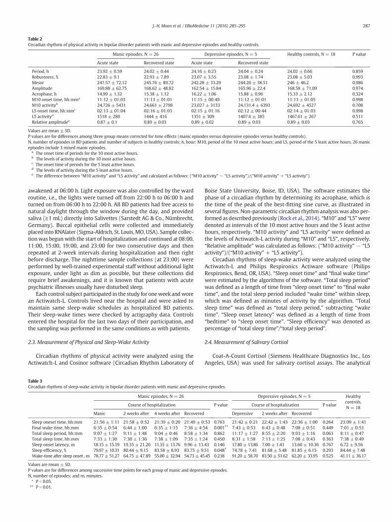
Table 2Circadian rhythms of physical activity in bipolar disorder patients with manic and depressive episodes and healthy controls.
Manic episodes, N = 26 Depressive episodes, N = 5 Healthy controls, N = 18 P value
Acute state Recovered state Acute state Recovered state
Period, h 23.92 ± 0.59 24.02 ± 0.44 24.16 ± 0.23 24.04 ± 0.24 24.02 ± 0.66 0.859Robustness, % 22.83 ± 9.1 22.93 ± 7.89 23.07 ± 3.55 23.08 ± 1.74 23.08 ± 5.03 0.993Mesor 247.57 ± 72.12 245.76 ± 89.72 242.28 ± 33.29 244.26 ± 38.51 246 ± 46.2 0.986Amplitude 169.88 ± 62.75 168.62 ± 48.82 162.54 ± 15.84 165.96 ± 22.4 168.58 ± 71.09 0.974Acrophase, h 14.99 ± 1.32 15.38 ± 1.12 16.22 ± 1.06 15.88 ± 0.96 15.33 ± 2.12 0.324M10 onset time, hh:mma 11:12 ± 01:03 11:13 ± 01:01 11:15 ± 00:49 11:12 ± 01:01 11:13 ± 01:05 0.998M10 activityb 24,726 ± 5431 24,661 ± 2798 23,027 ± 3133 24,131.4 ± 6393 24,692 ± 4527 0.708L5 onset time, hh:mmc 02:13 ± 01:04 02:16 ± 01:03 02:15 ± 01:16 02:12 ± 00:44 02:14 ± 01:03 0.998L5 activityd 1518 ± 280 1444 ± 416 1351 ± 309 1407.6 ± 385 1467.61 ± 267 0.511Relative amplitudee 0.87 ± 0.1 0.89 ± 0.03 0.89 ± 0.02 0.89 ± 0.03 0.89 ± 0.03 0.765
Values are mean ± SD.P values are for differences among three group means corrected for time effects (manic episodes versus depressive episodes versus healthy controls).N, number of episodes in BD patients and number of subjects in healthy controls; h, hour; M10, period of the 10 most active hours; and L5, period of the 5 least active hours. 26 manicepisodes include 3 mixed manic episodes.
a The onset time of periods for the 10 most active hours.b The levels of activity during the 10 most active hours.c The onset time of periods for the 5 least active hours.d The levels of activity during the 5 least active hours.e The difference between “M10 activity” and “L5 activity” and calculated as follows: (“M10 activity” − “L5 activity”)/(“M10 activity” + “L5 activity”).
287J.-H. Moon et al. / EBioMedicine 11 (2016) 285–295
awakened at 06:00 h. Light exposure was also controlled by the wardroutine, i.e., the lights were turned off from 22:00 h to 06:00 h andturned on from 06:00 h to 22:00 h. All BD patients had free access tonatural daylight through the window during the day, and providedsaliva (≥1 mL) directly into Salivettes (Sarstedt AG & Co., Nümbrecht,Germany). Buccal epithelial cells were collected and immediatelyplaced into RNAlater (Sigma-Aldrich, St. Louis,MO,USA). Sample collec-tionwas begunwith the start of hospitalization and continued at 08:00,11:00, 15:00, 19:00, and 23:00 for two consecutive days and thenrepeated at 2-week intervals during hospitalization and then rightbefore discharge. The nighttime sample collections (at 23:00) wereperformed by well-trained experimental staff without additional lightexposure, under light as dim as possible, but these collections didrequire brief awakenings, and it is known that patients with acutepsychiatric illnesses usually have disturbed sleep.
Each control subject participated in the study for oneweek andworean Actiwatch-L. Controls lived near the hospital and were asked tomaintain same sleep-wake schedules as hospitalized BD patients.Their sleep-wake times were checked by actigraphy data. Controlsentered the hospital for the last two days of their participation, andthe sampling was performed in the same conditions as with patients.
2.3. Measurement of Physical and Sleep-Wake Activity
Circadian rhythms of physical activity were analyzed using theActiwatch-L and Cosinor software (Circadian Rhythm Laboratory of
Table 3Circadian rhythms of sleep-wake activity in bipolar disorder patients with manic and depressi
Manic episodes, N = 26
Course of hospitalization
Manic 2 weeks after 4 weeks after Recovered
Sleep oneset time, hh:mm 21:56 ± 1:11 21:58 ± 0:52 21:39 ± 0:20 21:49 ± 0Final wake time, hh:mm 6:35 ± 0:54 6:44 ± 1:00 6:35 ± 1:15 7:36 ± 4:5Total sleep period, hh:mm 9:07 ± 1:27 9:11 ± 1:48 9:04 ± 0:46 8:58 ± 1:3Total sleep time, hh:mm 7:33 ± 1:30 7:30 ± 1:36 7:38 ± 1:09 7:35 ± 1:2Sleep onset latency, m 18.15 ± 15.19 19.35 ± 21.20 11.33 ± 13.76 9.96 ± 13Sleep efficiency, % 79.97 ± 10.31 80.44 ± 9.15 83.58 ± 8.93 83.75 ± 9Wake-time after sleep onset , m 78.77 ± 51.27 64.75 ± 47.89 55.00 ± 32.94 54.73 ± 45
Values are mean ± SD.P values are for differences among successive time points for each group of manic and depressN, number of episodes; and m, minutes.⁎ P b 0.05.⁎⁎ P b 0.01.
Boise State University, Boise, ID, USA). The software estimates thephase of a circadian rhythm by determining its acrophase, which isthe time of the peak of the best-fitting sine curve, as illustrated inseveral figures. Non-parametric circadian rhythm analysis was also per-formed as described previously (Rock et al., 2014). “M10” and “L5”weredenoted as intervals of the 10 most active hours and the 5 least activehours, respectively. “M10 activity” and “L5 activity” were defined asthe levels of Actiwatch-L activity during “M10” and “L5”, respectively.“Relative amplitude” was calculated as follows: (“M10 activity” − “L5activity”)/(“M10 activity” + “L5 activity”).
Circadian rhythms of sleep-wake activity were analyzed using theActiwatch-L and Philips Respironics Actiware software (PhilipsRespironics, Bend, OR, USA). “Sleep onset time” and “final wake time”were estimated by the algorithms of the software. “Total sleep period”was defined as a length of time from “sleep onset time” to “final waketime”, and the total sleep period included “wake time” within sleep,which was defined as minutes of activity by the algorithm. “Totalsleep time” was defined as “total sleep period,” subtracting “waketime”. “Sleep onset latency” was defined as a length of time from“bedtime” to “sleep onset time”. “Sleep efficiency” was denoted aspercentage of “total sleep time”/“total sleep period”.
2.4. Measurement of Salivary Cortisol
Coat-A-Count Cortisol (Siemens Healthcare Diagnostics Inc., LosAngeles, USA) was used for salivary cortisol assays. The analytical
ve episodes.
Depressive episodes, N = 5 Healthycontrols,N = 18
P value Course of hospitalization P value
Depressive 2 weeks after Recovered
:53 0.763 21:42 ± 0:21 22:42 ± 1:43 22:36 ± 1:00 0.264 23:09 ± 1:414 0.001⁎⁎ 7:43 ± 0:53 6:43 ± 0:48 7:08 ± 0:51 0.449 7:01 ± 0:534 0.862 11:17 ± 1:27 8:55 ± 2:20 9:03 ± 1:16 0.063 8:11 ± 0:474 0.450 8:31 ± 1:58 7:13 ± 1:25 7:08 ± 0:43 0.363 7:38 ± 0:49.43 0.146 17.80± 13.86 7.00 ± 1.41 13.60 ± 10.36 0.767 6.72 ± 9.56.51 0.048⁎ 74.78 ± 7.41 81.68 ± 5.48 81.85 ± 6.15 0.293 84.44 ± 7.48.45 0.238 91.20± 58.70 83.50 ± 51.62 62.20 ± 33.95 0.525 41.11 ± 36.17
ive episodes.

288 J.-H. Moon et al. / EBioMedicine 11 (2016) 285–295
289J.-H. Moon et al. / EBioMedicine 11 (2016) 285–295
sensitivity was 0.01 μg/dL. The intra-assay coefficient of variation was3% for samples of 0.19 ± 0.10 μg/dL and 4% for samples of 0.24 ±0.02 μg/dL. The inter-assay coefficient of variation was 12% for samplesof 1.85 ± 0.10 μg/dL and 14% for samples of 0.24 ± 0.02 μg/dL.
2.5. Measurement of Circadian Gene Expression
To observe circadian rhythms of gene expression, we tested fivecircadian clock genes (ARNTL, PER1, PER2, PER3, and NR1D1) extractedfrom buccal epithelial cells, and observed more relevant circadianrhythms of ARNTL and PER1 than in the other genes. The circadianrhythms of ARNTL and PER1 were inverse in phase to each other, inagreement with previous studies of circadian oscillation (Akashi et al.,2010; Chung et al., 2014; Guo et al., 2006; Novakova et al., 2015; Sonet al., 2008). Accordingly, to obtain the most distinguishable circadianrhythms, we investigated the relative gene expression of ARNTL andPER1, as performed and validated in a previous study (Cho et al., 2016;Guo et al., 2006). Briefly put, circadian gene expression levels of PER1and ARNTL were determined by RT-qPCR: then their ratios of PER1/ARNTL at each sampling time were used to describe circadian rhythms.Total RNA was isolated using RNeasy Micro Kit (Qiagen Inc., Valencia,CA, USA) from buccal epithelial cells, and only RNA samples concentrat-ed to N200 ng/μL were subjected to next step. The whole RNA samplewas reverse-transcribed using the Sensiscript Reverse TranscriptionKit (Qiagen). Then, cDNA was subjected to the Taqman PCR reactionusing an Applied Biosystems StepOnePlus Real-Time PCR Systems(ThermoFisher Scientific, Foster City, CA, USA). The primers and Taqmanprobes used in this experiment are as follows; PER1 (NM_002616):forward 5′-CTCACACAGCTCCTCCTCAG-3′, reverse 5′-TTTGTGCTCTTGCTGCTCTC-3′, probe 5′FAM-CGGCAAGGACTCAGCCCTGC-3′BHQ1;ARNTL (NM_001030272): forward 5′-TGCCTCGTCGCAATTGG-3′, re-verse 5′-ACCCTGATTTCCCCGTTCA-3′, probe 5’FAM-CGACTGCATTCTCATGTAGTTCCACAACCA-3′BHQ1.
2.6. Sine Regression Analysis
Biochemical rhythms were fitted with sine curves using SigmaPlotsoftware (Systat Software Inc., San Jose, CA, USA).
2.7. Statistical Analysis
Collected data were summarized using mean ± SD and frequenciesfor continuous variables and categorical variables, respectively. Weconducted Fisher's exact tests and Wilcoxon rank sum tests in order tocompare general characteristics of categorical and continuous variablesamong groups, respectively. For the analyses of physical, sleep-wakeactivity, cortisol and gene expression, we used marginal models withunstructured variance-covariance to compare groups and timesaccounting for within subject correlation. In addition, we also consid-ered the within-episode correlations, since episodes are nested withinsubjects in these models. Post-hoc tests were applied if needed usingTukey-Kramer tests which were proper for repeated measures data.All analyses were implemented by SAS version 9.4 for Windows (SASInstitute Inc., Cary, NC, USA).
Fig. 1. The shifting of acrophases of circadian rhythms in bipolar disorder patients. Note thattransitions of salivary cortisol circadian rhythms in bipolar patients compared to controls asARNTL expression circadian rhythms in bipolar patients compared to controls. The outerAcrophases of circadian rhythms of bipolar patients were observed over the course of hospievery two weeks into recovery states (at the end of hospitalization). The arrow presents threcovery state of each patient. If the duration of hospitalization was no longer than 2 weeks,solid circle at the time of the first acute acrophase assessment and the head of the arrow pointthere were sometimes intermediate and final assessments, with acrophases illustrated as tacrophase of circadian rhythms in manic episodes, green arrows indicate that of mixed episorange of acrophases for the healthy controls. The radius of each arc was arbitrarily chosen to ccircadian rhythms.
2.8. Role of the Funding Source
The funder of the study had no role in study design, data collection,data analysis, data interpretation, or writing of the report. The corre-sponding author had full access to all the data in the study and hadfinal responsibility for the decision to submit for publication.
3. Results
3.1. Circadian Rhythms of Physical and Sleep-Wake Activity
Patients did not show significant differences in any parametersfor physical activity when compared to healthy controls, neither inrecovered nor during acute mood exacerbations (Table 2).
Analyses of sleep parameters of manic episodes found significantchanges over time in group means for final wake time and sleepefficiency (Table 3). The final wake time was delayed about 1 h andthe sleep efficiency was increased about 4% during hospitalizationfrom admission to recovery. In contrast, analyses of depressive episodesfound no significant differences over time for any sleep parameters.
3.2. Biochemical Circadian Rhythms
Changes in biochemical circadian rhythms from acute to recoverystates of BD were observed (Fig. 1). Biochemical circadian rhythms ofacute mood episodes commenced with different phases compared tothose of the recovered states.
Fig. 1 and Table 4 clearly show that the biochemical circadianrhythms of acute manic and depressive episodes were different fromthose of controls. Notably, the biochemical circadian rhythms in salivarycortisol concentrations and PER1/ARNTL expression ratios had contrast-ing initial phases during the acute mood episodes, but transitioned bycomparable phase shifts during recovery, almost entirely arriving with-in the normal control ranges.
3.3. Healthy Controls
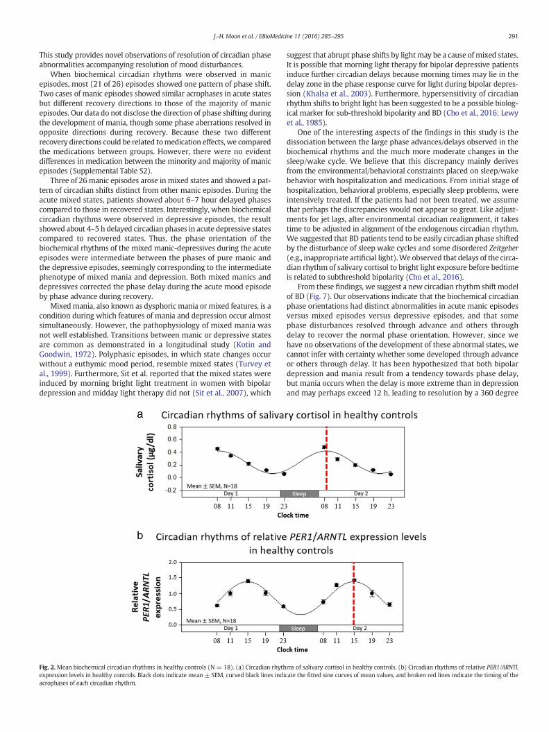
The biochemical circadian rhythms in control subjects are depictedin Table 4 and Fig. 2.
3.4. Manic Episodes
Of 26manic episodes, 21 episodes showed similar phase shifts of thebiochemical circadian rhythms during hospitalization (Figs. 1 and 3).Significant differences of acrophase of cortisol circadian rhythms wereobserved in acute manic states and after 2 weeks of hospitalizationwhen compared with recovered states (Table 4). Similarly, acrophasesof gene expression circadian rhythms also showed significant differ-ences between acute manic states and after 2 weeks of hospitalizationwhen compared with recovered states (Table 4). In sum, at admission,most of themanic episodes showed about 7 hour advanced biochemicalcircadian acrophases in acute states (equivalent to 17 hour delayed),and recovered through delays (clockwise), returning to the normalrange of the control group. Moreover, a significant increase in thePER1/ARNTL circadian amplitude accompanying recovery from mania
the acrophase is the timing of the peak of the best-fitting sine curve. (a) The acrophasethe circadian peaks shifted in timing. (b) The acrophase transitions of the relative PER1/circular axis indicates the clock time of each acrophase assessment for each patient.talization. Evaluation was performed at acute states (near start of hospitalization), ande direction of change in acrophase of the circadian rhythm from the acute state to thethere were only initial and final assessments, so the acrophase arc is an arrow that has aing to the recovery acrophase. If the duration of hospitalization was longer than 2 weeks,wo or three arrow heads along the arc. Red and orange arrows indicate the shifting ofdes, and blue arrows indicate that of depressive episodes. The gray sector indicates theontrast the different groups of participants, and does not represent the amplitudes of the
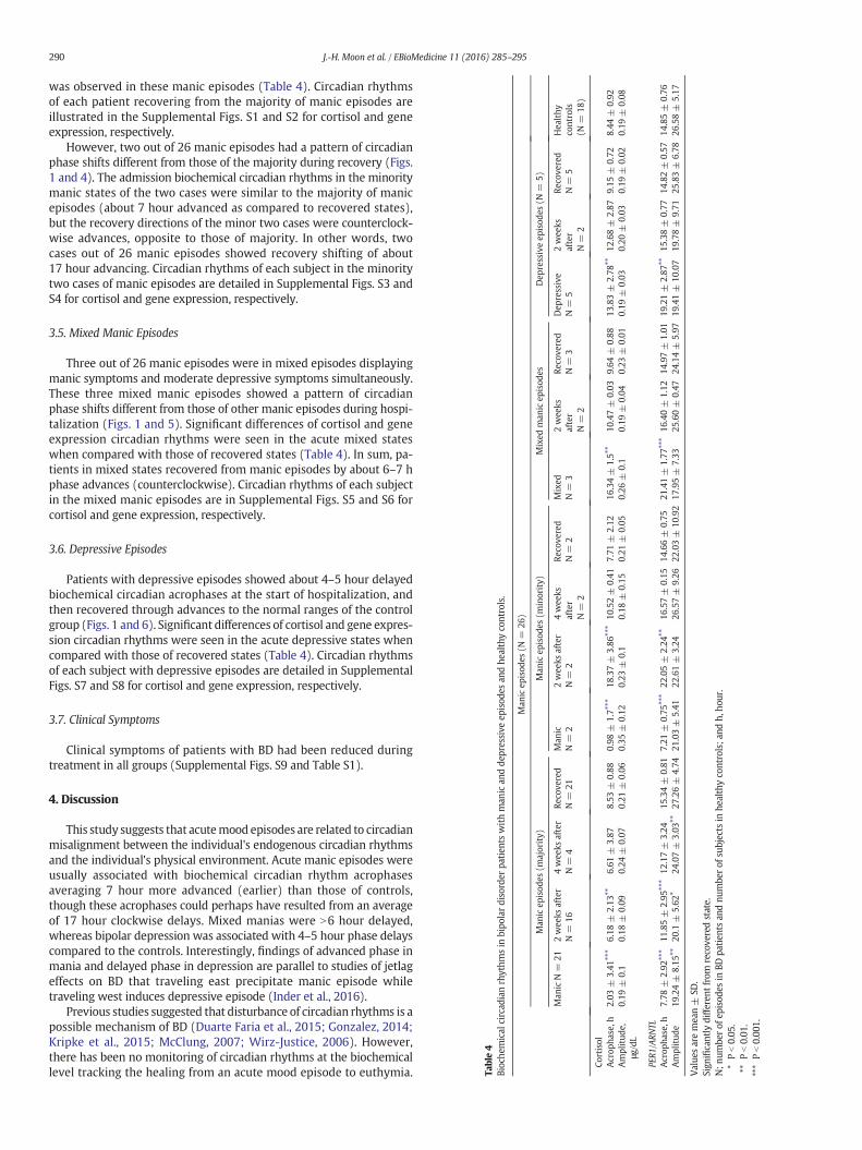
Table4
Bioche
mical
circad
ianrhythm
sin
bipo
lardisorder
patien
tswithman
ican
dde
pressive
episod
esan
dhe
althycontrols.
Man
icep
isod
es(N
=26
)
Man
icep
isod
es(m
ajority)
Man
icep
isod
es(m
inority)
Mixed
man
icep
isod
esDep
ressiveep
isod
es(N
=5)
Man
icN=
212wee
ksafter
N=
164wee
ksafter
N=
4Re
cove
red
N=
21Man
icN=
22wee
ksafter
N=
24wee
ksafter
N=
2
Reco
vered
N=
2Mixed
N=
32wee
ksafter
N=
2
Reco
vered
N=
3Dep
ressive
N=
52wee
ksafter
N=
2
Reco
vered
N=
5Hea
lthy
controls
(N=
18)
Cortisol
Acrop
hase,h
2.03
±3.41
⁎⁎⁎
6.18
±2.13
⁎⁎6.61
±3.87
8.53
±0.88
0.98
±1.7⁎⁎⁎
18.37±
3.86
⁎⁎⁎
10.52±
0.41
7.71
±2.12
16.34±
1.5⁎⁎
10.47±
0.03
9.64
±0.88
13.83±
2.78
⁎⁎12
.68±
2.87
9.15
±0.72
8.44
±0.92
Amplitud
e,μg
/dL
0.19
±0.1
0.18
±0.09
0.24
±0.07
0.21
±0.06
0.35
±0.12
0.23
±0.1
0.18
±0.15
0.21
±0.05
0.26
±0.1
0.19
±0.04
0.23
±0.01
0.19
±0.03
0.20
±0.03
0.19
±0.02
0.19
±0.08
PER1
/ARN
TLAcrop
hase,h
7.78
±2.92
⁎⁎⁎
11.85±
2.95
⁎⁎⁎
12.17±
3.24
15.34±
0.81
7.21
±0.75
⁎⁎⁎
22.05±
2.24
⁎⁎16
.57±
0.15
14.66±
0.75
21.41±
1.77
⁎⁎⁎
16.40±
1.12
14.97±
1.01
19.21±
2.87
⁎⁎15
.38±
0.77
14.82±
0.57
14.85±
0.76
Amplitud
e19
.24±
8.15
⁎⁎20
.1±
5.62
⁎24
.07±
3.03
⁎⁎27
.26±
4.74
21.03±
5.41
22.61±
3.24
26.57±
9.26
22.03±
10.92
17.95±
7.33
25.60±
0.47
24.14±
5.97
19.41±
10.07
19.78±
9.71
25.83±
6.78
26.58±
5.17
Value
saremean±
SD.
Sign
ificantly
diffe
rent
from
recove
redstate.
N;n
umbe
rof
episod
esin
BDpa
tien
tsan
dnu
mbe
rof
subjects
inhe
althyco
ntrols;a
ndh,
hour.
⁎Pb0.05
.⁎⁎
Pb0.01
.⁎⁎⁎
Pb0.00
1.
290 J.-H. Moon et al. / EBioMedicine 11 (2016) 285–295
was observed in these manic episodes (Table 4). Circadian rhythmsof each patient recovering from the majority of manic episodes areillustrated in the Supplemental Figs. S1 and S2 for cortisol and geneexpression, respectively.
However, two out of 26 manic episodes had a pattern of circadianphase shifts different from those of the majority during recovery (Figs.1 and 4). The admission biochemical circadian rhythms in the minoritymanic states of the two cases were similar to the majority of manicepisodes (about 7 hour advanced as compared to recovered states),but the recovery directions of the minor two cases were counterclock-wise advances, opposite to those of majority. In other words, twocases out of 26 manic episodes showed recovery shifting of about17 hour advancing. Circadian rhythms of each subject in the minoritytwo cases of manic episodes are detailed in Supplemental Figs. S3 andS4 for cortisol and gene expression, respectively.
3.5. Mixed Manic Episodes
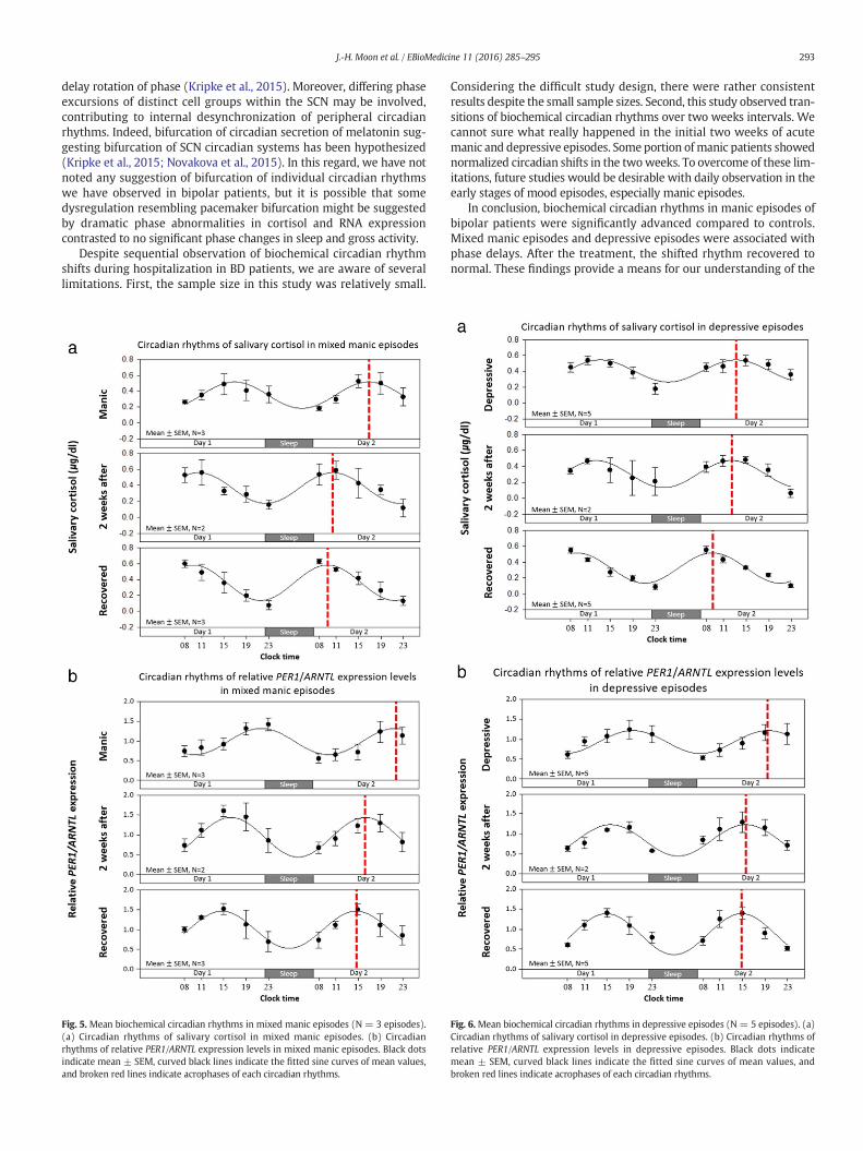
Three out of 26 manic episodes were in mixed episodes displayingmanic symptoms and moderate depressive symptoms simultaneously.These three mixed manic episodes showed a pattern of circadianphase shifts different from those of other manic episodes during hospi-talization (Figs. 1 and 5). Significant differences of cortisol and geneexpression circadian rhythms were seen in the acute mixed stateswhen compared with those of recovered states (Table 4). In sum, pa-tients in mixed states recovered from manic episodes by about 6–7 hphase advances (counterclockwise). Circadian rhythms of each subjectin the mixed manic episodes are in Supplemental Figs. S5 and S6 forcortisol and gene expression, respectively.
3.6. Depressive Episodes
Patients with depressive episodes showed about 4–5 hour delayedbiochemical circadian acrophases at the start of hospitalization, andthen recovered through advances to the normal ranges of the controlgroup (Figs. 1 and 6). Significant differences of cortisol and gene expres-sion circadian rhythms were seen in the acute depressive states whencompared with those of recovered states (Table 4). Circadian rhythmsof each subject with depressive episodes are detailed in SupplementalFigs. S7 and S8 for cortisol and gene expression, respectively.
3.7. Clinical Symptoms
Clinical symptoms of patients with BD had been reduced duringtreatment in all groups (Supplemental Figs. S9 and Table S1).
4. Discussion
This study suggests that acutemood episodes are related to circadianmisalignment between the individual's endogenous circadian rhythmsand the individual's physical environment. Acute manic episodes wereusually associated with biochemical circadian rhythm acrophasesaveraging 7 hour more advanced (earlier) than those of controls,though these acrophases could perhaps have resulted from an averageof 17 hour clockwise delays. Mixed manias were N6 hour delayed,whereas bipolar depression was associated with 4–5 hour phase delayscompared to the controls. Interestingly, findings of advanced phase inmania and delayed phase in depression are parallel to studies of jetlageffects on BD that traveling east precipitate manic episode whiletraveling west induces depressive episode (Inder et al., 2016).
Previous studies suggested that disturbance of circadian rhythms is apossible mechanism of BD (Duarte Faria et al., 2015; Gonzalez, 2014;Kripke et al., 2015; McClung, 2007; Wirz-Justice, 2006). However,there has been no monitoring of circadian rhythms at the biochemicallevel tracking the healing from an acute mood episode to euthymia.
291J.-H. Moon et al. / EBioMedicine 11 (2016) 285–295
This study provides novel observations of resolution of circadian phaseabnormalities accompanying resolution of mood disturbances.
When biochemical circadian rhythms were observed in manicepisodes, most (21 of 26) episodes showed one pattern of phase shift.Two cases of manic episodes showed similar acrophases in acute statesbut different recovery directions to those of the majority of manicepisodes. Our data do not disclose the direction of phase shifting duringthe development of mania, though some phase aberrations resolved inopposite directions during recovery. Because these two differentrecovery directions could be related tomedication effects, we comparedthe medications between groups. However, there were no evidentdifferences in medication between the minority and majority of manicepisodes (Supplemental Table S2).
Three of 26manic episodes arose in mixed states and showed a pat-tern of circadian shifts distinct from other manic episodes. During theacute mixed states, patients showed about 6–7 hour delayed phasescompared to those in recovered states. Interestingly, when biochemicalcircadian rhythms were observed in depressive episodes, the resultshowed about 4–5 h delayed circadian phases in acute depressive statescompared to recovered states. Thus, the phase orientation of thebiochemical rhythms of the mixed manic-depressives during the acuteepisodes were intermediate between the phases of pure manic andthe depressive episodes, seemingly corresponding to the intermediatephenotype of mixed mania and depression. Both mixed manics anddepressives corrected the phase delay during the acute mood episodeby phase advance during recovery.
Mixedmania, also known as dysphoric mania or mixed features, is acondition during which features of mania and depression occur almostsimultaneously. However, the pathophysiology of mixed mania wasnot well established. Transitions between manic or depressive statesare common as demonstrated in a longitudinal study (Kotin andGoodwin, 1972). Polyphasic episodes, in which state changes occurwithout a euthymic mood period, resemble mixed states (Turvey etal., 1999). Furthermore, Sit et al. reported that the mixed states wereinduced by morning bright light treatment in women with bipolardepression and midday light therapy did not (Sit et al., 2007), which
Fig. 2. Mean biochemical circadian rhythms in healthy controls (N = 18). (a) Circadian rhythexpression levels in healthy controls. Black dots indicate mean ± SEM, curved black lines indiacrophases of each circadian rhythm.
suggest that abrupt phase shifts by lightmay be a cause of mixed states.It is possible that morning light therapy for bipolar depressive patientsinduce further circadian delays because morning times may lie in thedelay zone in the phase response curve for light during bipolar depres-sion (Khalsa et al., 2003). Furthermore, hypersensitivity of circadianrhythm shifts to bright light has been suggested to be a possible biolog-ical marker for sub-threshold bipolarity and BD (Cho et al., 2016; Lewyet al., 1985).
One of the interesting aspects of the findings in this study is thedissociation between the large phase advances/delays observed in thebiochemical rhythms and the much more moderate changes in thesleep/wake cycle. We believe that this discrepancy mainly derivesfrom the environmental/behavioral constraints placed on sleep/wakebehavior with hospitalization and medications. From initial stage ofhospitalization, behavioral problems, especially sleep problems, wereintensively treated. If the patients had not been treated, we assumethat perhaps the discrepancies would not appear so great. Like adjust-ments for jet lags, after environmental circadian realignment, it takestime to be adjusted in alignment of the endogenous circadian rhythm.We suggested that BD patients tend to be easily circadian phase shiftedby the disturbance of sleep wake cycles and some disordered Zeitgeber(e.g., inappropriate artificial light).We observed that delays of the circa-dian rhythm of salivary cortisol to bright light exposure before bedtimeis related to subthreshold bipolarity (Cho et al., 2016).
From these findings, we suggest a new circadian rhythm shift modelof BD (Fig. 7). Our observations indicate that the biochemical circadianphase orientations had distinct abnormalities in acute manic episodesversus mixed episodes versus depressive episodes, and that somephase disturbances resolved through advance and others throughdelay to recover the normal phase orientation. However, since wehave no observations of the development of these abnormal states, wecannot infer with certainty whether some developed through advanceor others through delay. It has been hypothesized that both bipolardepression and mania result from a tendency towards phase delay,but mania occurs when the delay is more extreme than in depressionand may perhaps exceed 12 h, leading to resolution by a 360 degree
ms of salivary cortisol in healthy controls. (b) Circadian rhythms of relative PER1/ARNTLcate the fitted sine curves of mean values, and broken red lines indicate the timing of the
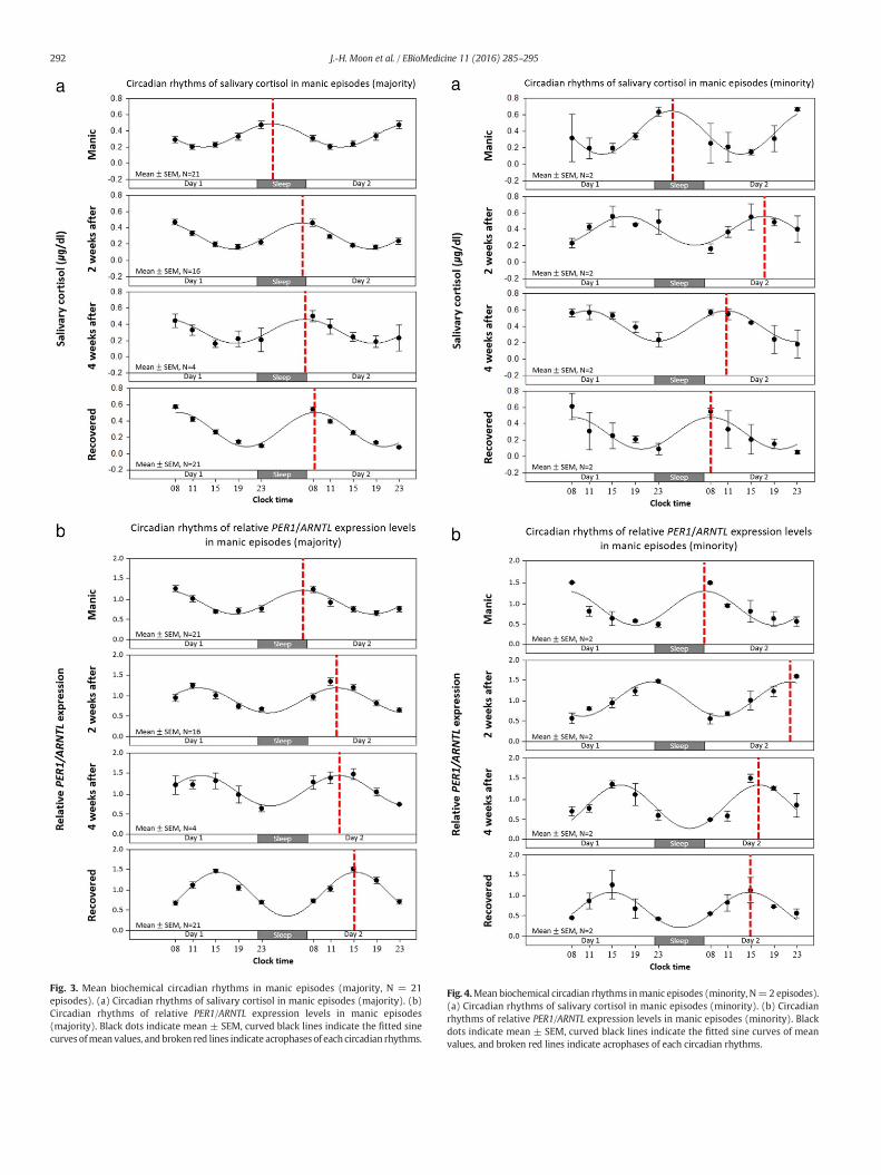
Fig. 3. Mean biochemical circadian rhythms in manic episodes (majority, N = 21episodes). (a) Circadian rhythms of salivary cortisol in manic episodes (majority). (b)Circadian rhythms of relative PER1/ARNTL expression levels in manic episodes(majority). Black dots indicate mean ± SEM, curved black lines indicate the fitted sinecurves ofmean values, and broken red lines indicate acrophases of each circadian rhythms.
Fig. 4.Mean biochemical circadian rhythms inmanic episodes (minority, N=2 episodes).(a) Circadian rhythms of salivary cortisol in manic episodes (minority). (b) Circadianrhythms of relative PER1/ARNTL expression levels in manic episodes (minority). Blackdots indicate mean ± SEM, curved black lines indicate the fitted sine curves of meanvalues, and broken red lines indicate acrophases of each circadian rhythms.
292 J.-H. Moon et al. / EBioMedicine 11 (2016) 285–295
293J.-H. Moon et al. / EBioMedicine 11 (2016) 285–295
delay rotation of phase (Kripke et al., 2015). Moreover, differing phaseexcursions of distinct cell groups within the SCN may be involved,contributing to internal desynchronization of peripheral circadianrhythms. Indeed, bifurcation of circadian secretion of melatonin sug-gesting bifurcation of SCN circadian systems has been hypothesized(Kripke et al., 2015; Novakova et al., 2015). In this regard, we have notnoted any suggestion of bifurcation of individual circadian rhythmswe have observed in bipolar patients, but it is possible that somedysregulation resembling pacemaker bifurcation might be suggestedby dramatic phase abnormalities in cortisol and RNA expressioncontrasted to no significant phase changes in sleep and gross activity.
Despite sequential observation of biochemical circadian rhythmshifts during hospitalization in BD patients, we are aware of severallimitations. First, the sample size in this study was relatively small.
Fig. 5. Mean biochemical circadian rhythms in mixed manic episodes (N = 3 episodes).(a) Circadian rhythms of salivary cortisol in mixed manic episodes. (b) Circadianrhythms of relative PER1/ARNTL expression levels in mixed manic episodes. Black dotsindicate mean ± SEM, curved black lines indicate the fitted sine curves of mean values,and broken red lines indicate acrophases of each circadian rhythms.
Considering the difficult study design, there were rather consistentresults despite the small sample sizes. Second, this study observed tran-sitions of biochemical circadian rhythms over two weeks intervals. Wecannot sure what really happened in the initial two weeks of acutemanic and depressive episodes. Some portion of manic patients showednormalized circadian shifts in the twoweeks. To overcome of these lim-itations, future studies would be desirable with daily observation in theearly stages of mood episodes, especially manic episodes.
In conclusion, biochemical circadian rhythms in manic episodes ofbipolar patients were significantly advanced compared to controls.Mixed manic episodes and depressive episodes were associated withphase delays. After the treatment, the shifted rhythm recovered tonormal. These findings provide a means for our understanding of the
Fig. 6.Mean biochemical circadian rhythms in depressive episodes (N = 5 episodes). (a)Circadian rhythms of salivary cortisol in depressive episodes. (b) Circadian rhythms ofrelative PER1/ARNTL expression levels in depressive episodes. Black dots indicatemean ± SEM, curved black lines indicate the fitted sine curves of mean values, andbroken red lines indicate acrophases of each circadian rhythms.
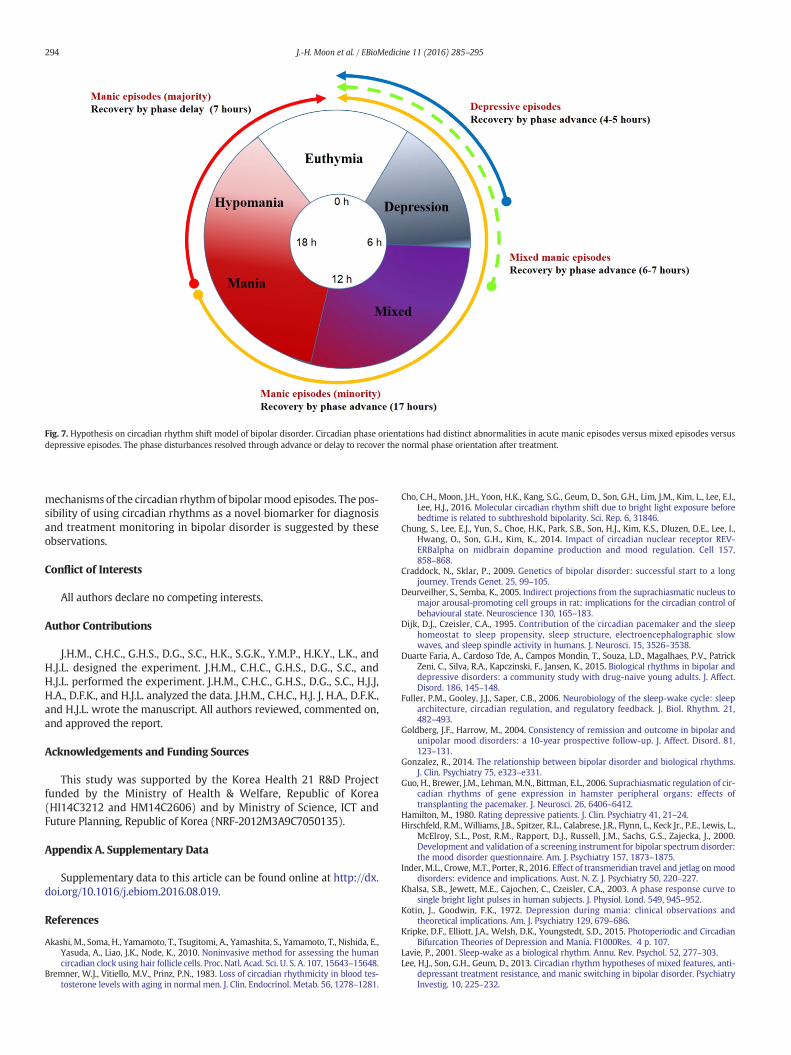
Fig. 7. Hypothesis on circadian rhythm shift model of bipolar disorder. Circadian phase orientations had distinct abnormalities in acute manic episodes versus mixed episodes versusdepressive episodes. The phase disturbances resolved through advance or delay to recover the normal phase orientation after treatment.
294 J.-H. Moon et al. / EBioMedicine 11 (2016) 285–295
mechanisms of the circadian rhythmof bipolarmood episodes. The pos-sibility of using circadian rhythms as a novel biomarker for diagnosisand treatment monitoring in bipolar disorder is suggested by theseobservations.
Conflict of Interests
All authors declare no competing interests.
Author Contributions
J.H.M., C.H.C., G.H.S., D.G., S.C., H.K., S.G.K., Y.M.P., H.K.Y., L.K., andH.J.L. designed the experiment. J.H.M., C.H.C., G.H.S., D.G., S.C., andH.J.L. performed the experiment. J.H.M., C.H.C., G.H.S., D.G., S.C., H.J.J,H.A., D.F.K., and H.J.L. analyzed the data. J.H.M., C.H.C., H.J. J, H.A., D.F.K.,and H.J.L. wrote the manuscript. All authors reviewed, commented on,and approved the report.
Acknowledgements and Funding Sources
This study was supported by the Korea Health 21 R&D Projectfunded by the Ministry of Health & Welfare, Republic of Korea(HI14C3212 and HM14C2606) and by Ministry of Science, ICT andFuture Planning, Republic of Korea (NRF-2012M3A9C7050135).
Appendix A. Supplementary Data
Supplementary data to this article can be found online at http://dx.doi.org/10.1016/j.ebiom.2016.08.019.
References
Akashi, M., Soma, H., Yamamoto, T., Tsugitomi, A., Yamashita, S., Yamamoto, T., Nishida, E.,Yasuda, A., Liao, J.K., Node, K., 2010. Noninvasive method for assessing the humancircadian clock using hair follicle cells. Proc. Natl. Acad. Sci. U. S. A. 107, 15643–15648.
Bremner, W.J., Vitiello, M.V., Prinz, P.N., 1983. Loss of circadian rhythmicity in blood tes-tosterone levels with aging in normal men. J. Clin. Endocrinol. Metab. 56, 1278–1281.
Cho, C.H., Moon, J.H., Yoon, H.K., Kang, S.G., Geum, D., Son, G.H., Lim, J.M., Kim, L., Lee, E.I.,Lee, H.J., 2016. Molecular circadian rhythm shift due to bright light exposure beforebedtime is related to subthreshold bipolarity. Sci. Rep. 6, 31846.
Chung, S., Lee, E.J., Yun, S., Choe, H.K., Park, S.B., Son, H.J., Kim, K.S., Dluzen, D.E., Lee, I.,Hwang, O., Son, G.H., Kim, K., 2014. Impact of circadian nuclear receptor REV-ERBalpha on midbrain dopamine production and mood regulation. Cell 157,858–868.
Craddock, N., Sklar, P., 2009. Genetics of bipolar disorder: successful start to a longjourney. Trends Genet. 25, 99–105.
Deurveilher, S., Semba, K., 2005. Indirect projections from the suprachiasmatic nucleus tomajor arousal-promoting cell groups in rat: implications for the circadian control ofbehavioural state. Neuroscience 130, 165–183.
Dijk, D.J., Czeisler, C.A., 1995. Contribution of the circadian pacemaker and the sleephomeostat to sleep propensity, sleep structure, electroencephalographic slowwaves, and sleep spindle activity in humans. J. Neurosci. 15, 3526–3538.
Duarte Faria, A., Cardoso Tde, A., Campos Mondin, T., Souza, L.D., Magalhaes, P.V., PatrickZeni, C., Silva, R.A., Kapczinski, F., Jansen, K., 2015. Biological rhythms in bipolar anddepressive disorders: a community study with drug-naive young adults. J. Affect.Disord. 186, 145–148.
Fuller, P.M., Gooley, J.J., Saper, C.B., 2006. Neurobiology of the sleep-wake cycle: sleeparchitecture, circadian regulation, and regulatory feedback. J. Biol. Rhythm. 21,482–493.
Goldberg, J.F., Harrow, M., 2004. Consistency of remission and outcome in bipolar andunipolar mood disorders: a 10-year prospective follow-up. J. Affect. Disord. 81,123–131.
Gonzalez, R., 2014. The relationship between bipolar disorder and biological rhythms.J. Clin. Psychiatry 75, e323–e331.
Guo, H., Brewer, J.M., Lehman, M.N., Bittman, E.L., 2006. Suprachiasmatic regulation of cir-cadian rhythms of gene expression in hamster peripheral organs: effects oftransplanting the pacemaker. J. Neurosci. 26, 6406–6412.
Hamilton, M., 1980. Rating depressive patients. J. Clin. Psychiatry 41, 21–24.Hirschfeld, R.M., Williams, J.B., Spitzer, R.L., Calabrese, J.R., Flynn, L., Keck Jr., P.E., Lewis, L.,
McElroy, S.L., Post, R.M., Rapport, D.J., Russell, J.M., Sachs, G.S., Zajecka, J., 2000.Development and validation of a screening instrument for bipolar spectrum disorder:the mood disorder questionnaire. Am. J. Psychiatry 157, 1873–1875.
Inder, M.L., Crowe, M.T., Porter, R., 2016. Effect of transmeridian travel and jetlag on mooddisorders: evidence and implications. Aust. N. Z. J. Psychiatry 50, 220–227.
Khalsa, S.B., Jewett, M.E., Cajochen, C., Czeisler, C.A., 2003. A phase response curve tosingle bright light pulses in human subjects. J. Physiol. Lond. 549, 945–952.
Kotin, J., Goodwin, F.K., 1972. Depression during mania: clinical observations andtheoretical implications. Am. J. Psychiatry 129, 679–686.
Kripke, D.F., Elliott, J.A., Welsh, D.K., Youngstedt, S.D., 2015. Photoperiodic and CircadianBifurcation Theories of Depression and Mania. F1000Res. 4 p. 107.
Lavie, P., 2001. Sleep-wake as a biological rhythm. Annu. Rev. Psychol. 52, 277–303.Lee, H.J., Son, G.H., Geum, D., 2013. Circadian rhythm hypotheses of mixed features, anti-
depressant treatment resistance, and manic switching in bipolar disorder. PsychiatryInvestig. 10, 225–232.
295J.-H. Moon et al. / EBioMedicine 11 (2016) 285–295
Lewy, A.J., Nurnberger Jr., J.I., Wehr, T.A., Pack, D., Becker, L.E., Powell, R.L., Newsome, D.A.,1985. Supersensitivity to light: possible trait marker for manic-depressive illness. Am.J. Psychiatry 142, 725–727.
Li, X., Bijur, G.N., Jope, R.S., 2002. Glycogen synthase kinase-3beta, mood stabilizers, andneuroprotection. Bipolar Disord. 4, 137–144.
Maciukiewicz, M., Dmitrzak-Weglarz, M., Pawlak, J., Leszczynska-Rodziewicz, A.,Zaremba, D., Skibinska, M., Hauser, J., 2014. Analysis of genetic association andepistasis interactions between circadian clock genes and symptom dimensions ofbipolar affective disorder. Chronobiol. Int. 31, 770–778.
McClung, C.A., 2007. Circadian genes, rhythms and the biology of mood disorders.Pharmacol. Ther. 114, 222–232.
Novakova, M., Prasko, J., Latalova, K., Sladek, M., Sumova, A., 2015. The circadian system ofpatients with bipolar disorder differs in episodes of mania and depression. BipolarDisord. 17, 303–314.
Nurnberger Jr., J.I., Adkins, S., Lahiri, D.K., Mayeda, A., Hu, K., Lewy, A., Miller, A., Bowman,E.S., Miller, M.J., Rau, L., Smiley, C., Davis-Singh, D., 2000. Melatonin suppression bylight in euthymic bipolar and unipolar patients. Arch. Gen. Psychiatry 57, 572–579.
Reppert, S.M., Weaver, D.R., 2002. Coordination of circadian timing in mammals. Nature418, 935–941.
Rock, P., Goodwin, G., Harmer, C., Wulff, K., 2014. Daily rest-activity patterns in the bipolarphenotype: a controlled actigraphy study. Chronobiol. Int. 31, 290–296.
Salvatore, P., Ghidini, S., Zita, G., De Panfilis, C., Lambertino, S., Maggini, C., Baldessarini,R.J., 2008. Circadian activity rhythm abnormalities in ill and recovered bipolar Idisorder patients. Bipolar Disord. 10, 256–265.
Saper, C.B., Scammell, T.E., Lu, J., 2005. Hypothalamic regulation of sleep and circadianrhythms. Nature 437, 1257–1263.
Schibler, U., Sassone-Corsi, P., 2002. A web of circadian pacemakers. Cell 111, 919–922.
Sit, D., Wisner, K.L., Hanusa, B.H., Stull, S., Terman, M., 2007. Light therapy for bipolardisorder: a case series in women. Bipolar Disord. 9, 918–927.
Son, G.H., Chung, S., Choe, H.K., Kim, H.D., Baik, S.M., Lee, H., Lee, H.W., Choi, S., Sun, W.,Kim, H., Cho, S., Lee, K.H., Kim, K., 2008. Adrenal peripheral clock controls the auton-omous circadian rhythm of glucocorticoid by causing rhythmic steroid production.Proc. Natl. Acad. Sci. U. S. A. 105, 20970–20975.
Spearing, M.K., Post, R.M., Leverich, G.S., Brandt, D., Nolen, W., 1997. Modification ofthe clinical global impressions (CGI) scale for use in bipolar illness (BP): the CGI-BP. Psychiatry Res. 73, 159–171.
Turvey, C.L., Coryell, W.H., Solomon, D.A., Leon, A.C., Endicott, J., Keller, M.B., Akiskal, H.,1999. Long-term prognosis of bipolar I disorder. Acta Psychiatr. Scand. 99, 110–119.
Wehr, T.A., Sack, D.A., Duncan, W.C., Mendelson, W.B., Rosenthal, N.E., Gillin, J.C.,Goodwin, F.K., 1985. Sleep and circadian rhythms in affective patients isolated fromexternal time cues. Psychiatry Res. 15, 327–339.
Wirz-Justice, A., 2006. Biological rhythm disturbances in mood disorders. Int. Clin.Psychopharmacol. 21 (Suppl 1), S11–S15.
Wood, J., Birmaher, B., Axelson, D., Ehmann, M., Kalas, C., Monk, K., Turkin, S., Kupfer, D.J.,Brent, D., Monk, T.H., Nimgainkar, V.L., 2009. Replicable differences in preferredcircadian phase between bipolar disorder patients and control individuals. PsychiatryRes. 166, 201–209.
Yoo, S., Kim, Y., Noh, J., Oh, K., Kim, C., Namkoong, K., Chae, J., Lee, G., Jeon, S., Min, K., 2006.Validity of Korean version of the mini-international neuropsychiatric interview.Anxiety Mood 2, 50–55.
Young, R.C., Biggs, J.T., Ziegler, V.E., Meyer, D.A., 1978. A rating scale for mania: reliability,validity and sensitivity. Br. J. Psychiatry 133, 429–435.