the inner ear
DESCRIPTION
Chapter from Kandel ER, et al. Principles of Neural Science. Fifth Edition. New York: McGraw-Hill Medical; 2013.TRANSCRIPT
30
The Inner Ear
The Ear Has Three Functional Parts
Hearing Commences with the Capture of Sound Energy by the Ear
The Hydrodynamic and Mechanical Apparatus of the Cochlea Delivers Mechanical Stimuli to the Receptor Cells
The Basilar Membrane Is a Mechanical Analyzer of Sound Frequency
The Organ of Corti Is the Site of Mechanoelectrical Transduction in the Cochlea
Hair Cells Transform Mechanical Energy into Neural Signals
Deflection of the Hair Bundle Initiates Mechanoelectrical Transduction
Mechanical Force Directly Opens Transduction Channels
Direct Mechanoelectrical Transduction Is Rapid
The Temporal Responsiveness of Hair Cells Determines Their Sensitivity
Hair Cells Adapt to Sustained Stimulation
Hair Cells Are Tuned to Specific Stimulus Frequencies
Sound Energy Is Mechanically Amplified in the Cochlea
Hair Cells Use Specialized Ribbon Synapses
Auditory Information Flows Initially Through the Cochlear Nerve
Bipolar Neurons in the Spiral Ganglion Innervate Cochlear Hair Cells
Cochlear Nerve Fibers Encode Stimulus Frequency and Intensity
Sensorineural Hearing Loss Is Common but Treatable
An Overall View
Human experience is enriched by our ability to distinguish a remarkable range of sounds—from the intimacy of a whisper to the warmth of
a conversation, from the complexity of a symphony to the roar of a stadium. Hearing begins when the cochlea, the snail-shaped receptor organ of the inner ear, trans-duces sound energy into electrical signals and forwards them to the brain. Our ability to recognize small differ-ences in sounds stems from the cochlea’s capacity to dis-tinguish among frequency components and to inform us of both the tones present and their amplitudes.
Deafness can be devastating. For the elderly, hearing loss can result in a painful and protracted estrangement from family, friends, and colleagues. Children may lack hearing as a result of pre- or perinatal infections and especially of genetic conditions, which affect one child in a thousand. Such children are often deprived of the normal avenue to the development of speech, and thus of reading and writing as well. It is for this reason that a modern pediatric examination must include an assess-ment of hearing. Many children thought to be cogni-tively impaired are found instead to be hard of hearing, and their intellectual development resumes its normal course when this problem is corrected.
Acute hearing loss in the intermediate years exacts an enormous price for two reasons. First, hearing plays an important, but often overlooked, role in our psycho-logical well-being. Daily conversation with family and colleagues helps to establish our social context. The abrupt loss of such social intercourse as a result of sud-den deafness leaves a person distressingly lonely and may lead to depression and even suicide. Hearing also serves us in another, more subtle way. Our auditory system is a remarkably efficient early warning system,
Chapter 30 / The Inner Ear 655
subconsciously informing us about our environment. For example, when other people enter the room or approach us we often hear them before we see them. More obviously, awareness of fire alarms and the sirens of emergency vehicles can be lifesaving. Deafness may leave a person with an ominous sense of vulnerability to unheard changes in the environment.
Hearing loss is often accompanied by another dis-tressing symptom, tinnitus, or ringing in the ears. By interfering with concentration and disrupting sleep, tinnitus can exasperate, depress, and even madden its victims. Because on rare occasions tinnitus stems from lesions to the auditory pathways, such as acous-tic neuromas, it is important in neurological diagno-sis to exclude such causes. Most tinnitus, however, is idiopathic: Its cause is uncertain. Some drugs trigger the condition; antimalarial drugs related to quinine and aspirin at the high dosages used in the treatment of rheumatoid arthritis are notorious for this. Often, however, tinnitus occurs at high frequencies to which a damaged ear is no longer sensitive. In these instances tinnitus may reflect hypersensitivity in the deaffer-ented central nervous system, a phenomenon analo-gous to phantom-limb pain (see Chapter 24).
Hearing depends on the remarkable properties of hair cells, the receptors of the internal ear. Hair cells receive mechanical inputs that correspond to sounds and transduce these signals into electrical responses
that are forwarded to the brain for interpretation. These cells can measure motions of atomic dimen-sions and transduce stimuli ranging from static inputs to those at frequencies of tens of kilohertz. Hair cells also serve as mechanical amplifiers that augment our auditory sensitivity. Each of the paired cochleas con-tains approximately 16,000 of these cells. Deterioration of hair cells accounts for most of the hearing loss that afflicts more than 30 million Americans.
The Ear Has Three Functional Parts
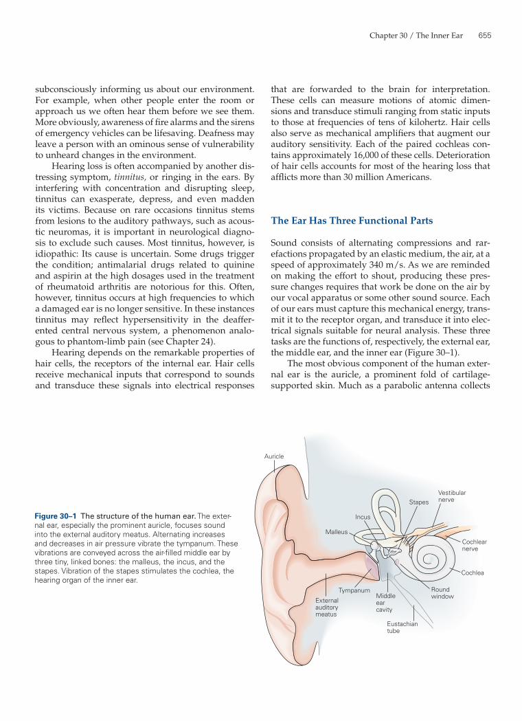
Sound consists of alternating compressions and rar-efactions propagated by an elastic medium, the air, at a speed of approximately 340 m/s. As we are reminded on making the effort to shout, producing these pres-sure changes requires that work be done on the air by our vocal apparatus or some other sound source. Each of our ears must capture this mechanical energy, trans-mit it to the receptor organ, and transduce it into elec-trical signals suitable for neural analysis. These three tasks are the functions of, respectively, the external ear, the middle ear, and the inner ear (Figure 30–1).
The most obvious component of the human exter-nal ear is the auricle, a prominent fold of cartilage-supported skin. Much as a parabolic antenna collects
Stapes
Incus
Malleus
Auricle
Vestibular nerve
Cochlear nerve
Cochlea
Round window
Eustachian tube
Middleearcavity
Tympanum
Externalauditorymeatus
Figure 30–1 The structure of the human ear. The exter-nal ear, especially the prominent auricle, focuses sound into the external auditory meatus. Alternating increases and decreases in air pressure vibrate the tympanum. These vibrations are conveyed across the air-filled middle ear by three tiny, linked bones: the malleus, the incus, and the stapes. Vibration of the stapes stimulates the cochlea, the hearing organ of the inner ear.
656 Part V / Perception
(Figure 30–2). The thin Reissner’s membrane, or ves-tibular membrane, divides the scala media from the scala vestibuli. The basilar membrane separates the scala media from the subjacent scala tympani; it is a complex structure involved in auditory transduction.
Hearing Commences with the Capture of Sound Energy by the Ear
Psychophysical experiments have established that we perceive an approximately equal increment in loudness for each tenfold increase in the amplitude of a sound stimulus. This type of relation is characteristic of many of our senses and is the basis of the Weber-Fechner law (see Chapter 21). A logarithmic scale is therefore useful in relating quantitatively sound intensity to perceived loudness. The sound-pressure level, L, of any sound may be expressed in decibels as
L = 20 · log10(P/PREF),
in which P, the magnitude of the stimulus, is the root-mean-square sound pressure (in units of pascals, abbreviated Pa, or newtons per square meter). For a sinusoidal stimulus the amplitude exceeds the root-mean-square value by a factor of √2. The reference level on this scale, 0 dB, corresponds to a root-mean-square sound pressure, PREF, of 20 µPa. This level repre-sents the approximate threshold of human hearing at 1 to 4 kHz, the frequency range in which our ears are most sensitive.
That sound consists of alternating changes in the local air pressure is evident when a loud noise rattles a window. The loudest sound tolerable to humans, with an intensity of approximately 120 dB, transiently alters the local atmospheric pressure by only ±0.01%. Acting on a window 1 m on each side, this oscillatory pres-sure nonetheless produces a force of ±14 newtons. To rattle the same window by pushing upon it, we would need to exert a comparable force, approximately ±3 lb. In contrast, a tone at the threshold level causes a frac-tional change in the local pressure of much less than one part in a billion.
Despite their small magnitude, sound-induced increases and decreases in air pressure push and pull effectively upon the tympanum, moving it inward and outward (Figure 30–3A, B). The subsequent motions of the ossicles are complex, depending on both the fre-quency and the intensity of sound. In simple terms, however, the actions of these bones may be understood as those of two interconnected levers (the malleus and incus) and a piston (the stapes). The vibration of the incus alternately drives the stapes deeper into the oval
electromagnetic radiation, the auricle acts as a reflec-tor to capture sound efficiently and to focus it into the external auditory meatus, or ear canal. The external auditory meatus ends at the tympanum or eardrum, a thin diaphragm approximately 9 mm in diameter.
The external ear is not uniformly effective at cap-turing sound from any direction; the auricle’s corru-gated surface collects sounds best when they originate at different, but specific, positions with respect to the head. Our capacity to localize sounds in space, espe-cially along the vertical axis, depends critically on the sound-gathering properties of the external ear.
The middle ear is an air-filled pouch connected to the pharynx by the Eustachian tube. Airborne sound traverses the middle ear as vibrations of three tiny ossicles, or bones: the malleus (hammer), incus (anvil), and stapes (stirrup). The base of the malleus is attached to the tympanic membrane; its other extreme makes a ligamentous connection to the incus, which is similarly connected to the stapes. The flattened termination of the stapes, the footplate, inserts in an opening—the oval window—in the bony covering of the cochlea. The first two ossicles are relics of evolution, for their antecedents served as components of the jaw in reptil-ian ancestors.
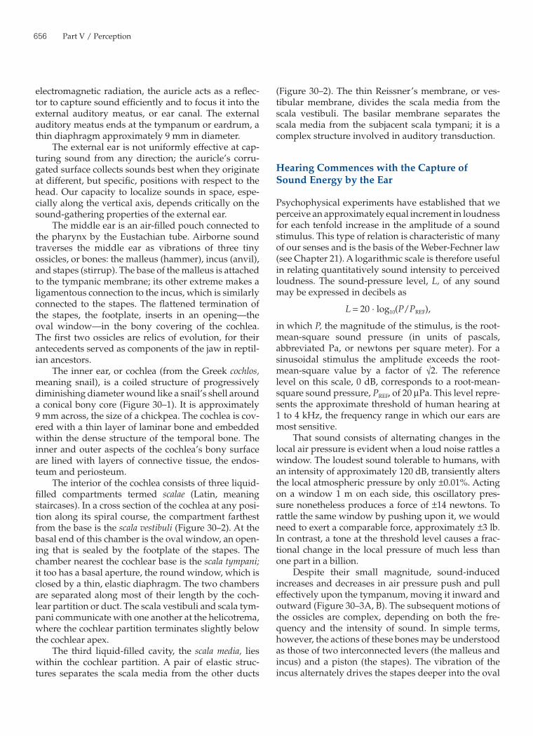
The inner ear, or cochlea (from the Greek cochlos, meaning snail), is a coiled structure of progressively diminishing diameter wound like a snail’s shell around a conical bony core (Figure 30–1). It is approximately 9 mm across, the size of a chickpea. The cochlea is cov-ered with a thin layer of laminar bone and embedded within the dense structure of the temporal bone. The inner and outer aspects of the cochlea’s bony surface are lined with layers of connective tissue, the endos-teum and periosteum.
The interior of the cochlea consists of three liquid-filled compartments termed scalae (Latin, meaning staircases). In a cross section of the cochlea at any posi-tion along its spiral course, the compartment farthest from the base is the scala vestibuli (Figure 30–2). At the basal end of this chamber is the oval window, an open-ing that is sealed by the footplate of the stapes. The chamber nearest the cochlear base is the scala tympani; it too has a basal aperture, the round window, which is closed by a thin, elastic diaphragm. The two chambers are separated along most of their length by the coch-lear partition or duct. The scala vestibuli and scala tym-pani communicate with one another at the helicotrema, where the cochlear partition terminates slightly below the cochlear apex.
The third liquid-filled cavity, the scala media, lies within the cochlear partition. A pair of elastic struc-tures separates the scala media from the other ducts
Chapter 30 / The Inner Ear 657
a sound stimulus thus evokes a cycle of up-and-down movement of a minuscule volume of liquid in each of the inner ear’s three chambers.
Because the energy associated with acoustic sig-nals is generally quite small, compromise of the mid-dle ear’s normal structure may lead to conductive hearing loss, of which two forms are especially com-mon. First, scar tissue caused by middle-ear infection (otitis media) can immobilize the tympanum or ossi-cles. Second, a proliferation of bone in the ligamentous attachments of the ossicles can deprive the ossicles of their normal freedom of motion. This chronic condi-tion of unknown origin, termed otosclerosis, can lead to severe deafness.
A clinician may test for conductive hearing loss by the simple Rinné test. A patient is asked to compare the loudness of a vibrating tuning fork held in the air near an affected ear with that perceived when the base of the tuning fork is pressed against the patient’s head just
window and retracts it. The footplate of the stapes thus serves as a piston that pushes and pulls cyclically upon the liquid in the scala vestibuli. The overall effect of the middle ear is to match the impedance of the air outside the ear to that of the cochlear partition, thus ensuring the efficient transfer of sound energy from the first medium to the second.
The action of the stapes at the oval window pro-duces changes in pressure that propagate throughout the liquid of the scala vestibuli at the speed of sound. Because the aqueous perilymph is virtually incom-pressible, however, the primary effect of the stapes’s motion is to displace the liquid in the scala vestibuli in the one direction that is not restricted by a rigid boundary: toward the elastic cochlear partition (Figure 30–3B). The deflection of the cochlear partition down-ward increases the pressure in the scala tympani. The enhanced pressure displaces a liquid mass that causes outward bowing of the round window. Each cycle of
Vestibular nerve
Cochlear nerve
Scala media
Organ of Corti
Basilarmembrane
Spiralganglion
Reissner’smembrane
Scala vestibuli
Spiral ganglion
HelicotremaRound window
Oval window
Anteriorvertical
Utricle
Saccule
Posteriorvertical
Semicircularcanals
Horizontal
Ampulla
Scala tympani
Figure 30–2 The structure of the cochlea. A cross section of the cochlea shows the arrangement of the three liquid-filled ducts or scalae, each of which is approximately 33 mm long. The scala vestibuli and scala tympani communicate through the heli-cotrema at the apex of the cochlea. At the base each duct is closed by a sealed aperture. The scala vestibuli is closed by the oval window, against which the stapes pushes in response to sound; the scala tympani is closed by the round window, a thin, flexible membrane. Between these two compartments lies the scala media, an endolymph-filled tube whose epithelial lining includes the 16,000 hair cells in the organ of Corti surmounting the basilar membrane. The cross section in the lower diagram has been rotated so that the cochlear apex is oriented toward the top.
658 Part V / Perception
Oval window
Stapes
IncusMalleus
Tympanum
Round windowmembrane
Scalamedia
Scalatympani
33 mm
100 Hz
1000 Hz
10,000 Hz
Oval windowmembrane
Basilarmembrane
Roundwindow
Tympanicmembrane
Rarefaction
Scalavestibuli
Helicotrema
Compression
Sound
Lowfrequency
B
C
D
F
E
G
A
Mediumfrequency
Highfrequency
Complexsound
Chapter 30 / The Inner Ear 659
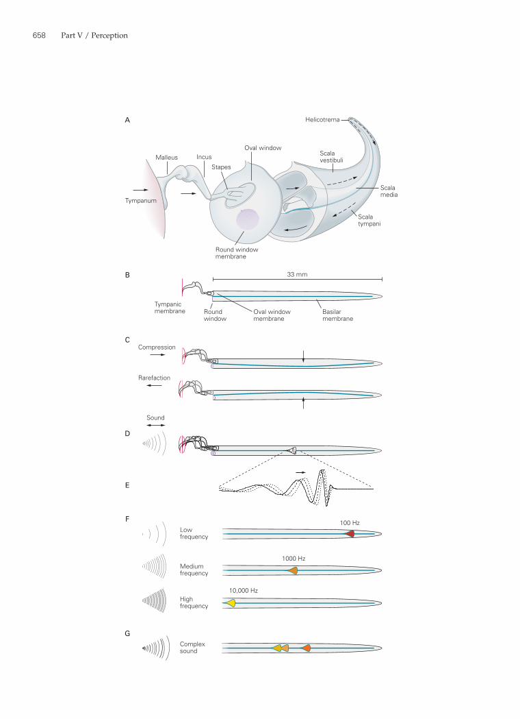
dimensions and mechanical properties along its entire length, approximately 33 mm. Under these conditions a fluctuating pressure difference between the scala vestibuli and the scala tympani would move the entire basilar membrane up and down with similar excur-sions at all points (Figure 30–3C).
This would occur regardless of the frequency of stimulation; any pressure difference between the scala vestibuli and the scala tympani would propa-gate throughout those chambers within microseconds, so the basilar membrane would be subjected to simi-lar forces at any position along its length. This simple form of basilar-membrane motion in fact occurs in the auditory organs of some reptiles.
In reality, however, the mechanical properties of the mammalian basilar membrane vary continu-ously along the cochlea’s length. The basilar mem-brane at the apex of the human cochlea is more than five times as broad as at the base. Thus, although the cochlear chambers become progressively larger from the organ’s apex toward its base, the basilar membrane decreases in width. Moreover, the basilar membrane is
behind the auricle. If the latter stimulus is perceived to be louder, the patient’s conductive pathway may be dam-aged but the internal ear is likely to be intact. In contrast, if bone conduction is not more efficient than airborne stim-ulation, the patient may have inner-ear damage, that is, sensorineural hearing loss. The diagnosis of conductive hearing loss is important because surgical intervention is highly effective: Removal of scar tissue or reconstitution of the conductive pathway with an inert prosthesis can in many instances restore excellent hearing.
The Hydrodynamic and Mechanical Apparatus of the Cochlea Delivers Mechanical Stimuli to the Receptor Cells
The Basilar Membrane Is a Mechanical Analyzer of Sound Frequency
The mechanical properties of the basilar membrane are key to the cochlea’s operation. To appreciate this, suppose that the basilar membrane had uniform
Figure 30–3 (Opposite) Motion of the basilar membrane.A. An uncoiled cochlea, with its base displaced to show its relation to the scalae, indicates the flow of stimulus energy. Sound vibrates the tympanum, which sets the three ossicles of the middle ear in motion. The piston-like action of the stapes, a bone inserted partially into the elastic oval window, produces oscillatory pressure differences that rapidly propagate along the scala vestibuli and scala tympani. Low-frequency pressure dif-ferences are shunted through the helicotrema, where the two ducts communicate. The oval and round windows do not actu-ally lie at the extreme base of the cochlea, but occur at oblique angles slightly toward the apex.B. The functional properties of the cochlea are conceptually simplified if the cochlea is viewed as a linear structure with only two liquid-filled compartments separated by the elastic basilar membrane.C. If the basilar membrane had uniform mechanical proper-ties along its full extent, a compression would drive the tympanum and ossicles inward, increasing the pressure in the scala vestibuli and forcing the basilar membrane down-ward. Opposite movements would occur during a rarefaction. The pressure changes in the scala tympani are relieved by bowing of the round-window membrane. The movements of the tympanum, ossicles, and basilar membrane are greatly exaggerated.D. In fact, the basilar membrane’s mechanical properties vary continuously along its length. The oscillatory stimulation of a sound causes a traveling wave on the basilar membrane, shown here within the envelope of maximal displacement over an entire cycle. The magnitude of movement is grossly exag-gerated in the vertical direction; the loudest tolerable sounds
move the basilar membrane by only ±150 nm, a scaled distance less than one-hundredth the width of the lines representing the basilar membrane in these figures.E. An enlargement of the active region in D demonstrates the motion of the basilar membrane in response to stimulation with sound of a single frequency. The continuous curve depicts a traveling wave at one instant; the vertical scale of basilar-membrane deflection is exaggerated about one-millionfold. The dashed and dotted curves portray the traveling wave at successively later times as it progresses from the cochlear base (left) toward the apex (right). As the wave approaches the characteristic place for the stimulus frequency, it slows and grows in amplitude. The stimulus energy is then transferred to hair cells at the position of the wave’s peak.F. Each frequency of stimulation excites maximal motion at a particular position along the basilar membrane. Low-frequency sounds produce basilar-membrane motion near the apex, where the membrane is relatively broad and flaccid. Mid-fre-quency sounds excite the membrane in its middle. The highest frequencies that we can hear excite the basilar membrane at its narrow, taut base. The mapping of sound frequency onto the basilar membrane is approximately logarithmic.G. The basilar membrane performs spectral analysis of complex sounds. In this example a sound with three prominent frequen-cies, such as the three formants of a vowel sound, excites basilar-membrane motion in three regions, each of which represents a particular frequency. Hair cells in the correspond-ing positions transduce the basilar-membrane oscillations into receptor potentials, which in turn excite the nerve fibers that innervate these particular regions.
660 Part V / Perception
basilar membrane’s operation is essentially the inverse of a piano’s. The piano synthesizes a complex sound by combining the pure tones produced by numerous vibrating strings; the cochlea deconstructs a complex sound by isolating each component tone at a discrete segment of the basilar membrane.
The arrangement of vibration frequencies along the basilar membrane is an example of a tonotopic map. The relation between frequency and position on the basilar membrane varies smoothly and monotonically but is not linear. Instead, the logarithm of the best fre-quency is roughly proportional to the distance from the cochlea’s apex. The frequencies from 20 Hz to 200 Hz, those between 200 Hz and 2 kHz, and those spanning 2 kHz to 20 kHz are each apportioned approximately one-third of the basilar membrane’s extent.
Analysis of the response to a complex sound illus-trates how the basilar membrane operates in daily life. A vowel sound in human speech, for example, ordinar-ily comprises three dominant frequency components termed formants. Measurement of the sound pressure outside an ear exposed to such a sound reveals a com-plex, seemingly chaotic signal. The movements of the tympanum and ossicles in response to a vowel sound likewise appear very complicated. The motion of the basilar membrane, however, is much simpler. Each fre-quency component of the stimulus establishes a traveling wave that, to a first approximation, is independent of the waves evoked by the others (Figure 30–3G). Each traveling wave reaches its peak excursion at a point on the basilar membrane appropriate for that frequency component. Moreover, the amplitude of each traveling wave is proportional, albeit in a complex way, to the intensity of the corresponding frequency component. The basilar membrane thus acts as a mechanical fre-quency analyzer by distributing specific stimulus ener-gies to hair cells arrayed along its length, and in doing so begins the encoding of the frequencies and intensities in a sound.
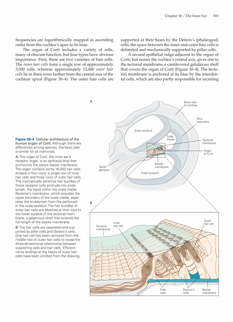
The Organ of Corti Is the Site of Mechanoelectrical Transduction in the Cochlea
The organ of Corti, a ridge of epithelium extending along the basilar membrane, is the receptor organ of the inner ear. Each organ of Corti contains approxi-mately 16,000 hair cells innervated by approximately 30,000 afferent nerve fibers, which carry information into the brain along the eighth cranial nerve. Like the basilar membrane itself, both the hair cells and the auditory nerve fibers are tonotopically organized: At any position along the basilar membrane the hair cells are most sensitive to a particular frequency, and these
relatively thin and floppy at the apex of the cochlea but thicker and tauter toward the base. Because its mechanical properties vary along its length, the basilar membrane does not oscillate like a single string on a musical instrument; instead, it more resembles a pano-ply of strings that vary from the coarsest string on a bass viol to the finest string on a violin.
Stimulation with a pure tone evokes a complex and elegant movement of the basilar membrane. Over one complete cycle of a sound, each affected segment along the basilar membrane undergoes a single cycle of vibration (Figure 30–3D, E). The various parts of the membrane do not, however, oscillate in phase with one another; instead, some portions of the membrane move upward while others move downward. As first demonstrated by Georg von Békésy using stroboscopic illumination, the overall pattern of motion of the mem-brane is that of successive traveling waves.
Because mechanical stimulation is applied at the cochlear base as a pressure difference between scala vestibuli and scala tympani, the traveling wave progresses from the cochlear base toward the apex. Each wave reaches its maximal amplitude at a par-ticular position appropriate for the frequency of stim-ulation, then declines rapidly in size as it advances toward the cochlear apex. A traveling wave ascending the basilar membrane resembles an ocean wave rolling toward the shore: As the wave nears the beach its crest grows to a maximal height, then breaks and rapidly fades away.
Although the analogy of an ocean wave gives some sense of the appearance of the basilar mem-brane’s motion, the connection between the cochlear traveling wave and the movement of an ocean wave is only metaphorical—the physical bases of the two waves are quite distinct. An ocean wave is the result of the momentum of a wind-blown mass of water. In contrast, movement of the basilar membrane is the result of motion of the liquid masses above and below the membrane. These liquids are continuously driven up and down by the energy supplied by the stapes’s piston-like movements at the oval window.
The variation in the mechanical properties of the mammalian basilar membrane explains why the mem-brane is tuned to different frequencies at each point along its length. In humans the membrane at the apex of the cochlea responds best to the lowest audible fre-quencies, down to approximately 20 Hz. At the coch-lear base it responds to frequencies as great as 20 kHz. The intervening frequencies are represented along the basilar membrane in a continuous array (Figure 30–3F). In the 19th century the German physiologist Hermann von Helmholtz was the first to appreciate that the
Chapter 30 / The Inner Ear 661
supported at their bases by the Deiters’s (phalangeal) cells; the space between the inner and outer hair cells is delimited and mechanically supported by pillar cells.
A second epithelial ridge adjacent to the organ of Corti, but nearer the cochlea’s central axis, gives rise to the tectorial membrane, a cantilevered gelatinous shelf that covers the organ of Corti (Figure 30–4). The tecto-rial membrane is anchored at its base by the interden-tal cells, which are also partly responsible for secreting
frequencies are logarithmically mapped in ascending order from the cochlea’s apex to its base.
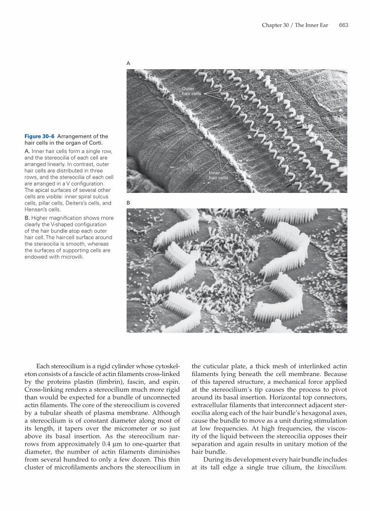
The organ of Corti includes a variety of cells, many of obscure function, but four types have obvious importance. First, there are two varieties of hair cells. The inner hair cells form a single row of approximately 3,500 cells, whereas approximately 12,000 outer hair cells lie in three rows farther from the central axis of the cochlear spiral (Figure 30–4). The outer hair cells are
A
B
Bony wallof cochlea
Stria vascularis
Scala vestibuli
Scala tympani
Basilarmembrane
Scalamedia
Spiral ganglion
Tectorialmembrane
Organ of Corti
Pillarcells
Deiters’scells
Basilarmembrane
Tectorialmembrane
Outerhair cellInner
hair cell
Figure 30–4 Cellular architecture of the human organ of Corti. Although there are differences among species, the basic plan is similar for all mammals.A. The organ of Corti, the inner ear’s receptor organ, is an epithelial strip that surmounts the elastic basilar membrane. The organ contains some 16,000 hair cells arrayed in four rows: a single row of inner hair cells and three rows of outer hair cells. The mechanically sensitive hair bundles of these receptor cells protrude into endo-lymph, the liquid within the scala media. Reissner’s membrane, which provides the upper boundary of the scala media, sepa-rates the endolymph from the perilymph in the scala vestibuli. The hair bundles of outer hair cells are attached at their tops to the lower surface of the tectorial mem-brane, a gelatinous shelf that extends the full length of the basilar membrane.B. The hair cells are separated and sup-ported by pillar cells and Deiters’s cells. One hair cell has been removed from the middle row of outer hair cells to reveal the three-dimensional relationship between supporting cells and hair cells. Efferent nerve endings at the bases of outer hair cells have been omitted from the drawing.
662 Part V / Perception
this extracellular structure. The tectorial membrane’s tapered distal edge forms a fragile connection with the organ of Corti. When the basilar membrane vibrates in response to a sound, the organ of Corti and the overly-ing tectorial membrane move with it. Because the basi-lar and tectorial membranes pivot about different lines of insertion, their up-and-down motion is accompa-nied by back-and-forth shearing motion of the upper surface of the organ of Corti and the lower surface of the tectorial membrane. This motion is detected by hair cells.
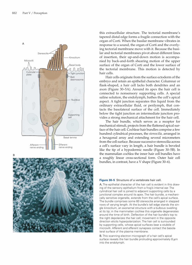
Hair cells originate from the surface ectoderm of the embryo and retain an epithelial character. Columnar or flask-shaped, a hair cell lacks both dendrites and an axon (Figure 30–5A). Around its apex the hair cell is connected to nonsensory supporting cells. A special saline solution, the endolymph, bathes the cell’s apical aspect. A tight junction separates this liquid from the ordinary extracellular fluid, or perilymph, that con-tacts the basolateral surface of the cell. Immediately below the tight junction an intermediate junction pro-vides a strong mechanical attachment for the hair cell.
The hair bundle, which serves as a receptor for mechanical stimuli, projects from the flattened apical sur-face of the hair cell. Cochlear hair bundles comprise a few hundred cylindrical processes, the stereocilia, arranged in a hexagonal array and extending several micro meters from the cell surface. Because successive stereocilia across a cell’s surface vary in length, a hair bundle is beveled like the tip of a hypodermic needle (Figure 30–5B). In the mammalian cochlea the inner hair cell bundles have a roughly linear cross-sectional form. Outer hair cell bundles, in contrast, have a V shape (Figure 30–6).
Microvilli
Cuticularplate
Kinocilium
Basal body
Tight junction
Beltdesmosome
Supportingcell
Basal lamina
Stereocilia
Nucleus
A
Efferentnerve ending
Afferentnerve ending
B
Figure 30–5 Structure of a vertebrate hair cell.A. The epithelial character of the hair cell is evident in this draw-ing of the sensory epithelium from a frog’s internal ear. The cylindrical hair cell is joined to adjacent supporting cells by a junctional complex around its apex. The hair bundle, a mechani-cally sensitive organelle, extends from the cell’s apical surface. The bundle comprises some 60 stereocilia arranged in stepped rows of varying length. At the bundle’s tall edge stands the sin-gle kinocilium, an axonemal structure with a bulbous swelling at its tip; in the mammalian cochlea this organelle degenerates around the time of birth. Deflection of the hair bundle’s top to the right depolarizes the hair cell; movement in the opposite direction elicits hyperpolarization. The hair cell is surrounded by supporting cells, whose apical surfaces bear a stubble of microvilli. Afferent and efferent synapses contact the basola-teral surface of the plasma membrane.B. This scanning electron micrograph of a hair cell’s apical surface reveals the hair bundle protruding approximately 8 µm into the endolymph.
Chapter 30 / The Inner Ear 663
the cuticular plate, a thick mesh of interlinked actin filaments lying beneath the cell membrane. Because of this tapered structure, a mechanical force applied at the stereocilium’s tip causes the process to pivot around its basal insertion. Horizontal top connectors, extracellular filaments that interconnect adjacent ster-eocilia along each of the hair bundle’s hexagonal axes, cause the bundle to move as a unit during stimulation at low frequencies. At high frequencies, the viscos-ity of the liquid between the stereocilia opposes their separation and again results in unitary motion of the hair bundle.
During its development every hair bundle includes at its tall edge a single true cilium, the kinocilium.
Each stereocilium is a rigid cylinder whose cytoskel-eton consists of a fascicle of actin filaments cross-linked by the proteins plastin (fimbrin), fascin, and espin. Cross-linking renders a stereocilium much more rigid than would be expected for a bundle of unconnected actin filaments. The core of the stereocilium is covered by a tubular sheath of plasma membrane. Although a stereocilium is of constant diameter along most of its length, it tapers over the micrometer or so just above its basal insertion. As the stereocilium nar-rows from approximately 0.4 µm to one-quarter that diameter, the number of actin filaments diminishes from several hundred to only a few dozen. This thin cluster of microfilaments anchors the stereocilium in
B
A
Innerhair cells
Outerhair cells
Figure 30–6 Arrangement of the hair cells in the organ of Corti.A. Inner hair cells form a single row, and the stereocilia of each cell are arranged linearly. In contrast, outer hair cells are distributed in three rows, and the stereocilia of each cell are arranged in a V configuration. The apical surfaces of several other cells are visible: inner spiral sulcus cells, pillar cells, Deiters’s cells, and Hensen’s cells.B. Higher magnification shows more clearly the V-shaped configuration of the hair bundle atop each outer hair cell. The hair-cell surface around the stereocilia is smooth, whereas the surfaces of supporting cells are endowed with microvilli.
664 Part V / Perception
This structure possesses at its core an axoneme, or array of nine paired microtubules, and sometimes an additional central pair of microtubules. The kinocilium is not essential for mechanoelectrical transduction, for in mammalian cochlear hair cells it degenerates around the time of birth.
Hair Cells Transform Mechanical Energy into Neural Signals
Deflection of the Hair Bundle Initiates Mechanoelectrical Transduction
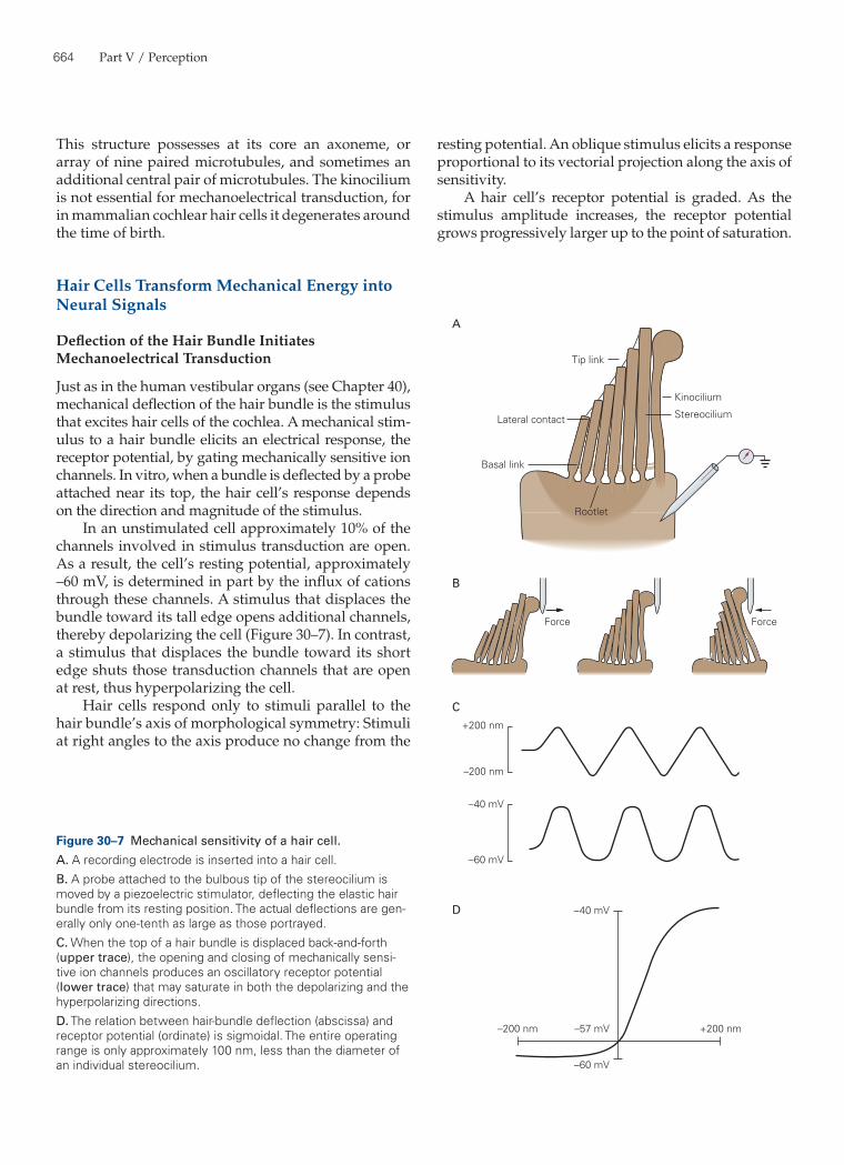
Just as in the human vestibular organs (see Chapter 40), mechanical deflection of the hair bundle is the stimulus that excites hair cells of the cochlea. A mechanical stim-ulus to a hair bundle elicits an electrical response, the receptor potential, by gating mechanically sensitive ion channels. In vitro, when a bundle is deflected by a probe attached near its top, the hair cell’s response depends on the direction and magnitude of the stimulus.
In an unstimulated cell approximately 10% of the channels involved in stimulus transduction are open. As a result, the cell’s resting potential, approximately –60 mV, is determined in part by the influx of cations through these channels. A stimulus that displaces the bundle toward its tall edge opens additional channels, thereby depolarizing the cell (Figure 30–7). In contrast, a stimulus that displaces the bundle toward its short edge shuts those transduction channels that are open at rest, thus hyperpolarizing the cell.
Hair cells respond only to stimuli parallel to the hair bundle’s axis of morphological symmetry: Stimuli at right angles to the axis produce no change from the
Kinocilium
Rootlet
Force
+200 nm
+200 nm
–200 nm
–200 nm –57 mV
–40 mV
–60 mV
–40 mV
–60 mV
Force
Tip link
Lateral contact
A
B
C
D
Basal link
Stereocilium
Figure 30–7 Mechanical sensitivity of a hair cell.A. A recording electrode is inserted into a hair cell.B. A probe attached to the bulbous tip of the stereocilium is moved by a piezoelectric stimulator, deflecting the elastic hair bundle from its resting position. The actual deflections are gen-erally only one-tenth as large as those portrayed.C. When the top of a hair bundle is displaced back-and-forth (upper trace), the opening and closing of mechanically sensi-tive ion channels produces an oscillatory receptor potential (lower trace) that may saturate in both the depolarizing and the hyperpolarizing directions.D. The relation between hair-bundle deflection (abscissa) and receptor potential (ordinate) is sigmoidal. The entire operating range is only approximately 100 nm, less than the diameter of an individual stereocilium.
resting potential. An oblique stimulus elicits a response proportional to its vectorial projection along the axis of sensitivity.
A hair cell’s receptor potential is graded. As the stimulus amplitude increases, the receptor potential grows progressively larger up to the point of saturation.
Chapter 30 / The Inner Ear 665
the transduction channels remains uncertain. Genetic and physiological evidence suggests, however, that proteins of the transmembrane channel family, specifi-cally TMC1 and TMC2, are involved in mechanoelec-trical transduction.
Mechanical Force Directly Opens Transduction Channels
The mechanism for gating of transduction channels in hair cells differs fundamentally from the mechanisms used for such electrical signals as the action potential or postsynaptic potential. Many ion channels respond to changes in membrane potential or to specific ligands (see Chapters 5, 7, 9–11). In contrast, the mechanoelec-trical transduction channels in the hair cell are acti-vated by mechanical strain.
Two lines of evidence suggest that the opening and closing of transduction channels is regulated by tension in elastic structures within the hair bundle. First, a bundle is stiffer along its axis of morphologi-cal symmetry, and hence of mechanical sensitivity, than at a right angle. This observation suggests that a portion of the work done in deflecting a bundle goes into elastic elements, termed gating springs, that pull on the molecular gates of the transduction channels. Because the gating springs contribute over half of a hair bundle’s stiffness, the transduction channels effi-ciently capture the energy supplied when a bundle is deflected. In addition, hair-bundle stiffness decreases during channel gating, a phenomenon expected if the channels are gated directly through a mechanical link-age to the hair bundle.
A second indication that transduction channels are directly controlled by gating springs is the rapidity with which hair cells respond. The response latency is so brief, only a few microseconds, that gating is more likely to be direct than to involve a second messenger (see Chapter 11). Moreover, the electrical responses of hair cells to a series of step stimuli of increasing mag-nitude become successively larger and faster. This behavior favors a kinetic scheme in which mechanical force controls the rate constant for channel gating. If the mechanical energy from a stimulus is stored in a spring attached to the channel’s gate, the rates of chan-nel opening and closing are determined by the prob-ability that the stored energy of the spring exceeds the transition-state energy for channel gating.
Mechanoelectrical transduction occurs near the tips of the stereocilia, as demonstrated by three experimen-tal techniques. First, the region where cations flow into a hair cell was inferred by measuring small differences
The receptor potential of an inner hair cell can be as great as 25 mV in peak-to-peak magnitude. The relation between a bundle’s deflection and the resulting electri-cal response is sigmoidal (Figure 30–7D). A displace-ment of only ±100 nm represents approximately 90% of the response range. During normal stimulation a hair bundle moves through an angle of ±1 degree or so, that is, by much less than the diameter of one stereocilium.
Hair cells are so sensitive that their response thresh-old is probably set by brownian motion; still weaker stimuli are lost in the thermal clatter of the ear’s com-ponents. When observed in vitro a hair bundle exhibits brownian motion of approximately ±3 nm. However, because the auditory system averages responses over several cycles to improve its signal-to-noise ratio, the threshold of hearing corresponds to hair-bundle deflec-tion of as little as ±0.3 nm. A stimulus of this magnitude evokes a receptor potential near 100 µV in amplitude.
The ion channels in hair cells that are involved in stimulus transduction are relatively nonselective, cation-passing pores with a conductance near 100 pS. Because small organic cations can support measurable current, and small fluorescent molecules can traverse the channel, the transduction channel’s pore is about 1.3 nm in diameter. Most of the transduction current is carried by K+, the cation with the highest concentra-tion in the endolymph bathing the hair bundle.
The large diameter and poor selectivity of the pore permit transduction channels to be blocked by aminoglycoside antibiotics, such as streptomycin, gen-tamicin, and tobramycin. When used in large doses to counter bacterial infections, these drugs have a toxic effect on hair cells; the antibiotics damage hair bundles and eventually kill hair cells. These drugs evidently insinuate themselves through transduction channels at a low rate and thus cause long-term toxic effects by interfering with protein synthesis on the mitochondrial ribosomes, which resemble prokaryotic ribosomes. Consistent with this hypothesis, human sensitivity to aminoglycosides is maternally inherited and in many instances reflects a single base change in the 12S ribo-somal RNA gene of the mitochondrion.
Single-channel recordings and noise analysis sug-gest that each hair cell possesses only a few hundred transduction channels. Because the number of channels is about the same as the number of stereocilia in a hair bundle and because the magnitude of the recep-tor potential is roughly proportional to the number of stereocilia remaining in a microdissected bundle, there are probably only two active transduction chan-nels per stereocilium. The paucity of channels along with the lack of high-affinity ligands with which to label them explains why the biochemical identity of
666 Part V / Perception
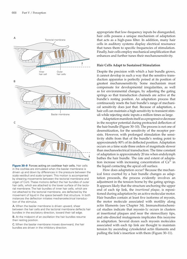
basilar membrane oscillates up and down, carrying the hair cells with it, a shearing motion occurs between the organ of Corti and the overlying tectorial membrane (Figure 30–9). The hair bundles of outer hair cells, whose tips are firmly attached to the tectorial mem-brane, are directly deflected by this movement. The hair bundles of inner hair cells, which do not contact the tectorial membrane, are deflected by movement of the liquid beneath the membrane. This mode of stimulation affords some mechanical amplification of the signals reaching hair bundles. At least for high-frequency stimuli, the movements of hair bundles are thought to be severalfold greater than that of the basi-lar membrane.
Direct Mechanoelectrical Transduction Is Rapid
In contrast to hair cells, many other sensory recep-tors, such as photoreceptors and olfactory neurons, use cyclic nucleotides or other second messengers in stimulus transduction. This strategy is advantageous in that the enzymatic pathway that generates a second messenger amplifies the signal, and feedback within the metabolic pathway readily permits adaptation and desensitization (see Chapter 11).
What is the advantage of transduction without the intervention of a second messenger? The answer prob-ably lies in its speed. Hair cells operate much more quickly than do other sensory receptor cells of the vertebrate nervous system, and indeed more quickly than neurons themselves. To deal with the frequencies of biologically relevant sounds, transduction by hair cells must be rapid. Given the behavior of sound in air and the dimensions of sound-emitting and sound-absorbing organs such as vocal cords and eardrums, optimal auditory communication occurs in the fre-quency range of 10 Hz to 100 kHz. Much higher fre-quencies propagate poorly through air; much lower frequencies are inefficiently produced and captured by animals of moderate size.
Locating sound sources, one of the most important functions of hearing, sets even more stringent limits on the speed of transduction. A sound from a source directly to one side of a person reaches the nearer ear somewhat sooner than the farther. Although this delay is at most 700 µs, a human observer can locate sound sources on the basis of much smaller delays, about 10 µs. For this to occur, hair cells must be capa-ble of detecting acoustic waveforms with microsecond-level resolution.
The ability of hair cells to discriminate high fre-quencies of stimulation implies that transduction chan-nels are gated very rapidly. Even in animals sensitive
in the extracellular potential around a stimulated hair bundle. The voltage signal is strongest at the bundle’s top; cations flowing toward transduction channels con-verge near the stereociliary tips. Second, aminoglyco-side antibiotics, which block these channels, have their greatest effect when applied from a microelectrode directed at the top of the hair bundle. Finally, Ca2+-sen-sitive fluorescent indicators initially signal Ca2+ entry near the apex of a deflected hair bundle. Measurements from cochlear hair cells at high temporal resolution suggest that the channels occur precisely at the stereo-ciliary tips. When transduction current enters the chan-nels, the ensuing change in membrane potential must propagate axially down the stereocilia before it changes the potential at the base of the cell body and thus influ-ences the rate of transmitter release. Because the stere-ocilia are short, however, their cable properties do not attenuate electrical signals significantly.
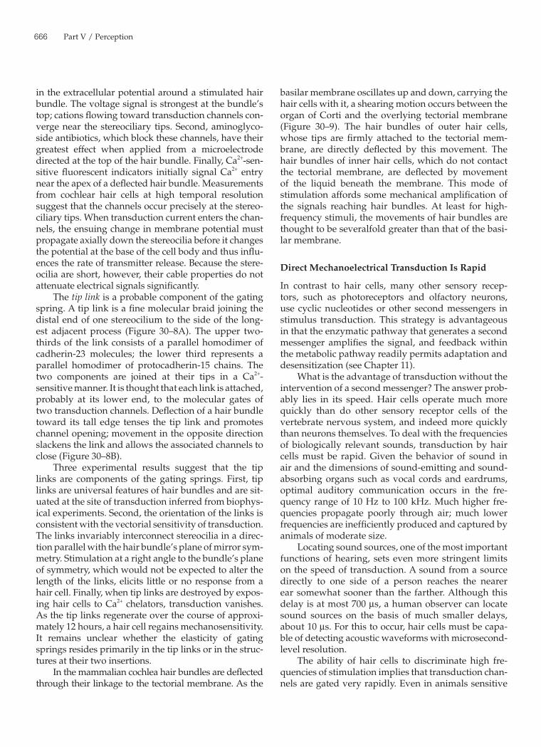
The tip link is a probable component of the gating spring. A tip link is a fine molecular braid joining the distal end of one stereocilium to the side of the long-est adjacent process (Figure 30–8A). The upper two-thirds of the link consists of a parallel homodimer of cadherin-23 molecules; the lower third represents a parallel homodimer of protocadherin-15 chains. The two components are joined at their tips in a Ca2+-sensitive manner. It is thought that each link is attached, probably at its lower end, to the molecular gates of two transduction channels. Deflection of a hair bundle toward its tall edge tenses the tip link and promotes channel opening; movement in the opposite direction slackens the link and allows the associated channels to close (Figure 30–8B).
Three experimental results suggest that the tip links are components of the gating springs. First, tip links are universal features of hair bundles and are sit-uated at the site of transduction inferred from biophys-ical experiments. Second, the orientation of the links is consistent with the vectorial sensitivity of transduction. The links invariably interconnect stereocilia in a direc-tion parallel with the hair bundle’s plane of mirror sym-metry. Stimulation at a right angle to the bundle’s plane of symmetry, which would not be expected to alter the length of the links, elicits little or no response from a hair cell. Finally, when tip links are destroyed by expos-ing hair cells to Ca2+ chelators, transduction vanishes. As the tip links regenerate over the course of approxi-mately 12 hours, a hair cell regains mechanosensitivity. It remains unclear whether the elasticity of gating springs resides primarily in the tip links or in the struc-tures at their two insertions.
In the mammalian cochlea hair bundles are deflected through their linkage to the tectorial membrane. As the
Chapter 30 / The Inner Ear 667
The Temporal Responsiveness of Hair Cells Determines Their Sensitivity
The mechanical sensitivity of hair cells is not constant; responsiveness varies in such a way that a given cell best detects behaviorally relevant stimuli. When it is
to relatively low frequencies, the response to a stimu-lus of moderate intensity has a time constant of only 80 µs at room temperature. For mammals to be able to respond to frequencies greater than 100 kHz, the hair cells evidently display gating rates that are an order of magnitude greater.
A
B
Tip link
Figure 30–8 Mechanoelectrical trans-duction by hair cells.A. A tip link connects each stereocilium to the side of the longest adjacent stere-ocilium, as seen in a scanning electron micrograph (left) and a transmission electron micrograph (right) of a hair bundle’s top surface. Each tip link is only 3 nm in diameter. The links appear stouter in the illustration on the left because of metallic coating during specimen prepara-tion. (Reproduced, with permission, from Assad, Shepherd, and Corey 1991; repro-duced, with permission, from Hudspeth and Gillespie 1994.)B. Top: Ion flux through the channel that underlies mechanoelectrical transduction in hair cells is regulated by a molecular gate. The opening and closing of the gate are controlled by the tension in an elastic element, the gating spring, that senses hair-bundle displacement. (Adapted, with permission, from Howard and Hudspeth 1988.)Bottom: When the hair bundle is at rest each transduction channel clatters between closed and open states, spend-ing most of its time shut. Displacement of the bundle in the positive direction increases the tension in the gating spring, here assumed to be in part a tip link, attached to each channel’s molecular gate. The enhanced tension promotes channel opening and the influx of cations, thereby producing a depolarizing receptor potential. (Adapted, with permission, from Hudspeth 1989.)
668 Part V / Perception
appropriate that low-frequency inputs be disregarded, hair cells possess a unique mechanism of adaptation that acts as a high-pass filter. In addition, many hair cells in auditory systems display electrical resonance that tunes them to specific frequencies of stimulation. Finally, hair cells employ mechanical amplification that enhances and further tunes their mechanosensitivity.
Hair Cells Adapt to Sustained Stimulation
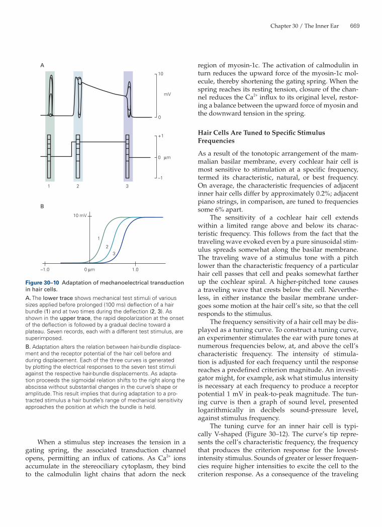
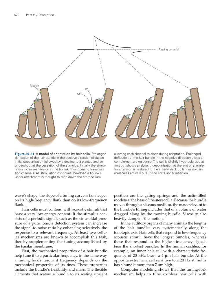
Despite the precision with which a hair bundle grows, it cannot develop in such a way that the sensitive trans-duction apparatus is perfectly poised at its position of greatest mechanosensitivity. Some mechanism must compensate for developmental irregularities, as well as for environmental changes, by adjusting the gating springs so that transduction channels are active at the bundle’s resting position. An adaptation process that continuously resets the hair bundle’s range of mechani-cal sensitivity does just that. Because of adaptation, a hair cell can maintain a high sensitivity to transient stim-uli while rejecting static inputs a million times as large.
Adaptation manifests itself as a progressive decrease in the receptor potential during protracted deflection of the hair bundle (Figure 30–10). The process is not one of desensitization, for the sensitivity of the receptor per-sists. However, with prolonged stimulation the sensi-tivity shifts from that of the bundle’s resting point to approximately 80% of its deflected position. Adaptation occurs on a time scale three orders of magnitude slower than mechanoelectrical transduction: The time constant of adaptation is approximately 20 ms when endolymph bathes the hair bundle. The rate and extent of adapta-tion increase with increasing concentration of Ca2+ in the liquid contacting the apical cell surface.
How does adaptation occur? Because the mechan-ical force exerted by a hair bundle changes as adap-tation proceeds, the process evidently involves an adjustment in the tension borne by the gating springs. It appears likely that the structure anchoring the upper end of each tip link, the insertional plaque, is reposi-tioned during adaptation by an active molecular motor. Hair bundles contain at least five isoforms of myosin, the motor molecule associated with motility along actin filaments (see Chapter 34). Immunohistochemi-cal studies indicate that myosin-1c occurs in clusters at insertional plaques and near the stereociliary tips, and site-directed mutagenesis implicates this isozyme in adaptation. Several dozen such myosin molecules associated with each tip link are thought to maintain tension by ascending cytoskeletal actin filaments and pulling the link’s insertion with them (Figure 30–11).
Tectorial membrane
Innerhair cell
Outerhair cells
Basilar membrane
A
B
C
Figure 30–9 Forces acting on cochlear hair cells. Hair cells in the cochlea are stimulated when the basilar membrane is driven up and down by differences in the pressure between the scala vestibuli and scala tympani. This motion is accompanied by shearing movements between the tectorial membrane and organ of Corti. These motions deflect the hair bundles of outer hair cells, which are attached to the lower surface of the tecto-rial membrane. The hair bundles of inner hair cells, which are not attached to the tectorial membrane, are deflected by the movement of liquid in the space beneath that structure. In both instances the deflection initiates mechanoelectrical transduc-tion of the stimulus.A. When the basilar membrane is driven upward, shear between the hair cells and the tectorial membrane deflects hair bundles in the excitatory direction, toward their tall edge.B. At the midpoint of an oscillation the hair bundles resume their resting position.C. When the basilar membrane moves downward, the hair bundles are driven in the inhibitory direction.
Chapter 30 / The Inner Ear 669
region of myosin-1c. The activation of calmodulin in turn reduces the upward force of the myosin-1c mol-ecule, thereby shortening the gating spring. When the spring reaches its resting tension, closure of the chan-nel reduces the Ca2+ influx to its original level, restor-ing a balance between the upward force of myosin and the downward tension in the spring.
Hair Cells Are Tuned to Specific Stimulus Frequencies
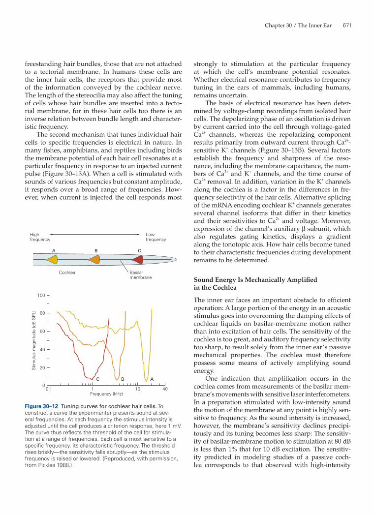
As a result of the tonotopic arrangement of the mam-malian basilar membrane, every cochlear hair cell is most sensitive to stimulation at a specific frequency, termed its characteristic, natural, or best frequency. On average, the characteristic frequencies of adjacent inner hair cells differ by approximately 0.2%; adjacent piano strings, in comparison, are tuned to frequencies some 6% apart.
The sensitivity of a cochlear hair cell extends within a limited range above and below its charac-teristic frequency. This follows from the fact that the traveling wave evoked even by a pure sinusoidal stim-ulus spreads somewhat along the basilar membrane. The traveling wave of a stimulus tone with a pitch lower than the characteristic frequency of a particular hair cell passes that cell and peaks somewhat farther up the cochlear spiral. A higher-pitched tone causes a traveling wave that crests below the cell. Neverthe-less, in either instance the basilar membrane under-goes some motion at the hair cell’s site, so that the cell responds to the stimulus.
The frequency sensitivity of a hair cell may be dis-played as a tuning curve. To construct a tuning curve, an experimenter stimulates the ear with pure tones at numerous frequencies below, at, and above the cell’s characteristic frequency. The intensity of stimula-tion is adjusted for each frequency until the response reaches a predefined criterion magnitude. An investi-gator might, for example, ask what stimulus intensity is necessary at each frequency to produce a receptor potential 1 mV in peak-to-peak magnitude. The tun-ing curve is then a graph of sound level, presented logarithmically in decibels sound-pressure level, against stimulus frequency.
The tuning curve for an inner hair cell is typi-cally V-shaped (Figure 30–12). The curve’s tip repre-sents the cell’s characteristic frequency, the frequency that produces the criterion response for the lowest- intensity stimulus. Sounds of greater or lesser frequen-cies require higher intensities to excite the cell to the criterion response. As a consequence of the traveling
When a stimulus step increases the tension in a gating spring, the associated transduction channel opens, permitting an influx of cations. As Ca2+ ions accumulate in the stereociliary cytoplasm, they bind to the calmodulin light chains that adorn the neck
10
mV
µm
0 µm
A
B
10 mV
0
0
+1
–1
321
1
23
–1.0 1.0
Figure 30–10 Adaptation of mechanoelectrical transduction in hair cells.A. The lower trace shows mechanical test stimuli of various sizes applied before prolonged (100 ms) deflection of a hair bundle (1) and at two times during the deflection (2, 3). As shown in the upper trace, the rapid depolarization at the onset of the deflection is followed by a gradual decline toward a plateau. Seven records, each with a different test stimulus, are superimposed.B. Adaptation alters the relation between hair-bundle displace-ment and the receptor potential of the hair cell before and during displacement. Each of the three curves is generated by plotting the electrical responses to the seven test stimuli against the respective hair-bundle displacements. As adapta-tion proceeds the sigmoidal relation shifts to the right along the abscissa without substantial changes in the curve’s shape or amplitude. This result implies that during adaptation to a pro-tracted stimulus a hair bundle’s range of mechanical sensitivity approaches the position at which the bundle is held.
670 Part V / Perception
position are the gating springs and the actin-filled rootlets at the base of the stereocilia. Because the bundle moves through a viscous medium, the mass relevant to the bundle’s tuning includes that of a volume of water dragged along by the moving bundle. Viscosity also heavily dampens the motion.
In the auditory organs of many animals the lengths of the hair bundles vary systematically along the tonotopic axis. Hair cells that respond to low-frequency acoustic stimuli have the longest bundles, whereas those that respond to the highest-frequency signals bear the shortest bundles. In the human cochlea, for example, an inner hair cell with a characteristic fre-quency of 20 kHz bears a 4 µm hair bundle. At the opposite extreme, a cell sensitive to a 20 Hz stimulus has a bundle more than 7 µm high.
Computer modeling shows that the tuning-fork mechanism helps to tune cochlear hair cells with
wave’s shape, the slope of a tuning curve is far steeper on its high-frequency flank than on its low-frequency flank.
Hair cells must contend with acoustic stimuli that have a very low energy content. If the stimulus con-sists of a periodic signal, such as the sinusoidal pres-sure of a pure tone, a detection system can increase the signal-to-noise ratio by enhancing selectively the response to a relevant frequency. At least two cellu-lar mechanisms are known to accomplish this task, thereby supplementing the tuning accomplished by the basilar membrane.
First, the mechanical properties of a hair bundle help tune it to a particular frequency, in the same way a tuning fork’s resonant frequency depends on the mechanical properties of its tines. These properties include the bundle’s flexibility and mass. The flexible elements that restore a bundle to its resting upright
Myosin
Resting potential
Ca2+
K+
Figure 30–11 A model of adaptation by hair cells. Prolonged deflection of the hair bundle in the positive direction elicits an initial depolarization followed by a decline to a plateau and an undershoot at the cessation of the stimulus. Initially the stimu-lation increases tension in the tip link, thus opening transduc-tion channels. As stimulation continues, however, a tip link’s upper attachment is thought to slide down the stereocilium,
allowing each channel to close during adaptation. Prolonged deflection of the hair bundle in the negative direction elicits a complementary response. The cell is slightly hyperpolarized at first but shows a rebound depolarization at the end of stimula-tion; tension is restored to the initially slack tip link as myosin molecules actively pull up the link’s upper insertion.
Chapter 30 / The Inner Ear 671
freestanding hair bundles, those that are not attached to a tectorial membrane. In humans these cells are the inner hair cells, the receptors that provide most of the information conveyed by the cochlear nerve. The length of the stereocilia may also affect the tuning of cells whose hair bundles are inserted into a tecto-rial membrane, for in these hair cells too there is an inverse relation between bundle length and character-istic frequency.
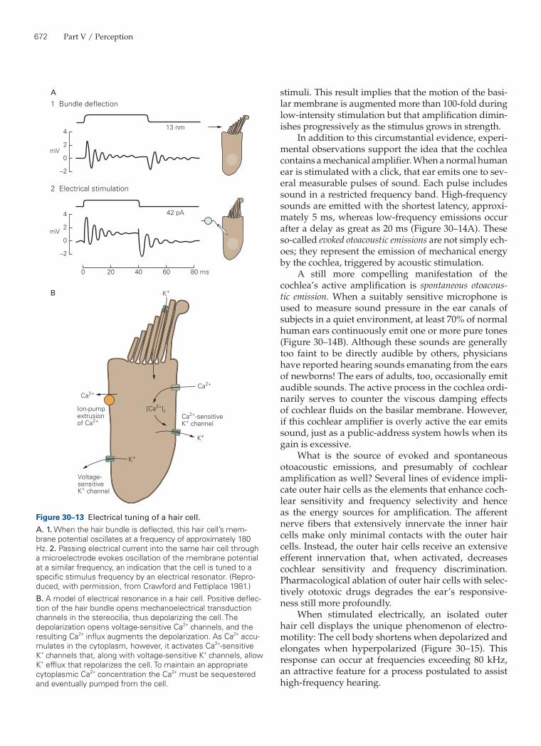
The second mechanism that tunes individual hair cells to specific frequencies is electrical in nature. In many fishes, amphibians, and reptiles including birds the membrane potential of each hair cell resonates at a particular frequency in response to an injected current pulse (Figure 30–13A). When a cell is stimulated with sounds of various frequencies but constant amplitude, it responds over a broad range of frequencies. How-ever, when current is injected the cell responds most
100
60
Stim
ulus
mag
nitu
de (d
B S
PL)
40
20
00.1
Highfrequency
A B C
ABC
Cochlea Basilarmembrane
Lowfrequency
1Frequency (kHz)
10 40
80
Figure 30–12 Tuning curves for cochlear hair cells. To construct a curve the experimenter presents sound at sev-eral frequencies. At each frequency the stimulus intensity is adjusted until the cell produces a criterion response, here 1 mV. The curve thus reflects the threshold of the cell for stimula-tion at a range of frequencies. Each cell is most sensitive to a specific frequency, its characteristic frequency. The threshold rises briskly—the sensitivity falls abruptly—as the stimulus frequency is raised or lowered. (Reproduced, with permission, from Pickles 1988.)
strongly to stimulation at the particular frequency at which the cell’s membrane potential resonates. Whether electrical resonance contributes to frequency tuning in the ears of mammals, including humans, remains uncertain.
The basis of electrical resonance has been deter-mined by voltage-clamp recordings from isolated hair cells. The depolarizing phase of an oscillation is driven by current carried into the cell through voltage-gated Ca2+ channels, whereas the repolarizing component results primarily from outward current through Ca2+-sensitive K+ channels (Figure 30–13B). Several factors establish the frequency and sharpness of the reso-nance, including the membrane capacitance, the num-bers of Ca2+ and K+ channels, and the time course of Ca2+ removal. In addition, variation in the K+ channels along the cochlea is a factor in the differences in fre-quency selectivity of the hair cells. Alternative splicing of the mRNA encoding cochlear K+ channels generates several channel isoforms that differ in their kinetics and their sensitivities to Ca2+ and voltage. Moreover, expression of the channel’s auxiliary β subunit, which also regulates gating kinetics, displays a gradient along the tonotopic axis. How hair cells become tuned to their characteristic frequencies during development remains to be determined.
Sound Energy Is Mechanically Amplified in the Cochlea
The inner ear faces an important obstacle to efficient operation: A large portion of the energy in an acoustic stimulus goes into overcoming the damping effects of cochlear liquids on basilar-membrane motion rather than into excitation of hair cells. The sensitivity of the cochlea is too great, and auditory frequency selectivity too sharp, to result solely from the inner ear’s passive mechanical properties. The cochlea must therefore possess some means of actively amplifying sound energy.
One indication that amplification occurs in the cochlea comes from measurements of the basilar mem-brane’s movements with sensitive laser interferometers. In a preparation stimulated with low-intensity sound the motion of the membrane at any point is highly sen-sitive to frequency. As the sound intensity is increased, however, the membrane’s sensitivity declines precipi-tously and its tuning becomes less sharp: The sensitiv-ity of basilar-membrane motion to stimulation at 80 dB is less than 1% that for 10 dB excitation. The sensitiv-ity predicted in modeling studies of a passive coch-lea corresponds to that observed with high-intensity
672 Part V / Perception
stimuli. This result implies that the motion of the basi-lar membrane is augmented more than 100-fold during low-intensity stimulation but that amplification dimin-ishes progressively as the stimulus grows in strength.
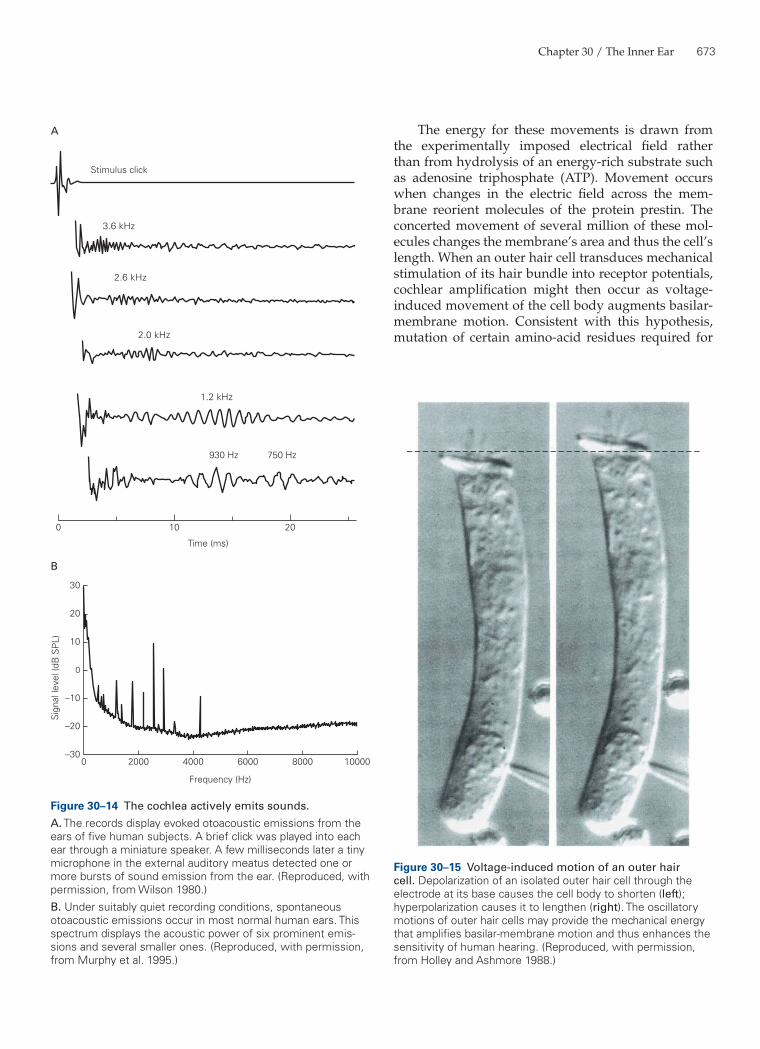
In addition to this circumstantial evidence, experi-mental observations support the idea that the cochlea contains a mechanical amplifier. When a normal human ear is stimulated with a click, that ear emits one to sev-eral measurable pulses of sound. Each pulse includes sound in a restricted frequency band. High-frequency sounds are emitted with the shortest latency, approxi-mately 5 ms, whereas low-frequency emissions occur after a delay as great as 20 ms (Figure 30–14A). These so-called evoked otoacoustic emissions are not simply ech-oes; they represent the emission of mechanical energy by the cochlea, triggered by acoustic stimulation.
A still more compelling manifestation of the cochlea’s active amplification is spontaneous otoacous-tic emission. When a suitably sensitive microphone is used to measure sound pressure in the ear canals of subjects in a quiet environment, at least 70% of normal human ears continuously emit one or more pure tones (Figure 30–14B). Although these sounds are generally too faint to be directly audible by others, physicians have reported hearing sounds emanating from the ears of newborns! The ears of adults, too, occasionally emit audible sounds. The active process in the cochlea ordi-narily serves to counter the viscous damping effects of cochlear fluids on the basilar membrane. However, if this cochlear amplifier is overly active the ear emits sound, just as a public-address system howls when its gain is excessive.
What is the source of evoked and spontaneous otoacoustic emissions, and presumably of cochlear amplification as well? Several lines of evidence impli-cate outer hair cells as the elements that enhance coch-lear sensitivity and frequency selectivity and hence as the energy sources for amplification. The afferent nerve fibers that extensively innervate the inner hair cells make only minimal contacts with the outer hair cells. Instead, the outer hair cells receive an extensive efferent innervation that, when activated, decreases cochlear sensitivity and frequency discrimination. Pharmacological ablation of outer hair cells with selec-tively ototoxic drugs degrades the ear’s responsive-ness still more profoundly.
When stimulated electrically, an isolated outer hair cell displays the unique phenomenon of electro-motility: The cell body shortens when depolarized and elongates when hyperpolarized (Figure 30–15). This response can occur at frequencies exceeding 80 kHz, an attractive feature for a process postulated to assist high-frequency hearing.
B
1 Bundle deflection
2 Electrical stimulation
13 nm
42 pA
mV
mV
A
4
2
0
–2
4
2
0
0 20 40 60 80 ms
–2
K+
K+
K+
Ca2+Ca2+
Ca2+-sensitiveK+ channel
Voltage-sensitiveK+ channel
Ion-pumpextrusionof Ca2+
[Ca2+]i
Figure 30–13 Electrical tuning of a hair cell.A. 1. When the hair bundle is deflected, this hair cell’s mem-brane potential oscillates at a frequency of approximately 180 Hz. 2. Passing electrical current into the same hair cell through a microelectrode evokes oscillation of the membrane potential at a similar frequency, an indication that the cell is tuned to a specific stimulus frequency by an electrical resonator. (Repro-duced, with permission, from Crawford and Fettiplace 1981.)B. A model of electrical resonance in a hair cell. Positive deflec-tion of the hair bundle opens mechanoelectrical transduction channels in the stereocilia, thus depolarizing the cell. The depolarization opens voltage-sensitive Ca2+ channels, and the resulting Ca2+ influx augments the depolarization. As Ca2+ accu-mulates in the cytoplasm, however, it activates Ca2+-sensitive K+ channels that, along with voltage-sensitive K+ channels, allow K+ efflux that repolarizes the cell. To maintain an appropriate cytoplasmic Ca2+ concentration the Ca2+ must be sequestered and eventually pumped from the cell.
Chapter 30 / The Inner Ear 673
The energy for these movements is drawn from the experimentally imposed electrical field rather than from hydrolysis of an energy-rich substrate such as adenosine triphosphate (ATP). Movement occurs when changes in the electric field across the mem-brane reorient molecules of the protein prestin. The concerted movement of several million of these mol-ecules changes the membrane’s area and thus the cell’s length. When an outer hair cell transduces mechanical stimulation of its hair bundle into receptor potentials, cochlear amplification might then occur as voltage-induced movement of the cell body augments basilar-membrane motion. Consistent with this hypothesis, mutation of certain amino-acid residues required for
Stimulus click
3.6 kHz
2.6 kHz
2.0 kHz
1.2 kHz
750 Hz930 Hz
0 10 20
Time (ms)
Sig
nal l
evel
(dB
SP
L)
A
B
Frequency (Hz)
0 2000 4000 6000 8000 10000
30
20
10
0
–10
–20
–30
Figure 30–14 The cochlea actively emits sounds.A. The records display evoked otoacoustic emissions from the ears of five human subjects. A brief click was played into each ear through a miniature speaker. A few milliseconds later a tiny microphone in the external auditory meatus detected one or more bursts of sound emission from the ear. (Reproduced, with permission, from Wilson 1980.)B. Under suitably quiet recording conditions, spontaneous otoacoustic emissions occur in most normal human ears. This spectrum displays the acoustic power of six prominent emis-sions and several smaller ones. (Reproduced, with permission, from Murphy et al. 1995.)
Figure 30–15 Voltage-induced motion of an outer hair cell. Depolarization of an isolated outer hair cell through the electrode at its base causes the cell body to shorten (left); hyperpolarization causes it to lengthen (right). The oscillatory motions of outer hair cells may provide the mechanical energy that amplifies basilar-membrane motion and thus enhances the sensitivity of human hearing. (Reproduced, with permission, from Holley and Ashmore 1988.)
674 Part V / Perception
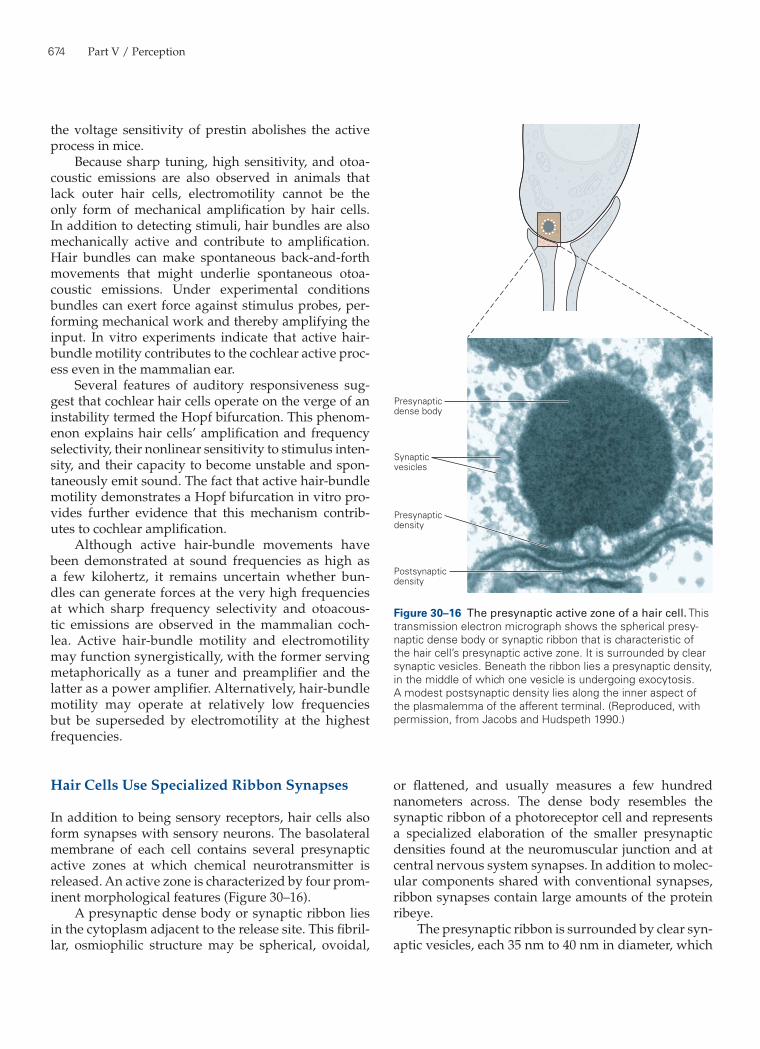
or flattened, and usually measures a few hundred nanometers across. The dense body resembles the synaptic ribbon of a photoreceptor cell and represents a specialized elaboration of the smaller presynaptic densities found at the neuromuscular junction and at central nervous system synapses. In addition to molec-ular components shared with conventional synapses, ribbon synapses contain large amounts of the protein ribeye.
The presynaptic ribbon is surrounded by clear syn-aptic vesicles, each 35 nm to 40 nm in diameter, which
the voltage sensitivity of prestin abolishes the active process in mice.
Because sharp tuning, high sensitivity, and otoa-coustic emissions are also observed in animals that lack outer hair cells, electromotility cannot be the only form of mechanical amplification by hair cells. In addition to detecting stimuli, hair bundles are also mechanically active and contribute to amplification. Hair bundles can make spontaneous back-and-forth movements that might underlie spontaneous otoa-coustic emissions. Under experimental conditions bundles can exert force against stimulus probes, per-forming mechanical work and thereby amplifying the input. In vitro experiments indicate that active hair-bundle motility contributes to the cochlear active proc-ess even in the mammalian ear.
Several features of auditory responsiveness sug-gest that cochlear hair cells operate on the verge of an instability termed the Hopf bifurcation. This phenom-enon explains hair cells’ amplification and frequency selectivity, their nonlinear sensitivity to stimulus inten-sity, and their capacity to become unstable and spon-taneously emit sound. The fact that active hair-bundle motility demonstrates a Hopf bifurcation in vitro pro-vides further evidence that this mechanism contrib-utes to cochlear amplification.
Although active hair-bundle movements have been demonstrated at sound frequencies as high as a few kilohertz, it remains uncertain whether bun-dles can generate forces at the very high frequencies at which sharp frequency selectivity and otoacous-tic emissions are observed in the mammalian coch-lea. Active hair-bundle motility and electromotility may function synergistically, with the former serving metaphorically as a tuner and preamplifier and the latter as a power amplifier. Alternatively, hair-bundle motility may operate at relatively low frequencies but be superseded by electromotility at the highest frequencies.
Hair Cells Use Specialized Ribbon Synapses
In addition to being sensory receptors, hair cells also form synapses with sensory neurons. The basolateral membrane of each cell contains several presynaptic active zones at which chemical neurotransmitter is released. An active zone is characterized by four prom-inent morphological features (Figure 30–16).
A presynaptic dense body or synaptic ribbon lies in the cytoplasm adjacent to the release site. This fibril-lar, osmiophilic structure may be spherical, ovoidal,
Presynapticdense body
Synapticvesicles
Presynapticdensity
Postsynapticdensity
Figure 30–16 The presynaptic active zone of a hair cell. This transmission electron micrograph shows the spherical presy-naptic dense body or synaptic ribbon that is characteristic of the hair cell’s presynaptic active zone. It is surrounded by clear synaptic vesicles. Beneath the ribbon lies a presynaptic density, in the middle of which one vesicle is undergoing exocytosis. A modest postsynaptic density lies along the inner aspect of the plasmalemma of the afferent terminal. (Reproduced, with permission, from Jacobs and Hudspeth 1990.)
Chapter 30 / The Inner Ear 675
channels (SK channels), whose opening leads to a pro-tracted hyperpolarization. The cytoplasm of a hair cell immediately beneath each efferent terminal holds a single cisterna of smooth endoplasmic reticulum. This structure may be involved in the reuptake of the Ca2+ that enters in response to efferent stimulation.
The best-understood role of efferent input in the cochlea is its effect on hair cells that employ electri-cal resonance for frequency tuning. Stimulation of the efferent nerve fibers hyperpolarizes a hair cell. The associated increase in membrane conductance perturbs the critically tuned resonance circuit in the cell’s mem-brane, thus decreasing both the sharpness of frequency selectivity and the gain of electrical amplification. In the mammalian cochlea, where efferent fibers contact outer hair cells, the efferent system desensitizes the cochlea by turning down the active process.
Auditory Information Flows Initially Through the Cochlear Nerve
Bipolar Neurons in the Spiral Ganglion Innervate Cochlear Hair Cells
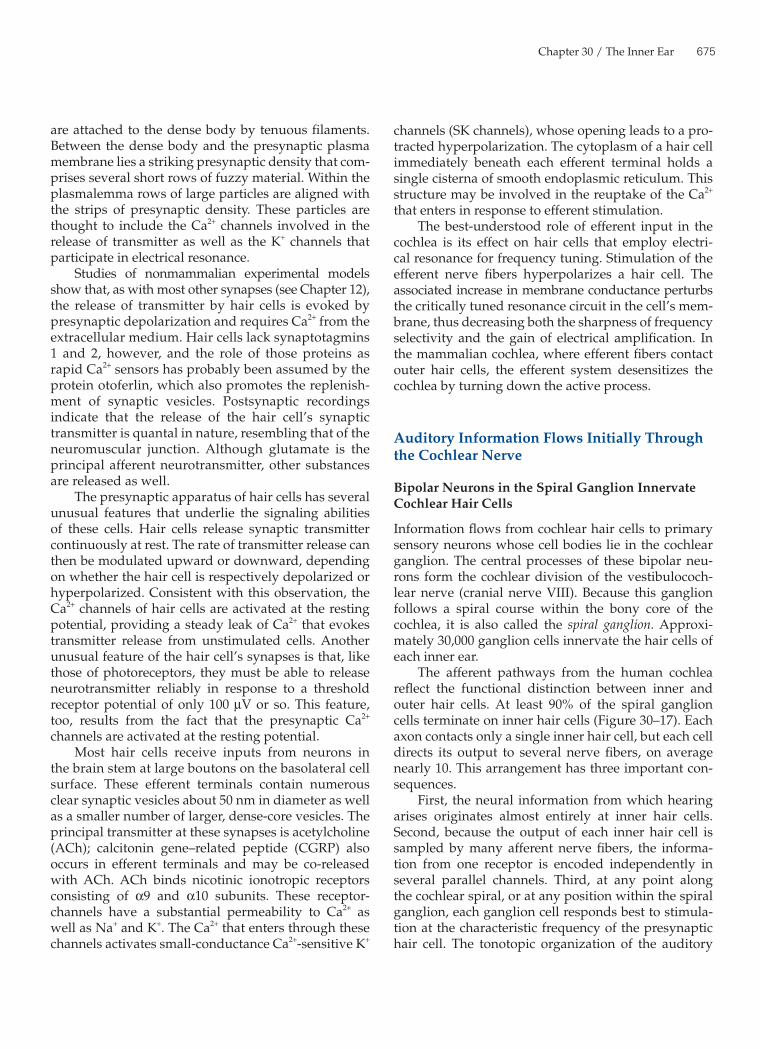
Information flows from cochlear hair cells to primary sensory neurons whose cell bodies lie in the cochlear ganglion. The central processes of these bipolar neu-rons form the cochlear division of the vestibulococh-lear nerve (cranial nerve VIII). Because this ganglion follows a spiral course within the bony core of the cochlea, it is also called the spiral ganglion. Approxi-mately 30,000 ganglion cells innervate the hair cells of each inner ear.
The afferent pathways from the human cochlea reflect the functional distinction between inner and outer hair cells. At least 90% of the spiral ganglion cells terminate on inner hair cells (Figure 30–17). Each axon contacts only a single inner hair cell, but each cell directs its output to several nerve fibers, on average nearly 10. This arrangement has three important con-sequences.
First, the neural information from which hearing arises originates almost entirely at inner hair cells. Second, because the output of each inner hair cell is sampled by many afferent nerve fibers, the informa-tion from one receptor is encoded independently in several parallel channels. Third, at any point along the cochlear spiral, or at any position within the spiral ganglion, each ganglion cell responds best to stimula-tion at the characteristic frequency of the presynaptic hair cell. The tonotopic organization of the auditory
are attached to the dense body by tenuous filaments. Between the dense body and the presynaptic plasma membrane lies a striking presynaptic density that com-prises several short rows of fuzzy material. Within the plasmalemma rows of large particles are aligned with the strips of presynaptic density. These particles are thought to include the Ca2+ channels involved in the release of transmitter as well as the K+ channels that participate in electrical resonance.
Studies of nonmammalian experimental models show that, as with most other synapses (see Chapter 12), the release of transmitter by hair cells is evoked by presynaptic depolarization and requires Ca2+ from the extracellular medium. Hair cells lack synaptotagmins 1 and 2, however, and the role of those proteins as rapid Ca2+ sensors has probably been assumed by the protein otoferlin, which also promotes the replenish-ment of synaptic vesicles. Postsynaptic recordings indicate that the release of the hair cell’s synaptic transmitter is quantal in nature, resembling that of the neuromuscular junction. Although glutamate is the principal afferent neurotransmitter, other substances are released as well.
The presynaptic apparatus of hair cells has several unusual features that underlie the signaling abilities of these cells. Hair cells release synaptic transmitter continuously at rest. The rate of transmitter release can then be modulated upward or downward, depending on whether the hair cell is respectively depolarized or hyperpolarized. Consistent with this observation, the Ca2+ channels of hair cells are activated at the resting potential, providing a steady leak of Ca2+ that evokes transmitter release from unstimulated cells. Another unusual feature of the hair cell’s synapses is that, like those of photoreceptors, they must be able to release neurotransmitter reliably in response to a threshold receptor potential of only 100 µV or so. This feature, too, results from the fact that the presynaptic Ca2+ channels are activated at the resting potential.
Most hair cells receive inputs from neurons in the brain stem at large boutons on the basolateral cell surface. These efferent terminals contain numerous clear synaptic vesicles about 50 nm in diameter as well as a smaller number of larger, dense-core vesicles. The principal transmitter at these synapses is acetylcholine (ACh); calcitonin gene–related peptide (CGRP) also occurs in efferent terminals and may be co-released with ACh. ACh binds nicotinic ionotropic receptors consisting of α9 and α10 subunits. These receptor-channels have a substantial permeability to Ca2+ as well as Na+ and K+. The Ca2+ that enters through these channels activates small-conductance Ca2+-sensitive K+
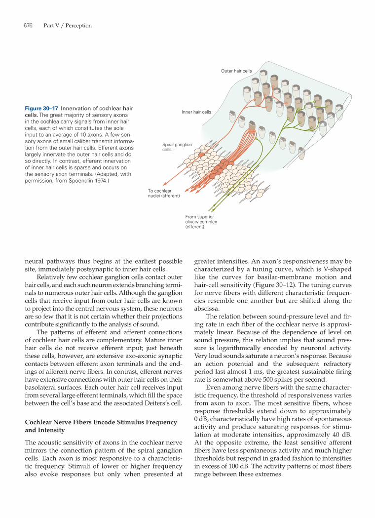
676 Part V / Perception
greater intensities. An axon’s responsiveness may be characterized by a tuning curve, which is V-shaped like the curves for basilar-membrane motion and hair-cell sensitivity (Figure 30–12). The tuning curves for nerve fibers with different characteristic frequen-cies resemble one another but are shifted along the abscissa.
The relation between sound-pressure level and fir-ing rate in each fiber of the cochlear nerve is approxi-mately linear. Because of the dependence of level on sound pressure, this relation implies that sound pres-sure is logarithmically encoded by neuronal activity. Very loud sounds saturate a neuron’s response. Because an action potential and the subsequent refractory period last almost 1 ms, the greatest sustainable firing rate is somewhat above 500 spikes per second.
Even among nerve fibers with the same character-istic frequency, the threshold of responsiveness varies from axon to axon. The most sensitive fibers, whose response thresholds extend down to approximately 0 dB, characteristically have high rates of spontaneous activity and produce saturating responses for stimu-lation at moderate intensities, approximately 40 dB. At the opposite extreme, the least sensitive afferent fibers have less spontaneous activity and much higher thresholds but respond in graded fashion to intensities in excess of 100 dB. The activity patterns of most fibers range between these extremes.
neural pathways thus begins at the earliest possible site, immediately postsynaptic to inner hair cells.
Relatively few cochlear ganglion cells contact outer hair cells, and each such neuron extends branching termi-nals to numerous outer hair cells. Although the ganglion cells that receive input from outer hair cells are known to project into the central nervous system, these neurons are so few that it is not certain whether their projections contribute significantly to the analysis of sound.
The patterns of efferent and afferent connections of cochlear hair cells are complementary. Mature inner hair cells do not receive efferent input; just beneath these cells, however, are extensive axo-axonic synaptic contacts between efferent axon terminals and the end-ings of afferent nerve fibers. In contrast, efferent nerves have extensive connections with outer hair cells on their basolateral surfaces. Each outer hair cell receives input from several large efferent terminals, which fill the space between the cell’s base and the associated Deiters’s cell.
Cochlear Nerve Fibers Encode Stimulus Frequency and Intensity
The acoustic sensitivity of axons in the cochlear nerve mirrors the connection pattern of the spiral ganglion cells. Each axon is most responsive to a characteris-tic frequency. Stimuli of lower or higher frequency also evoke responses but only when presented at
Outer hair cells
Inner hair cells
Spiral ganglioncells
To cochlearnuclei (afferent)
From superiorolivary complex(efferent)
Figure 30–17 Innervation of cochlear hair cells. The great majority of sensory axons in the cochlea carry signals from inner hair cells, each of which constitutes the sole input to an average of 10 axons. A few sen-sory axons of small caliber transmit informa-tion from the outer hair cells. Efferent axons largely innervate the outer hair cells and do so directly. In contrast, efferent innervation of inner hair cells is sparse and occurs on the sensory axon terminals. (Adapted, with permission, from Spoendlin 1974.)
Chapter 30 / The Inner Ear 677
The afferent neurons of lowest sensitivity contact the surface of an inner hair cell nearest the axis of the cochlear spiral. The most sensitive afferent neurons contact the hair cell’s opposite side. The multiple inner-vation of each inner hair cell is therefore not wholly redundant. Instead, the output from a given hair cell is directed into several parallel channels of differing sen-sitivity and dynamic range.
The firing pattern of fibers in the eighth cranial nerve has both phasic and tonic components. Brisk firing occurs at the onset of a tone presented for a few seconds. As adaptation occurs, however, the firing rate declines to a plateau level over a few tens of millisec-onds. When stimulation ceases there is usually a tran-sitory cessation of activity with a similar time course before resumption of the spontaneous firing rate (Figure 30–18).
When a periodic stimulus such as a pure tone is pre-sented, the firing pattern of a cochlear nerve fiber encodes information about the periodicity of the stimulus. For example, a relatively low-frequency tone at a moder-ate intensity might produce one spike in a nerve fiber during each cycle of stimulation. The phase of firing is also stereotyped. Each action potential might occur, for example, during the compressive phase of the stimu-lus. As the stimulation frequency rises, the stimuli eventually become so rapid that the nerve fiber can no longer produce action potentials on a cycle-by-cycle basis. Up to a frequency in excess of 4 kHz, however, phase-locking persists; a fiber may produce an action potential only every few cycles of the stimulus, but its firing continues to occur at a particular phase in the stimulus cycle.
Periodicity in neuronal firing enhances the infor-mation about the stimulus frequency. Any pure tone of sufficient intensity evokes firing in numerous coch-lear nerve fibers. Those fibers whose characteristic fre-quency coincides with the frequency of the stimulus respond at the lowest stimulus level, but respond still more briskly for stimuli of moderate intensity. Other nerve fibers with characteristic frequencies close to the stimulus also respond, although somewhat less vigorously. Regardless of their characteristic frequen-cies, however, all the responsive fibers display phase locking: Each tends to fire during a particular part of the stimulus cycle.
The central nervous system can therefore gain information about stimulus frequency in two ways. First, there is a place code; the fibers are arrayed in a tonotopic map in which the position is related to char-acteristic frequency. Second, there is a frequency code; the fibers fire at a rate that signals the frequency of the stimulus. Frequency coding is of particular importance
10 dB
20 dB
30 dB
40 dB
0 250Time (ms)
Duration of tone at characteristic frequency
Figure 30–18 The firing pattern of a cochlear nerve fiber. A cochlear nerve fiber is stimulated for somewhat more than 250 ms with a tone burst at about 5 kHz, the cell’s characteris-tic frequency. After a quiet period the stimulus is repeated. Histograms show the average response patterns of the fiber as a function of stimulus intensity. The sample period is divided into discrete temporal bins, and the number of spikes occurring in each bin is displayed. An initial, phasic increase in firing is correlated with the onset of the stimulus. The discharge contin-ues during the remainder of the stimulus following adaptation, but decreases following termination. This pattern is evident when the stimulus is 20 dB or more above threshold. Activ-ity gradually returns to baseline during the interval between stimuli. (Adapted, with permission, from Kiang 1965.)
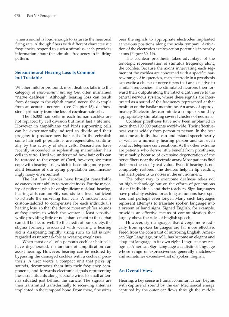
678 Part V / Perception
bear the signals to appropriate electrodes implanted at various positions along the scala tympani. Activa-tion of the electrodes excites action potentials in nearby axons (Figure 30–19).
The cochlear prosthesis takes advantage of the tonotopic representation of stimulus frequency along the cochlea. Because the axons innervating each seg-ment of the cochlea are concerned with a specific, nar-row range of frequencies, each electrode in a prosthesis can excite a cluster of nerve fibers that are sensitive to similar frequencies. The stimulated neurons then for-ward their outputs along the intact eighth nerve to the central nervous system, where these signals are inter-preted as a sound of the frequency represented at that position on the basilar membrane. An array of approx-imately 20 electrodes can mimic a complex sound by appropriately stimulating several clusters of neurons.
Cochlear prostheses have now been implanted in more than 100,000 patients worldwide. Their effective-ness varies widely from person to person. In the best outcome an individual can understand speech nearly as well as a normally hearing person and can even conduct telephone conversations. At the other extreme are patients who derive little benefit from prostheses, presumably because of extensive degeneration of the nerve fibers near the electrode array. Most patients find their prostheses of great value. Even if hearing is not completely restored, the devices help in lip reading and alert patients to noises in the environment.
The other way to overcome deafness relies not on high technology but on the efforts of generations of deaf individuals and their teachers. Sign languages have probably existed for as long as humans have spo-ken, and perhaps even longer. Many such languages represent attempts to translate spoken language into a system of hand signs. Signed English, for example, provides an effective means of communication that largely obeys the rules of English speech.
However, sign languages that diverge more radi-cally from spoken languages are far more effective. Freed from the constraint of mirroring English, Ameri-can Sign Language, or ASL, has become an elegant and eloquent language in its own right. Linguists now rec-ognize American Sign Language as a distinct language whose range of expressiveness generally matches—and sometimes exceeds—that of spoken English.
An Overall View
Hearing, a key sense in human communication, begins with capture of sound by the ear. Mechanical energy captured by the outer ear flows through the middle
when a sound is loud enough to saturate the neuronal firing rate. Although fibers with different characteristic frequencies respond to such a stimulus, each provides information about the stimulus frequency in its firing pattern.
Sensorineural Hearing Loss Is Common but Treatable
Whether mild or profound, most deafness falls into the category of sensorineural hearing loss, often misnamed “nerve deafness.” Although hearing loss can result from damage to the eighth cranial nerve, for example from an acoustic neuroma (see Chapter 45), deafness stems primarily from the loss of cochlear hair cells.
The 16,000 hair cells in each human cochlea are not replaced by cell division but must last a lifetime. However, in amphibians and birds supporting cells can be experimentally induced to divide and their progeny to produce new hair cells. In the zebrafish some hair cell populations are regenerated continu-ally by the activity of stem cells. Researchers have recently succeeded in replenishing mammalian hair cells in vitro. Until we understand how hair cells can be restored to the organ of Corti, however, we must cope with hearing loss, which is becoming more prev-alent because of our aging population and increas-ingly noisy environment.
The last few decades have brought remarkable advances in our ability to treat deafness. For the major-ity of patients who have significant residual hearing, hearing aids can amplify sounds to a level sufficient to activate the surviving hair cells. A modern aid is custom-tailored to compensate for each individual’s hearing loss, so that the device most amplifies sounds at frequencies to which the wearer is least sensitive while providing little or no enhancement to those that can still be heard well. To the credit of our society, the stigma formerly associated with wearing a hearing aid is dissipating rapidly; using such an aid is now regarded as unremarkable as wearing eyeglasses.
When most or all of a person’s cochlear hair cells have degenerated, no amount of amplification can assist hearing. However, hearing can be restored by bypassing the damaged cochlea with a cochlear pros-thesis. A user wears a compact unit that picks up sounds, decomposes them into their frequency com-ponents, and forwards electronic signals representing these constituents along separate wires to small anten-nas situated just behind the auricle. The signals are then transmitted transdermally to receiving antennas implanted in the temporal bone. From there, fine wires
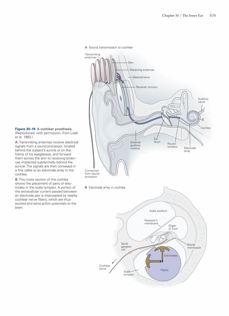
Chapter 30 / The Inner Ear 679
Spiralganglioncell
Cochlearnerve
Skin
Receiving antennas
Mastoid bone
Receiver circuitry
Cochlea
Auditorynerve
Electrodearray
Roundwindow
EardrumExternal
auditorymeatus
Connectionfrom soundprocessor
Transmittingantennas
A Sound transmission to cochlea
B Electrode array in cochlea
B
Scala vestibuli
Reissner’smembrane
Organof Corti
Scalatympani
Basilarmembrane
Electrodes
Plastic
Figure 30–19 A cochlear prosthesis. (Reproduced, with permission, from Loeb et al. 1983.)A. Transmitting antennas receive electrical signals from a sound processor, located behind the subject’s auricle or on the frame of his eyeglasses, and forward them across the skin to receiving anten-nas implanted subdermally behind the auricle. The signals are then conveyed in a fine cable to an electrode array in the cochlea.B. This cross section of the cochlea shows the placement of pairs of elec-trodes in the scala tympani. A portion of the extracellular current passed between an electrode pair is intercepted by nearby cochlear nerve fibers, which are thus excited and send action potentials to the brain.
680 Part V / Perception
Kazmierczak P, Sakaguchi H, Tokita J, Wilson-Kubalek EM, Milligan RA, Müller U, Kachar B. 2007. Cadherin 23 and protocadherin 15 interact to form tip-link filaments in sen-sory hair cells. Nature 449:87–91.
Loeb GE. 1985. The functional replacement of the ear. Sci Am 252:104–111.
Pickles JO. 2008. An Introduction to the Physiology of Hearing, 3rd ed. New York: Academic.
ReferencesArt JJ, Crawford AC, Fettiplace R, Fuchs PA. 1985. Efferent
modulation of hair cell tuning in the cochlea of the turtle. J Physiol 360:397–421.
Ashmore JF. 1987. A fast motile response in guinea-pig outer hair cells: the cellular basis of the cochlear amplifier. J Physiol 388:323–347.
Assad JA, Shepherd GM, Corey DP. 1991. Tip-link integrity and mechanical transduction in vertebrate hair cells. Neu-ron 7:985–994.
Beurg M, Fettiplace R, Nam J-H, Ricci AJ. 2009. Localiza-tion of inner hair cell mechanotransducer channels using high-speed calcium imaging. Nat Neurosci 12:553–558.
Chan DK, Hudspeth AJ. 2005. Ca2+ current-driven nonlin-ear amplification by the mammalian cochlea in vitro. Nat Neurosci 8:149–155.
Crawford AC, Fettiplace R. 1985. The mechanical properties of ciliary bundles of turtle cochlear hair cells. J Physiol 364:359–379.
Dallos P, Harris D. 1978. Properties of auditory nerve responses in absence of outer hair cells. J Neurophysiol 41:365–383.
Dallos P, Wu X, Cheatham MA, Gao J, Zheng J, Anderson CT, Jia S, et al. 2008. Prestin-based outer hair cell motility is necessary for mammalian cochlear amplification. Neuron 58:333–339.
Helmholtz HLF. [1877] 1954. On the Sensations of Tone as a Physiological Basis for the Theory of Music. New York: Dover.
Holley MC, Ashmore JF. 1988. On the mechanism of a high-frequency force generator in outer hair cells isolated from the guinea pig cochlea. Proc R Soc Lond B Biol Sci 232:413–429.
Howard J, Hudspeth AJ. 1988. Compliance of the hair bun-dle associated with gating of mechanoelectrical transduc-tion channels in the bullfrog’s saccular hair cell. Neuron 1:189–199.
Hudspeth AJ. 1982. Extracellular current flow and the site of transduction by vertebrate hair cells. J Neurosci 2:1–10.
Hudspeth AJ, Gillespie PG. 1994. Pulling springs to tune transduction: adaptation by hair cells. Neuron 12:1–9.
Jacobs RA, Hudspeth AJ. 1990. Ultrastructural correlates of mechanoelectrical transduction in hair cells of the bullfrog’s internal ear. Cold Spring Harbor Symp Quant Biol 55:547–561.
Johnson SL, Beurg M, Marcotti W, Fettiplace R. 2011. Prestin-driven cochlear amplification is not limited by the outer hair cell membrane time constant. Neuron 70:1143–1154.
ear to the cochlea, where it causes the elastic basilar membrane to oscillate. Aligned along the basilar mem-brane in a tonotopic array, 16,000 hair cells detect the frequency components of a stimulus and transduce them into receptor potentials that cause sensory neu-rons to fire.
As the population ages and as society becomes more concerned about hearing loss, physicians will increasingly confront patients and families who are experiencing the social difficulties associated with deafness. This subject is at present politically charged. On the one hand, the rapid technical improvement in cochlear prostheses leads their developers to advo-cate use of the devices whenever practical, including for young children. Many members of the deaf com-munity, on the other hand, believe that widespread implantation of cochlear prostheses, particularly in children, will foster a generation of individuals whose ability to communicate is dependent on technological support of as yet unproved durability. The extensive use of prostheses might also diminish the use of Amer-ican Sign Language and thus reverse the deaf commu-nity’s remarkable recent advances.
Although this debate will not soon subside, it is worthwhile to emphasize the most positive aspect of the issue. A few decades ago there were no widely effective ways of coping with profound deafness; now there are two. Moreover, these solutions are not mutu-ally exclusive; with a cochlear prosthesis a deaf indi-vidual can benefit from bilingualism in spoken English and American Sign Language.
A. J. Hudspeth
Selected ReadingsCrawford AC, Fettiplace R. 1981. An electrical tuning mecha-
nism in turtle cochlear hair cells. J Physiol 312:377–412.Holt JR, Gillespie SKH, Provance DW, Jr., Shah K, Shokat
KM, Corey DP, Mercer JA, Gillespie PG. 2002. A chemical-genetic strategy implicates myosin-1c in adaptation by hair cells. Cell 108:371–381.
Hudspeth AJ. 1989. How the ear’s works work. Nature 341:397–404.
Hudspeth AJ. 2008. Making an effort to listen: mechanical amplification in the ear. Neuron 59:530–545.
Hudspeth, AJ, Jülicher F, Martin P. 2010. A critique of the critical cochlea: Hopf—a bifurcation—is better than none. J Neurophysiol 104:1219–1229.
Chapter 30 / The Inner Ear 681
a human deafness form, is essential for exocytosis at the auditory ribbon synapse. Cell 127:277–289.
Ruggero MA. 1992. Responses to sound of the basilar membrane of the mammalian cochlea. Curr Opin Neurobiol 2:449–456.
Rzadzinska AK, Schneider ME, Davies C, Riordan GP, Kachar B. 2004. An actin molecular treadmill and myosins maintain stereocilia functional architecture and self-renewal. J Cell Biol 164:887–897.
Spoendlin H. 1974. Neuroanatomy of the cochlea. In: E Zwicker, E Terhardt (eds.). Facts and Models in Hearing, pp. 18–32. New York: Springer-Verlag.
Stauffer EA, Scarborough JD, Hirono M, Miller ED, Shah K, Mercer JA, Holt JR, Gillespie PG. 2005. Fast adaptation in vestibular hair cells requires myosin-1c activity. Neuron 47:541–553.
Tilney LG, Tilney MS, Saunders JS, DeRosier DJ. 1986. Actin filaments, stereocilia, and hair cells of the bird cochlea. III. The development and differentiation of hair cells and stereocilia. Dev Biol 116:100–118.
von Békésy G. 1960. Experiments in Hearing. EG Wever (ed, transl). New York: McGraw-Hill.
Wilson JP. 1980. Evidence for a cochlear origin for acoustic re-emissions, threshold fine-structure and tonal tinnitus. Hear Res 2:233–252.
Zhang DS, Piazza V, Perrin BJ, Rzadzinska AK, Poczatek JC, Wang M, Prosser HM, et al. 2012. Multi-isotope imaging mass spectrometry reveals slow protein turnover in hair-cell stereocilia. Nature 481:520-524.
Zheng J, Shen W, He DZZ, Long KB, Madison LD, Dallos P. 2000. Prestin is the motor protein of cochlear outer hair cells. Nature 405:149–155.
Kawashima Y, Géléoc GS, Kurima K, Labay V, Lelli A, Asai Y, Makishima T, et al. 2011. Mechanotransduction in mouse inner ear hair cells requires transmembrane channel-like genes. J Clin Invest 2011 121:4796–4809.
Kiang NY-S. 1965. Discharge Patterns of Single Fibers in the Cat’s Auditory Nerve. Cambridge, MA: MIT Press.
Kozlov AS, Baumgart J, Risler T, Versteegh CPC, Huds-peth AJ. 2011. Forces between clustered stereocilia mini-mize friction in the ear on a subnanometre scale. Nature 474:376–379.
Liberman MC. 1982. Single-neuron labeling in the cat audi-tory nerve. Science 216:1239–1241.
Loeb GE, Byers CL, Rebscher SJ, Casey DE, Fong MM, Schin-dler RA, Gray RF, Merzenich MM. 1983. Design and fab-rication of an experimental cochlear prosthesis. Med Biol Eng Comput 21:241–254.
Lumpkin EA, Hudspeth AJ. 1998. Regulation of free Ca2+ con-centration in hair-cell stereocilia. J Neurosci 18:6300–6318.
Martin P, Hudspeth AJ. 1999. Active hair-bundle movements can amplify a hair cell’s response to oscillatory mechani-cal stimuli. Proc Natl Acad Sci U S A 96:14306–14311.
Murphy WJ, Tubis A, Talmadge CL, Long GR. 1995. Relaxa-tion dynamics of spontaneous otoacoustic emissions per-turbed by external forces. II. Suppression of interacting emissions. J Acoust Soc Am 97:3711–3720.
Rosenblatt KP, Sun Z-P, Heller S, Hudspeth AJ. 1997. Dis-tribution of Ca2+ activated K+ channel isoforms along the tonotopic gradient of the chicken’s cochlea. Neuron 19:1061–1075.
Roux I, Safieddine S, Nouvian R, Grati M, Simmle M-C, Bahloul A, Perfettii I, et al. 2006. Otoferlin, defective in