the influence of hip strength on lower-limb, pelvis, …€¦armour smith j, popovich jm jr, kulig...
TRANSCRIPT

Chapman UniversityChapman University Digital Commons
Physical Therapy Faculty Articles and Research Physical Therapy
2014
The Influence of Hip Strength on Lower-Limb,Pelvis, and Trunk Kinematics and CoordinationPatterns During Walking and Hopping in HealthyWomenJo Armour SmithChapman University, [email protected]
John M. PopovichMichigan State University
Kornelia KuligUniversity of Southern California
Follow this and additional works at: http://digitalcommons.chapman.edu/pt_articles
Part of the Musculoskeletal System Commons, Physical Therapy Commons, and the Women'sHealth Commons
This Article is brought to you for free and open access by the Physical Therapy at Chapman University Digital Commons. It has been accepted forinclusion in Physical Therapy Faculty Articles and Research by an authorized administrator of Chapman University Digital Commons. For moreinformation, please contact [email protected].
Recommended CitationArmour Smith J, Popovich JM Jr, Kulig K. The influence of hip strength on lower limb, pelvis and trunk kinematics during walking andhopping in healthy women. Journal of Orthopaedic and Sports Physical Therapy. 2014; 44 (7): 525-531.DOI: 10.2519/jospt.2014.5028

The Influence of Hip Strength on Lower-Limb, Pelvis, and TrunkKinematics and Coordination Patterns During Walking and Hopping inHealthy Women
CommentsThis is a pre-copy-editing, author-produced PDF of an article accepted for publication in Journal ofOrthopaedic and Sports Physical Therapy, volume 44, issue 7, in 2014 following peer review. The definitivepublisher-authenticated version is available online at DOI: 10.2519/jospt.2014.5028
CopyrightJournal of Orthopaedic & Sports Physical Therapy 2014. For personal use only. No other uses withoutpermission. All rights reserved.
This article is available at Chapman University Digital Commons: http://digitalcommons.chapman.edu/pt_articles/45

Review Copy
3
Key Words: coordination, muscle performance, hopping, gait. 24
Page 17 of 43
JOSPT, 1033 N. Fairfax St., Suite 304, Alexandria, VA 22314, ph. 877-766-3450
Journal of Orthopaedic & Sports Physical Therapy

Review Copy
4
INTRODUCTION 25
Musculoskeletal disorders of the lower limbs are often associated with both poor hip 26
muscle performance and altered kinematics during dynamic tasks. However, it is still unclear 27
whether altered lower limb or pelvis/trunk motion as a result of hip weakness contributes to the 28
development of musculoskeletal pathology and pain.13,25 During the stance phases of activities 29
such as walking, running or hopping, the hip extensors and abductors play a complex role in 30
control of the lower extremities, pelvis and trunk. This includes deceleration of hip internal 31
rotation and adduction16 and maintenance of the equilibrium of the pelvis and trunk over the 32
stance limb.8 Additionally, motion at the hip, pelvis and trunk influences kinematics and kinetics 33
at the knee.13,25 Therefore, weakness of the hip musculature may be associated with altered 34
kinematics at the knee, hip, pelvis or trunk. 35
36
A number of studies have examined the relationship between diminished hip muscle 37
performance and kinematics in patients with musculoskeletal dysfunction. For example, females 38
with patellofemoral pain syndrome (PFP) have decreased maximum hip abductor and extensor 39
torque and increased peak knee external rotation and increased hip adduction during the stance 40
phase of running compared with healthy controls.30,34 Similarly, hip osteoarthritis is associated 41
with decreased hip abductor strength as well as increased pelvic drop and hip internal rotation 42
during the stance phase of walking.2,33 However, cross sectional studies of patient populations do 43
not discriminate between weakness resulting from musculoskeletal pain or pathology and 44
weakness that may have contributed to the original development of the disorder.4,25 45
46
Page 18 of 43
JOSPT, 1033 N. Fairfax St., Suite 304, Alexandria, VA 22314, ph. 877-766-3450
Journal of Orthopaedic & Sports Physical Therapy

Review Copy
5
Existing studies that have investigated the relationship between hip strength and single 47
joint/segment kinematics in healthy subjects have failed to account for the confounding influence 48
of trunk motion in persons with weak hip musculature.4,14,19,25 In the frontal plane, subjects with 49
weak hip abductors often demonstrate increased trunk motion towards the stance limb,23,25 50
resulting in altered moments at the hip and knee.4,21 In addition, existing studies utilizing mixed 51
samples of male and female subjects may also have been confounded by sex-specific differences 52
in kinematics during dynamic tasks.4,6,16,25,26 Therefore, the effect of hip muscle performance on 53
peak kinematics of the lower limbs, pelvis and trunk in the absence of musculoskeletal 54
pathology remains unclear. 55
56
Analysis of the relative timing, or coordination, of motion occurring between joints 57
or segments may facilitate identification of subtle adaptations in lower limb, pelvis or trunk 58
motion associated with diminished hip muscle performance during sub-maximal tasks.11,12 59
Adaptations in patterns of joint or segmental coordination have the potential to alter joint 60
loading during the stance phase of dynamic activities and therefore may also be associated 61
with the development of lower limb pathologies.5,11,32 Continuous methods of analyzing 62
coordination, such as the vector coding method, quantify patterns of coordination between 63
segments (inter-segmental coordination) or joints (inter-joint coordination) across the time-64
series of a task.1,27 These types of coordination analyses may have greater sensitivity to 65
detect subtle kinematic differences between groups of subjects, or between modes of gait 66
with varying mechanical demands, than single joint/segment kinematics. 67
68
Page 19 of 43
JOSPT, 1033 N. Fairfax St., Suite 304, Alexandria, VA 22314, ph. 877-766-3450
Journal of Orthopaedic & Sports Physical Therapy

Review Copy
6
The purpose of this study was to investigate kinematics in healthy women with strong 69
and weak hip muscle performance during the stance phase of walking at self-selected speed and 70
rate controlled single-legged hopping. We hypothesized that during both walking and hopping, 71
women with weak hip musculature would demonstrate greater peak lower limb, trunk and pelvis 72
angular motion in the frontal and transverse planes in addition to different patterns of 73
coordination compared to women with strong musculature. 74
75
METHODS 76
All participants provided written informed consent and the University of Southern 77
California Institutional Review Board approved the study procedures. Eligible participants were 78
free from any history of injury or surgery to the lower extremities and spine or other medical 79
conditions affecting physical activity. 80
81
Isometric hip abductor and extensor strength were tested bilaterally in healthy women 82
using a dynamometer (Primus RS, BTE Technologies, Hanover, MD). Hip abduction strength 83
was tested in a side lying position with the test limb in neutral hip alignment and full knee 84
extension. Hip extension strength was tested in a prone position with 30° and 90° of hip and knee 85
flexion respectively.23,30 Participants performed three trials with each leg. Peak torque was 86
averaged across the three trials and was normalized to participant body mass. Participants were 87
given three practice trials prior to testing, and consistent verbal encouragement was 88
provided during each trial. This protocol has high test-retest reliability.17 89
90
Page 20 of 43
JOSPT, 1033 N. Fairfax St., Suite 304, Alexandria, VA 22314, ph. 877-766-3450
Journal of Orthopaedic & Sports Physical Therapy

Review Copy
7
Participants were stratified to a weak or strong group if the normalized peak torque of 91
both hip abduction and extension on their dominant limb fell outside of a 95% confidence 92
interval. This confidence interval was calculated from the distribution of abduction and extension 93
torque values from a database of the first 30 female participants tested in this study (age 25.8 ± 94
1.8 years, height 1.68 ± 0.01 m, weight 64.3 ± 8.2 kg). Threshold values for the strong group 95
(SG) were 2.74 and 1.63 N m·kg-1 for extension and abduction respectively. Threshold values for 96
the weak group (WG) were 1.35 and 0.77 N m·kg-1. The dominant limb was defined as the 97
preferred leg for kicking a ball.23,28 The hip performance of 150 women was tested in order to 98
find 22 that met the criteria for either the SG or the WG. These women were retained for 99
the second phase of the study, consisting of the complete biomechanical assessment. These 100
data were collected as part of a broader study investigating kinematics and EMG during a 101
number of dynamic activities that included drop jumps and running in addition to walking 102
and hopping. The EMG and kinematic data from the drop jump task have been presented 103
elsewhere.23 A-priori power analysis was completed for the drop jump task utilizing pilot 104
data for lumbopelvic excursion and indicated that a total sample of 16 participants was 105
required to achieve a power of 80% at an alpha level of 0.05. A conservative recruitment 106
goal of 22 participants was selected to account for attrition. 107
108
Instrumentation 109
Lower extremity, pelvis and trunk kinematic data were collected using a ten-camera 110
three-dimensional motion capture system sampling at 250 Hz (Qualisys AB, Gothenburg, 111
Sweden). Retro-reflective markers were placed on bony landmarks to define the local coordinate 112
frames of the lower extremities, pelvis and trunk. Motion of the pelvis segment was tracked by 113
Page 21 of 43
JOSPT, 1033 N. Fairfax St., Suite 304, Alexandria, VA 22314, ph. 877-766-3450
Journal of Orthopaedic & Sports Physical Therapy

Review Copy
8
markers on the bilateral anterior superior iliac spines, iliac crests and at the L5/S1 interspinous 114
space. A rigid cluster of markers placed over the spinous process of T3 was used to track the 115
motion of the trunk, and clusters of markers on the heel counter of the shoe, shanks and lateral 116
thighs were used to track segmental motion of the lower extremities. 117
118
Experimental tasks 119
For walking gait, participants walked along a walkway at self-selected speed. Average 120
speed during the walking trials was calculated from the time taken to pass between two 121
photoelectric triggers. For the hopping task, participants performed consecutive hops on a 122
46cm by 51cm force plate (AMTI OR-6, Watertown, MA, sampling rate 1500Hz) in time 123
with a metronome. Hops were performed at a rate of 100 hops per minute. This hopping rate is 124
slower than typical self-selected hopping rate, and induces greater demand on the knee than self-125
selected hopping.29 Participants were required to land with the support foot fully within the force 126
plate for at least 20 consecutive hops. All hops were performed on the participant’s dominant leg 127
and the arms were crossed over the chest for the duration of the trial. 128
129
130
Data processing 131
Marker coordinates and force plate data were processed using Visual 3DTM (C-Motion 132
Inc., MD). For walking, stance phase initiation and termination on the dominant leg were 133
identified using the heel marker trajectories. For hopping, support phase initiation and 134
termination were identified as the moment the vertical ground reaction force exceeded or 135
dropped below 20 N respectively. A model consisting of the feet, shanks, femurs, pelvis and 136
Page 22 of 43
JOSPT, 1033 N. Fairfax St., Suite 304, Alexandria, VA 22314, ph. 877-766-3450
Journal of Orthopaedic & Sports Physical Therapy

Review Copy
9
trunk was constructed. Motion of the lower extremity segments was referenced to the proximal 137
segment. Motion of the trunk and pelvis segments was referenced to the global coordinate frame 138
and was normalized to a static calibration trial to account for individual postural alignment.23 139
Peak angles of the knee and hip joints and the pelvis and trunk segments in the frontal and 140
transverse planes were calculated for ten stance phases on the dominant leg for walking and for 141
the first ten hops for hopping and were averaged across the repeated trials for each subject. The 142
first ten hops were selected in order to maximize the consistency of the task performance. 143
Coordination between lower extremity joints and between the trunk and pelvis segments was 144
quantified using the vector coding technique.5,10,20 Vector coding is based on methods 145
originally described by Sparrow et al. 31 to quantify coordination behavior using angle-146
angle plots. Coordination between two segments or joints is calculated as the angle of the 147
vector between successive points on the angle-angle plot relative to the right horizontal. 148
This provides an angle, called the coupling angle, between 0 and 360 degrees for each 149
successive interval on the time series. The pattern of coordination for each time interval 150
across the time series can then be defined as in-phase (both segments/joints moving in the 151
same direction at the same time,); anti-phase (both segments/joints moving in the opposite 152
direction at the same time); proximal phase (motion occurring primarily in the proximal 153
joint/segment); distal phase (motion occurring primarily in the distal joint/segment) using 154
45° bin widths (Figure 1a).1,5 In-phase coordination is represented by coupling angles 155
between 22.5 – 67.5° and 202.5 – 247.5°. Anti-phase coordination is represented by 156
coupling angles between 112.5 – 157.5° and 292.5 – 337.5°. Proximal phase coordination is 157
represented by coupling angles between 157.5 – 202.5° and 337.5 – 360°. Distal phase 158
coordination is represented by coupling angles between 67.5 – 112.5° and 247.5 to 292.5°.5 159
Page 23 of 43
JOSPT, 1033 N. Fairfax St., Suite 304, Alexandria, VA 22314, ph. 877-766-3450
Journal of Orthopaedic & Sports Physical Therapy

Review Copy
10
The vector coding technique was utilized in this study as, unlike other continuous methods 160
of coordination analysis such as continuous relative phase, it does not require amplitude 161
normalization of kinematic data and therefore can be more easily interpreted relative to 162
the original kinematics, and is appropriate for both discrete and oscillatory motor tasks.22 163
For both walking and hopping, coordination was quantified between the following 164
joint/segment pairs: Coupling 1: Hip/knee motion in the frontal plane (positive values = 165
abduction); Coupling 2: Hip/knee motion in the transverse plane (positive values = rotation 166
ipsilateral to the stance limb); Coupling 3: Pelvis/trunk motion in the frontal plane 167
(positive values = tilt towards the side of the stance limb); Coupling 4: Pelvis/trunk motion 168
in the transverse plane (positive values = rotation towards the side of the stance limb) 169
(Figure 1b). The amount of each coordination pattern utilized during walking and hopping 170
for each coupling segment/joint pair was quantified as a percentage of the total 171
coordination. This indicates the amount of each movement cycle that was spent in each of 172
the four coordination patterns. 173
174
Statistics 175
Individual two-way repeated measures ANOVA were used to examine the main effects of 176
group (between subjects factor; SG, WG) and the interaction effects of group by task (within 177
subjects factor; walk, hop) on the dependent variables. Post-hoc comparisons on significant 178
group main effects were made using t-tests for independent samples with a Bonferroni correction 179
for multiple comparisons, with statistical significance set at p ≤ 0.05. Effect sizes for pairwise 180
comparisons were calculated using Cohen’s d (PASW Statistics 18, IBM Corp., Armonk, NY). 181
182
Page 24 of 43
JOSPT, 1033 N. Fairfax St., Suite 304, Alexandria, VA 22314, ph. 877-766-3450
Journal of Orthopaedic & Sports Physical Therapy

Review Copy
11
RESULTS 183
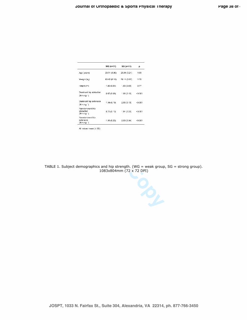
There was no significant difference in age, height or weight between the groups (Table 184
1). Hip abductor and extensor strength was significantly greater in the SG than in the WG on 185
both the dominant and the non-dominant limb (Table 1). Kinematic data from three participants 186
were excluded due to technical issues leaving a total of 19 subjects (SG n = 10, WG n = 9). 187
Mean (SD) self-selected walking speed for the entire sample was 1.32 (0.18) m·s-1 and was not 188
significantly different between groups (p = 0.49). 189
190
Single joint/segment kinematics 191
The only significant main effect of group for peak single-joint/segment kinematics was in 192
frontal plane trunk motion (F = 13.19, p = 0.002). Post-hoc analyses indicated that there was no 193
significant difference between groups during walking (WG = 2.5 (1.6)°, SG = 1.3 (1.5)°, 194
adjusted p = 0.234). However, the WG had significantly greater trunk lateral bend towards the 195
stance limb during the hopping task than the SG (WG = 7.9 (2.1)°, SG = 4.1 (2.0)°; adjusted p = 196
0.002, effect size d = 1.88). In addition, the WG demonstrated a significantly greater change in 197
peak trunk motion during hopping compared with walking than the strong group (ordinal 198
interaction, F = 8.657, p = 0.009). A disordinal group by task interaction was also evident for 199
ipsilateral pelvic tilt. The WG demonstrated less ipsilateral pelvic tilt than the SG during walking 200
and a greater amount of ipsilateral tilt during hopping (Walking, WG = 2.0 (1.3)°, SG = 2.5 201
(1.1)°; Hopping, WG = 11.0 (2.1)°, SG = 9.0 (2.0)°, F = 8.079, p = 0.011). 202
203
Coordination 204
Page 25 of 43
JOSPT, 1033 N. Fairfax St., Suite 304, Alexandria, VA 22314, ph. 877-766-3450
Journal of Orthopaedic & Sports Physical Therapy

Review Copy
12
There was a significant effect of group for hip/knee transverse plane coordination 205
(coupling 2; anti-phase (F = 7.376, p = 0.015), in-phase (F = 8.22. p = 0.011), hip phase (F = 206
10.311 p = 0.005)). During walking, the WG utilized less in-phase coordination between the hip 207
and knee in the transverse plane (WG = 22.4 (6.4)%, SG = 29.4 (2.7)%, adjusted p = 0.036, d = 208
1.45) and greater primarily hip motion than the SG (WG = 23.2 (6.1)%, SG = 15.7 (2.0)%, 209
adjusted p = 0.036, d = 1.70) (Figure 2). The WG had significantly greater anti-phase 210
coordination between the hip and knee in the transverse plane during hopping than the SG (WG 211
= 30.2 (7.1)%; SG = 17.0 (10.4)%, adjusted p = 0.03, d = 1.47) (Figures 2 and 3). There was also 212
a significant effect of group for coordination between the pelvis and the trunk in the frontal plane 213
(coupling 3; in-phase coordination, F = 5.44, p = 0.032). The WG tended to utilize more in-phase 214
coordination between the trunk and the pelvis in the frontal plane than the SG during hopping 215
(WG = 10.0 (5.3)%, SG = 5.4 (1.8)%, adjusted p = 0.066, d = 1.19). In addition, the WG 216
demonstrated a smaller change in the amount of in-phase coordination utilized in the transverse 217
plane between the pelvis and the thorax between walking and hopping than the SG (ordinal 218
interaction, p = 0.026). 219
220
DISCUSSION 221
This study indicates that in healthy young women, hip muscle performance does not 222
affect peak kinematics of the hip or knee during walking or rate-controlled hopping. 223
However, women with strong or weak hip musculature do demonstrate significantly 224
different patterns of coordination between the hip and knee and the trunk and pelvis. 225
226
Page 26 of 43
JOSPT, 1033 N. Fairfax St., Suite 304, Alexandria, VA 22314, ph. 877-766-3450
Journal of Orthopaedic & Sports Physical Therapy

Review Copy
13
By demonstrating little relationship between isometric strength and peak hip and knee 227
joint kinematics, this present research supports the findings of other studies investigating 228
subjects at the extremes of typical hip muscle performance.4,15,19 Some previous studies using 229
healthy subjects have demonstrated changes in lower extremity kinematics after the hip 230
musculature is fatigued.3,9 However, the kinematics observed after fatigue in these studies 231
may in part represent a short-term response to a novel, localized loss of muscle 232
performance rather than the purely habitual movement strategy for that subject. 233
234
In this study the weaker participants did demonstrate increased frontal plane trunk 235
motion in the direction of the stance limb during hopping. It is possible that if this trunk lateral 236
bend had been constrained during hopping a greater group difference in peak lower limb 237
kinematics would have emerged. The fact that this strategy was not evident during walking gait 238
is reflective of the higher mechanical demands of the rate-controlled slow hopping task. 239
240
The quantification of coordination patterns in this study permitted greater insight into 241
differences between groups than the single joint/segment peak kinematics. During weight-242
bearing tasks, the coordination between joints or segments is in part constrained by the 243
morphology of the joints and associated soft tissues.7,32 However, the kinematics of multiple 244
segments or joints are also coordinated as part of a motor control strategy or synergy.18 Despite 245
the lack of group differences in peak hip or knee kinematics, the coordination analyses indicated 246
differences in patterns of lower extremity coordination between the SG and the WG. The weak 247
subjects demonstrated significantly greater anti-phase coordination between the hip and knee in 248
the transverse plane compared with the SG during hopping. The anti-phase coordination pattern, 249
Page 27 of 43
JOSPT, 1033 N. Fairfax St., Suite 304, Alexandria, VA 22314, ph. 877-766-3450
Journal of Orthopaedic & Sports Physical Therapy

Review Copy
14
consisting of simultaneous hip internal rotation and knee external rotation, occurred during both 250
the deceleration and acceleration components of the hop stance phase in the WG. This pattern of 251
coordination may result in increased patellofemoral joint stress25,34 and therefore suggests a 252
mechanism for the development of PFP in subjects with weak hip musculature. 253
254
Interestingly, in this present study there were also differences between the groups in transverse 255
plane lower extremity coordination during the less mechanically demanding walking task. The 256
WG used less in-phase hip and knee rotation than the SG, and also spent a greater amount of 257
time utilizing primarily hip motion (hip phase) than the SG. These differences were driven 258
primarily by a pattern of relative external rotation of the hip during mid-stance in the WG that 259
did not occur in the SG. Powers et al.,24 also demonstrated decreased hip internal rotation during 260
walking in subjects with PFP compared with controls. They suggested that this may be a 261
compensatory mechanism to minimize the lateral forces on the patella. This present study 262
indicates that this finding may also be related to hip muscle performance. 263
264
265
Limitations 266
This study utilized a relatively small sample size. However, the large effect sizes for group 267
differences in a number of variables suggest that the study was adequately powered. As our 268
study aimed to investigate women with contrasting hip muscle performance, the 269
generalizability of these results to individuals with less extreme muscle performance may 270
be limited. The strength thresholds for inclusion in the study were calculated a priori after 271
testing only an initial 30 participants. However, utilizing strength data calculated from all 272
Page 28 of 43
JOSPT, 1033 N. Fairfax St., Suite 304, Alexandria, VA 22314, ph. 877-766-3450
Journal of Orthopaedic & Sports Physical Therapy

Review Copy
15
150 study participants would have resulted in a smaller sample due to larger standard 273
deviations in the entire cohort data. Further, due to the time required to screen all 150 274
subjects, retaining subjects for biomechanical testing might have been difficult. It should 275
also be noted that as the criterion for stratification to the SG and WG in this study was the 276
performance of the hip extensors and abductors, it is possible that differing performance in 277
other lower extremity or trunk musculature may have contributed to the group differences. 278
In particular, the adaptations in transverse plane coordination patterns may also be 279
associated with poor hip rotator performance. In addition, this study did not control for 280
habitual physical or sporting activity in the participants and did not investigate the non-281
dominant limb. 282
283
This study helps to clarify the relationship between hip muscle performance and lower 284
limb, pelvis, and trunk kinematics in young women. In the absence of the confounding 285
influences of pain or pathology, hip weakness is not associated with significant differences in 286
peak kinematics in the lower limbs, pelvis, or trunk during walking. Compensatory frontal plane 287
trunk motion in weak subjects may reduce the effect of weak hip musculature on lower limb 288
kinematics during hopping. The significantly different lower limb and pelvis/trunk coordination 289
patterns during both walking and hopping in the weak participants suggest subtle adaptations to 290
diminished hip performance even in young, healthy women during sub-maximal motor tasks. 291
Further research is needed to establish the relationships between these coordination adaptations 292
and joint loading or the development of musculoskeletal pathology. 293
294
295
Page 29 of 43
JOSPT, 1033 N. Fairfax St., Suite 304, Alexandria, VA 22314, ph. 877-766-3450
Journal of Orthopaedic & Sports Physical Therapy

Review Copy
16
296
Key Points: 297
Findings: Healthy women with poor hip muscle performance have different coordination, but 298
not different peak lower limb kinematics during walking and hopping compared with women 299
with strong hip muscle performance. 300
Implications: The differences in kinematics previously observed in patients with 301
musculoskeletal disorders may be more related to pain or pathology than hip muscle weakness. 302
However, the adaptations in trunk motion and in patterns of lower limb and trunk coordination 303
evident in this study may contribute to the development of musculoskeletal disorders. 304
Caution: This study only investigated young, healthy women performing sub-maximal tasks. In 305
addition, the interpretation of the data relies on a premise that functional tasks require a common 306
pattern of coordination. 307
308
Page 30 of 43
JOSPT, 1033 N. Fairfax St., Suite 304, Alexandria, VA 22314, ph. 877-766-3450
Journal of Orthopaedic & Sports Physical Therapy

Review Copy
17
REFERENCES
1. Armour Smith J, Siemienski A, Popovich JM Jr, Kulig K. Intra-task variability of trunk coordination during a rate-controlled bipedal dance jump. J Sports Sci. 2012;30(2):139–147.
2. Arokoski MH, Arokoski JP, Haara M, et al. Hip muscle strength and muscle cross sectional area in men with and without hip osteoarthritis. J Rheumatol. 2002;29(10):2185–2195.
3. Carcia C, Eggen J, Shultz S. Hip-abductor fatigue, frontal-plane landing angle, and excursion during a drop jump. J Sport Rehabil. 2005;14(4):321-331.
4. Cashman GE. The effect of weak hip abductors or external rotators on knee valgus kinematics in healthy subjects: a systematic review. J Sport Rehabil. 2012;12:273–284.
5. Chang R, van Emmerik REA, Hamill J. Quantifying rearfoot–forefoot coordination in human walking. J Biomech. 2008;41(14):3101–3105.
6. Chumanov ES, Wall-Scheffler C, Heiderscheit BC. Gender differences in walking and running on level and inclined surfaces. Clin Biomech. 2008;23(10):1260–1268.
7. DeLeo AT, Dierks TA, Ferber R, Davis IS. Lower extremity joint coupling during running: a current update. Clin Biomech. 2004;19(10):983–991.
8. Eng JJ, Winter DA. Kinetic analysis of the lower limbs during walking: what information can be gained from a three-dimensional model? J Biomech. 1995;28(6):753–758.
9. Geiser CF, O’Connor KM, Earl JE. Effects of Isolated Hip Abductor Fatigue on Frontal Plane Knee Mechanics. Med Sci Sports Exerc. 2010;42(3):535–545.
10. Hamill J, Haddad J, McDermott WJ. Issues in quantifying variability from a dynamical systems perspective. J Appl Biomech. 2000;16:407–418.
11. Hamill J, van Emmerik REA, Heiderscheit BC, Li L. A dynamical systems approach to lower extremity running injuries. Clin Biomech. 1999;14(5):297–308.
12. Heiderscheit B, Hamill J, van Emmerik REA. Variability of stride characteristics and joint coordination among individuals with unilateral patellofemoral pain. J Appl Biomech. 2002;18:110–121.
13. Heiderscheit BC. Lower Extremity Injuries: Is It Just About Hip Strength? J Orthop Sports Phys Ther. 2010;40(2): 39-41.
14. Hollman JH, Hohl JM, Kraft JL, Strauss JD, Traver KJ. Effects of hip extensor fatigue on lower extremity kinematics during a jump-landing task in women: A controlled laboratory study. Clin Biomech. 2012;27(9):903–909.
Page 31 of 43
JOSPT, 1033 N. Fairfax St., Suite 304, Alexandria, VA 22314, ph. 877-766-3450
Journal of Orthopaedic & Sports Physical Therapy

Review Copy
18
15. Homan KJ, Norcross MF, Goerger BM, Prentice WE, Blackburn JT. The influence of hip strength on gluteal activity and lower extremity kinematics. J Electromyogr Kinesiol. 2012.
16. Jacobs CA, Uhl TL, Mattacola CG, Shapiro R, Rayens WS. Hip abductor function and lower extremity landing kinematics: sex differences. J Athl Train. 2007;42(1):76–83.
17. Kulig K, Popovich JM, Noceti-Dewit LM, Reischl SF, Kim D. Women with posterior tibial tendon dysfunction have diminished ankle and hip muscle performance. J Orthop Sports Phys Ther. 2011;41(9):687–694.
18. Latash ML, Anson JG. Synergies in health and disease: relations to adaptive changes in motor coordination. Phys Ther. 2006;86(8):1151–1160.
19. Lawrence RK III, Kernozek TW, Miller EJ, Torry MR, Reuteman P. Influences of hip external rotation strength on knee mechanics during single-leg drop landings in females. Clin Biomech. 2008;23(6):806–813.
20. Miller RH, Chang R, Baird JL, van Emmerik REA, Hamill J. Variability in kinematic coupling assessed by vector coding and continuous relative phase. J Biomech. 2010;43(13):2554–2560.
21. Mündermann A, Asay JL, Mündermann L, Andriacchi TP. Implications of increased medio-lateral trunk sway for ambulatory mechanics. J Biomech. 2008;41(1):165–170.
22. Peters BT, Haddad JM, Heiderscheit BC, van Emmerik REA, Hamill J. Limitations in the use and interpretation of continuous relative phase. J Biomech. 2003;36(2):271–274.
23. Popovich JM, Kulig K. Lumbopelvic landing kinematics and EMG in women with contrasting hip strength. Med Sci Sports Exerc. 2012;44(1):146–153.
24. Powers CM, Chen P-Y, Reischl SF, Perry J. Comparison of foot pronation and lower extremity rotation in persons with and without patellofemoral pain. Foot Ankle Int. 2002;23(7):634–640.
25. Powers CM. The influence of abnormal hip mechanics on knee injury: a biomechanical perspective. J Orthop Sports Phys Ther. 2010;2:42–51.
26. Schache A, Blanch P, Rath D, Wrigley T, Bennell K. Differences between the sexes in the three-dimensional angular rotations of the lumbo-pelvic-hip complex during treadmill running. J Sports Sci. 2003;21(2):105–118.
27. Seay JF, van Emmerik REA, Hamill J. Influence of Low Back Pain Status on Pelvis-Trunk Coordination During Walking and Running. Spine. 2011;36(16):E1070–E1079.
28. Snyder KR, Earl JE, O’Connor KM, Ebersole KT. Resistance training is accompanied by increases in hip strength and changes in lower extremity biomechanics during running. Clin Biomech. 2009;24(1):26–34.
Page 32 of 43
JOSPT, 1033 N. Fairfax St., Suite 304, Alexandria, VA 22314, ph. 877-766-3450
Journal of Orthopaedic & Sports Physical Therapy

Review Copy
19
29. Souza RB, Arya S, Pollard CD, Salem G, Kulig K. Patellar tendinopathy alters the distribution of lower extremity net joint moments during hopping. J Appl Biomech. 2010;26(3):249–255.
30. Souza RB, Powers CM. Differences in hip kinematics, muscle strength, and muscle activation between subjects with and without patellofemoral pain. J Orthop Sports Phys Ther. 2009;39(1):12–19.
31. Sparrow W, Donovan E, van Emmerik REA, Barry EB. Using relative motion plots to measure changes in intra-limb and inter-limb coordination. J Mot Behav. 1987; 19(1):115-129.
32. Tiberio D. The effect of excessive subtalar joint pronation on patellofemoral mechanics: a theoretical model. J Orthop Sports Phys Ther. 1987;9(4):160–165.
33. Watelain E, Dujardin F, Babier F, Dubois D, Allard P. Pelvic and lower limb compensatory actions of subjects in an early stage of hip osteoarthritis. Arch Phys Med Rehabil. 2001;82(12):1705–1711.
34. Willson JD, Davis IS. Lower extremity mechanics of females with and without patellofemoral pain across activities with progressively greater task demands. Clin Biomech. 2008;23(2):203–211.
Page 33 of 43
JOSPT, 1033 N. Fairfax St., Suite 304, Alexandria, VA 22314, ph. 877-766-3450
Journal of Orthopaedic & Sports Physical Therapy

Review Copy
20
TABLE 1. Subject demographics and hip strength. (WG = weak group, SG = strong group).
Page 34 of 43
JOSPT, 1033 N. Fairfax St., Suite 304, Alexandria, VA 22314, ph. 877-766-3450
Journal of Orthopaedic & Sports Physical Therapy

Review Copy
21
FIGURE 1(a). Exemplar angle-angle plot and detail from plot demonstrating calculation of
coupling angle and categorization of coupling angles for a single coupling pair into coordination
patterns using 45° bin widths. In-phase coordination, coupling angles between 22.5 – 67.5° and
202.5 – 247.5°; anti-phase coordination, coupling angles 112.5 – 157.5° and 292.5 – 337.5°;
proximal phase coordination, coupling angles 157.5 – 202.5° and 337.5 – 360°; distal phase
coordination, coupling angles 67.5 – 112.5° and 247.5 to 292.5°. FIGURE 1(b). Coupling
joint/segment pairs in the frontal (1 & 3) and transverse (2 & 4) planes. Direction of arrows
indicates direction of motion with positive values.
Page 35 of 43
JOSPT, 1033 N. Fairfax St., Suite 304, Alexandria, VA 22314, ph. 877-766-3450
Journal of Orthopaedic & Sports Physical Therapy

Review Copy
22
FIGURE 2. Coordination pattern between the hip and knee in the transverse plane during stance
phase of hopping and walking; weak group (WG, n = 9) and strong group (SG, n = 10), each
coordination pattern expressed as a % of total stance phase. * = significant difference between
groups.
Page 36 of 43
JOSPT, 1033 N. Fairfax St., Suite 304, Alexandria, VA 22314, ph. 877-766-3450
Journal of Orthopaedic & Sports Physical Therapy

Review Copy
23
FIGURE 3: Angle-angle plots between the hip and knee in the transverse plane during hopping;
weak group (WG, n = 9) and strong group (SG, n = 10). IC = initial contact, TO = toe-off, arrows
indicate direction of motion.
Page 37 of 43
JOSPT, 1033 N. Fairfax St., Suite 304, Alexandria, VA 22314, ph. 877-766-3450
Journal of Orthopaedic & Sports Physical Therapy
Review Copy
TABLE 1. Subject demographics and hip strength. (WG = weak group, SG = strong group). 1083x804mm (72 x 72 DPI)
Page 38 of 43
JOSPT, 1033 N. Fairfax St., Suite 304, Alexandria, VA 22314, ph. 877-766-3450
Journal of Orthopaedic & Sports Physical Therapy

Review Copy
FIGURE 1(a). Exemplar angle-angle plot and detail from plot demonstrating calculation of coupling angle and categorization of coupling angles for a single coupling pair into coordination patterns using 45° bin widths. In-phase coordination, coupling angles between 22.5 – 67.5° and 202.5 – 247.5°; anti-phase coordination, coupling angles 112.5 – 157.5° and 292.5 – 337.5°; proximal phase coordination, coupling angles 157.5 – 202.5° and 337.5 – 360°; distal phase coordination, coupling angles 67.5 – 112.5° and 247.5 to 292.5°.
FIGURE 1(b). Coupling joint/segment pairs in the frontal (1 & 3) and transverse (2 & 4) planes. Direction of arrows indicates direction of motion with positive values.
1083x804mm (72 x 72 DPI)
Page 39 of 43
JOSPT, 1033 N. Fairfax St., Suite 304, Alexandria, VA 22314, ph. 877-766-3450
Journal of Orthopaedic & Sports Physical Therapy

Review Copy
FIGURE 2. Coordination pattern between the hip and knee in the transverse plane during stance phase of hopping and walking; weak group (WG, n = 9) and strong group (SG, n = 10), each coordination pattern
expressed as a % of total stance phase. * = significant difference between groups. 1083x804mm (72 x 72 DPI)
Page 40 of 43
JOSPT, 1033 N. Fairfax St., Suite 304, Alexandria, VA 22314, ph. 877-766-3450
Journal of Orthopaedic & Sports Physical Therapy

Review Copy
FIGURE 3: Angle-angle plots between the hip and knee in the transverse plane during hopping; weak group (WG, n = 9) and strong group (SG, n = 10). IC = initial contact, TO = toe-off, arrows indicate direction of
motion. 1083x804mm (72 x 72 DPI)
Page 41 of 43
JOSPT, 1033 N. Fairfax St., Suite 304, Alexandria, VA 22314, ph. 877-766-3450
Journal of Orthopaedic & Sports Physical Therapy