the in vitro and in vivo evaluation of the...
TRANSCRIPT

This content has been downloaded from IOPscience. Please scroll down to see the full text.
Download details:
IP Address: 134.129.115.40
This content was downloaded on 03/07/2014 at 05:20
Please note that terms and conditions apply.
The in vitro and in vivo evaluation of the biocompatibility of Mg alloys
View the table of contents for this issue, or go to the journal homepage for more
2014 Biomed. Mater. 9 015006
(http://iopscience.iop.org/1748-605X/9/1/015006)
Home Search Collections Journals About Contact us My IOPscience

Biomedical Materials
Biomed. Mater. 9 (2014) 015006 (11pp) doi:10.1088/1748-6041/9/1/015006
The in vitro and in vivo evaluation of thebiocompatibility of Mg alloys
J Walker1,4, S Shadanbaz1, T B F Woodfield2, M P Staiger3 and G J Dias1
1 Department of Anatomy and Structural Biology, University of Otago, Dunedin, New Zealand2 Department of Orthopaedic Surgery, University of Otago, Christchurch, New Zealand3 Department of Mechanical Engineering, University of Canterbury, Christchurch, New Zealand
E-mail: [email protected]
Received 8 August 2013, revised 13 October 2013Accepted for publication 27 November 2013Published 16 December 2013
AbstractMagnesium (Mg) and its alloys are being widely investigated for their potential use asresorbable biomaterials for orthopaedic applications. However, the natural corrosion of themetals results in potentially harmful perturbations to the physiological environment, whichrequires a comprehensive understanding of their biocompatibility. Currently, mostinvestigations proceed directly from in vitro biocompatibility studies to intraosseousimplantation. However, this can result in the unnecessary elimination of appropriate materialsdue to over sensitive in vitro methods or the implantation of potentially harmful materials. Thisstudy involved the development of a relevant in vitro cell culture method, and an in vivo softtissue implantation technique to provide an intermediate step between basic cell culturemethods and large animal intraosseous investigations. A Live/Dead fluorescent assay wasused to investigate the viability of both L929 and SaOS-2 cells exposed to Mg alloys, with theresults compared to those seen with the intramuscular implantation of the same materials inLewis rats. These methods were able to successfully provide data on the corrosion of Mgalloys, allowing the identification of slowly and safely corroding materials that may be used infuture intraosseous investigations.
Keywords: in vitro, in vivo, intramuscular, magnesium, corrosion, cell culture
(Some figures may appear in colour only in the online journal)
1. Introduction
Magnesium (Mg) alloys are now widely being researchedfor their potential application as orthopaedic biomaterials.The appeal lies in the corrosion that occurs when Mgbased materials are exposed to a physiological environment.However, it is the corrosion that results in significantdifficulties in assessing and predicting the implant behaviourand the subsequent tissue response. For this reason,it is particularly important that a range of appropriatemethodologies are designed and utilized to specifically assessthe biocompatibility of Mg based materials. The difficulty inthe development of orthopaedic biomaterials in particular,is the necessity to investigate the tissue response in an
4 Author to whom any correspondence should be addressed.
intraosseous environment. This typically requires expensiveand complex surgery utilizing larger animal models such asrabbits, dogs, goats, sheep or pigs [1]. It is therefore importantboth ethically and financially that the materials selected forsuch investigations demonstrate excellent biocompatibility.
The logical first step towards this goal is to investigate thebehaviour of the selected materials in an in vitro cell cultureenvironment. Most commonly, this is carried out by eitherdirect or indirect testing methods adapted from Internationalstandards. Although these standards seek to regulate testing,the unique corrosion behaviour of Mg that can result in acytotoxic increase in pH and osmolality, indicates that thesemethods may not be appropriate for Mg based materials. Thisdoes not entirely invalidate the use of in vitro techniques,provided appropriate testing protocols are adopted to takeinto account potential environmental perturbations due to Mg
1748-6041/14/015006+11$33.00 1 © 2014 IOP Publishing Ltd Printed in the UK
Biomed. Mater. 9 (2014) 015006 J Walker et al
corrosion. In vitro methods can still allow the comparisonof different Mg based materials to one another. However,the results gathered from in vitro testing cannot effectivelyreplicate an in vivo environment, and therefore should notbe used alone to assess the potential biocompatibility of amaterial.
In the field of Mg biomaterials, it is common toproceed directly from such in vitro methods to intraosseousinvestigations. However, this does not generally allow theassessment of a vast number of materials due to ethicaland financial barriers. Furthermore, there are significantdiscrepancies between the behaviour of Mg based materialsin in vitro environments when compared to in vivo [2]. It istherefore important that an intermediate step between the invitro and intraosseous investigation of Mg alloys be developed.
The aim of this study was to develop an appropriate in vitrocell culture method, the results of which could be assessedin conjunction with the results from complimentary in vivoinvestigations of biocompatibility. To this end an in vitro directcontact method using multiple cell lines was developed andutilized alongside an in vivo intramuscular implantation in arat model to assess the biocompatibility of four Mg alloys.
2. Materials and methods
2.1. Materials
Rectangular ingots of pure Mg (99.99%), Mg–0.4Ca, Mg–0.8Ca, Mg–0.5Mn and Mg–1Zn were cast using a purposebuilt induction furnace. Medical grade Ti6Al4V was suppliedby Enztec Limited. For all materials, samples were machinedinto discs (10 mm diameter, 2 mm height), and polished to1200 grit with silicon carbide paper. The samples were thencleaned ultrasonically in 100% ethanol, mild detergent anddistilled water before drying and sterilization with gamma-irradiation (standard dosage 25 kGy, Schering-Plough AnimalHealth).
2.2. Cellular viability/cytotoxicity evaluation
The biocompatibility of the Mg alloys was assessed usinga modified direct method with two cell lines in conjunctionwith the LIVE/DEAD R© cell viability/cytotoxicity assay(L-3224, Molecular Probes, Invitrogen). The mouseconnective tissue cell line L929, was cultured in MEMsupplemented with 10% FBS and 2.2 g L−1 NaHCO3 ina humidified, 37 ◦C, 5% CO2 environment. The humanosteosarcoma cell line SaOS-2, was cultured in MEM-αsupplemented with 10% FBS and 2.2 g L−1 NaHCO3 in ahumidified, 37 ◦C, 5% CO2 environment.
For the assay, discs of pure Mg, Mg–0.4Ca, Mg–0.8Ca,Mg–0.5Mn, Mg–1Zn, and Ti6Al4V were first immersed inthe appropriate culture media for 24 h prior to cell exposure.On experimental day 0, the discs were removed and placedin six-well cell culture plates. Cells were then directly seededonto each disc at a density of either 6000 SaOS-2, or 2000L929 cells per disc, suspended in 100 μL of appropriatemedia (cell densities were previously identified as reachingappropriate levels of confluence over the experimental time
period for each cell line). The cells were then left to adhereto the discs for 60 min in a 37 ◦C, 5% CO2 environment,before 5 mL of the appropriate media was added to each well.The cells were maintained for 24, 48 or 96 h with total mediareplacement every 24 h. At these time points, assessment of thenumber of both live and dead cells was carried out usingthe previously mentioned viability/cytotoxicity assay usingthe following protocol. The media was removed from eachwell, and the cells washed twice with Dulbecco’s phosphatebuffered saline (DPBS). The wash solution was removedbefore 100 μL of assay solution containing 4 μM calceinAM and 4 μM ethidium homodimer-1 in DPBS was pipettedonto each disc. These were incubated for 30 min at 37 ◦Cprior to visualization with confocal laser-scanning microscopy(Zeis LSM 710 Confocal Microscope; Zen 2009 software,Carl Zeiss MicroImaging GmBH, Germany). Living cells wereidentified due to the enzymatic conversion of calcein AM tocalcein (excitation 494 nm, emission 517 nm). Dead cells wereidentified due to the binding of ethidium homodimer-1 to thenucleic acids of cell with damaged cell membranes (excitation528 nm, emission 617 nm).
For quantitative analysis, cell counts were performedon a minimum of three randomly identified fields per disc(850 μm2 per field), and for three discs of each material at eachtime point (n � 9). The random identification of fields, andthe subsequent image capturing were performed by a blind,independent party. The viability of cells in each image wascalculated using the following formula
Cell viability(%) =Number of live cells
Total cell number× 100.
The average viability for each material was then calculated andpresented alongside the average total cell number (number oflive cells and dead cells) ( ± SE). A one-way ANOVA withDunnett’s post hoc analysis was used to compare the cellviability and total cell numbers of all Mg based materialsto Ti6Al4V.
2.3. Animal implantation
For the in vivo analysis of biocompatibility an intramuscularimplantation model in the rat was developed and utilized.Ethical approval was gained from the Otago University AnimalEthics committee (73/10).
For this study 48 mature male Lewis rats weighingbetween 310–452 g were randomly allocated to two groupsof 24 for the four and eight week time points investigated.Each group of 24 rats was randomly divided into six groups(four animals per group). Each group of four rats receivedbilateral intramuscular implants of either pure Mg, Mg–0.4Ca,Mg–0.8Ca, Mg–0.5Mn, Mg–1Zn, or Ti6Al4V discs (i.e. eachanimal received two implants of the same material). Therewere therefore four animals implanted with two samples eachof a single material for each time point.
Anaesthesia was induced with a combination of4% halothane and 2 L min−1 oxygen, and maintainedwith a reduced level of 2% halothane and 1 L min−1
oxygen. Analgesic relief was provided by subcutaneousadministration of a non-steroidal anti-inflammatory analgesic
2

Biomed. Mater. 9 (2014) 015006 J Walker et al
Table 1. Semi-quantitative grading scale applied to assess the tissue reaction to pure Mg, Mg alloys and Ti–6Al–4V. The grade was allocatedper high-powered field of view (1200 × 900 μm).
Scale
0 1 2 3 4
Lymphocytes 0 Rare 1–5 cells Moderate 5–10 cells Heavy infiltrate PackedMacrophages 0 Rare 1–5 cells Moderate 5–10 cells Heavy infiltrate PackedNeo-vascularization 0 1–3 capillary profiles 4–7 capillary profiles Broad band Extensive band
of capillaries of capillariesHydrogen production None Minimal distension, Mild distension Moderate distension Severe distension
no bubbles and bubbles and bubbles and bubbles
(carprofen, 5 mg kg−1) 20 min prior to surgery and 24 hpost-operatively. Strepsin (0.1 mL, procaine penicillin G anddihydrostreptomycin sulphate) was injected 30 min prior tosurgery as a prophylactic antibiotic. The dorsal lower thoracicregion of the rat was shaved, disinfected with chlorhexidinegluconate (Hibitane) and covered with a sterile drape exposingonly the surgical area. Surgical scissors were then used to makea midline vertical skin incision from the level of the twelfththoracic vertebra to approximately 10 mm above the level ofthe sacrum. Ten millimetre vertical incisions were then madebilaterally in the epimysium of the paravertebral muscles. Apocket approximately 12 mm in length was made laterally bybluntly dissecting between muscle fibres within the musclebelly, and the implants were inserted. The external epimysiumwas then closed with a single interrupted suture (4/0, RB-1needle, Ethicon Vicryl Rapide). The skin incision was thenclosed with 5–6 interrupted sutures as required (3/0, FS-1needle, Ethicon Vicryl, Amtech Medical, New Zealand).
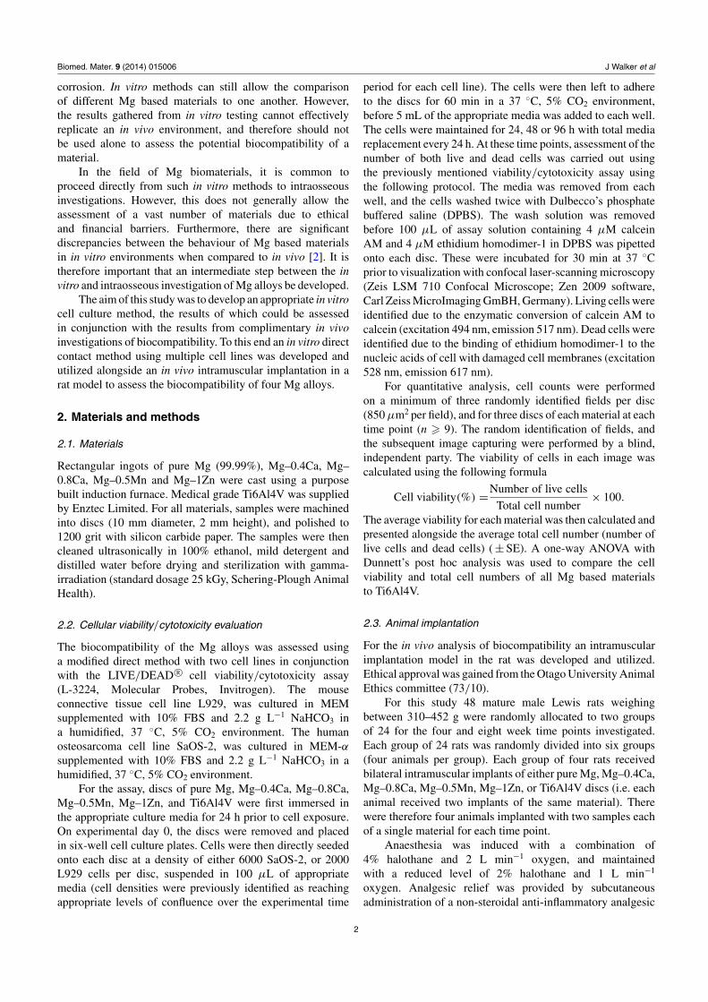
The animals were housed individually and monitoredfor food intake and hydration for 10 days post surgery,before rehoming in group cages. The animals were thenmonitored primarily for indications of hydrogen evolutionand pain behaviour every 48 h until the conclusion of theinvestigation. At either 4 or 8 weeks post-implantation, theanimals were euthanized by carbon dioxide inhalation. Theimplant and surrounding tissue were then excised and fixedin 10% neutral buffered formalin (10% NBF) for 48 h forhistological analysis. After fixation the discs were carefullyremoved from the surrounding tissue, immersed in a chromicacid solution (200 g L−1 CrO3 and 10 g L−1 AgNO3) for 15 minto remove corrosion products, and weighed to assess corrosion.Tissue samples were then processed using a standard waxembedding protocol, and 4 μM sections were cut 2.5 mm,5 mm and 7.5 mm from the medial edge of the disc spacewith a rotary microtome. The sections were then stainedwith haematoxylin and eosin (H&E) and visualized withlight microscopy. For analysis three randomly identified high-magnification fields were examined (40 × objective lens,each field 1200 × 900 μm). A representative field of viewis presented in figure 1. In each field the width of the fibrouscapsule was assessed by averaging the depth of the fibrousand inflammatory tissue layer adjacent to the implant at threerandomly allocated points. A semi-quantitative scale wasused to assess the infiltration of lymphoctyes, macrophagesand neovascularization based on the scoring system definedby ISO 10993–6 (2007) (table 1). Other features of tissue
Figure 1. A representative field of view used for the assessment ofin vivo biocompatibility. The white space towards the top of theimage is the area where the implant was present prior to removal.The black arrow indicates the fibrous capsule that has formedadjacent to the implant. This region is lighter in colour as it isprimarily composed of collagen as opposed to the darker skeletalmuscle.
reaction that were not measured within this study recognizedin ISO 10993–6 such as polymorphonuclear cells, plasmacells, and giant cells were identified extremely rarely, andno tissue necrosis was identified in any of the tissue sectionsexamined. The production of hydrogen gas was also assessedat low magnification with a semi-quantitative scale based onthe presence of bubble shaped spaces within the tissue, anddistension of the implant space for each entire tissue section(table 1). A one-way ANOVA with Dunnett’s post hoc analysiswas used to compare the results of all Mg based materials tothose of Ti6Al4V.
3. Results
3.1. Assessing the in vitro biocompatibility of magnesiumalloys
The viability results for L929 and SaOS-2 cells cultured on thefour Mg alloys, pure Mg and Ti–6Al–4V at 24, 48 and 96 hare presented in figures 2 and 3, respectively. The viabilityresults from each time point must be assessed alongside thetotal cell numbers present to attain a comprehensive view of thecellular response to the materials investigated. This prevents
3

Biomed. Mater. 9 (2014) 015006 J Walker et al
Figure 2. Results from the Live/Dead R© assay 24, 48, and 96 h afterthe seeding of L929 cells onto Ti–6Al–4V, pure Mg or Mg alloydiscs. ∗ Indicates a statistically significant difference when the Mgbased materials were compared to Ti–6Al–4V (p < 0.01) using a1-way ANOVA with Dunnett’s post hoc analysis.
the misrepresentation of a material as cytocompatible, if forexample 80% of the cells are viable, but only five cells arepresent (the low numbers of cells would therefore representsignificant toxicity).
The results for the L929 cells at 24 h show a statisticallysignificant difference in the viability of cells between Ti–6Al–4V and Mg–0.4Ca, Mg–0.8Ca and Mg–1Zn (p < 0.01).Whereas at this time point there was no significant differencebetween Ti–6Al–4V and Pure Mg or Mg–0.5Mn (p >
0.01). However, the total cell numbers indicate a statisticallysignificant difference between Ti–6Al–4V and all of the Mgbased materials (p < 0.01). This reduction in total cell numberfor all Mg based materials at this early stage is indicativeof difficulties in the initial adherence of cells to the Mgbased substrates. This reduced initial attachment has importantramifications on the total cell numbers present at later timepoints. Figure 4 displays representative confocal images oflive and dead L929 cells after 96 h cultured on Mg basedmaterials or Ti–6Al–4V.
Figure 3. Results from the Live/Dead R© assay 24, 48, and 96 h afterthe seeding of SaOS-2 cells onto Ti–6Al–4V, pure Mg or Mg alloydiscs. ∗ Indicates a statistically significant difference when the Mgbased materials were compared to Ti–6Al–4V (p < 0.01) using a1-way ANOVA with Dunnett’s post hoc analysis.
A similar trend can be identified after 48 h, withstatistically significant difference in viability between Ti–6Al–4V and Mg–0.4Ca, Mg–0.8Ca and Mg–1Zn. Total cellnumbers are similar with a significant difference identifiablebetween Ti6Al4V and Pure Mg, Mg–0.4Ca, Mg–0.8Ca andMg–0.5Mn. At this stage Mg–1Zn alone exhibited a total cellnumber that was not significantly different from the titaniumalloy. After 96 h, the total cell numbers present on the titaniumalloy had reached 1300 with a viability 99.5%. However, PureMg is the only material providing a viability significantly lowerthan Ti–6Al–4V at this time point. In contrast, all the Mg basedmaterials exhibited a statistically significant reduction in totalcell number when compared to Ti–6Al–4V.
The results of the SaOS-2 assay indicate similar results. Atthe 24 and 48 h time points, all the magnesium based materialsexhibited significantly reduced viability and total cell numberwhen compared to Ti–6Al–4V, with the viability of Pure Mgthe only exception. After 96 h, the same trend is followed withthe only difference being the lack of a significant reduction
4

Biomed. Mater. 9 (2014) 015006 J Walker et al
(A) (B)
(C) (D)
(E) (F)
Figure 4. Confocal micrographs of L929 cells cultured on thesurface of Mg alloy, pure Mg and Ti–6Al–4V after 96 h. Greenprofiles indicate living cells (calcein AM). Red profiles indicatedying or dead cells (ethidium homodimer-1). (A) Mg–0.4Ca.(B) Mg–0.8Ca. (C) Mg–0.5Mn. (D) Mg–1Zn. (E). Pure Mg.(F) Ti–6Al–4V.
between the total cell numbers on Ti–6Al–4V when comparedto Mg–1Zn. Figure 5 displays representative confocal imagesof live and dead SaOS-2 cells after 96 h cultured on Mg basedmaterials or Ti–6Al–4V.
3.2. The biocompatibility of magnesium alloys in anintramuscular environment
The average width of the fibrous capsule, and the averagegrade allocated for lymphocyte and macrophage infiltration inassociation with the intramuscular implantation of Ti–6Al–4V,Pure Mg, Mg–0.4Ca, Mg–0.8Ca, Mg–0.5Mn, and Mg–1Zn at4 and 8 weeks are presented in figure 6. Statistical analysisindicated no significant differences between Ti–6Al–4V andthe Mg based materials for any variable at either time point(p > 0.01).
The average grade allocated for neovascularization,hydrogen production and corrosion in association with theintramuscular implantation of Ti–6Al–4V, Pure Mg, Mg–0.4Ca, Mg–0.8Ca, Mg–0.5Mn, and Mg–1Zn at 4 and 8 weeks
(A)
(C )
(B)
(E ) (F )
(D)
Figure 5. Confocal micrographs of SaOS-2 cells cultured on thesurface of Mg alloy, pure Mg and Ti–6Al–4V after 96 h. Greenprofiles indicate living cells (calcein AM). Red profiles indicatedying or dead cells (ethidium homodimer-1). (A) Mg–0.4Ca.(B) Mg–0.8Ca. (C) Mg–0.5Mn. (D) Mg–1Zn. (E). Pure Mg.(F) Ti–6Al–4V.
are presented in figure 7. In concurrence with the results fromthe fibrous encapsulation, and lymphocyte and macrophageinfiltration, there were no statistically significant differencesbetween the average grade allocated for neovascularizationwhen Ti–6Al–4V was compared to the Mg based materials(p > 0.01). In contrast, there was a statistically significantdifference between Ti–6Al–4V and all of the Mg basedmaterials for hydrogen production and corrosion (p < 0.01).However, this result was expected as the titanium alloyneither corrodes nor produces hydrogen gas. Figure 8 showslow magnification micrographs indicating tissue distensionin response to hydrogen production with the implantation ofTi–6Al–4V and Mg–0.4Ca.
4. Discussion
4.1. General comments on the In Vitro methodology
A multitude of in vitro cell culture techniques exist forthe assessment of the biocompatibility of magnesium as
5

Biomed. Mater. 9 (2014) 015006 J Walker et al
Figure 6. The average width of the fibrous capsule, and average grades allocated to lymphocyte infiltration and macrophage infiltration inthe tissue adjacent to Ti–6Al–4V, pure Mg or Mg alloy implants. There were no statistically significant differences for any measure when theMg based materials were compared to Ti–6Al–4V (p > 0.01) using a 1-way ANOVA with Dunnett’s post hoc analysis.
a biomaterial. Primarily, these techniques are based onthe methods outlined by international standards with slightvariations in experimental set-up and the assessment of cellular
behaviour. In particular, ISO-10993–5 suggests the use ofeither an indirect method in which cells are exposed toextracts of the materials, or the direct method in which the
6

Biomed. Mater. 9 (2014) 015006 J Walker et al
Figure 7. The average grade allocated to neovascularization, hydrogen production, and implant corrosion with implantation of Ti–6Al–4V,pure Mg or Mg alloy implants. There were no statistically significant differences between the Mg based materials and Ti–6Al–4V forneovascularization (p > 0.01, 1-way ANOVA with Dunnett’s post hoc analysis). All Mg based materials exhibited statistically significantincreases in both hydrogen production and corrosion when compared to Ti–6Al–4V (p < 0.01, 1-way ANOVA with Dunnett’s post hocanalysis).
cells come into contact with the materials [3]. Unfortunately,the standards currently available are designed for theassessment of the in vitro biocompatibility of primarily non-
degrading biomaterials. This leads to difficulties in applyingthese methods to rapidly corroding materials such as Mgalloys.
7

Biomed. Mater. 9 (2014) 015006 J Walker et al
(A)
(B)
Figure 8. Low magnification micrographs of the implant spaces leftafter the removal of an intramuscular implant (H & E). (A) Thewhite space is the area left after removal of a Ti–6Al–4V implantwith clearly identifiable margins within the tissue. This received agrade of 0 on the previously identified scoring system for hydrogenevolution. (B) The space left after removal of a Mg–0.4Ca implantwith some distension of the space, and obscured margins due toprobable hydrogen bubbles within the tissue. This received a grade 3on the previously identified scoring system.
The indirect method is used relatively frequently withMg based materials in one of two ways. Either extracts areproduced and diluted to various concentrations [4–16], or amodification to the method is adopted in which metal saltsare added to the culture media to replicate the toxicity ofthe released Mg and alloying component ions [17–19]. Theprimary issue with the first of these methods is that any toxicityidentified is primarily due to the osmolality of the solution [20].Whilst this can be argued to be of primary importance to thebiocompatibility of the material, it is likely that in an in vivoenvironment constant diffusion would mitigate this effect to adegree. Additionally, when this method is adopted, the extractsare often diluted to a point for which the increased osmolalityis no longer toxic [4, 7, 10, 14, 16], somewhat nullifying theuse of the method in the first place.
The production of an extract solution also requires theimmersion of a Mg sample in media for a period of time.The resulting corrosion that occurs would likely cause anincrease in the pH of the extract solution [8, 21], which left un-adjusted would certainly result in cytotoxicity. However, thispoint is often left unmentioned in studies using the technique[6, 9, 10, 12–16, 22], or there is reliance on pH adjustmentto prevent this occurrence [5]. The aforementioned additionof Mg and alloying component salts dissolved in cell culturemedia is also problematic due to the effects on the osmolalityof the solution. This method can be of use when investigatingalloying elements not found within cell culture media such asrare earth elements, but is less useful for assessing the effectof ions already found within physiological solutions such asMg and Ca. Additionally, the most common technique usedto assess the viability of cells with the indirect method is touse a colorimetric assay based on the reduction of tetrazoliumsalts to formazan dyes within living cells [7–9, 11, 12, 14–19, 22, 23]. However, this method can be affected by various
factors including both the pH and osmolality of the cell cultureenvironment, reducing the value of the results when used withMg based materials [20, 24].
For the above reasons, a modified direct method usingthe Live/Dead R© assay was adopted for this investigation. Thechoice to seed cells directly onto the surfaces of the materialswas based upon a desire to assess whether cells could attachand stay viable on an actively corroding material. This waspreferable to the protocol outlined in ISO 10993–5 for thedirect method in which the test material is placed onto anadherent cell layer, as it was believed that for Mg this wastoo similar to the indirect method in which the pH change andincrease in osmolality would have the greatest influence on cellsurvival. Additionally, no evidence existed that the corrodingMg materials would interfere with the calcein AM or ethidiumhomodimer utilized for the assay.
Several other aspects of the methodology used withinthis investigation require comment. Firstly, the materials werepre-soaked to allow the formation of a passivation layer onthe surface of the implant. Whilst it could be argued thatthis step is masking the potentially toxic effects of the rapidinitial corrosion of the materials, initial experiments indicatedthat without this allowance initial cellular adhesion would begreatly diminished. Eliminating this step would drasticallyreduce any ability to investigate the in vitro biocompatibilityof Mg alloys, and it was therefore included in the protocol.
Secondly, it is important to note that the media for eachmaterial was replaced daily. This step was taken to reduce anytoxicity occurring due to an increase in the pH of the staticcell culture environment. Again, this could be argued to bemasking the toxic effect of the pH change associated with Mgcorrosion. However, it is believed that in a dynamic in vivoenvironment, the pH in the area associated with a Mg alloyimplant would not fluctuate as significantly as in an in vitroenvironment. However, there would still be a marked increasein pH at the surface of the material in vitro that would also occurin an in vivo environment. Therefore, the media replacementregime utilized should not have further increased the innatediscrepancies between in vitro and in vivo methodologies.
Thirdly, the choice of cells used in the investigationof in vitro biocompatibility is important. In this study twoimmortalized cell lines were used, L929 (mouse fibroblast) andSaOS-2 (human osteosarcoma). The L929 line was selectedas a standardized, and commonly employed cell type forbiocompatibility studies [25–28]. The SaOS-2 cells werealso included as a cell line derived from the more clinicallyapplicable human osteosarcoma [29]. These cells are alsocommonly used for the assessment of the biocompatibilityof potential orthopaedic materials due to their osteoblasticphenotype, ability to differentiate and produce mineralizedextracellular matrix [30–33]. Within this experiment the SaOS-2 cells were not exposed to a differentiating media, whichin retrospect could have improved adherence, and thereforeincreased cell number. Although this could potentially havealtered the results gathered, the use of both cell lines is still ofvital importance as the cellular response to any given materialhas been observed to differ vastly depending on the cell type[8, 17, 29, 34, 35].
8

Biomed. Mater. 9 (2014) 015006 J Walker et al
The corrosion associated with Mg alloys, and the resultingby-products of this corrosion make the assessment of anyaspect of alloy behaviour difficult in an in vitro environment.The assessment of in vitro biocompatibility is particularlydifficult, due to the sensitive nature of cells growing inculture. The points mentioned above highlight these issuesand the difficulties in developing methods that avoid celldeath due to environmental perturbations, without disguisingthe potential cytotoxic effects of the Mg alloys. For thesereasons the assessment of the biocompatibility of the Mg alloysinvestigated in this study focused on the viability/cytotoxicity.More in depth investigations of cellular behaviour anddifferentiation are commonly carried out. However, it isbelieved that the discrepancies and difficulties in replicatingan in vivo environment in vitro, is likely to provide results forthese studies only barely comparable to those that would beseen with in vivo implantation of the materials.
4.2. Comparison between the in vitro biocompatibility of Mgalloys
The results of the in vitro analysis of the biocompatibility ofMg–0.4Ca, Mg–0.8Ca, Mg–0.5Mn, Mg–1Zn, pure Mg and Ti–6Al–4V indicate a general trend towards lower viability andtotal cell number on the Mg based materials when comparedto Ti–6Al–4V. This result was not unexpected due to theactive corrosion occurring when the Mg based materials areexposed to a physiological solution. The resultant increasein osmolality, increase in pH and the physical disruption tocells by hydrogen gas evolution, are all factors that wouldreduce cellular adhesion and viability in a closed cell cultureenvironment [21]. However, despite a relatively inhospitableenvironment, some cellular adhesion and active cell divisiondid occur when cells were seeded directly onto the Mg alloysbeing investigated. Even at the reduced viability and totalcell numbers observed, this can be seen as an exceptionallypositive result for the potential biocompatibility of these alloysin the more dynamic and adjustable in vivo environment.Consequently, despite the significant differences between theresults from the Ti–6Al–4V and the Mg based materials, theresults of these studies can still be used to an extent for thecomparison of the in vitro biocompatibility of the Mg alloys.
The results from the L929 experiments generally indicatedrelatively good performance of the Mg–0.5Mn and Mg–1Znalloys with increasing time. The cells on these two materialsappeared to recover after the initial low total cell number seenat 24 h. In contrast, the Ca containing alloys exhibited verylow total cell counts for the entirety of the experiment. Similarresults were observed with the SaOS-2 cells for which muchhigher total cell numbers were seen with Mg–0.5Mn, Mg–1Znand pure Mg when compared to the Ca containing alloys.
The assessment of the in vitro biocompatibility of alloyscomparable to those investigated in this study is relativelyrare, and more infrequent still is the use of a direct method.Most commonly pure Mg is included as a control materialin studies investigating a range of alloys [5, 36], surfacetreatments [21, 37], coatings [38], or fabrication methods[16]. The studies using a direct method for the assessment of
the cytocompatibility of pure Mg show variable results, fromextremely toxic [21, 38] to highly biocompatible [36, 37].This highlights the continuing difficulties with comparingthe behaviour of Mg based materials between in vitroinvestigations in which small differences in variables suchas surface roughness, cell type, media type or surface area tovolume ratio can dramatically affect corrosion behaviour, andtherefore cellular behaviour.
Even less evidence exists in the literature for the behaviourof cells on the relevant Mg alloys. One study indicated asignificantly reduced total protein concentration, for a Mg–0.5Ca alloy when compared to a negative control (tissueculture slide). The same study also indicated Mg–0.8Caprovided a result statistically indistinct to the control, perhapssuggesting Mg–0.8Ca was more biocompatible than Mg–0.5Ca [36]. These results are somewhat in contrast to thoseseen within this investigation in which the lower calciumpercentage-containing alloy generally exhibited slightlyimproved biocompatibility. However this experiment utilizedtotal protein primarily as an indicator of osteogenic capacityrather than to assess cell number, and was also measuredafter eight days in culture, both of which make comparisonto this investigation difficult. The in vitro biocompatibilityof a binary Mg–6Zn alloy has also been investigated, withresults showing acceptable adhesion and some cellular division[39, 40]. However these studies do not compare the results toan inert control material preventing any comparison to otherinvestigations. No research has been published investigatingthe in vitro biocompatibility of a binary Mg–Mn alloy.
There are many inherent difficulties in both testingthe biocompatibility of Mg alloys in vitro, and attemptingto compare the results between studies. If a conservativeapproach was taken in the assessment of the results seenwithin this investigation, Mg alloys would be assumed highlytoxic materials. However, use of similar alloys and pure Mg inan in vivo environment has indicated good biocompatibility[15, 22, 41]. It is therefore important that a more liberalinterpretation of the in vitro results be taken, and to ensure thatthe conclusions from such studies be made alongside otherevidence such as the results from in vivo investigations.
4.3. General comments on the in vivo methodology
Analysis of the biocompatibility of Mg alloys in asoft tissue environment is relatively uncommon, with themajority of studies progressing from in vitro corrosion andbiocompatibility investigations directly to their assessmentin an intraosseous location. The reason for this avoidanceof standard biocompatibility protocols remains unclear. BothISO and ASTM standards outline protocols for the short-termassessment of the local effects of implantation of biomaterialsin either a subcutaneous or intramuscular location [42–44].For this investigation, the inclusion of a soft tissue studywas deemed important for several reasons. Firstly, it washypothesized that an intramuscular environment would beable to tolerate the production of hydrogen gas better than arigid intraosseous environment, a factor considered importantgiven the limited knowledge available regarding the effect
9

Biomed. Mater. 9 (2014) 015006 J Walker et al
of hydrogen production in an in vivo environment. Secondly,soft tissue implantation provided the ability to accuratelyassess the corrosion of the materials by mass loss. Thirdly,many orthopaedic biomaterials will directly contact muscleand connective tissue as well as bone. The assessment ofthe reaction of soft tissues to Mg alloys being developed fororthopaedic application is therefore important.
4.4. Comparison between the in vivo biocompatibility of Mgalloys
The in vivo biocompatibility of Mg–0.4Ca, Mg–0.8Ca,Mg–0.5Mn, Mg–1Zn, pure Mg and Ti–6Al–4V in anintramuscular environment was analysed through theassessment of the width of the fibrous capsule, the lymphocyteand macrophage infiltration, and neovascularization in thetissue adjacent to the implants. An overwhelming trend couldbe identified for all these measures indicating no significantdifferences between any of the Mg based materials and theTi–6Al–4V. As this Ti alloy is widely used as a medical implantmaterial in both medicine and dentistry [45–47], these resultsindicate comparable biocompatibility for these Mg alloys.
The assessment of in vivo biocompatibility of Mg alloysin a soft tissue environment is rare, with very few articlesavailable for the comparison of results. The majority of thosethat have been performed rely on brief descriptive results ofthe histology to indicate the tissue reaction to the materials[13, 48–50]. One study however, has thoroughly assessed thereaction of muscle tissue to a Mg–0.8Ca implant, using asemi-quantitative scale to assess fibrous encapsulation, tissuecavity formation, necrosis and the infiltration of macrophages,giant cells, heterophilic granulocytes, B-lymphocytes andT-lymphoctyes [51]. This extensive analysis indicated a similarresult to that seen within this investigation, with Mg–0.8Caperforming similarly to a stainless steel 316L control material.
4.5. The corrosion of Mg alloys in an intramuscularenvironment
One of the benefits in performing a soft tissue implantation ofMg alloys was the ability to remove the samples and assessthe corrosion by gravimetric analysis. The results indicatedMg–1Zn as corroding slower than the other Mg based materialsat the four-week time point. After eight-weeks of implantation,the corrosion of the alloys followed a similar trend, indicatingMg–1Zn to be the slowest corroding of the alloys investigated.
5. Conclusion
The primary aim of the in vitro and in vivo investigationof Mg alloy biocompatibility was to develop and refine aseries of simple, rapid and reproducible methods to assessMg alloy biocompatibility. The provision of an intermediatestep between in vitro corrosion analysis and intraosseousimplantation of Mg alloys allows the investigation of a largervariety of materials in an in vivo location without the expenseand complexity of an intraosseous study. This could preventboth the intraosseous implantation of potentially harmful
materials, and the inappropriate exclusion of potentially usefulmaterials due to conservative in vitro corrosion analysis.
Additionally it was identified that whilst in vitrotechniques can be used as an indicator of the biocompatibilityof Mg alloys, the results must be analysed with anunderstanding that the corrosion of the materials can lead tofalse positive results for toxicity. It is therefore important thatin vitro analysis of biocompatibility be assessed alongside theresults of a corresponding in vivo method.
References
[1] Pearce A I, Richards R G, Milz S, Schneider E and Pearce S G2007 Animal models for implant biomaterial research inbone: a review Eur. Cells Mater. 13 1–10
[2] Witte F et al 2006 In vitro and in vivo corrosion measurementsof magnesium alloys Biomaterials 27 1013–8
[3] ISO 1999 Biological Evalutation of Medical Devices Part 5:Tests for In Vitro Cytotoxicity ISO 10993–5 (Geneva:International Organization for Standardization)
[4] Fischer J, Profrock D, Hort N, Willumeit R and Feyerabend F2011 Reprint of: Improved cytotoxicity testing ofmagnesium materials Mater. Sci. Eng. B 176 1773–7
[5] Yang C, Yuan G, Zhang J, Tang Z, Zhang X and Dai K 2010Effects of magnesium alloys extracts on adult human bonemarrow-derived stromal cell viability and osteogenicdifferentiation Biomed. Mater. 5 045005
[6] Zheng Y F, Gu X N, Xi Y L and Chai D L 2010 In vitrodegradation and cytotoxicity of Mg/Ca compositesproduced by powder metallurgy Acta Biomater. 6 1783–91
[7] Wang D-W, Cao Y, Qiu H and Bi Z-G 2011 Improved bloodcompatibility of Mg–1.0Zn–1.0Ca alloy by micro-arcoxidation J. Biomed. Mater. Res. A 99 166–72
[8] Gu X, Zheng Y, Cheng Y, Zhong S and Xi T 2009 In vitrocorrosion and biocompatibility of binary magnesium alloysBiomaterials 30 484–98
[9] Huan Z G, Leeflang M a, Zhou J, Fratila-Apachitei L Eand Duszczyk J 2010 In vitro degradation behavior andcytocompatibility of Mg–Zn–Zr alloys J. Mater. Sci., Mater.Med. 21 2623–35
[10] Li Y et al 2012 Mg–Zr–Sr alloys as biodegradable implantmaterials Acta Biomater. 8 3177–88
[11] Park R S et al 2012 Corrosion behavior and cytotoxicity ofMg–35Zn–3Ca alloy for surface modified biodegradableimplant material J. Biomed. Mater. Res. B 100 911–23
[12] Wang Y et al 2012 In vitro degradation and biocompatibilityof Mg–Nd–Zn–Zr alloy Chin. Sci. Bull. 57 2163–70
[13] Hanzi A C, Gerber I, Schinhammer M, Loffler J Fand Uggowitzer P J 2010 On the in vitro and in vivodegradation performance and biological response of newbiodegradable Mg–Y–Zn alloys Acta Biomater. 6 1824–33
[14] Zhang E, Yin D, Xu L, Yang L and Yang K 2009Microstructure, mechanical and corrosion properties andbiocompatibility of Mg–Zn–Mn alloys for biomedicalapplication Mater. Sci. Eng. C 29 987–93
[15] Li Z, Gu X, Lou S and Zheng Y 2008 The development ofbinary Mg–Ca alloys for use as biodegradable materialswithin bone Biomaterials 29 1329–44
[16] Gu X N, Zhou W R, Zheng Y F, Liu Y and Li Y X 2010Degradation and cytotoxicity of lotus-type porous puremagnesium as potential tissue engineering scaffold materialMater. Lett. 64 1871–4
[17] Feyerabend F et al 2010 Evaluation of short-term effects ofrare earth and other elements used in magnesium alloys onprimary cells and cell lines Acta Biomater. 6 1834–42
10

Biomed. Mater. 9 (2014) 015006 J Walker et al
[18] Feser K, Kietzmann M, Baumer W, Krause C and Bach F W2011 Effects of degradable Mg–Ca alloys on dendritic cellfunction J. Biomater. Appl. 25 685–97
[19] Zhang E, Yang L, Xu J and Chen H 2010 Microstructure,mechanical properties and bio-corrosion properties ofMg-Si(-Ca, Zn) alloy for biomedical application ActaBiomater. 6 1756–62
[20] Witte F et al 2008 Degradable biomaterials based onmagnesium corrosion Curr. Opin. Solid State Mater. Sci.12 63–72
[21] Lorenz C, Brunner J G, Kollmannsberger P, Jaafar L, Fabry Band Virtanen S 2009 Effect of surface pre-treatmentson biocompatibility of magnesium Acta Biomater.5 2783–9
[22] Zhang S et al 2010 Research on an Mg–Zn alloy as adegradable biomaterial Acta Biomater. 6 626–40
[23] Witte F et al 2007 Biodegradable magnesium-hydroxyapatite metal matrix composites Biomaterials28 2163–74
[24] Fischer J, Prosenc MH, Wolff M, Hort N, Willumeit Rand Feyerabend F 2010 Interference of magnesiumcorrosion with tetrazolium-based cytotoxicity assays ActaBiomater. 6 1813–23
[25] Turner T D, Spyratou O and Schmidt R J 1989Biocompatibility of wound management products:standardization of and determination of cell growthrate in L929 fibroblast cultures J. Pharm. Pharmacol.41 775–80
[26] Acharya C, Ghosh S K and Kundu S C 2008 Silk fibroinprotein from mulberry and non-mulberry silkworms:cytotoxicity, biocompatibility and kinetics of L929murine fibroblast adhesion J. Mater. Sci., Mater. Med.19 2827–36
[27] Jorge J H, Giampaolo E T, Vergani C E, Machado A L,Pavarina A C and Carlos I Z 2007 Biocompatibility ofdenture base acrylic resins evaluated in culture of L929cells. Effect of polymerisation cycle and post-polymerisation treatments Gerodontology 24 52–7
[28] Serrano M C et al 2004 In vitro biocompatibility assessment ofpoly(epsilon-caprolactone) films using L929 mousefibroblasts Biomaterials 25 5603–11
[29] Kirkpatrick C J, Krump-Konvalinkova V, Unger R E,Bittinger F, Otto M and Peters K 2002 Tissue response andbiomaterial integration: the efficacy of in vitro methodsBiomol. Eng. 19 211–7
[30] Anderson H C et al 1995 The mechanism of bone inductionand bone healing by human osteosarcoma cell extracts Clin.Orthop. Relat. Res. 313 129–34
[31] Okumura A et al 2001 Substrate affects the initial attachmentand subsequent behavior of human osteoblastic cells(Saos-2) Biomaterials 22 2263–71
[32] Gronowicz G and McCarthy M B 1996 Response of humanosteoblasts to implant materials: integrin-mediated adhesionJ. Orthop. Res. 14 878–87
[33] Yu X, Botchwey E a, Levine E M, Pollack S Rand Laurencin C T 2004 Bioreactor-based bone tissueengineering: the influence of dynamic flow on osteoblastphenotypic expression and matrix mineralization Proc. NatlAcad. Sci. USA 101 11203–8
[34] Wataha JC, Hanks CT and Sun Z 1994 Effect of cell line onin vitro metal ion cytotoxicity Dent. Mater. 10 156–61
[35] Thonemann B, Schmalz G, Hiller K and Schweikl H 2002Responses of L929 mouse fibroblasts, primary andimmortalized bovine dental papilla-derived cell lines todental resin components Dent. Mater. 18 318–23
[36] Pietak A, Mahoney P, Dias G J and Staiger M P 2008Bone-like matrix formation on magnesium and magnesiumalloys J. Mater. Sci., Mater. Med. 19 407–15
[37] Li L, Gao J and Wang Y 2004 Evaluation of cyto-toxicity andcorrosion behaviour of alkali-heat-treated magnesium insimulated body fluid Surf. Coat. Technol. 185 92–8
[38] Jo J-H et al 2011 Hydroxyapatite coating on magnesium withMgF(2) interlayer for enhanced corrosion resistance andbiocompatibility J. Mater. Sci., Mater. Med. 22 2437–47
[39] Li J et al 2010 In vitro responses of human bone marrowstromal cells to a fluoridated hydroxyapatite coatedbiodegradable Mg–Zn alloy Biomaterials 31 5782–8
[40] Zhang S X et al 2009 In vitro degradation, hemolysis andMC3T3-E1 cell adhesion of biodegradable Mg–Zn alloyMater. Sci. Eng. C 29 1907–12
[41] Xu L P, Yu G N, Zhang E, Pan F and Yang K 2007 In vivocorrosion behavior of Mg–Mn–Zn alloy for bone implantapplication J. Biomed. Mater. Res. A 83 703–11
[42] ISO 2007 Biological Evaluation of Medical Devices Part 6:Tests for local effects after implantation ISO 10993–6(Geneva: International Organization for Standardization)
[43] ASTM 2004 Standard Practice for Short-Term Screening ofImplant Materials ASTM F763–04 (West Conshohocken,PA: American Society for Testing and Materials)
[44] ASTM 2008 Standard Practice for Subcutaneous ScreeningTest for Implant Materials ASTM 1408–97 (WestConshohocken, PA: American Society for Testing andMaterials)
[45] Deligianni D D, Katsala N, Ladas S, Sotiropoulou D,Amedee J and Missirlis Y F 2001 Effect of surfaceroughness of the titanium alloy Ti–6Al–4V on human bonemarrow cell response and on protein adsorptionBiomaterials 22 1241–51
[46] Park J-W, Kim H-K, Kim Y-J, Jang J-H, Song H andHanawa T 2010 Osteoblast response and osseointegrationof a Ti–6Al–4V alloy implant incorporating strontium ActaBiomater. 6 2843–51
[47] Niinomi M 1998 Mechanical properties of biomedicaltitanium alloys Mater. Sci. Eng. A 243 231–6
[48] Hussl H, Papp C, Hopfel-Kreiner I, Rumpl E andWilflingseder P 1981 Resorption time and tissue reactionswith magnesium rods in rats and rabbits Eur. J. Plast. Surg.6 117–26
[49] Xue D, Yun Y, Tan Z, Dong Z and Schulz M J 2012 In vivoand in vitro degradation behavior of magnesium alloys asbiomaterials J. Mater. Sci. Technol. 28 261–7
[50] Aghion E, Levy G and Ovadia S 2012 In vivo behavior ofbiodegradable Mg–Nd–Y–Zr–Ca alloy J. Mater. Sci., Mater.Med. 23 805–12
[51] Erdmann N et al 2010 Evaluation of the soft tissuebiocompatibility of MgCa0.8 and surgical steel 316Lin vivo: a comparative study in rabbits Biomed. Eng. online9 63
11