biocompatibility studies on lanthanum oxide nanoparticles · biocompatibility studies on lanthanum...
TRANSCRIPT
Toxicology Research
PAPER
Cite this: DOI: 10.1039/c4tx00198b
Received 8th November 2014,Accepted 27th April 2015
DOI: 10.1039/c4tx00198b
www.rsc.org/toxicology
Biocompatibility studies on lanthanum oxidenanoparticles
B. Brabu,a,b,c S. Haribabu,a,b M. Revathy,a,b S. Anitha,d M. Thangapandiyan,e
K. R. Navaneethakrishnan,a,b C. Gopalakrishnan,a,c S. S. Murugana,b andT. S. Kumaravel*a,b,f
Lanthanum oxide nanoparticles (LONP), a rare earth metal oxide, have unique properties that make them
a suitable candidate for several biomedical applications. We investigated certain key in vitro and in vivo
biocompatibility endpoints on LONP. LONP were cytotoxic in in vitro assays and predominantly exerted
their action via release of reactive oxygen species. These nanoparticles were neither irritants nor sensi-
tizers in a rabbit model. LONP extracts did not exert any acute systemic toxicity effects in mice. On the
other hand LONP exerted toxicity to the liver following oral administration, suggesting that these particles
are absorbed from the gastrointestinal (GI) tract and deposited in the hepatobiliary system. LONP did not
induce any mutation in the Ames test both in the presence or absence of S-9. These observations provide
a base line biocompatibility and toxicity data on LONP. The current findings will also be useful in defining
standards for nanoparticle containing devices.
Introduction
Lanthanum oxide nanoparticles (LONP), a rare earth metaloxide, have unique properties that make them a suitable candi-date for several biomedical applications. Probes coated withLONP are being developed as implantable sensors of variousmolecules such as glucose, phosphate and uric acid.1 In com-bination with other elements, lanthanum oxide (La2O3) isbeing developed as an optical sensing system for measuringvariations in human body temperature.2 Because of its para-magnetic properties, it is also being developed for the mag-netic field controlled targeted release of drugs within thebody. La2O3 suppresses bacteria,3 viruses,4 and fluorescencedyes,5 selectively binds to several proteins,6 suppressescalcium channels7 and has light emitting properties.8 While itis important to note that there are several potential appli-cations of this nanoparticle in medical device technologies, no
biocompatibility data are currently available on LONP toassure their safety.
In this manuscript, we investigated biocompatibility ofLONP in line with well established ISO 10993 standards.9
Specifically we investigated cytotoxicity, irritation, sensiti-zation, acute systemic toxicity and mutagenicity potential ofLONP, which are critical biocompatibility indicators for anymedical devices.9 Several guidance documents on the safetyevaluation of nanomaterials to be used in food/feed, cosmeticsand medical devices have been published by European FoodSafety Authority (EFSA), Scientific Committee on ConsumerSafety (SCCS) and Scientific Committee on Emerging andNewly Identified Health Risks (SCENIHR) as Scientific Com-mittees of the European Commission. While InternationalOrganization for Standardization (ISO) ISO 10993 standardsdo not give specific guidance on testing the biocompatibilityof materials in the nanoscale range, we adopted these broadprinciples and combined them with the usual techniquesemployed in nanotoxicology to evaluate the biocompatibility ofLONP. It should be noted that ISO is currently developing gui-dance on evaluating medical devices containing nanoparticles(ISO/NP TR 10993-22 under development) and our results mayfeed into this process. Also our results will be helpful in evalu-ating the safety of medical device technologies using LONP asa raw material. It should be noted that there is an increasinginterest in biocompatibility of nanomaterials.10–12 This workwill further add to the knowledge base of biocompatibility ofnanomaterials.
aNanotechnology Research Center and GLR Laboratories, Academic–Industry Joint
Collaborative Research Unit, SRM University, Chennai, IndiabGLR Laboratories Private Limited, Chennai 600060, IndiacNanotechnology Research Center, SRM University, Chennai 603203, IndiadUNAM-National Nanotechnology Research Centre, Bilkent University, Ankara,
06800, TurkeyeDepartment of Veterinary Pathology, Madras Veterinary College, Chennai 600007,
IndiafGLR Laboratories Private Limited, UK. E-mail: [email protected]
This journal is © The Royal Society of Chemistry 2015 Toxicol. Res.
Publ
ishe
d on
27
Apr
il 20
15. D
ownl
oade
d by
Uni
leve
r R
&D
mul
tisite
on
13/0
5/20
15 1
5:43
:30.
View Article OnlineView Journal

Experimental sectionMaterials and methods
Characterization of lanthanum oxide particles. La2O3 bulkand nanoparticles were purchased from Ottokemi, Mumbai,India and Nanoshel USA, respectively. The certificate of analy-sis from the manufacturer indicated the purity as 99.99%. Thelist of impurities were Fe2O3, CaO, SiO2, Cl and others, at con-centrations of 0.002, 0.002, 0.01, 0.001 and 0.004 ppm, respecti-vely. These impurities were unlikely to affect the resultsobtained. The morphology and size of the particles werecarried out using a High-Resolution Transmission ElectronMicroscope (HRTEM-JEOL 3010). The surface charge and thehydrodynamic diameter of the La2O3 particles were measuredby using a Dynamic Light Scattering analyzer (Nanoparticaanalyzer SZ-100). The zeta potential of the samples wasmeasured at 25 °C and 37 °C to characterize the particles atroom temperature and at physiological temperature.
Cytotoxicity. Cytotoxicity was assessed by the direct contactmethod. We used the MTT assay (from the battery of cytotoxi-city assays described in ISO 10993, part 5)9,13 to assess thecytotoxicity of LONP. In addition we used assays that reflectother pathways of cytotoxicity such as the LDH assay (mem-brane damage), Caspase 3/7 (apoptosis), adenosine triphos-phate levels (ATP, metabolic competency of cells) andmalondialdehyde levels (MDA, measure of lipid peroxidation).We also conducted these cytotoxicity assays in the presence of25 µM ascorbic acid to evaluate whether or not the cytotoxiceffects seen were mediated by the release of reactive oxygenspecies (ROS).
Balb/c 3T3 (clone A31) mouse fibroblast cells originallywere obtained from American Type Culture Collection and areroutinely used in our laboratory for cytotoxicity studies. Thesecells are cultured in culture flasks containing Dulbecco’s
modified Eagle medium (DMEM) supplemented with 10%newborn calf serum, 4 mM glutamine and penicillin/strepto-mycin at 37 ± 1 °C under an atmosphere of 5% CO2. A total of1 × 105 cells per mL were treated with LONP or La2O3 bulkmaterials in 96-well plates and various endpoints assessed asgiven in Tables 1 and 2. Commercially available kits were usedto evaluate the MTT assay (Sigma Aldrich, UK), LDH assay(Roche Applied Sciences, Germany), ATP levels (Sigma Aldrich,UK) and Caspase 3/7 (Promega BioSciences, USA). Lipid peroxi-dation was measured in cultures treated with various concen-trations of LONP quantify the amount of oxidative damage tophospholipid membranes. For this experiment, the cells wereseeded at 5 × 10⁷ cells per T25 flask. At the end of 24 hourstreatment, 200 µL of the medium supernatant was taken forlipid peroxidation measurements, and the cultures werewashed thrice with PBS. The cells were then trypsinized andcounted. Around 106 cells were also taken for cellular lipid per-oxidation measurements. The lipid peroxide samples (MDA)were stabilised by adding butylated hydroxytoluene and thio-barbiturate to each sample. The samples were then heated to95 °C for 1 hour and absorbance was measured at 540 nm. Asimilar experiment was repeated with 25 µM ascorbic acid pre-treatment. The lipid peroxidation experiments were repeatedthrice.
Irritation. All animal studies were carried out following theapproval from the Institutional Animal Ethics Committee. Askin irritation test was carried out in rabbits as described inOECD 404. Briefly 0.5 g of LONP was applied to the skin of therabbit for 4 hours and the skin was observed for 14 days forsigns of irritation and corrosion.14 Furthermore, an intracuta-neous test in rabbits was also carried out using polar (saline)and non-polar (cotton seed oil) extracts (0.2 g mL−1) preparedat 121 °C for 1 hour.12,13 Rabbits were injected intracuta-neously with 0.1 mL of the extract and the injected site was
Table 1 Experimental design for cytotoxicity endpoints
Pretreatment Treatmenta Concentrationsb Endpoints tested
Group 1 — La2O3 nanoparticles 0, 20, 60, 100, 200, 400, 600, 800, and 1000 µg mL−1 1. LDHGroup 2 — La2O3 bulk 0, 20, 60, 100, 200, 400, 600, 800 and 1000 µg mL−1 2. MTT assayGroup 3 25 µM ascorbic acidc La2O3 nanoparticles 0, 40, 80, 400, 800 and 1000 µg mL−1 3. ATP levels
4. Caspase 3/7 activation
a Treatment was for 24 hours at 37 °C in a humidified atmosphere of 5% CO2.b Concentrations for various groups were selected based on
preliminary trials. c Pretreatment was for 3 hours and continued during the 24 hours treatment with test articles.
Table 2 Experimental design for lipid peroxidation experiments
Pretreatment Treatmenta Concentrationsb Endpoints tested
Experiment 1 — La2O3 nanoparticles 0, 40, 80, 400, 800 and 1000 µg mL−1 Lipid peroxidationExperiment 2 25 µM ascorbic acidc La2O3 nanoparticles 0, 40, 80, 400, 800 and 1000 µg mL−1
a Treatment was for 24 hours at 37 °C in a humidified atmosphere of 5% CO2.b Concentrations for various groups were selected based on
preliminary trials. c Pretreatment was for 3 hours and continued during the 24 hours treatment with the test article.
Paper Toxicology Research
Toxicol. Res. This journal is © The Royal Society of Chemistry 2015
Publ
ishe
d on
27
Apr
il 20
15. D
ownl
oade
d by
Uni
leve
r R
&D
mul
tisite
on
13/0
5/20
15 1
5:43
:30.
View Article Online
observed for 72 hours. Solvents were also injected whichserved as negative controls.
Sensitization. Both Beuhler’s (LONP) and guinea pig maxi-mization tests (GPMT; saline and cotton seed oil extracts ofLONP; 0.2 g mL−1; 121 °C for 1 hour) were used to evaluatetype 4 sensitization reactions. Cinnamic aldehyde served aspositive controls.15,16
Acute toxicity. Acute systemic toxicity studies were carriedout, using saline (IV route) and cotton seed oil (IP route)extracts (0.2 g mL−1; 121 °C for 1 hour), in mice.17 Also theacute oral toxicity study of LONP was performed in mice.Initially two mice were treated with 300 mg per kg body weightand observed for 14 days. Even though there was no mortality,animals showed some signs of discomfort and lethargy. There-fore they were tested with two additional lower doses (50 and5 mg per kg body weight). Blood samples were taken fromthese animals at days 7 and 14 for hematology and biochemis-try. Gross and histopathology were also carried out. Higherdoses of 2000 mg kg−1 were also tested.
Ames test. The genotoxicity of LONP (extracts as well as thenanoparticle itself ) was assessed using five strains of the bac-terium Salmonella Typhimurium (TA98, TA100, TA102, TA1535and TA1537), following the procedures described by Maronand Ames.18,19 Appropriate positive control chemicals wereused as shown in Table 3.
Results and discussionCharacterization of lanthanum oxide particles
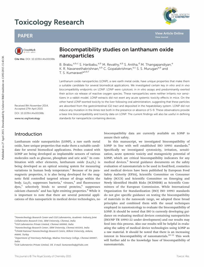
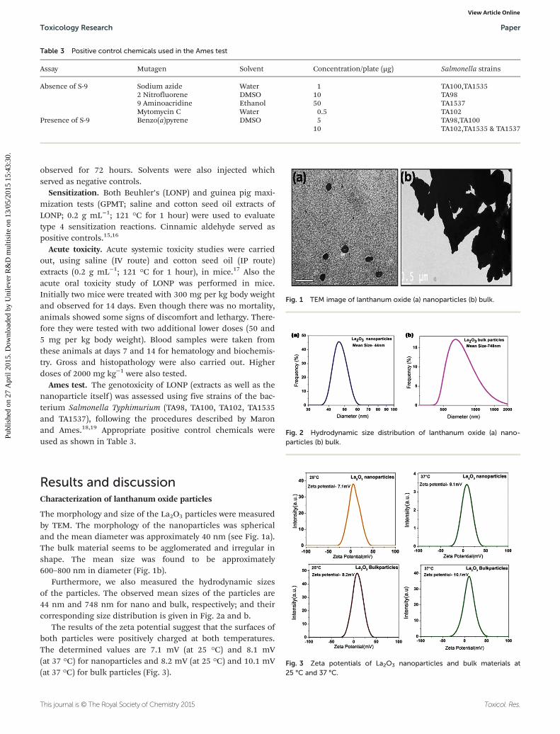
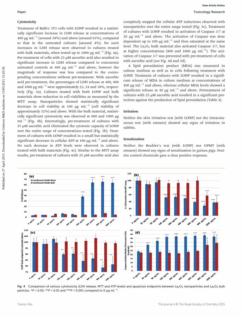
The morphology and size of the La2O3 particles were measuredby TEM. The morphology of the nanoparticles was sphericaland the mean diameter was approximately 40 nm (see Fig. 1a).The bulk material seems to be agglomerated and irregular inshape. The mean size was found to be approximately600–800 nm in diameter (Fig. 1b).
Furthermore, we also measured the hydrodynamic sizesof the particles. The observed mean sizes of the particles are44 nm and 748 nm for nano and bulk, respectively; and theircorresponding size distribution is given in Fig. 2a and b.
The results of the zeta potential suggest that the surfaces ofboth particles were positively charged at both temperatures.The determined values are 7.1 mV (at 25 °C) and 8.1 mV(at 37 °C) for nanoparticles and 8.2 mV (at 25 °C) and 10.1 mV(at 37 °C) for bulk particles (Fig. 3).
Fig. 1 TEM image of lanthanum oxide (a) nanoparticles (b) bulk.
Fig. 2 Hydrodynamic size distribution of lanthanum oxide (a) nano-particles (b) bulk.
Table 3 Positive control chemicals used in the Ames test
Assay Mutagen Solvent Concentration/plate (µg) Salmonella strains
Absence of S-9 Sodium azide Water 1 TA100,TA15352 Nitrofluorene DMSO 10 TA989 Aminoacridine Ethanol 50 TA1537Mytomycin C Water 0.5 TA102
Presence of S-9 Benzo(a)pyrene DMSO 5 TA98,TA10010 TA102,TA1535 & TA1537
Fig. 3 Zeta potentials of La2O3 nanoparticles and bulk materials at25 °C and 37 °C.
Toxicology Research Paper
This journal is © The Royal Society of Chemistry 2015 Toxicol. Res.
Publ
ishe
d on
27
Apr
il 20
15. D
ownl
oade
d by
Uni
leve
r R
&D
mul
tisite
on
13/0
5/20
15 1
5:43
:30.
View Article Online
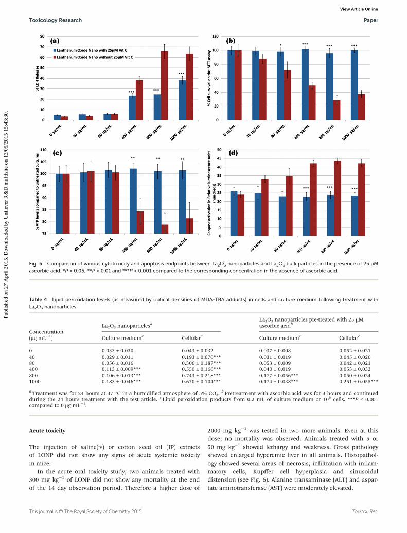
Cytotoxicity
Treatment of Balb/c 3T3 cells with LONP resulted in a statisti-cally significant increase in LDH release at concentrations of400 µg mL−1 (around 38%) and above (around 65%), comparedto that in the untreated cultures (around 4%). No suchincreases in LDH release were observed in cultures treatedwith bulk materials, when tested up to 1000 µg mL−1 (Fig. 4a).Pre-treatment of cells with 25 µM ascorbic acid also resulted insignificant increase in LDH release compared to concurrentuntreated controls at 400 µg mL−1 and above, however themagnitude of response was less compared to the corres-ponding concentrations without pre-treatment. With ascorbicacid pre-treatment, the percentages of LDH release at 400, 800and 1000 µg mL−1 were approximately 23, 24 and 38%, respect-ively (Fig. 5a). Cultures treated with both LONP and bulkmaterials show reduction in cell viabilities as measured by theMTT assay. Nanoparticles showed statistically significantdecrease in cell viability at 100 µg mL−1 (cell viability ofapproximately 52%) and above. With the bulk material, statisti-cally significant cytotoxicity was observed at 800 and 1000 µgmL−1 (Fig. 4b). Interestingly, pre-treatment of cultures with25 µM ascorbic acid eliminated the cytotoxic capacity of LONPover the entire range of concentrations tested (Fig. 5b). Treat-ment of cultures with LONP resulted in a small but statisticallysignificant decrease in cellular ATP at 100 µg mL−1 and above.No such decrease in ATP levels were observed in culturestreated with bulk materials (Fig. 4c). Similar to the MTT assayresults, pre-treatment of cultures with 25 µM ascorbic acid also
completely stopped the cellular ATP reductions observed withnanoparticles over the entire range tested (Fig. 5c). Treatmentof cultures with LONP resulted in activation of Caspase 3/7 at20 µg mL−1 and above. The activation of Caspase was dosedependent up to 100 µg mL−1 and then saturated at the samelevel. The La2O3 bulk material also activated Caspase 3/7, butat higher concentrations (800 and 1000 µg mL−1). The acti-vation of Caspase 3/7 was prevented with pre-treatment of cellswith ascorbic acid (see Fig. 4d and 5d).
A lipid peroxidation product (MDA) was measured inculture medium as well as in cells following treatment withLONP. Treatment of cultures with LONP resulted in a signifi-cant release of MDA in culture medium at concentrations of400 µg mL−1 and above, whereas cellular MDA levels showed asignificant release at 40 µg mL−1 and above. Pretreatment ofcultures with 25 µM ascorbic acid resulted in a significant pro-tection against the production of lipid peroxidation (Table 4).
Irritation
Neither the skin irritation test (with LONP) nor the intracuta-neous test (with extracts) showed any signs of irritation inrabbits.
Sensitization
Neither the Beuhler’s test (with LONP) nor GPMT (withextracts) showed any signs of sensitization in guinea pigs. Posi-tive control chemicals gave a clear positive response.
Fig. 4 Comparison of various cytotoxicity (LDH release, MTT and ATP levels) and apoptosis endpoints between La2O3 nanoparticles and La2O3 bulkparticles. *P < 0.05; **P < 0.01 and ***P < 0.001 compared to 0 µg mL−1.
Paper Toxicology Research
Toxicol. Res. This journal is © The Royal Society of Chemistry 2015
Publ
ishe
d on
27
Apr
il 20
15. D
ownl
oade
d by
Uni
leve
r R
&D
mul
tisite
on
13/0
5/20
15 1
5:43
:30.
View Article Online
Acute toxicity
The injection of saline(IV) or cotton seed oil (IP) extractsof LONP did not show any signs of acute systemic toxicityin mice.
In the acute oral toxicity study, two animals treated with300 mg kg−1 of LONP did not show any mortality at the endof the 14 day observation period. Therefore a higher dose of
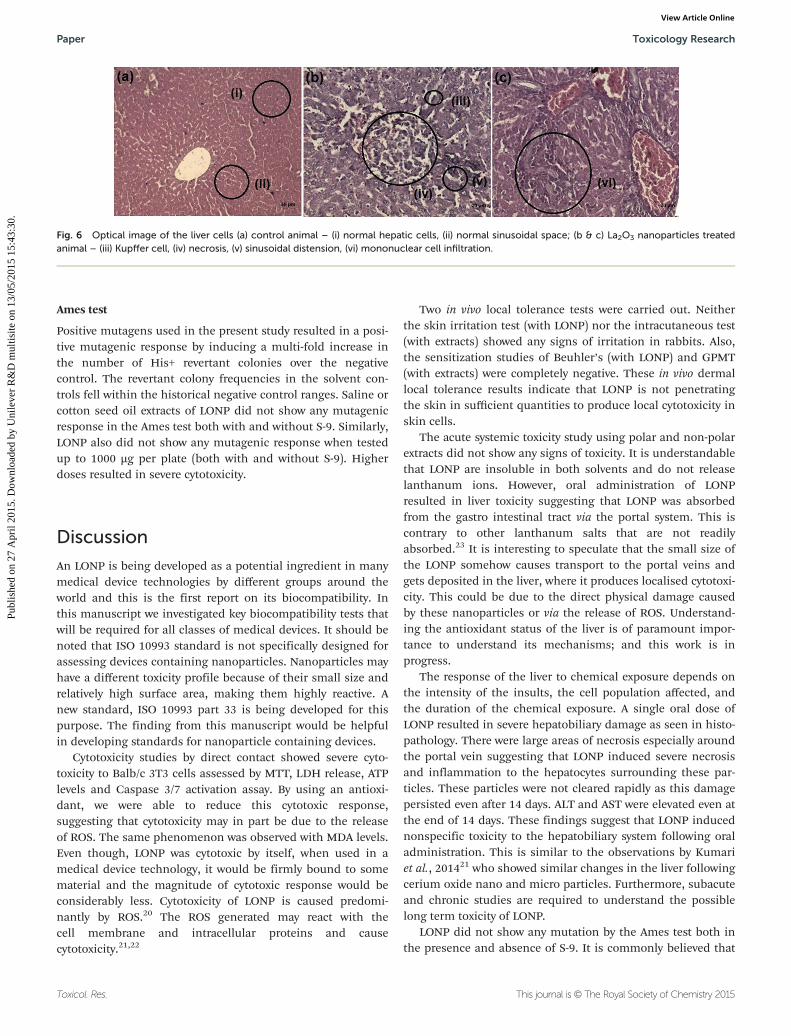
2000 mg kg−1 was tested in two more animals. Even at thisdose, no mortality was observed. Animals treated with 5 or50 mg kg−1 showed lethargy and weakness. Gross pathologyshowed enlarged hyperemic liver in all animals. Histopathol-ogy showed several areas of necrosis, infiltration with inflam-matory cells, Kupffer cell hyperplasia and sinusoidaldistension (see Fig. 6). Alanine transaminase (ALT) and aspar-tate aminotransferase (AST) were moderately elevated.
Fig. 5 Comparison of various cytotoxicity and apoptosis endpoints between La2O3 nanoparticles and La2O3 bulk particles in the presence of 25 µMascorbic acid. *P < 0.05; **P < 0.01 and ***P < 0.001 compared to the corresponding concentration in the absence of ascorbic acid.
Table 4 Lipid peroxidation levels (as measured by optical densities of MDA-TBA adducts) in cells and culture medium following treatment withLa2O3 nanoparticles
Concentration(µg mL−1)
La2O3 nanoparticlesa
La2O3 nanoparticles pre-treated with 25 µMascorbic acidb
Culture mediumc Cellularc Culture mediumc Cellularc
0 0.033 ± 0.030 0.043 ± 0.032 0.037 ± 0.008 0.052 ± 0.02140 0.029 ± 0.011 0.193 ± 0.070*** 0.031 ± 0.019 0.045 ± 0.02080 0.056 ± 0.016 0.306 ± 0.187*** 0.053 ± 0.009 0.042 ± 0.021400 0.113 ± 0.009*** 0.550 ± 0.166*** 0.040 ± 0.019 0.053 ± 0.032800 0.106 ± 0.013*** 0.743 ± 0.218*** 0.177 ± 0.056*** 0.050 ± 0.0241000 0.183 ± 0.046*** 0.670 ± 0.104*** 0.174 ± 0.038*** 0.251 ± 0.055***
a Treatment was for 24 hours at 37 °C in a humidified atmosphere of 5% CO2.b Pretreatment with ascorbic acid was for 3 hours and continued
during the 24 hours treatment with the test article. c Lipid peroxidation products from 0.2 mL of culture medium or 106 cells. ***P < 0.001compared to 0 µg mL−1.
Toxicology Research Paper
This journal is © The Royal Society of Chemistry 2015 Toxicol. Res.
Publ
ishe
d on
27
Apr
il 20
15. D
ownl
oade
d by
Uni
leve
r R
&D
mul
tisite
on
13/0
5/20
15 1
5:43
:30.
View Article Online
Ames test
Positive mutagens used in the present study resulted in a posi-tive mutagenic response by inducing a multi-fold increase inthe number of His+ revertant colonies over the negativecontrol. The revertant colony frequencies in the solvent con-trols fell within the historical negative control ranges. Saline orcotton seed oil extracts of LONP did not show any mutagenicresponse in the Ames test both with and without S-9. Similarly,LONP also did not show any mutagenic response when testedup to 1000 µg per plate (both with and without S-9). Higherdoses resulted in severe cytotoxicity.
Discussion
An LONP is being developed as a potential ingredient in manymedical device technologies by different groups around theworld and this is the first report on its biocompatibility. Inthis manuscript we investigated key biocompatibility tests thatwill be required for all classes of medical devices. It should benoted that ISO 10993 standard is not specifically designed forassessing devices containing nanoparticles. Nanoparticles mayhave a different toxicity profile because of their small size andrelatively high surface area, making them highly reactive. Anew standard, ISO 10993 part 33 is being developed for thispurpose. The finding from this manuscript would be helpfulin developing standards for nanoparticle containing devices.
Cytotoxicity studies by direct contact showed severe cyto-toxicity to Balb/c 3T3 cells assessed by MTT, LDH release, ATPlevels and Caspase 3/7 activation assay. By using an antioxi-dant, we were able to reduce this cytotoxic response,suggesting that cytotoxicity may in part be due to the releaseof ROS. The same phenomenon was observed with MDA levels.Even though, LONP was cytotoxic by itself, when used in amedical device technology, it would be firmly bound to somematerial and the magnitude of cytotoxic response would beconsiderably less. Cytotoxicity of LONP is caused predomi-nantly by ROS.20 The ROS generated may react with thecell membrane and intracellular proteins and causecytotoxicity.21,22
Two in vivo local tolerance tests were carried out. Neitherthe skin irritation test (with LONP) nor the intracutaneous test(with extracts) showed any signs of irritation in rabbits. Also,the sensitization studies of Beuhler’s (with LONP) and GPMT(with extracts) were completely negative. These in vivo dermallocal tolerance results indicate that LONP is not penetratingthe skin in sufficient quantities to produce local cytotoxicity inskin cells.
The acute systemic toxicity study using polar and non-polarextracts did not show any signs of toxicity. It is understandablethat LONP are insoluble in both solvents and do not releaselanthanum ions. However, oral administration of LONPresulted in liver toxicity suggesting that LONP was absorbedfrom the gastro intestinal tract via the portal system. This iscontrary to other lanthanum salts that are not readilyabsorbed.23 It is interesting to speculate that the small size ofthe LONP somehow causes transport to the portal veins andgets deposited in the liver, where it produces localised cytotoxi-city. This could be due to the direct physical damage causedby these nanoparticles or via the release of ROS. Understand-ing the antioxidant status of the liver is of paramount impor-tance to understand its mechanisms; and this work is inprogress.
The response of the liver to chemical exposure depends onthe intensity of the insults, the cell population affected, andthe duration of the chemical exposure. A single oral dose ofLONP resulted in severe hepatobiliary damage as seen in histo-pathology. There were large areas of necrosis especially aroundthe portal vein suggesting that LONP induced severe necrosisand inflammation to the hepatocytes surrounding these par-ticles. These particles were not cleared rapidly as this damagepersisted even after 14 days. ALT and AST were elevated even atthe end of 14 days. These findings suggest that LONP inducednonspecific toxicity to the hepatobiliary system following oraladministration. This is similar to the observations by Kumariet al., 201421 who showed similar changes in the liver followingcerium oxide nano and micro particles. Furthermore, subacuteand chronic studies are required to understand the possiblelong term toxicity of LONP.
LONP did not show any mutation by the Ames test both inthe presence and absence of S-9. It is commonly believed that
Fig. 6 Optical image of the liver cells (a) control animal – (i) normal hepatic cells, (ii) normal sinusoidal space; (b & c) La2O3 nanoparticles treatedanimal – (iii) Kupffer cell, (iv) necrosis, (v) sinusoidal distension, (vi) mononuclear cell infiltration.
Paper Toxicology Research
Toxicol. Res. This journal is © The Royal Society of Chemistry 2015
Publ
ishe
d on
27
Apr
il 20
15. D
ownl
oade
d by
Uni
leve
r R
&D
mul
tisite
on
13/0
5/20
15 1
5:43
:30.
View Article Online
nanoparticles are not easily taken in by prokaryocytes andtherefore the Ames test may not be of value in assessing thegenotoxicity of nanoparticles.24 On the contrary, there areseveral published literature suggesting that nanoparticles areinternalised by bacteria and other prokaryotic cells.25–27 There-fore the notion that nanoparticles will generally be negative inthe Ames test would require further investigations. Further-more a battery of mutagenicity assays are required to under-stand the genetic toxicity potential of LONP.
In conclusion, we have generated some key in vitro andin vivo biocompatibility data on LONP in anticipation of poten-tial use of this nanoparticle in medical device technologies.Like many other nanoparticles, LONP also showed non-specificcytotoxicity, most probably due to its size effect and by releas-ing ROS. One unexpected finding from this study is that LONPis rapidly absorbed from the gastrointestinal tract and gets de-posited in the liver, where it produces persistent nonspecifichepatoxicity for up to two weeks. Furthermore repeated dosetoxicity studies are underway to understand more about itsuptake.
Notes and references
1 N. K. Mogha, V. Sahu, M. Sharma, R. K. Sharma andD. T. Masram, Sensitive and reliable ascorbic acid sensingby lanthanum oxide/reduced graphene oxide nano-composite, Appl. Biochem. Biotechnol., 2014, 174(3), 1010–1020.
2 S. V. Yap, R. M. Ranson, W. M. Cranton andD. Koutsogeorgis, Decay time characteristics of La2O2S:Euand La2O2S:Tb for use within an optical sensor for humanskin temperature measurement, Appl. Opt., 2008, 47(27),4895–4899.
3 B. Brabu, G. K. Yamuna, S. Anitha, C. Gopalakrishnan,S. S. Murugan and T. S. Kumaravel, Characterization andbacterial toxicity of lanthanum oxide bulk and nano-particles, J. Rare Earths, 2012, 30(12), 1298–1302.
4 J. Liu, W. Mei, Y. Li, E. Wang, L. Ji and P. Tao, Antiviralactivity of mixed valence rare earth borotungstate hetero-poly blues against influenza virus in mice, Antiviral Chem.Chemother., 2000, 11(6), 367–372.
5 A. Guha and A. Basu, Role of rare earth oxide nanoparticles(CeO2 and La2O3) in suppressing the photobleaching offluorescent organic dyes, J. Fluoresc., 2014, 24(3), 683–687.
6 Z. Ning, Y. Ran, Z. Libin, G. Guanhua, S. Rongrong,Q. Guanzhou and L. Xiaohe, Lanthanide hydroxide nano-rods and their thermal decomposition to lanthanide oxidenanorods, Mater. Chem. Phys., 2009, 114, 160–167.
7 A. Corti, C. Mannarino, R. Mazza, T. Angelone, R. Longhiand B. Tota, Chromogranin A N-terminal fragments vaso-statin-1 and the synthetic CGA 7-57 peptide act as cardio-statins on the isolated working frog heart, Gen. Comp.Endocrinol., 2004, 136(2), 217–224.
8 J. Fang, M. Saunders, Y. Guo, G. Lu, C. L. Raston andK. S. Iyer, Green light-emitting LaPO4: Ce3+:Tb3+ koosh
nanoballs assembled by p-sulfonato-calix[6]arene coatedsuperparamagnetic Fe3O4, Chem. Commun., 2010, 46(18),3074–3076.
9 Biological Evaluation of Medical Devices – Part 1, Evalu-ation and testing within a risk management process, ISO10993-1, 2009/Cor 1:2010.
10 X. Zhang, M. Liu, X. Zhang, F. Deng, C. Zhou, J. Hui,W. Liu and Y. Wei, Interaction of tannic acid with carbonnanotubes: enhancement of dispersibility and biocompat-ibility, Toxicol. Res., 2015, 4, 160–168.
11 J. H. Liu, T. Wang, H. Wang, Y. Gu, Y. Xu, H. Tang, G. Jiaand Y. Liu, Biocompatibility of graphene oxide intra-venously administrated in mice—effects of dose, size andexposure protocols, Toxicol. Res., 2015, 4, 83–91.
12 X. Zhang, S. Wang, M. Liu, J. Hui, B. Yang, L. Tao andY. Wei, Surfactant-dispersed nanodiamond: biocompatibil-ity evaluation and drug delivery applications, Toxicol. Res.,2013, 2, 335–342.
13 Biological Evaluation of Medical Devices – Part 5: Tests forIn vitro Cytotoxicity, ISO 10993-5, 2009.
14 OECD Guidelines for the Testing of Chemicals, Section 4 –
Health Effects. Test No. 404: Acute Dermal Irritation/Corrosion, adopted 2002.
15 Biological Evaluation of Medical Devices – Part 10,Tests for irritation and skin sensitization, ISO 10993-10,2010.
16 Biological Evaluation of Medical Devices – Part 12,Sample preparation and reference materials, ISO 10993 12,2012.
17 Biological Evaluation of Medical Devices – Part 11, Testsfor systemic toxicity ISO 10993 11, 2009.
18 D. M. Maron and B. N. Ames, Revised methods for theSalmonella mutagenicity test, Mutat. Res., 1983, 113, 173–215.
19 OECD Guidelines for Testing of Chemicals, Section 4 –
Health Effects. Test No. 471: Bacterial Reverse MutationTest, adopted 1997.
20 N. Lewinski, V. Colvin and R. Drezek, Cytotoxicity of nano-particles, Small, 2008, 4(1), 2649.
21 M. Kumari, S. I. Kumari, S. S. K. Kamal and P. Grover,Genotoxicity assessment of cerium oxide nanoparticles infemale Wistar rats after acute oral exposure, Mutat. Res.,Genet. Toxicol. Environ. Mutagen., 2014, 775–776, 7–19.
22 F. Marano, S. Hussain, F. Rodrigues-Lima, A. Baeza-Squiban and S. Boland, Nanoparticles: molecular targetsand cell signalling, Arch. Toxicol., 2011, 85(7), 733–741.
23 G. J. Behets, G. Dams, S. J. Damment, P. Martin,D. M. E. Broe and P. C. D’Haese, Differences in gastrointes-tinal calcium absorption after the ingestion of calcium-freephosphate binders, Am. J. Physiol.: Renal Physiol., 2014,306(1), F61–F67. Erratum in: Am. J. Physiol. Renal Physiol.,2014, 306(5), F568.
24 Z. Magdolenova, A. Collins, A. Kumar, A. Dhawan, V. Stoneand M. Dusinska, Mechanisms of genotoxicity. A review ofin vitro and in vivo studies with engineered nanoparticles,Nanotoxicology, 2014, 8, 233–278.
Toxicology Research Paper
This journal is © The Royal Society of Chemistry 2015 Toxicol. Res.
Publ
ishe
d on
27
Apr
il 20
15. D
ownl
oade
d by
Uni
leve
r R
&D
mul
tisite
on
13/0
5/20
15 1
5:43
:30.
View Article Online
25 R. Sadiq, Q. M. Khan, A. Mobeen and A. J. Hashmat, Invitro toxicological assessment of iron oxide, aluminiumoxide and copper nanoparticles in prokaryotic and eukary-otic cell types, Drug Chem. Toxicol., 2014, 4, 1–10.
26 M. Premanathan, K. Karthikeyan, K. Jeyasubramanian andG. Manivannan, Selective toxicity of ZnO nanoparticles
toward Gram-positive bacteria and cancer cells by apopto-sis through lipid peroxidation, Nanomedicine, 2011, 7(2),184–192.
27 B. V. Aken, Gene expression changes in plants and micro-organisms exposed to nanomaterials, Curr. Opin. Bio-technol., 2015, 33, 206–219.
Paper Toxicology Research
Toxicol. Res. This journal is © The Royal Society of Chemistry 2015
Publ
ishe
d on
27
Apr
il 20
15. D
ownl
oade
d by
Uni
leve
r R
&D
mul
tisite
on
13/0
5/20
15 1
5:43
:30.
View Article Online