the immunopathology of lung fibrosis: amphiregulin
TRANSCRIPT

REVIEW
The immunopathology of lung fibrosis: amphiregulin-producingpathogenic memory T helper-2 cells control the airway fibroticresponses by inducing eosinophils to secrete osteopontin
Kiyoshi Hirahara1,2 & Ami Aoki1 & Yuki Morimoto1& Masahiro Kiuchi1 & Mikiko Okano1
& Toshinori Nakayama1,3
Received: 24 February 2019 /Accepted: 14 March 2019 /Published online: 9 April 2019# Springer-Verlag GmbH Germany, part of Springer Nature 2019
AbstractFibrosis is defined as excessive deposition of the extracellular matrix (ECM) in the parenchyma of various organs, and sometimesleads to irreversible organ malfunction such as idiopathic pulmonary fibrosis (IPF), a fatal disorder of the lung. Chronicinflammatory stimuli induce fibrotic responses in various organs. Various immune cells, including T helper (Th) cells in thelung, protect the host from different harmful particles, including pathogenic microorganisms. However, the dysregulation of thefunction of these immune cells in the lung sometimes causes inflammatory diseases, such as lung fibrosis. In this review, we willintroduce an outline of the cellular and molecular mechanisms underlying the pathogenic fibrotic responses in the lung. We willalso introduce the concept of the BPathogenic Th population disease induction model,^ in which unique subpopulations of certainTh cell subsets control the pathology of immune-mediated inflammatory diseases. Finally, we introduce our recent findings,which demonstrate that amphiregulin-producing pathogenic memory Th2 cells control airway fibrosis through the osteopontinproduced by inflammatory eosinophils. The identification of this new pathogenic Th cell population supports the concept ofBPathogenic Th population disease induction model^, and will provide novel strategies for treating intractable diseases, includinglung fibrosis.
Keywords Fibrosis . Extracellular matrix (ECM) . Asthma . BPathogenic Th population disease induction model^ .
Amphiregulin-producing pathogenicmemory Th2 cells . Osteopontin . Inflammatory eosinophils
Themolecular and cellular mechanism of lungfibrosis
The lungs and bronchus are continuously exposed to exoge-nous stimuli from sources such as smoking, infection frommicroorganisms, foreign materials, and drugs. Thus, the
bronchi and alveolar epithelial cells are at risk of being dam-aged and need to be repaired repeatedly. Excessive acute in-flammation leads to massive damage in lung tissue, inducingpulmonary alveolar and airway endothelial cells, which causesadvanced fibrosis.Moreover, the chronic inflammation inducedvarious sources of stimulation, including tissue injury, infec-tion, autoimmune responses, exogenous foreign agents, tumors,aging, and a genetic predisposition cause fibrotic responsesin vivo, in other words: Bno inflammation, no fibrosis^ [1, 2].
The tissue regeneration occurs after inflammation and tis-sue damage through two mechanisms: (1) the proliferation ofcommon differentiated cells and (2) the differentiation of stemcells or progenitor cells. In alveolar epithelial injury, commondifferentiated cells proliferate; however, rare airway stem cellshave also been shown to be induced by injury and to play arole in tissue regeneration [3]. The airway stem cells, whichare lineage specific marker-negative, cytokeratin 5-positive,can repair the influenza infection-damaged tissue via Notchsignaling in mice [4]. Excess Notch signaling is also known topromote fibrotic responses such as cystic honeycombing [4].
Kiyoshi Hirahara and Ami Aoki contributed equally to this work.
This article is a contribution to the special issue on The Pathogenicity ofAcquired Immunity in Human Diseases - Guest Editor: Kiyoshi Hirahara
* Kiyoshi [email protected]
1 Department of Immunology, Graduate School of Medicine, ChibaUniversity, 1-8-1 Inohana, Chuo-ku, Chiba 260-8670, Japan
2 AMED-PRIME, AMED, 1-8-1 Inohana, Chuo-ku, Chiba 260-8670,Japan
3 AMED-CREST, AMED, 1-8-1 Inohana, Chuo-ku, Chiba 260-8670,Japan
Seminars in Immunopathology (2019) 41:339–348https://doi.org/10.1007/s00281-019-00735-6
Acute inflammation damages vascular endothelial cells,which is followed by the secretion of various types of growthfactors that recruit fibroblasts and immune-related cells suchas T cells, macrophages, neutrophils, and eosinophils into theinflamed lung. At the same time, activated inflammatory cellsproduce various types of pro-inflammatory cytokines, includ-ing Interleukin-1β (IL-1β), IL-6, IL-25, IL-33, thymic stro-mal lymphopoietin (TSLP), tumor necrosis factor-α (TNF-α),and granulocyte-macrophage colony stimulating factor (GM-CSF) [5]. The damage of vascular endothelial cells also in-duces innate wound-healing responses, such as coagulationresponses by activated platelets. These tissue repair responsesin the vascular endothelium are proceeded by activated plate-lets and immune cells that show the enhanced production ofseveral growth factors (e.g., platelet-derived growth factor(PDGF), vascular endothelial growth factor (VEGF), and fi-broblast growth factor (FGF)). Tissue-resident fibroblasts pro-liferate in the inflammatory tissue and differentiate intomyofibroblasts, which are a key cell population in the fibroticresponse. Activated myofibroblasts show the enhanced ex-pression of α-smooth muscle actin (SMA) accompanied bythe massive production of the extracellular matrix (ECM). Theintratracheal transfer of resident fibroblasts clearly showedthat the transferred resident fibroblasts were activated andproduced a significant amount of collagen in bleomycin-induced pulmonary fibrosis [6].
Both myofibroblasts and fibroblasts play important roles inpathogenic fibrotic responses and wound repair [7]; however,the origin of myofibroblasts remains controversial. Originally,it was reported that myofibroblasts were derived from tissueresident fibroblasts [8]. Subsequently, fibrocytes, which arecirculating monocytic progenitor cells with characteristic fea-tures of both monocytes and fibroblasts, were reported totransform into myofibroblasts [9, 10]. Fibrocytes are knownto be derived from the bone marrow and circulate in the pe-ripheral blood with the expression of CD34 (a stem cell mark-er), CD45 (a pan-hematopoietic marker), CD14, and CD11(both monocyte markers) and produce collagen l, collagenlll, and vimentin [10]. In healthy subjects, fibrocytes makeup among approximately 0.5% of the peripheral blood. Pro-inflammatory and fibrogenic chemokines, such as CXCL12,CCL12, CCR3, and CCR5, are involved in the recruitmentand infiltration of fibrocytes to the injured lung [11, 12].Interestingly, fibrocytes from patients with idiopathic pulmo-nary fibrosis (IPF) show higher expression levels of CXCR4and CXCL12 (a ligand for CXCR4), in the peripheral bloodand the lung [13]. Thus, hyperactivation of fibrocytes may beinvolved in shaping the pathology of the fibrotic responses inIPF patients. Lung epithelial cells or endothelial cells are an-other source for myofibroblasts, as these cell populationstransform into myofibroblasts via epithelial/endothelial mes-enchymal transition (EMT) [14, 15]. Recent technological de-velopments have allowed us to investigate the RNA
expression landscape at the single-cell level using single-cellRNA sequencing (scRNA-Seq) [16]. An scRNA-Seq analysisidentified a rare cell population, Foxi1+ ionocytes, in the lung[17]. Ionocytes express high levels of cystic fibrosis trans-membrane conductance (Cftr), in which more than 1000 mu-tations have been identified in patients with cystic fibrosis[17]. Thus, this rare cell population is involved in the pathol-ogy of lung fibrotic disease (Fig. 1).
Oxidative stress is one of the crucial chemical stresses thatare involved in the induction of pathogenic fibrotic responses.Glutathione (GSH) is decreased in the lungs of patients withIPF, which causes an imbalance between oxidants and antiox-idants [18]. N-Acetyl-L-cysteine (NAC), a precursor of GSH,was developed as a treatment for patients with IPF; however,the clinical trial failed due to insufficient efficacy [19].Interestingly however, glutaredoxin-1 (GLRX), which re-verses a post-translational modification of GSH, S-glutathionylation (PSSG), was reported as a potential thera-peutic agent in recent years. The intratracheal administrationof exogenous GLRX to mice with bleomycin-lung fibrosisreversed increases in collagen in the lungs [20].
Lessons from recent mouse models of fibrosis
Bleomycin, an anti-cancer medication, acts by inducingDNA strand breaks. The intratracheal administration ofbleomycin into the lung can induce massive inflammationfollowed by the severe fibrotic responses [21]. Thus, micewith a bleomycin-induced lung fibrosis are among the an-imal models most commonly used to investigate the pathol-ogy of lung fibrosis. In addition to this model, in recentyears, other mouse models have been used to investigatethe fibrotic responses in the lung. The lungs of geneticallyengineered mice with the overexpression of AP-1 (activatorprotein-1) family members show a spontaneous fibrotic re-sponse. The AP-1 family is formed by a large number ofproteins, including Fos proteins (c-Fos, FosB, Fra-1, Fra-2)and Jun (JunB, c-Jun, JunD) proteins. Fos and Jun proteins,which are activated by various types of stress signals, con-trol a variety of cellular responses, including proliferation,apoptosis, inflammation, and wound healing [22]. Fra2-transgenic mice show skin and lung fibrosis with vasculop-athy that are similar to systemic sclerosis in humans [23,24]. Interestingly, the expression of osteopontin, an AP-1targeting gene, is increased in Fra2-transgenic mice.Osteopontin is a phosphoglycoprotein and is induced invarious types of myeloid cells and epithelial cells by in-flammation, infection, and irradiation. Osteopontin existsas two isoforms, intracellular osteopontin and secretedosteopontin, which have distinct functions. Intracellularosteopontin regulates myeloid cells and works as a scaffoldprotein in signal transduction pathways [25]. Secreted
340 Semin Immunopathol (2019) 41:339–348
osteopontin controls lymphocytes to maintain the balanceof the leukocyte population in hematopoiesis and is associ-ated with fibrosis [25]. Systematic fibrosis is also inducedin c-Jun transgenic mice [26]. These data suggest that AP-1transcription factor is one of the central mediators of fibro-sis. A cell surface molecule, tetraspanin is another essentialprotein for the pathology of pulmonary fibrosis.Tetraspanins consist of four transmembrane domains thatbind adhesion proteins, such as integrins, and thereby gen-erate a hierarchical network of interactions. CD151, amember of the Tetraspanin family, is predominantlyexpressed in alveolar epithelial cells and CD151-deficientmice spontaneously exhibit pulmonary fibrosis with age[27]. The deletion of CD151 causes the loss of epithelialintegrity that is associated with an exacerbation of pulmo-nary fibrosis. Telomere dysfunction is also associated withfibrosis. Genetically engineered mice with short telomeres(telomerase-deficient mice) show spontaneous pulmonaryfibrosis due to severe telomerase dysfunction in alveolartype II cells, which suggests that cellular senescence isinvolved in the pathology of pulmonary fibrosis [28].
The involvement of immune cellsin the pathology of fibrosis
Various types of immune cells are involved in the pathology offibrosis. It is well known that type 2 immunity contributes tothe development of fibrotic responses [29]. Th2 responsesresult in tissue injury and fibrotic responses, while the Th1response ameliorates fibrotic responses [2]. Pulmonary alve-olar macrophages produce profibrotic cytokines, includingPDGF, transforming growth factor-β (TGF-β), and IL-13 inthe fibrogenic phase. TGF-β promotes the differentiation offibroblasts to myofibroblasts and enhances the production ofECM from differentiated myofibroblasts [7]. IL-13 is alsoinvolved in shaping the pathology of the fibrotic responsesboth directly and indirectly [1]. Group 2 innate lymphoid cells(ILC2) are also involved in the development of fibrosis ininfluenza virus and S. mansoni infection [30, 31].
M2 macrophages, which are a macrophage subpopu-lation, are activated by Th2 cytokines such as IL-4 andIL-13, which are involved in shaping the pathology offibrosis [32]. M2 macrophages express high levels of
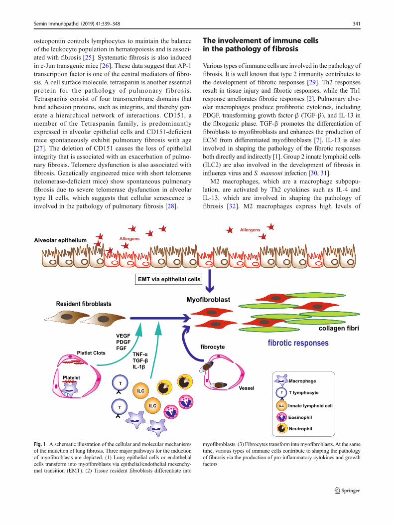
Fig. 1 A schematic illustration of the cellular and molecular mechanismsof the induction of lung fibrosis. Three major pathways for the inductionof myofibroblasts are depicted. (1) Lung epithelial cells or endothelialcells transform into myofibroblasts via epithelial/endothelial mesenchy-mal transition (EMT). (2) Tissue resident fibroblasts differentiate into
myofibroblasts. (3) Fibrocytes transform into myofibroblasts. At the sametime, various types of immune cells contribute to shaping the pathologyof fibrosis via the production of pro-inflammatory cytokines and growthfactors
Semin Immunopathol (2019) 41:339–348 341

arginase-1(Arg1) that control the production of L-pro-line, which is required for collagen synthesis [33]. AnscRNA-Seq analysis of macrophages from the lungs ofmice with bleomycin-induced fibrosis revealed a popu-lation of disease-associated CX3CR1+SiglecF+ macro-phages with a unique gene expression profile [34]. Aspecific subpopulation of Ceacam1+Msr1+Ly6C−F4/80−Mac1+ monocytes called segregated-nucleus-containing atypical monocytes (SatM) has also beenshown to be crucial for the induction of fibrosis [35].
The pathogenic Th population diseaseinduction model
Leading-edge techniques, including scRNA-Seq, have in-creased our knowledge regarding the complexity and hetero-geneity of immune cells and we now need to revisit issuesregarding the pathogenicity of immune-related inflammatorydiseases. In the case of T cells, a functional imbalance ofhelper T cell subsets has been suggested to cause the pathol-ogy of immune-related inflammatory diseases (‘Th1/Th2 bal-ance disease induction model). However, the diversity ofCD4 T cells suggests us that a unique subpopulation of Thcells controls the pathology of specific inflammatory dis-eases. Recently, we proposed a Bpathogenic Th populationdisease induction model,^ in which—despite the balance be-tween Th1 and Th2 cells—the pathogenesis of so-called Th1-and Th2-mediated diseases was mostly dependent on theBpathogenic subpopulations^ of each helper T cell subsetgenerated in vivo and possessed an effector function, whichwas a distinct feature [36]. In the model, a pathogenic sub-population of T helper cells that was induced under certainconditions was found to be critical for the pathogenesis ofimmune-mediated diseases rather than the balance among Thelper cell subsets [36]. In particular, type 2 immunity-mediated pathologies, including atopic dermatitis, pollen al-lergy, allergic airway inflammatory disease, and IL-5-producing Th2 cell subpopulations, are considered to be path-ogenic populations. Indeed, several groups—including ourown—have revealed that identified distinct pathogenic helperT cell subpopulations that play key roles in shaping the pa-thology of various types of immune-mediated inflammatorydiseases in both mice and humans. For example, IL-5-producing Th2 cells, so-called pathogenic Th2 (Tpath2) cells,are identified in eosinophilic gastrointestinal diseases, allergicairway inflammation, pollen allergy, and eosinophilic chronicrhinosinusitis in both mice and humans [36–38]. Th1 cells,which express high levels of CXCR3, are crucial for thepathogenicity of type 1 diabetes [39]. In the case of Th17cells, pathogenic Th17 cells are induced without TGF-β1 orwith TGF-β3 [40, 41]. AIM (CD5L) also controls Th17 cellpathogenicity through the regulation of lipid biosynthesis
[42]. In the case of human Th17 cells, the transcription factorc-MAF controls the immunopathology of Th17 cells [43].Thus, a comprehensive understanding of the BpathogenicTh population disease induction model^ is a key to elucidat-ing the precise pathogenicity of immune-mediated inflamma-tory diseases.
Pathogenic Th2 cells are inducedby the epithelial cytokine IL-33
Various environmental stimuli are known to induce pathogen-ic populations of helper T cells [36]. IL-33, a member of theepithelial cytokines, was identified as a ligand of ST2 (an IL-1receptor) [44]. IL-33 works as a transcriptional repressor in thenuclei of many types of cells under a steady state because it isconstitutively localized in the nuclei and is associated withchromatin by a chromatin-binding motif [45]. The finding thatIL-33 has no signal sequence suggests that IL-33 differs fromconventional secreted cytokines [46]. In contrast to being se-creted, mechanical damage, cellular activation through ATPsignaling, or necrotic cell death induces the release of IL-33into the extracellular space [47, 48]. The expression patternand the mode of IL-33 release differ between mice andhumans (i.e., vascular endothelial cells do not express IL-33constitutively in mice, while human endothelial cells preserveIL-33 universally) [49–51].
In humans, IL-33 is involved in different types of diseases,such as bacterial and viral infections, cardiovascular disease,allergies, and metabolic disorders [51]. At the same time, theST2 and IL-33 gene loci are often associated with asthma indifferent genome-wide association studies (GWAS) [52].Moreover, polymorphism in the IL-33 signal pathway is asso-ciated with the clinical phenotype of childhood asthma [53].IL-33 induces potent type 2 immune responses accompaniedby massive eosinophil infiltration through the activation ofboth innate and adaptive immune cells, including mast cells,eosinophils, and ILC2s [54–57]. Moreover, we recently foundthat IL-5-producing pathogenic Th2 (Tpath2) cells selectivelyexpress a high level of ST2, and the exposure of memory Th2cells to IL-33 resulted in a significant increase in the produc-tion of IL-5 in both mice and humans [58]. Thus, the IL-33-ST2 axis is crucial for the induction and enhancement of thepathogenicity of memory Th2 cells in allergic airwayinflammation.
Maintenance of memory Tpath2 cellswithin the iBALT
After the induction of airway inflammation, a type of ectopiclymphoid tissue called inducible bronchus-associated lym-phoid tissue (iBALT), which consists of T cells, B cells,
342 Semin Immunopathol (2019) 41:339–348

dendritic cells (DCs), and follicular DCs (FDCs), is induced inthe lung parenchyma [59, 60]. A recent study revealed thattype I interferon (IFN) after influenza A infection causes theactivation of a subpopulation of lung fibroblasts, which canconvert non-lymphoid tissue into functional tertiary lymphoidstructure formations [61]. FDCs represent a unique cell pop-ulation that is required for the development of proper B cells[62]. Furthermore, CD11c+ DCs are involved in maintainingthe iBALT structure [63]. Antigen-specific memory CD4 Tcells, including Tpath2 cells, are maintained in the iBALT[59, 64]. During chronic allergic airway inflammation,Thy1-positive IL-7-producing lymphatic endothelial cells(LECs), which are localized within iBALT structures, are akey population for the maintenance of antigen-specific mem-ory Tpath2 cells [59]. Lung-infiltrating memory Tpath2 cellspreferentially localize within the iBALT and are attached toIL-7-producing LECs, which increase in number under in-flammatory conditions. Thus, the survival of antigen-specificmemory Tpath2 cells is induced by the modification of thelung microenvironment. Interestingly, Thy1+IL-7-producingLECs also produce IL-33, which is critical for maintainingthe pathogenic ability (e.g., type 2 cytokine production) mem-ory of Tpath2 cells [59]. Taken together, Thy1-positive IL-7-producing LECs show dual effects on memory Tpath2 cells(i.e., IL-7 from LECs supports the survival of memory Tpath2cells, while IL-33 is responsible for maintaining the pathogen-ic functions of Tpath2 cells in the airway).
IL-33 induces the enhanced productionof IL-15 by tissue-resident memory Th2 cellswithin iBALT
Three major subpopulations are known to exist in memory Th2cells: central memory T cells (TCM), effector memory T (TEM)cells, and tissue resident memory T (TRM) cells, which haverecently been identified [65, 66]. TCM cells with the high ex-pression of CCR7 and CD62L can respond rapidly to patho-gens in the lymphoid organs [67]. TEM cells show low expres-sion levels of CD62L and CCR7 and circulate among non-lymphoid tissue, secondary lymphoid organs and the blood[67]. In contrast, TRM cells permanently reside in the peripheraltissue with the expression of CD69 and CD103 [68]. CD4+
TRM cells reside in various mucosal organs, including the re-productive organs, skins, and lungs [69–71]. Interestingly, lungTRM cells are retained in the lungs, and migrate back to the lungin the adaptive transfer mouse model [72]. Lung CD4+ TRMcells play crucial roles in shaping the pathology of allergicairway inflammation [73]. Visceral adipose tissue has been re-ported to be a reservoir of CD4 TRM cells [74]. The involve-ment of CD4 TRM cells in shaping the pathology of lung fibro-sis has been uncertain. Further investigations are needed toclarify the precise roles of CD4 TRM cells in the lung.
Amphiregulin-producing pathogenic memoryT helper-2 cells drive airway fibrosis
Asthma is a common disease worldwide, and is characterizedby chronic airway inflammation with persistent cough andairflow limitation. Long-standing asthma causes airway re-modeling and exacerbates bronchial hyperresponsivenesswith various symptoms and airway narrowing. Airway re-modeling involves a loss of normal bronchial epithelial cells,mucous-gland hyperplasia, and deposition of the collagensubepithelial layer [75]. Adult patients with severe asthmashow a decreased lung function (FEV1 or FEV1 /VC < 75%predicted) accompanied by fibrotic changes [76].
Various stimuli, such as allergens, viruses, bacteria, andfungi, which cause exacerbations of asthma, injure the bron-chus and induce the release of epithelial cytokines (IL-25, IL-33, and TSLP). These epithelial cytokines activate both innateand adaptive immune cells, including ILC2s and memory-type Tpath2 cells that produce large amounts of Th2 cytokinesand induce massive infiltration of eosinophils in the airway. Itwas reported that the fibrotic responses in allergic airway in-flammation were associated with IL-25, IL-33, and TSLP[77]. Fibrotic responses involve the massive deposition ofcollagenous and non-collagenous extracellular matrix compo-nents in the lung parenchyma as a result of the activation andproliferation of fibroblasts, myofibroblasts, and various typesof immune cells, as discussed previously. Th2 cells, especiallyTpath2 cells, which have the ability to produce large amountsof IL-5, are central players in shaping the pathology of bothallergic airway inflammation and fibrosis [36]. Eosinophils, akey population for allergic inflammation, can develop, infil-trate inflammatory sites, and are activated through IL-5 stim-ulation [78]. Eosinophils are involved in the pathogenesis ofairway remodeling characterized by collagen fibril deposition[79]. Activated eosinophils produce inflammatory mediators,including cytokines, lipid mediators, and reactive oxygen spe-cies (ROS), which damage airway tissue and nerves. This isfollowed by inflammation accompanied by enhanced airwayhyper responsiveness [80]. However, the precise cellular andmolecular mechanisms underlying the pathology of fibroticresponses have not been clarified.
We used an experimental mouse model in which repet-itive exposure of house dust mite (HDM) induced massivefibrotic responses together with eosinophilic airway in-flammation, to investigate this point. The repeated expo-sure of the HDM mice increased the expression of Il33,Il25, and Tslp and molecules relevant to the fibrotic re-sponse, such as Spp1, Tenascin C (Tnc), collagen type 1alpha 1 (Col1a1), and Actin alpha 2 smooth muscle aorta(Acta 2) in the inflamed lung. The genetic deletion of Il33or Il1rl1 encoding ST2 (a component of the IL-33 recep-tor) resulted in a decreased fibrotic response. At the sametime, the lungs of Il33-deficient mice that received OVA-
Semin Immunopathol (2019) 41:339–348 343

specific memory Th2 cells or wild-type mice that receivedIl1rl1-deficient OVA-specific memory Th2 cells showedsignificantly decreased in collagen deposition. Thus, the
IL-33-ST2 axis in memory Th2 cells plays an importantrole in the induction of the fibrotic responses in allergicinflammation.
Fig. 2 Amphiregulin-producing Tpath2 cells induce pathogenic fibroticresponses by instructing inflammatory eosinophils to produceosteopontin. Amphiregulin produced by pathogenic memory Th2 cellsreprogrammed eosinophils to produce osteopontin and trigger airwayfibrosis. The image illustrates amphiregulin-producing pathogenic
memory Th2 cells as a Bwitch^ sharing Bpoisoned apples^(amphiregulin) to Bdwarves^ (eosinophils). The dwarves who ate a poi-soned apple are confused and are fixing the lung of a robot with improperitems such as Btin cans^ (osteopontin)
344 Semin Immunopathol (2019) 41:339–348
To identify the key molecule(s) of the fibrotic responses,we performed an RNA sequencing (RNA-Seq) analysis usingOVA-specific ST2hi memory Th2 cells stimulated with IL-33in vitro. IL-33 stimulation induced the high expression ofAreg, encoding amphiregulin, in ST2hi memory Th2 cells.In vivo, memory Th2 cells producing amphiregulin increasedin the lung after exposure to HDM. The production ofamphiregulin by memory Th2 cells stimulated by IL-33 de-creases with the application anti-ST2 antibodies, which blocksthe IL-33-ST2 signaling pathway. At the same time, lung fi-brotic responses in Areg-deficient mice were ameliorated withHDM inhalation, although the expression levels of Il33, ll5,and ll13 were comparable between wild-type and Areg-defi-cient mice. Furthermore, the fibrotic responses decreased inrecipients of Areg-deficient memory Th2 cells. These datasuggest that IL-33 induces the production of amphiregulinby ST2hi memory Th2 cells and that amphiregulin is a keymolecule in the induction of fibrotic responses in vivo.
Amphiregulin is a member of the epithelial growth factorproteins and is a key molecule for tissue repair [81]. Variousimmune cells, including mast cells, ILC2s, and Treg cells, alsoproduce amphiregulin [82–84]. In the brain, amphiregulinfrom Treg cells contributes to the suppression of neurotoxicastrogliosis [84]. In the lung, the stimulation of epithelial cellswith amphiregulin results in proliferation and enhancedmucinproduction [82, 85]. Amphiregulin also induces the prolifera-tion of fibroblasts in the lung [86]. In addition to these well-known target cells, we found that eosinophils expressed EGFreceptor and that amphiregulin stimulation resulted in globaltranscriptional reprogramming in eosinophils, marked by theupregulation of a number of inflammatory genes. Spp1, whichencodes osteopontin (a major component of non-collagenousECM) was one of the genes upregulated by amphiregulinstimulation in eosinophils. Osteopontin is known to contributeto shaping the pathogenesis of fibrosis [87, 88]. Thus, eosin-ophils directly produce non-collagenous ECM and contributeto tissue fibrosis in allergic airway inflammation
Finally, we wanted to determine whether the observed IL-33-ST2-amphiregulin pathway was important for shaping thefibrotic pathology in inflammatory lesions of human chronicallergic diseases. Eosinophilic chronic rhinosinusitis (ECRS)is a chronic upper airway inflammatory condition that is ac-companied by the formation of nasal polyps with the massiveinfiltration of eosinophils [89]. Patients with ECRS often suf-fer from allergic airway inflammation such as asthma, andtheir polyps exhibit eosinophilic infiltration, suggesting thatexcessive type 2 immunity is involved in shaping the pathol-ogy of ECRS [90]. Thus, the pathogenesis of asthma is closelyrelated to that of ECRS [91]. Interestingly, we found thatpolyps from patients with eosinophilic chronic rhinosinusitis(ECRS) showed enhanced fibronectin disposition. We alsofound that CD45RO+CRTH2hiCD161hiCD4+ T cells inpolyps specifically produced amphiregulin. Furthermore, the
eosinophils infiltrating the polyps showed higher osteopontinexpression levels in comparison to the eosinophils in periph-eral circulation. Taken together, the IL-33-amphiregulin-osteopontin axis controls the fibrotic responses in chronic al-lergic inflammation in both mouse and human systems, andthese molecules may be potential therapeutic targets for intrac-table chronic allergic diseases (Fig. 2).
Conclusion
The regeneration of fibrotic organs has not yet been achieved.Because of the poor understanding of the pathology underly-ing the development of fibrosis, the diagnosis and therapy areunder development; however, the prognosis of pulmonary fi-brosis remains poor, with a median survival period of 3–5 years. The small molecule receptor tyrosine kinase inhibitornintedanib is one of the few Food and Drug Administration-approved treatments for lung fibrosis is. Unfortunately, thecurrent goal of anti-fibrotic treatment is to delay the progres-sion of fibrosis rather than cure the disease [92–94]. Thus, wemust explore not only novel biomarkers of the progression offibrosis but also effective therapeutic strategies for lung fibro-sis. To this end, intensive studies are needed to understand themechanisms underlying pathogenic fibrotic changes.
Acknowledgments We appreciate all of the members in Department ofImmunology, Graduate School of Medicine, Chiba University, Japan.
Funding information This work was supported by the following grants:Ministry of Education, Culture, Sports, Science and Technology (MEXTJapan) Grants-in-Aid for Scientific Research (S) 26221305, (C)17K08876; Practical Research Project for Allergic Diseases andImmunology (Research on Allergic Diseases and Immunology) fromthe Japan Agency for Medical Research and Development, AMED(No. JP18ek0410030, JP18ek0410045); AMED-PRIME, AMED (No.JP18gm6110005); AMED-CREST, AMED (No. JP18gm1210003);Mochida Memorial Foundation for Medical and PharmaceuticalResearch, The Ichiro Kanehara Foundation for the Promotion ofMedical Sciences and Medical Care and Takeda Science Foundation.
Compliance with ethical standards
Competing interests The authors declare that they have no conflict ofinterest.
References
1. Wynn TA (2004) Fibrotic disease and the T(H)1/T(H)2 paradigm.Nat Rev Immunol 4(8):583–594
2. Wick G, Grundtman C, Mayerl C, Wimpissinger TF, Feichtinger J,Zelger B, Sgonc R,Wolfram D (2013) The immunology of fibrosis.Annu Rev Immunol 31:107–135
3. ZuoW, Zhang T, Wu DZ, Guan SP, Liew AA, Yamamoto Y, WangX, Lim SJ, Vincent M, Lessard M, Crum CP, Xian W, McKeon F
Semin Immunopathol (2019) 41:339–348 345

(2015) p63(+)Krt5(+) distal airway stem cells are essential for lungregeneration. Nature 517(7536):616–620
4. Vaughan AE, Brumwell AN, Xi Y, Gotts JE, Brownfield DG,Treutlein B, Tan K, Tan V, Liu FC, Looney MR, Matthay MA,Rock JR, Chapman HA (2015) Lineage-negative progenitors mo-bilize to regenerate lung epithelium after major injury. Nature517(7536):621–625
5. Zhang Y, Lee TC, Guillemin B, Yu MC, Rom WN (1993)Enhanced IL-1 beta and tumor necrosis factor-alpha release andmessenger RNA expression in macrophages from idiopathic pul-monary fibrosis or after asbestos exposure. J Immunol 150(9):4188–4196
6. Tsukui T, Ueha S, Abe J, Hashimoto S, Shichino S, Shimaoka T,Shand FH, Arakawa Y, Oshima K, Hattori M, Inagaki Y, TomuraM, Matsushima K (2013) Qualitative rather than quantitativechanges are hallmarks of fibroblasts in bleomycin-induced pulmo-nary fibrosis. Am J Pathol 183(3):758–773
7. Tomasek JJ, Gabbiani G, Hinz B, Chaponnier C, Brown RA (2002)Myofibroblasts and mechano-regulation of connective tissue re-modelling. Nat Rev Mol Cell Biol 3(5):349–363
8. Hinz B, Phan SH, Thannickal VJ, Galli A, Bochaton-Piallat ML,Gabbiani G (2007) The myofibroblast: one function, multiple ori-gins. Am J Pathol 170(6):1807–1816
9. Bucala R, Spiegel LA, Chesney J, Hogan M, Cerami A (1994)Circulating fibrocytes define a new leukocyte subpopulation thatmediates tissue repair. Mol Med 1(1):71–81
10. Herzog EL, Bucala R (2010) Fibrocytes in health and disease. ExpHematol 38(7):548–556
11. Phillips RJ, Burdick MD, Hong K, Lutz MA, Murray LA, Xue YY,Belperio JA, Keane MP, Strieter RM (2004) Circulating fibrocytestraffic to the lungs in response to CXCL12 and mediate fibrosis. JClin Invest 114(3):438–446
12. Moore BB,Murray L, Das A,Wilke CA, Herrygers AB, Toews GB(2006) The role of CCL12 in the recruitment of fibrocytes and lungfibrosis. Am J Respir Cell Mol Biol 35(2):175–181
13. Moeller A, Gilpin SE, Ask K, Cox G, Cook D, Gauldie J, MargettsPJ, Farkas L, Dobranowski J, Boylan C, O'Byrne PM, Strieter RM,Kolb M (2009) Circulating fibrocytes are an indicator of poor prog-nosis in idiopathic pulmonary fibrosis. Am J Respir Crit Care Med179(7):588–594
14. Kim KK, Kugler MC, Wolters PJ, Robillard L, Galvez MG,Brumwell AN, Sheppard D, Chapman HA (2006) Alveolar epithe-lial cell mesenchymal transition develops in vivo during pulmonaryfibrosis and is regulated by the extracellular matrix. Proc Natl AcadSci U S A 103(35):13180–13185
15. Hashimoto N, Phan SH, Imaizumi K, Matsuo M, Nakashima H,Kawabe T, Shimokata K, Hasegawa Y (2010) Endothelial-mesenchymal transition in bleomycin-induced pulmonary fibrosis.Am J Respir Cell Mol Biol 43(2):161–172
16. Stuart T, Satija R (2019) Integrative single-cell analysis. Nat RevGenet. https://doi.org/10.1038/s41576-019-0093-7
17. Montoro DT, Haber AL, Biton M, Vinarsky V, Lin B, Birket SE,Yuan F, Chen S, LeungHM, Villoria J, Rogel N, Burgin G, TsankovAM, Waghray A, Slyper M, Waldman J, Nguyen L, Dionne D,Rozenblatt-Rosen O, Tata PR, Mou H, Shivaraju M, Bihler H,Mense M, Tearney GJ, Rowe SM, Engelhardt JF, Regev A,Rajagopal J (2018) A revised airway epithelial hierarchy includesCFTR-expressing ionocytes. Nature 560(7718):319–324
18. Cantin AM, Hubbard RC, Crystal RG (1989) Glutathione deficien-cy in the epithelial lining fluid of the lower respiratory tract inidiopathic pulmonary fibrosis. Am Rev Respir Dis 139(2):370–372
19. N. Idiopathic Pulmonary Fibrosis clinical research, Martinez FJ, deAndrade JA, Anstrom KJ, King TE Jr, Raghu G (2014)Randomized trial of acetylcysteine in idiopathic pulmonary fibro-sis. N Engl J Med 370(22):2093–2101
20. Anathy V, Lahue KG, Chapman DG, Chia SB, Casey DT,Aboushousha R, van der Velden JLJ, Elko E, Hoffman SM,McMillan DH, Jones JT, Nolin JD, Abdalla S, Schneider R,Seward DJ, Roberson EC, Liptak MD, Cousins ME, Butnor KJ,Taatjes DJ, Budd RC, Irvin CG, Ho YS, Hakem R, Brown KK,Matsui R, BachschmidMM, Gomez JL, Kaminski N, van der VlietA, Janssen-Heininger YMW (2018) Reducing protein oxidationreverses lung fibrosis. Nat Med 24(8):1128–1135
21. Fleischman RW, Baker JR, Thompson GR, Schaeppi UH, IllievskiVR, Cooney DA, Davis RD (1971) Bleomycin-induced interstitialpneumonia in dogs. Thorax 26(6):675–682
22. Eferl R, Wagner EF (2003) AP-1: a double-edged sword in tumor-igenesis. Nat Rev Cancer 3(11):859–868
23. Eferl R, Hasselblatt P, Rath M, Popper H, Zenz R, Komnenovic V,Idarraga MH, Kenner L, Wagner EF (2008) Development of pul-monary fibrosis through a pathway involving the transcription fac-tor Fra-2/AP-1. Proc Natl Acad Sci U S A 105(30):10525–10530
24. Maurer B, Distler JH, Distler O (2013) The Fra-2 transgenic mousemodel of systemic sclerosis. Vasc Pharmacol 58(3):194–201
25. KanayamaM, Xu S, Danzaki K, Gibson JR, InoueM, Gregory SG,Shinohara ML (2017) Skewing of the population balance of lym-phoid and myeloid cells by secreted and intracellular osteopontin.Nat Immunol 18(9):973–984
26. Wernig G, Chen SY, Cui L, Van Neste C, Tsai JM, Kambham N,Vogel H, NatkunamY, GillilandDG, Nolan G,Weissman IL (2017)Unifying mechanism for different fibrotic diseases. Proc Natl AcadSci U S A 114(18):4757–4762
27. Tsujino K, Takeda Y, Arai T, Shintani Y, Inagaki R, Saiga H,Iwasaki T, Tetsumoto S, Jin Y, Ihara S, Minami T, Suzuki M,Nagatomo I, Inoue K, Kida H, Kijima T, Ito M, Kitaichi M, InoueY, Tachibana I, Takeda K, Okumura M, Hemler ME, KumanogohA (2012) Tetraspanin CD151 protects against pulmonary fibrosisby maintaining epithelial integrity. Am J Respir Crit Care Med186(2):170–180
28. Povedano JM, Martinez P, Flores JM, Mulero F, Blasco MA (2015)Mice with pulmonary fibrosis driven by telomere dysfunction. CellRep 12(2):286–299
29. Gieseck RL 3rd, Wilson MS, Wynn TA (2018) Type 2 immunity intissue repair and fibrosis. Nat Rev Immunol 18(1):62–76
30. Monticelli LA, Sonnenberg GF, Abt MC, Alenghat T, Ziegler CG,Doering TA, Angelosanto JM, LaidlawBJ, YangCY, SathaliyawalaT, Kubota M, Turner D, Diamond JM, Goldrath AW, Farber DL,Collman RG, Wherry EJ, Artis D (2011) Innate lymphoid cellspromote lung-tissue homeostasis after infection with influenza vi-rus. Nat Immunol 12(11):1045–1054
31. Hams E, Armstrong ME, Barlow JL, Saunders SP, Schwartz C,Cooke G, Fahy RJ, Crotty TB, Hirani N, Flynn RJ, Voehringer D,McKenzie AN, Donnelly SC, Fallon PG (2014) IL-25 and type 2innate lymphoid cells induce pulmonary fibrosis. Proc Natl AcadSci U S A 111(1):367–372
32. El Kasmi KC, Qualls JE, Pesce JT, Smith AM, Thompson RW,Henao-Tamayo M, Basaraba RJ, Konig T, Schleicher U, Koo MS,Kaplan G, Fitzgerald KA, Tuomanen EI, Orme IM, KannegantiTD, Bogdan C, Wynn TA, Murray PJ (2008) Toll-like receptor-induced arginase 1 in macrophages thwarts effective immunityagainst intracellular pathogens. Nat Immunol 9(12):1399–1406
33. Hesse M, Modolell M, La Flamme AC, Schito M, Fuentes JM,Cheever AW, Pearce EJ, Wynn TA (2001) Differential regulationof nitric oxide synthase-2 and arginase-1 by type 1/type 2 cytokinesin vivo: granulomatous pathology is shaped by the pattern of L-arginine metabolism. J Immunol 167(11):6533–6544
34. Aran D, Looney AP, Liu L, Wu E, Fong V, Hsu A, Chak S,Naikawadi RP, Wolters PJ, Abate AR, Butte AJ, Bhattacharya M(2019) Reference-based analysis of lung single-cell sequencing re-veals a transitional profibrotic macrophage. Nat Immunol 20(2):163–172
346 Semin Immunopathol (2019) 41:339–348

35. Satoh T, Nakagawa K, Sugihara F, Kuwahara R, Ashihara M,Yamane F, Minowa Y, Fukushima K, Ebina I, Yoshioka Y,Kumanogoh A, Akira S (2017) Identification of an atypical mono-cyte and committed progenitor involved in fibrosis. Nature541(7635):96–101
36. Nakayama T, Hirahara K, Onodera A, Endo Y, Hosokawa H,Shinoda K, Tumes DJ, Okamoto Y (2017) Th2 cells in health anddisease. Annu Rev Immunol 35:53–84
37. Mitson-Salazar A, Yin Y, Wansley DL, Young M, Bolan H, ArceoS, Ho N, Koh C, Milner JD, Stone KD,Wank SA, Prussin C (2016)Hematopoietic prostaglandin D synthase defines a proeosinophilicpathogenic effector human T(H)2 cell subpopulation with enhancedfunction. J Allergy Clin Immunol 137(3):907–18 e9
38. Wambre E, Bajzik V, DeLong JH, O'Brien K, Nguyen QA, SpeakeC, Gersuk VH, DeBerg HA, Whalen E, Ni C, Farrington M, JeongD, Robinson D, Linsley PS, Vickery BP, Kwok WW (2017) Aphenotypically and functionally distinct human TH2 cell subpopu-lation is associated with allergic disorders. Sci Transl Med 9(401):eaam9171
39. Antonelli A, Ferrari SM, Corrado A, Ferrannini E, Fallahi P (2014)CXCR3, CXCL10 and type 1 diabetes. Cytokine Growth FactorRev 25(1):57–65
40. Ghoreschi K, Laurence A, Yang XP, Hirahara K, O'Shea JJ (2011)T helper 17 cell heterogeneity and pathogenicity in autoimmunedisease. Trends Immunol 32(9):395–401
41. Lee Y, Awasthi A, Yosef N, Quintana FJ, Xiao S, Peters A, Wu C,Kleinewietfeld M, Kunder S, Hafler DA, Sobel RA, Regev A,Kuchroo VK (2012) Induction and molecular signature of patho-genic TH17 cells. Nat Immunol 13(10):991–999
42. Wang C, Yosef N, Gaublomme J, Wu C, Lee Y, Clish CB,Kaminski J, Xiao S, Horste GMZ, Pawlak M, Kishi Y, Joller N,Karwacz K, Zhu C, Ordovas-Montanes M, Madi A, Wortman I,Miyazaki T, Sobel RA, Park H, Regev A, Kuchroo VK (2015)CD5L/AIM regulates lipid biosynthesis and restrains Th17 cellpathogenicity. Cell 163(6):1413–1427
43. Aschenbrenner D, Foglierini M, Jarrossay D, Hu D, Weiner HL,Kuchroo VK, Lanzavecchia A, Notarbartolo S, Sallusto F (2018)An immunoregulatory and tissue-residency program modulated byc-MAF in human TH17 cells. Nat Immunol 19(10):1126–1136
44. Schmitz J, Owyang A, Oldham E, Song Y, Murphy E,McClanahanTK, Zurawski G, Moshrefi M, Qin J, Li X, Gorman DM, Bazan JF,Kastelein RA (2005) IL-33, an interleukin-1-like cytokine that sig-nals via the IL-1 receptor-related protein ST2 and induces T helpertype 2-associated cytokines. Immunity 23(5):479–490
45. Carriere V, Roussel L, Ortega N, Lacorre DA, Americh L, AguilarL, Bouche G, Girard JP (2007) IL-33, the IL-1-like cytokine ligandfor ST2 receptor, is a chromatin-associated nuclear factor in vivo.Proc Natl Acad Sci U S A 104(1):282–287
46. Keller M, Ruegg A, Werner S, Beer HD (2008) Active caspase-1 isa regulator of unconventional protein secretion. Cell 132(5):818–831
47. Cayrol C, Girard JP (2009) The IL-1-like cytokine IL-33 isinactivated after maturation by caspase-1. Proc Natl Acad Sci U SA 106(22):9021–9026
48. Cayrol C, Girard JP (2014) IL-33: an alarmin cytokine with crucialroles in innate immunity, inflammation and allergy. Curr OpinImmunol 31:31–37
49. Moussion C, Ortega N, Girard JP (2008) The IL-1-like cytokine IL-33 is constitutively expressed in the nucleus of endothelial cells andepithelial cells in vivo: a novel 'alarmin'? PLoS One 3(10):e3331
50. PicheryM,Mirey E,Mercier P, Lefrancais E, Dujardin A, Ortega N,Girard JP (2012) Endogenous IL-33 is highly expressed in mouseepithelial barrier tissues, lymphoid organs, brain, embryos, and in-flamed tissues: in situ analysis using a novel Il-33-LacZ gene trapreporter strain. J Immunol 188(7):3488–3495
51. Liew FY, Girard JP, Turnquist HR (2016) Interleukin-33 in healthand disease. Nat Rev Immunol 16(11):676–689
52. Gudbjartsson DF, Bjornsdottir US, Halapi E, Helgadottir A, SulemP, Jonsdottir GM, Thorleifsson G, Helgadottir H, Steinthorsdottir V,Stefansson H, Williams C, Hui J, Beilby J, Warrington NM, JamesA, Palmer LJ, Koppelman GH, Heinzmann A, Krueger M, BoezenHM, Wheatley A, Altmuller J, Shin HD, Uh ST, Cheong HS,Jonsdottir B, Gislason D, Park CS, Rasmussen LM, Porsbjerg C,Hansen JW, Backer V, Werge T, Janson C, Jonsson UB, Ng MC,Chan J, SoWY,MaR, Shah SH, Granger CB, Quyyumi AA, LeveyAI, Vaccarino V, Reilly MP, Rader DJ, Williams MJ, van Rij AM,Jones GT, Trabetti E, Malerba G, Pignatti PF, Boner A,Pescollderungg L, Girelli D, Olivieri O, Martinelli N, LudvikssonBR, Ludviksdottir D, Eyjolfsson GI, Arnar D, Thorgeirsson G,Deichmann K, Thompson PJ, Wjst M, Hall IP, Postma DS,Gislason T, Gulcher J, Kong A, Jonsdottir I, Thorsteinsdottir U,Stefansson K (2009) Sequence variants affecting eosinophil num-bers associate with asthma and myocardial infarction. Nat Genet41(3):342–347
53. Savenije OE, Mahachie John JM, Granell R, Kerkhof M, Dijk FN,de Jongste JC, Smit HA, Brunekreef B, Postma DS, Van Steen K,Henderson J, Koppelman GH (2014) Association of IL33-IL-1receptor-like 1 (IL1RL1) pathway polymorphisms with wheezingphenotypes and asthma in childhood. J Allergy Clin Immunol134(1):170–177
54. CherryWB,Yoon J, Bartemes KR, Iijima K, Kita H (2008) A novelIL-1 family cytokine, IL-33, potently activates human eosinophils.J Allergy Clin Immunol 121(6):1484–1490
55. Klein Wolterink RG, Serafini N, van Nimwegen M, VosshenrichCA, deBruijnMJ, Fonseca Pereira D, Veiga FernandesH, HendriksRW, Di Santo JP (2013) Essential, dose-dependent role for thetranscription factor Gata3 in the development of IL-5+ and IL-13+ type 2 innate lymphoid cells. Proc Natl Acad Sci U S A110(25):10240–10245
56. Allakhverdi Z, Smith DE, Comeau MR, Delespesse G (2007)Cutting edge: the ST2 ligand IL-33 potently activates and drivesmaturation of human mast cells. J Immunol 179(4):2051–2054
57. Moro K, Yamada T, TanabeM, Takeuchi T, Ikawa T, Kawamoto H,Furusawa J, Ohtani M, Fujii H, Koyasu S (2010) Innate productionof T(H)2 cytokines by adipose tissue-associated c-Kit(+)Sca-1(+)lymphoid cells. Nature 463(7280):540–544
58. Endo Y, Hirahara K, Iinuma T, Shinoda K, Tumes DJ, Asou HK,Matsugae N, Obata-Ninomiya K, Yamamoto H, Motohashi S,Oboki K, Nakae S, Saito H, Okamoto Y, Nakayama T (2015) Theinterleukin-33-p38 kinase axis confers memory T helper 2 cell path-ogenicity in the airway. Immunity 42(2):294–308
59. Shinoda K, Hirahara K, Iinuma T, Ichikawa T, Suzuki AS, SugayaK, Tumes DJ, Yamamoto H, Hara T, Tani-Ichi S, Ikuta K, OkamotoY, Nakayama T (2016) Thy1+IL-7+ lymphatic endothelial cells iniBALT provide a survival niche for memory T-helper cells in aller-gic airway inflammation. Proc Natl Acad Sci U S A 113(20):E2842–E2851
60. Kuroda E, Ozasa K, Temizoz B, Ohata K, Koo CX, Kanuma T,Kusakabe T, Kobari S, Horie M, Morimoto Y, Nakajima S,Kabashima K, Ziegler SF, Iwakura Y, Ise W, Kurosaki T,Nagatake T, Kunisawa J, Takemura N, Uematsu S, Hayashi M,Aoshi T, Kobiyama K, Coban C, Ishii KJ (2016) Inhaled fine par-ticles induce alveolar macrophage death and interleukin-1alpha re-lease to promote inducible bronchus-associated lymphoid tissueformation. Immunity 45(6):1299–1310
61. Denton AE, Innocentin S, Carr EJ, Bradford BM, Lafouresse F,Mabbott NA, Morbe U, Ludewig B, Groom JR, Good-JacobsonKL, Linterman MA (2019) Type I interferon induces CXCL13 tosupport ectopic germinal center formation. J ExpMed 216:621–637
62. Randall TD (2010) Bronchus-associated lymphoid tissue (BALT)structure and function. Adv Immunol 107:187–241
Semin Immunopathol (2019) 41:339–348 347

63. Shinoda K, Hirahara K, Nakayama T (2017) Maintenance of path-ogenic Th2 cells in allergic disorders. Allergol Int 66(3):369–376
64. Moyron-Quiroz JE, Rangel-Moreno J, Hartson L, Kusser K, TigheMP, Klonowski KD, Lefrancois L, Cauley LS, Harmsen AG, LundFE, Randall TD (2006) Persistence and responsiveness of immuno-logic memory in the absence of secondary lymphoid organs.Immunity 25(4):643–654
65. Sallusto F, Lenig D, Forster R, Lipp M, Lanzavecchia A (1999)Two subsets of memory T lymphocytes with distinct homing po-tentials and effector functions. Nature 401(6754):708–712
66. Gebhardt T, Wakim LM, Eidsmo L, Reading PC, Heath WR,Carbone FR (2009) Memory T cells in nonlymphoid tissue thatprovide enhanced local immunity during infection with herpes sim-plex virus. Nat Immunol 10(5):524–530
67. Sallusto F, Geginat J, Lanzavecchia A (2004) Central memory andeffector memory T cell subsets: function, generation, and mainte-nance. Annu Rev Immunol 22:745–763
68. Mueller SN, Gebhardt T, Carbone FR, Heath WR (2013) MemoryT cell subsets, migration patterns, and tissue residence. Annu RevImmunol 31:137–161
69. Turner DL, Bickham KL, Thome JJ, Kim CY, D'Ovidio F, WherryEJ, Farber DL (2014) Lung niches for the generation and mainte-nance of tissue-resident memory T cells. Mucosal Immunol 7(3):501–510
70. Koelle DM, SchomogyiM, Corey L (2000) Antigen-specific Tcellslocalize to the uterine cervix in women with genital herpes simplexvirus type 2 infection. J Infect Dis 182(3):662–670
71. Mackay LK, Stock AT, Ma JZ, Jones CM, Kent SJ, Mueller SN,Heath WR, Carbone FR, Gebhardt T (2012) Long-lived epithelialimmunity by tissue-resident memory T (TRM) cells in the absenceof persisting local antigen presentation. Proc Natl Acad Sci U S A109(18):7037–7042
72. Teijaro JR, Turner D, PhamQ,Wherry EJ, Lefrancois L, Farber DL(2011) Cutting edge: tissue-retentive lung memory CD4 T cellsmediate optimal protection to respiratory virus infection. JImmunol 187(11):5510–5514
73. Hondowicz BD, An D, Schenkel JM, Kim KS, Steach HR,Krishnamurty AT, Keitany GJ, Garza EN, Fraser KA, Moon JJ,Altemeier WA, Masopust D, Pepper M (2016) Interleukin-2-dependent allergen-specific tissue-resident memory cells driveasthma. Immunity 44(1):155–166
74. Han SJ, Glatman Zaretsky A, Andrade-Oliveira V, Collins N,Dzutsev A, Shaik J, Morais da Fonseca D, Harrison OJ,Tamoutounour S, Byrd AL, Smelkinson M, Bouladoux N, BliskaJB, Brenchley JM, Brodsky IE, Belkaid Y (2017) White adiposetissue is a reservoir for memory T cells and promotes protectivememory responses to infection. Immunity 47(6):1154–1168 e6
75. Russell RJ, Brightling C (2017) Pathogenesis of asthma: implica-tions for precision medicine. Clin Sci (Lond) 131(14):1723–1735
76. ten Brinke A, Zwinderman AH, Sterk PJ, Rabe KF, Bel EH (2001)Factors associated with persistent airflow limitation in severe asth-ma. Am J Respir Crit Care Med 164(5):744–748
77. Vannella KM, Ramalingam TR, Borthwick LA, Barron L, HartKM, Thompson RW, Kindrachuk KN, Cheever AW, White S,Budelsky AL, Comeau MR, Smith DE, Wynn TA (2016)Combinatorial targeting of TSLP, IL-25, and IL-33 in type 2cytokine-driven inflammation and fibrosis. Sci Transl Med8(337):337ra65
78. Rothenberg ME, Hogan SP (2006) The eosinophil. Annu RevImmunol 24:147–174
79. Humbles AA, Lloyd CM, McMillan SJ, Friend DS, Xanthou G,McKenna EE, Ghiran S, Gerard NP, Yu C, Orkin SH, Gerard C(2004) A critical role for eosinophils in allergic airways remodeling.Science 305(5691):1776–1779
80. Rosenberg HF, Dyer KD, Foster PS (2013) Eosinophils: changingperspectives in health and disease. Nat Rev Immunol 13(1):9–22
81. Stoll S, Garner W, Elder J (1997) Heparin-binding ligands mediateautocrine epidermal growth factor receptor activation in skin organculture. J Clin Invest 100(5):1271–1281
82. Okumura S, Sagara H, Fukuda T, Saito H, Okayama Y (2005)FcepsilonRI-mediated amphiregulin production by human mastcells increases mucin gene expression in epithelial cells. J AllergyClin Immunol 115(2):272–279
83. Zaiss DM, Gause WC, Osborne LC, Artis D (2015) Emergingfunctions of amphiregulin in orchestrating immunity, inflammation,and tissue repair. Immunity 42(2):216–226
84. Ito M, Komai K, Mise-Omata S, Iizuka-Koga M, Noguchi Y,Kondo T, Sakai R, Matsuo K, Nakayama T, Yoshie O,Nakatsukasa H, Chikuma S, Shichita T, Yoshimura A (2019)Brain regulatory T cells suppress astrogliosis and potentiate neuro-logical recovery. Nature 565(7738):246–250
85. Zaiss DM, Yang L, Shah PR, Kobie JJ, Urban JF, Mosmann TR(2006) Amphiregulin, a TH2 cytokine enhancing resistance to nem-atodes. Science 314(5806):1746
86. Al-Muhsen S, Johnson JR, Hamid Q (2011) Remodeling in asthma.J Allergy Clin Immunol 128(3):451–462 quiz 463-4
87. Denhardt DT, Noda M, O'Regan AW, Pavlin D, Berman JS (2001)Osteopontin as a means to cope with environmental insults: regu-lation of inflammation, tissue remodeling, and cell survival. J ClinInvest 107(9):1055–1061
88. Sabo-Attwood T, Ramos-NinoME, Eugenia-Ariza M, MacphersonMB, Butnor KJ, Vacek PC, McGee SP, Clark JC, Steele C,Mossman BT (2011) Osteopontin modulates inflammation, mucinproduction, and gene expression signatures after inhalation of as-bestos in a murine model of fibrosis. Am J Pathol 178(5):1975–1985
89. Nakayama T, Yoshikawa M, Asaka D, Okushi T, Matsuwaki Y,Otori N, Hama T, Moriyama H (2011) Mucosal eosinophilia andrecurrence of nasal polyps - new classification of chronicrhinosinusitis. Rhinology 49(4):392–396
90. Hamilos DL (2015) Drivers of chronic rhinosinusitis: inflammationversus infection. J Allergy Clin Immunol 136(6):1454–1459
91. Kobayashi Y, Asako M, Ooka H, Kanda A, Tomoda K, Yasuba H(2015) Residual exhaled nitric oxide elevation in asthmatics is as-sociated with eosinophilic chronic rhinosinusitis. J Asthma 52(10):1060–1064
92. Richeldi L, du Bois RM, Raghu G, Azuma A, Brown KK, CostabelU, Cottin V, Flaherty KR, Hansell DM, Inoue Y, Kim DS, Kolb M,Nicholson AG, Noble PW, Selman M, Taniguchi H, Brun M, LeMaulf F, Girard M, Stowasser S, Schlenker-Herceg R, Disse B,Collard HR, Investigators IT (2014) Efficacy and safety ofnintedanib in idiopathic pulmonary fibrosis. N Engl J Med370(22):2071–2082
93. Fisher M, Nathan SD, Hill C, Marshall J, Dejonckheere F,Thuresson PO, Maher TM (2017) Predicting life expectancy forpirfenidone in idiopathic pulmonary fibrosis. J Manag Care SpecPharm 23(3-b Suppl):S17–S24
94. Brunnemer E, Walscher J, Tenenbaum S, Hausmanns J, Schulze K,Seiter M, Heussel CP, Warth A, Herth FJF, Kreuter M (2018) Real-world experience with nintedanib in patients with idiopathic pul-monary fibrosis. Respiration 95(5):301–309
Publisher’s note Springer Nature remains neutral with regard to jurisdic-tional claims in published maps and institutional affiliations.
348 Semin Immunopathol (2019) 41:339–348