the human microglial hmc3 cell line: where do we stand? a
TRANSCRIPT

REVIEW Open Access
The human microglial HMC3 cell line:where do we stand? A systematicliterature reviewCinzia Dello Russo1,2* , Natalia Cappoli1, Isabella Coletta3, Daniele Mezzogori4, Fabiola Paciello5,Giacomo Pozzoli1,2, Pierluigi Navarra1,2 and Alessandra Battaglia6
Abstract
Microglia, unique myeloid cells residing in the brain parenchyma, represent the first line of immune defense withinthe central nervous system. In addition to their immune functions, microglial cells play an important role in othercerebral processes, including the regulation of synaptic architecture and neurogenesis. Chronic microglial activationis regarded as detrimental, and it is considered a pathogenic mechanism common to several neurological disorders.Microglial activation and function have been extensively studied in rodent experimental models, whereas thecharacterization of human cells has been limited due to the restricted availability of primary sources of humanmicroglia. To overcome this problem, human immortalized microglial cell lines have been developed. Thehuman microglial clone 3 cell line, HMC3, was established in 1995, through SV40-dependent immortalization ofhuman embryonic microglial cells. It has been recently authenticated by the American Type Culture Collection(ATCC®) and distributed under the name of HMC3 (ATCC®CRL-3304). The HMC3 cells have been used in sixresearch studies, two of which also indicated by ATCC® as reference articles. However, a more accurate literature revisionsuggests that clone 3 was initially distributed under the name of CHME3. In this regard, several studies havebeen published, thus contributing to a more extensive characterization of this cell line. Remarkably, the samecell line has been used in different laboratories with other denominations, i.e., CHME-5 cells and C13-NJ cells.In view of the fact that “being now authenticated by ATCC®” may imply a wider distribution of the cells, weaimed at reviewing data obtained with the human microglia cell line clone 3, making the readers aware ofthis complicated nomenclature. In addition, we also included original data, generated in our laboratory withthe HMC3 (ATCC®CRL-3304) cells, providing information on the current state of the culture together with supplementarydetails on the culturing procedures to obtain and maintain viable cells.
Keywords: Human microglial cell line, HMC3, HMC-3, CHME3, CHME-3, CHME-5, C13-NJ, Molecular phenotype, Molecularsignature, Functional properties, IL-6, Chemokines, Free oxygen radicals
BackgroundMicroglial cells are unique myeloid cells residing in theparenchyma of the healthy central nervous system(CNS). These cells arise from erythro-myeloid precur-sors in the yolk sac and enter the brain early duringdevelopment. Although sharing a common lineage with
monocyte-derived macrophages, microglia retain uniquemolecular signatures [1, 2] and acquire specific func-tional properties which make them substantially differ-ent from other populations of myeloid cells present inthe brain (i.e., perivascular macrophages, meningealmacrophages, and choroid plexus macrophages) [3]. Asthe resident immune cells of the brain parenchyma,microglia are constantly monitoring the CNS micro-environment, being able to detect extracellular changesand become rapidly activated in response to differentnoxious stimuli (effector function). This activity isimportant in the regulation of brain homeostasis during
* Correspondence: [email protected] of Pharmacology, Università Cattolica del S. Cuore, L.go F Vito 1,00168 Rome, Italy2Pharmacology Unit, Fondazione Policlinico Universitario A. Gemelli IRCCS,Rome, ItalyFull list of author information is available at the end of the article
© The Author(s). 2018 Open Access This article is distributed under the terms of the Creative Commons Attribution 4.0International License (http://creativecommons.org/licenses/by/4.0/), which permits unrestricted use, distribution, andreproduction in any medium, provided you give appropriate credit to the original author(s) and the source, provide a link tothe Creative Commons license, and indicate if changes were made. The Creative Commons Public Domain Dedication waiver(http://creativecommons.org/publicdomain/zero/1.0/) applies to the data made available in this article, unless otherwise stated.
Dello Russo et al. Journal of Neuroinflammation (2018) 15:259 https://doi.org/10.1186/s12974-018-1288-0

development and in the adult brain, both in physio-logical conditions as well as pathology [4]. In addition totheir immune functions, microglial cells seem to exertan important role in other cerebral processes, includingthe regulation of synaptic architecture [5–8] and neuro-genesis [9–11], to name a few. Microglia can indeed pro-duce several mediators, among which cytokines (withboth pro- and anti-inflammatory activities) and chemo-kines, but also growth factors and neurotrophins. Inaddition, they can perform phagocytosis and produce re-active oxygen and nitrogen species, including nitricoxide (NO) mainly produced by the upregulation of theinducible form of NO synthase (iNOS, also known asNOS2). Chronic microglial activation seems to be acommon pathogenic mechanism underlying severalneurological disorders [12, 13]; therefore, studies on theregulatory mechanisms of microglial activation are im-portant for a large spectrum of cerebral diseases. Ac-cordingly, numerous experimental models have beenestablished to address the issue.In vitro, microglial functions have been extensively
characterized using rodent cultures of microglial cells,either rat primary cultures derived from brain cortices of1- to 2-day-old newborn animals [14] or immortalizedmurine cell lines [15]. However, rodent microglia displayimportant biochemical and pharmacological differencescompared to human microglia [16]. On the other hand,microglia research using human cells has been limiteddue to the restricted availability of primary sources ofhuman microglia, including aborted fetal tissue, biopsiesfrom epileptic patients, normal tissue from brain tumorexcisions, or postmortem brain tissue. In this respect,human microglial cell lines allow to overcome this prob-lem and can be considered a valuable experimentalmodel. Several cell lines have been generated in differentlaboratories [17, 18], including more recent lines derivedfrom adult brain tissue [19]. All these cell lines weredeveloped through SV40 immortalization of humanprimary microglial cells, whereas a fraction of SV40-im-mortalized adult microglial cells were further engineeredto express the human telomerase reverse transcriptase(hTERT) and reduce the proliferation rate [19]. To the bestof our knowledge, two cell lines are commercially available,the human microglial clone 3 cell line, HMC3 [17, 20–22]and the “Immortalized Human Microglia - SV40”, devel-oped and distributed by Applied Biological Materials (Van-couver, Canada) [23–28]. The HMC3 has been recentlyauthenticated by the American Type Culture Collection(ATCC®), a leading nonprofit organization in the authenti-cation and distribution of biologic material, microorgan-isms, and standards. The cell line is distributed by ATCC®under the catalog designation of HMC3 (ATCC®CRL-3304).Interestingly, a PubMed search (database accessed on June4, 2018), using (HMC3) OR (HMC-3) as keywords
retrieved 24 papers, among which only seven are indeed ar-ticles related to the human microglial HMC3 cell line, i.e.,six original studies [20, 21, 29–32] and one review article[22]. Two of these original papers are also included in thereference list reported in the HMC3 (ATCC®CRL-3304) cellline’s data sheet [20, 21]. However, a more accurate litera-ture revision would suggest that clone 3 circulated amongdifferent laboratories also under the name of CHME3microglial cells. In fact, a PubMed search including(CHME3) OR (CHME-3) as keywords allowed us to iden-tify 21 additional articles, published between 1999 and2018. All these studies employed the CHME3 cell line, thuscontributing to a more extensive characterization of the hu-man microglial clone 3. In Fig. 1, it is reported a historicalreconstruction of the distribution process of this cell lineacross different laboratories, based on published articles.Remarkably, the same cell line has been used in several la-boratories with other denominations, i.e., C13-NJ cells andCHME-5 cells. Since “being now authenticated by ATCC®”may imply a wider distribution of the cells, we aimed atreviewing data obtained with the human microglia cell lineclone 3, making the readers aware of this complicated de-nomination. In addition, we also included original data,generated in our laboratory with the HMC3(ATCC®CRL-3304) cell line, providing information on thecurrent state of the culture together with additional detailson the culturing procedures to obtain and maintain viablecells. We believe that a systematic revision of this literaturecan be regarded as an important starting point for re-searchers planning to use this cell line in their experimentalparadigms.
The human microglial HMC3 cell lineThe human microglial clone 3 cell line, HMC3 [20], wasestablished in the laboratory of Prof. Tardieu in 1995,through SV40-dependent immortalization of humanmicroglial cells [17]. The detailed methodology for thepreparation of primary cultures of human microglial cellswas previously published [33]. Briefly, microglial cells wereisolated by circular shaking from primary mixed culturesof human spinal cord and cortical cells, derived from 8-to12-week-old embryos and kept in vitro for 10–15 days.Microglial cells were described as slowly diving cells, ableto reach confluency in 8–12 days after seeding. The cellsshowed a complex morphology, with different shapes, avacuolated cytoplasm, and short processes. In addition,microglial cells were stained positive for several myeloidspecific markers, including cluster of differentiation(CD)68, CD11b, and CD14. Interestingly, for the detectionof CD68, different antibodies were used, i.e., clones Ki-M7and Ki-M6 from Behring (Rueil-Malmaison, France) andclone EBM/11 from Dako Corporation (Santa Barbara,CA). More importantly, quantification of CD68 expressionvaried with the primary antibody used. At in vitro day 1,
Dello Russo et al. Journal of Neuroinflammation (2018) 15:259 Page 2 of 24

83.3 ± 2.9 (± SD) % were detected as CD68 Ki-M7-positivecells, whereas only 33.5 ± 12% were CD68 EBM/11 posi-tive and 18 ± 2.8% CD68 Ki-M6 positive. In addition, pri-mary human microglial cells tended to lose the positivityfor CD68 Ki-M6 and EBM/11 during in vitro culturing.Similarly, the expression of CD14 (34 ± 12% of positivecells at in vitro day 1) disappeared after 10 days in culture(< 1% positive cells). Microglial cells were virtually nega-tive for class II major histocompatibility complex (MHCII,4 ± 4% of positive cells at in vitro day 1, < 1% after 10 daysin culture) and CD4 antigen expression. Conversely, mostof the cells steadily displayed nonspecific esterase activity(NSE, 90% positive cells at day 1 vs 81 ± 1% at day 10) andwere able to phagocytize zymosan particles (97% at day 1vs 81 ± 1% at day 10) [33].Immortalized microglial cells were generated by trans-
fection of the SV40 T antigen in primary human micro-glial cultures, derived from 8- to 10-week old embryos.Several clones of immortalized cells were isolated, albeitclonality could not be totally confirmed due to inabilityof the cells to grow at very low density [17]. It should
also be pointed out that primary CNS cultures are notnecessarily restricted to parenchymal microglia, andother myeloid populations may be present in these cul-tures, possibly contributing to the culture heterogeneity.Immortalized cells acquired rapid growth capacity (withdoubling times ranging between 24 and 48 h) andretained most of the phenotypical and morphologicalproperties of the primary microglial cell source, exceptfor a higher percentage of CD68 EBM/11-positive cellsand lower phagocytic activity. Antigenic expression wasconfirmed to be stable for 35 passages in vitro (data notshown). As summarized in Table 1, the human micro-glial clone 3 (HMC3 cells) was originally characterizedas NSE, CD68, and CD11b positive (80–90%), andCD14, MHCII, CD4 negative under basal conditions[17]. However, the expression level of MHCII increasedin response to treatment with human recombinantinterferon-γ (IFNγ, 100 U/ml for 18 h; Boeringher-Mannheim, Mayland France) (Table 1). The percentageof MHCII-positive cells (43 ± 10%, ± SD) was higher inHMC3 cells in comparison to other clones (4–13% in
Fig. 1 Historical reconstruction of the distribution process of the human microglial clone 3 cell line. The human microglial clone 3 cell line wasdeveloped in the laboratory of Prof. M Tardieu, Paris, in 1995 (red circle). As shown in the picture, clone 3 has been distributed worldwide, withthe acronym of CHME3 cells (blue boxes) or HMC3 cells (green boxes). Distribution followed two main pathways, either directly from Prof.Tardieu’s laboratory (black thick arrows) or indirectly by the first recipient laboratory (black dotted arrows). A second main distributor of theCHME3 cell line is the laboratory of Prof. A Basu, National Brain Research Centre (NBRC), India (purple circle). Since 2014, this laboratory appears to bethe main distributor of the CHME3 cells in India. However, we could not trace on the timeline when the cell line was transferred from the laboratory ofProf. Tardieu to NBRC. In addition, we identified several studies (not reported in the schematic), in which the CHME3 cells were used withoutany indication of the source, and one study in which the cell line was provided by an Academic institution without any link to published data.In 2016, the HMC3 cells were transferred to ATCC®, USA (orange box) and authenticated and distributed under the catalog designation ofHMC3 (ATCC®CRL-3304)
Dello Russo et al. Journal of Neuroinflammation (2018) 15:259 Page 3 of 24

Table 1 Antigenic profile of the human microglial clone 3 cell line
Markers Original characterization CHME3 cells HMC3 cells References
Resting Stimulated Resting Stimulated Resting Stimulated
Myeloid Markers
CD68 Ki-M7 +++ [17]
CD68 Ki-M6 – [17]
CD68 EBM/11 ++ + [17, 35]
CD68 +++ −/++ IFNγ: ↑ [20, 29, 38]
CD11b +++ + (and also atmRNA level)
Aβ1–42: ↑ – IFNγ: ↑ [17, 20, 29, 39, 40]
CD45 + [29]
IBA1 + [20, 29]
MHCII – IFNγ: ↑ + −/+ IFNγ: ↑ [17, 20, 29, 37, 39]
CD14 – + [17, 20, 29]
MCSF-R + (westernimmunoblot)
[41]
TLR1 + [40]
TLR 2 and 6 +++/++ HCV NS3protein: ↑
[40]
Polarization markers
CD40 +/++ Aβ1–42: ↓/↑ [37, 39, 43]
CD80 + [37]
CD86 + Aβ1–42:no effect
[37, 39, 43]
CD163 + Aβ1–42:no effect
[37, 43]
CD206 + Aβ1–42:no effect
[37, 43]
Chemokine receptors
CCR1 + [29]
CCR2 – + [29, 38]
CCR3 ++ (MFI) TNFα: ↑IFNγ: ↑
[38]
CCR4 ++ [29]
CCR5 + +++ [29, 38]
CCR10 + (mRNA level) TNFα:↑(mRNA level)
[38]
CXCR1 ++ (MFI) ++ [29, 38]
CXCR3 +++ (MFI) TNFα: slight ↑IFNγ: slight↑
+++ [29, 38]
CX3CR1 +++ [29]
Other markers
CD4 – [17]
NSE +++ [17]
GFAP – - (including atmRNA level)
– [17, 20, 35, 40]
NF70KD – [17]
Α7nAcR + [44]
Resting conditions: +, 1–33% of the cell population (or a positive immunocytochemistry); ++, 34–66%; +++, > 66%; −, < 1%. Inflammatory stimuli: ↑,upregulation; ↓, downregulation. MFI, mean fluorescence intensity
Dello Russo et al. Journal of Neuroinflammation (2018) 15:259 Page 4 of 24

clones 1, 2, and 4) and closer to what observed in primarycultures (50%) after stimulation with IFNγ. All the immor-talized cells were negative for the specific astrocytemarker, glial fibrillary acidic protein (GFAP), and for theneuronal neurofilament staining (NF70KD) (Table 1). At a
functional level, immortalized cells produced and releasedsizable amounts of interleukin (IL)-6 under basal condi-tions (Table 2). Interestingly, the HMC3 cells secretedhigher amounts in comparison to the other clones [17].Unfortunately, a direct comparison with primary microglial
Table 2 Production of cytokines, chemokines, and other inflammatory mediators
Markers Original characterization CHME3 cells HMC3 cells References
Resting Stimulated Resting Stimulated Resting Stimulated
Pro-inflammatory molecules
IL-6 1553 ± 142 IL-1α: > 2500LPS: 2110 ± 111
20–950/sizableamount
LPS: 2–4 fold ↑IL-1β: 50 fold ↑IFNγ: modest ↑IL-1β + IFNγ:additional ↑Aβ1–40: 2–12 fold ↑Aβ1–40 + IFNγ:no- additional ↑Aβ1–42: no effectα-MSH: ↑EPA: ↑HCV NS3: ↑
Sizable amount /(mRNA level)
HIV-vector: ↑HIV-U937: ↑
[17, 20, 21, 30, 36,37, 40, 43, 47, 48]
TNFα ND ND 4–8/ND Aβ1–40: no effectAβ1–42: modest ↑HCV NS3: ↑
(mRNA level) [17, 21, 37, 40, 43,44, 47]
IL-1α ND Aβ1–40: no effect [47]
IL-1β ND Aβ1–40: no effectHCV NS3: ↑
ND /(mRNA level) [21, 30, 40, 47]
Caspase-1 ND Aβ1–40: no effect [47]
IL-12 LPS:↑ [52]
IFNγ LPS:↑ ND [30, 52]
iNOS 16% of cells(positiveimmunoreactivity)
(mRNA level) [20, 36]
ROS Sizable amount HIV TAT-Cprotein:modest ↑
[20, 21]
Antinflammatory molecules
IL-4 ND [30]
IL-10 5,4 Aβ1–42: no effect ND [30, 43]
TGFβ1 ~ 8 (mRNA level) [20, 37]
TGFβ2 ~ 100 [37]
Chemokines
CCL2 HIV-U937: ↑ [30]
CCL5 Sizable amount HIV-vector: ↑HIV-U937: ↑
[30]
IL-8 HCV NS3: ↑ [40]
CXCL10 HIV-vector: ↑HIV-U937: ↑
[30]
Levels of cytokines and chemokines assessed in the incubation media are reported. Data are expressed as pg/ml ± SD secreted in 24-h incubation experiments.ND, not-detectable. ↑, increased production. Several inflammatory genes (i.e., iNOS, IL-1β, TNFα, IL-6, MHCII antigens, ARG1, and IL-10) were found to be expressedat the mRNA levels, in the CHME3 cells when co-cultured with differentiated neuronal SH-SY5Y wild type cells for 24, 48, 72 h. These markers were significantlyupregulated in presence of the SHswe differentiated neuronal cells, with major modifications observed for IL-1β and IL-6 gene expression at 72 h [50]. In addition,the mRNA levels of both pro-inflammatory (IL-1β, IL-6, and TNFα) and anti-inflammatory (IFNβ, IL-4, and IL-10) cytokines increased in response to CHME3 cellinfection with the Japanese Encephalitis Virus (JEV) [53, 54]
Dello Russo et al. Journal of Neuroinflammation (2018) 15:259 Page 5 of 24

cells was not included in the paper, and it is difficult toextrapolate from a previous study [34], in which a bio-logical assay was employed to measure the cytokine’s pro-duction in place of the enzyme-linked immunosorbentassay (ELISA) adopted later. However, in all the immortal-ized microglial clones, including the HMC3 cells, basalproduction of IL-6 was consistently increased by 24-htreatments with human recombinant IL-1α (10 U/ml,Boeringher-Mannheim) or by lipopolysaccharide (LPS)from Salmonella typhimurium (Sigma; 10 μg/ml) (Table 2).Again, a direct comparison with primary microglial cul-tures appears difficult due to substantial differences in theamount of IL-1α/LPS used for the stimulation and theassay employed to assess IL-6 production. However, itseems that the immortalized cell lines were less responsiveto LPS in comparison to primary cultures [17, 34].Similarly to primary cells, all the immortalized microglialcell clones were unable to produce tumor necrosis α(TNFα, data not shown), neither spontaneously nor afterpro-inflammatory activation [17]. The production of TNFαwas evaluated with a biological assay. Interestingly, lack ofTNFα production and CD14 expression was considered aspecific property of human embryonic microglia.
Cell morphologyA description of the morphology of the immortalizedmicroglial cells was initially reported by Janabi and col-leagues [17]. The authors did not distinguish amongdifferent clones, and in general they described the im-mortalized cells as “globular or elongated with thickprocesses and numerous dark granulation in a largeand clear cytoplasm”, page 106. Cells were originallymaintained in Eagle’s minimum essential medium(EMEM), containing 6 g/l glucose, 10% fetal calf serum(FCS), 2 mM glutamine, and antibiotics. When culturedon fibronectin-coated coverslips, the human microglialCHME3 cells showed mostly a globular morphology,detected by rhodamine-phalloidin staining, with a moreintense signal surrounding the nucleus [35]. Interest-ingly, changes in the actin polymerization were inducedby short term exposure (60 min) to human chemokines(MCP-1 or RANTES, 20 ng/ml), thus suggesting theability of human microglia to migrate in response tochemotactic gradients. In contrast to the CHME3 cells,rat microglia displayed a more complex and branchedmorphology in response to rat chemokines, with an in-tense rhodamine-phalloidin staining in pseudopodia-like structures. However, both human and rodentmicroglia were able to migrate in response to severalhuman chemokines showing a similar pattern, albeitthe CHME3 cells responded to lower concentrationsand in shorter times [35]. The CHME3 cells have beenmostly maintained in Dulbecco’s modified MEM(DMEM), retaining their morphological features (i.e.,
globular cells with dark cytoplasmic vacuoles) [36, 37].On the other hand, a phase-contrast image of theHMC3 cells, published later by Etemad and colleagues[29], showed the presence of different morphologicalphenotypes in the same culture, including globular,bipolar, and very elongated cells. Noteworthy, thesecells were maintained in a more enriched medium(i.e., DMEM-F12) containing also a higher percentageof FCS (15% FCS).
Antigenic profileThe antigenic profile of the human microglial clone 3cells, as it emerges from published studies, is outlinedin Table 1. In line with the original characterization[17], it was later confirmed that the CHME3 cell linewere CD68 EBM/11 positive and GFAP negative [35].Consistently, the expression of CD68 was detected inresting CHME3 cells using a different primary antibody(DAKO, UK) [38]. In addition, a small percentage(around 5%) of CHME3 cells was found to express theMCHII antigen under basal conditions [37], and restingCHME3 cells were described as MHCII and CD11bpositive by flow-cytometry [39]. Expression of CD11band lack of GFAP was further confirmed at transcrip-tional level [40]. In addition, it was shown that restingCHME3 cells expressed different toll-like receptors(TLRs), including TLR1, TLR2, and TLR 6; and thatexposure to the HCV NS3 viral protein significantlyincreased the expression level of TLR2 and 6 [40].Expression of the M-CSF receptor was detected in theCHME3 cells, by western immunoblot analysis [41].Although the concept of microglial polarization hasbeen recently challenged [42], the expression of specificM1- (like CD40 and CD86) and M2- (like CD163 andCD206) polarization markers has been detected in theCHME3 cells [37, 39, 43]. The majority of the CHME3cells (approximately 30–60%) indeed express the CD40antigen, whereas other markers (i.e., CD86; CD80;MHCII) were found in approximately 5–20% cellsunder basal conditions [37, 43]. Interestingly, differen-tial expression of these markers was linked to differentmicroglial activation states and functions. The stimula-tion of CHME3 cells with the amyloid beta peptide,Aβ1–42 (1 μg/ml, consisting mainly of monomers anddimers), reduced the expression of CD40, leavinglargely unaffected the levels of other polarizationmarkers [43]. Interestingly, microglial cells phagocytos-ing Aβ1–42 mostly expressed M2-polarization markers,i.e., CD163 and CD206 [43]. At higher concentrations(10–100 μg/ml), the Aβ1–42 tended to form larger oligo-mers, together with fibrillary aggregates [44]. In theseconditions, the Aβ1–42 peptide (10 μg/ml) seemed tofavor microglial M1-polarization, as demonstrated byincreased expression of CD40 and CD11b, and no
Dello Russo et al. Journal of Neuroinflammation (2018) 15:259 Page 6 of 24

substantial effects on the expression levels of severalM2-polarization markers, including CD163, CD206,CD200R (a deactivating receptor) and CD33 (an endo-cytic marker) [39]. When cultured in presence of neur-onal precursor cells (NPCs), the CHME3 significantlyupregulated the expression of MHCII antigens (up to30%) together with the M2-polarization marker, CD206[37]. Finally, treatments of CHME3 with conditioned-medium (CM) harvested from different glioma celllines, i.e., U251, U87, LN229, U373, and A172 cells, re-sulted in the down-regulation of M1-specific (TNFαand CXCL10) and up-regulation of M2-specificpolarization markers (IL-1 receptor antagonist andCD204) [41]. These authors further show that M-CSFreleased by glioma cells increased the pro-angiogeneticproperties of microglial CHME3 cells with no signifi-cant effects on their polarization [41].The research group of Prof. Krause (Department of
Pathology and Immunology; Faculty of Medicine,University of Geneva), i.e., the depositor of the HMC3(ATCC®CRL-3304) cell line, further characterized theantigenic profile of the HMC3 cells by immunofluores-cence staining for different myeloid lineage markersand for resting/activated microglial receptors. The au-thors reported that “resting HMC3 cells were stronglypositive for the microglia/macrophage marker IBA1(Fig. 1a), positive for the endotoxin receptor CD14(Fig. 1b), but negative for the astrocyte marker GFAP(Fig. 1c and d). Markers of activated microglia, namelyMHCII (Fig. 1e and f ), CD68 (Fig. 1g and h) and CD11b(Fig. 1i and j), were negative in resting HMC3 cells, butupregulated after activation by IFNγ (10 ng/ml,24 h)”, page 575 [20]. Of note, the same description isreported in the data sheet of the HMC3(ATCC®CRL-3304) cell line. In our opinion, data shownby Li and colleagues in figure 1 of their paper [20] seemto indicate a similar low level of expression of bothIBA1 and CD14 in the HMC3 cells under resting con-ditions. Moreover, in contrast with the originalcharacterization [17], resting HMC3 cells were foundnegative for the expression of CD68 and CD11b, albeitthese activation markers were induced by IFNγ. Takentogether, these data suggest that the HMC3 may havelost some of the original antigenic characteristics [17].However, it should be noted that the expression level ofCD68 in this culture appears to vary in function of thespecific primary antibody adopted, as discussed above.Consistently, a basal expression of CD68 was observedin the HMC3 cells (provided by Prof. Krause) by flowcytometric analysis using a specific antibody producedby BD-Pharmingen, CA, USA [29]. In this study, it wasalso confirmed that resting HMC3 cells express IBA1,and at very low level other myeloid markers, includingMHCII (HLA-DR), CD14, and CD45 [29]. The latter is
a transmembrane glycoprotein expressed by cells ofhematopoietic origin, except erythrocytes. It has fre-quently been used to distinguish CNS resident micro-glia (CD11b+/CD45low) from peripheral macrophages(CD11b+/CD45high) [45]. However, it has recentlybeen shown that the expression level of CD45 onmicroglial cells may vary under pathological condi-tions, and novel specific microglial biomarkers havebeen identified [1, 2].In addition, the cell line was characterized for the ex-
pression of several chemokine receptors. Particularlyhigh levels of the CCR3, CXCR1, and CXCR3 receptorswere expressed by the CHME3 cells, whereas the CCR2receptor was barely detected [38]. Compared to primaryhuman microglial cells, harvested from adult humanbrain tissue, the expression pattern of chemokine recep-tors in the CHME3 cells was very similar, with CCR3,CXCR1, and CXCR3 mainly distributed on the cell sur-face, and CCR5 only detectable in intracellular vesicles.Moreover, 24-h treatments with the pro-inflammatorycytokines, TNFα (25 ng/ml) or IFNγ (200 U/ml), in-duced a significant upregulation of CCR3 and a slight in-crease in the expression levels of CXCR3, suggesting apossible role of these receptors in CHME3 inflammatoryactivation [38]. These evaluations were performed byflow cytometry and, more importantly, all the antibodiesused were specific for human antigens, i.e., no cross-re-activity with rodent antigens reported in their respectivedatasheets. This can be taken as an indirect evidence ofthe human origin of these cells, thus excluding hypothet-ical contaminations with rodent cells [19]. Analysis ofthe mRNA steady state levels largely confirmed cytomet-ric observations and documented in addition a signifi-cant increase in the mRNA levels of CCR10 in responseto TNFα. Though the CCR10 receptor protein could notbe measured by flow cytometry (due to the lack of spe-cific antibodies at that time), an indirect evidence of theexpression of this receptor on the surface of the CHME3cells was obtained by evaluating the migration capacityof the cells in response to IP-10 [38]. Noteworthy, itshould be considered that in this study, the human pri-mary microglial cells were maintained in medium con-taining GM-CSF (Leucomax, Norvartis), at final aconcentration of 25 ng/ml. Moreover, it is stated thatthe CHME3 cells were grown in the same conditions asprimary cultures. Thus, it is possible that the expressionprofile of chemokine receptors reflects the exposure tothis relevant growth factor. Consistently, it has beenshown that the HMC3 cells express at high levels theCXCR1, CXCR3, and CCR5 chemokine receptors, albeitalso in this study, the cells were maintained in a highlyenriched medium and at higher (15%) FCS concentra-tions [29]. In these conditions, the expression level ofCCR2 was very low, whereas the HMC3 cells expressed
Dello Russo et al. Journal of Neuroinflammation (2018) 15:259 Page 7 of 24

at higher levels, other chemokines receptors, includingthe CX3CR1 [29]. The latter is currently regarded as amore specific microglial marker [2].Finally, the expression of classic neurotransmitter re-
ceptors has been detected in resting CHME3 cells, withrespect to the cholinergic receptor α7nAcR [44]. Inter-estingly, the expression level of this receptor varied inresponse to different inflammatory stimuli, including anup-regulation with LPS treatments and down-regulationin response to 10–100 μg/ml Aβ1–42. On the other hand,the observation of a direct inhibition of cFos expressionobtained with dexmedetomidine, a specific α2 adrenergicreceptor agonist, would suggest the presence of these re-ceptors on the HMC3 cell membranes [32]. In addition,it has been documented the expression of functionalneurotensin receptor 3 in the aliquot of cells namedC13-NJ [46]. Accordingly, neurotensin stimulated micro-glial migration and cytoskeleton remodeling, via activa-tion of ERK signaling.
Functional propertiesCytokine and chemokine productionA summary of the inflammatory mediators produced bythe cell line is provided in Table 2. In agreement withthe original characterization [17], the CHME3 cells werefound to release sizable amounts of IL-6 in the incuba-tion medium, with a very low or undetectable secretionof other inflammatory molecules, including TNFα,IL-1α, IL-1β, and caspase-1 [47]. Consistently, the pro-portion of resting cells showing a positive immunoreac-tivity for IL-1β and its receptor, IL-1RI, was quantifiedas 2.9% and 12.5% of the total, respectively [36]. Theproduction of TNFα was later estimated to be in therange of 4–28 pg/ml, 24 h after plating under basal con-ditions [43, 44]. On the other hand, basal IL-6 produc-tion by CHME3 cells was far more abundant and variedbetween 20 and 950 pg/ml in serum free medium after24-h incubation [36, 43]. Similar results with respect toIL-6 and TNFα basal production were also reported byLiu J and collaborators [37]. Finally, it was detectedunder basal conditions, a marginal secretion ofanti-inflammatory cytokines, including IL-10 (around5.4 pg/ml) [43], TGFβ1, and TGFβ2 [37].Interestingly, the CHME3 cells responded to different
pro-inflammatory stimuli with significant increases inthe production of IL-6. Two- to four-fold increases ofbasal production were measured in response to the bac-terial endotoxin (LPS), used in the concentration rangeof 10–5000 ng/ml [48]. Similarly, the Aβ1–40 peptide,another peptide involved in the Alzheimer’s diseasepathogenesis [49], induced a two-fold increase of basalIL-6 production at 10 μM and a 10–12-fold increase at20–60 μM. The effects of Aβ1–40 were selective on theIL-6 pathway, and the secretion of other inflammatory
mediators (i.e., TNFα, IL-1α, IL-1β, and caspase-1) wasnot modified [47]. However, while LPS did not affectcell viability, treatment with Aβ1–40 significantly re-duced CHME3 viability, assessed by the MTT reductionassay [47, 48]. Consistently, the CHME3 microglial cellsdisplayed a pro-inflammatory phenotype whenco-cultured with human neuroblastoma cells SH-SY5Ystably expressing the amyloid beta precursor protein(APP) harboring the APP695 Swedish mutation (SHswe)[50]. Similarly to wild type cells, the SHswe cells can bedifferentiated into neurons by exposure to 10 μM retin-oic acid for 7 days. However, the SHswe neurons pro-duce increased amounts of immature APP, soluble APP,and the Aβ1–40 peptide than wild type cells, and thiscan explain their pro-inflammatory effects whenco-cultured with CHME3 cells. Briefly, the CHME3cells were found to express, at the mRNA levels, sev-eral inflammatory genes (i.e., iNOS, IL-1β, TNFα, IL-6,MHCII antigens, ARG1, and IL-10) when co-culturedwith differentiated neuronal SH-SY5Y wild type cellsfor 24, 48, and 72 h. These markers were significantlyupregulated in presence of the SHswe differentiatedneuronal cells, with major modifications observed forIL-1β and IL-6 gene expression at 72 h [50]. Interest-ingly, the expression of several microRNAs (miRNAs)implicated in microglial polarization (i.e., miR-155,miR-146a, and miR-124), together with their respectivedownstream signaling pathway, was modulated in theCHME3 cells co-cultured with SHswe neuronal cells[50]. Conversely, the Aβ1–42 peptide, at concentrationslower than 5 μg/ml, did not exert any stimulatory ef-fect on IL-6 production [36, 43], neither modified basalTNFα and IL-10 secretion by the CHME3 cells [43].Only a modest increase of basal TNFα production(from 4 pg/ml to 6 pg/ml) was observed in response tohigher concentrations (10 μg/ml) of the Aβ1–42 peptide[44]. With respect to IL-6 production, IL-1β seems tobe the most effective inducer, increasing by 50 timesthe basal release (when used at 50 ng/ml, for 24 h)[36]. On the other hand, IFNγ per se displayed onlymodest stimulatory effects [36, 47]. In contrast to whatwas observed in rodent models [51], IFNγ did notmodify the response of the CHME3 cells to 10 μMAβ1–40 peptide [47], whereas it significantly increasedthe pro-inflammatory activity of IL-1β (both cytokinesused in combination at 50 ng/ml) [36]. Neither IL-1βnor IFNγ per se affected microglia viability (MTTassay) [36, 47], albeit the cytokines used in combin-ation significantly reduced cell viability [36]. Finally, amodest stimulatory effect on IL-6 production was ob-served in response to the α-melanocyte stimulatinghormone (αMSH), and this was potentiated by a spe-cific receptor agonist [47]. The ω-3 fatty acid, eicosa-pentenoic acid (EPA), displayed stimulatory effects
Dello Russo et al. Journal of Neuroinflammation (2018) 15:259 Page 8 of 24
only at lower concentrations (5–10 nM), an effect thatwas reversed by cell exposure to Aβ1–42 peptide [43].Another relevant ω-3 fatty acid, docosahexaenoic acid(DHA), did not show any significant stimulatory effecton IL-6 production [43]. In addition, the CHME3 cellswere found to release other cytokines, including IL-12and IFNγ, in response to LPS [52].A potent inducer of CHME3 cell activation is the
non-structural NS3 protein of the hepatitis virus C(HCV). The recombinant HCV NS3 (at 2–20 ng/ml)promoted the upregulation and significantly increasedthe release of several cytokines, i.e., IL-1β, IL-6, andTNFα, and the chemokine IL-8 [40]. The effects of NS3protein were mediated by binding to TLRs, as TLR2 andTLR6, followed by activation of downstream NFκBsignaling pathway. TLR specific agonists inhibited thepro-inflammatory effect of the HCV NS3 protein bypromoting the release of anti-inflammatory IL-10 [40].Similarly, the mRNA levels of several cytokines, withboth pro-inflammatory (IL-1β, IL-6, and TNFα) andanti-inflammatory (IL-4 and IL-10) activities, increasedin response to microglial infection with the Japanese en-cephalitis virus (JEV) [53, 54], together with a significantup-regulation of type I interferon, IFNβ. Increased levelsof IFNβ mRNA were mediated by activation of the tran-scription factor IRF3 [55] and subsequent expression ofIRF8 [53]. JEV-mediated microglial infection is also char-acterized by the induction of TRIM21, which exerts aninhibitory role on microglial antiviral responses [55]. Inaddition, a more comprehensive characterization of thedynamic changes occurring in the human microglialmicroRNAome and transcriptome in response to JEV in-fection can be found in the paper by Kumari and collab-orators [54]. This study further extends previousobservations, confirming the immune regulatory role ofdifferent microRNAs, i.e., miR-155 [53], miR-146a [56],and miRNA-432 [57]. Consistently, microarray data ana-lysis showed the downregulation of STAT1 gene duringJEV infection, which suggests impaired signaling down-stream of cytokine receptors/JAK activation [58]. Thus,a complex interplay between miRNAs, transcription fac-tors and mRNAs occur during JEV infection, contribut-ing to the inhibition of microglial antiviral responses(as for example secretion of type I interferon) andfacilitating viral replication in CHME3 cells.The expression of different inflammatory molecules,
including IL-1β, IL-6, TNFα, iNOS, vascular endothelialgrowth factor (VEGF), and TGFβ1, was documented, al-beit only at the mRNA level, in the HMC3 cells [20, 21].The expression levels of IL-6 were reduced by silencingthe isoform 4 of NADPH oxidase (NOX4), thus, suggest-ing that the endogenous production of free oxygen radi-cals is linked to basal expression of IL-6 [20]. The basalmRNA levels of other genes, i.e., iNOS, VEGF, and
TGFβ1, was unaffected by downregulation of NOX4.Consistently with data obtained using the CHME3 cells,the mRNA steady state level of the pro-inflammatorycytokines (IL-1β, IL-6, and TNFα) was modulated by aspecific microRNA (miR-17, as discussed in more detailin the next section) [21]. In addition, it has been shownthat the HMC3 cells (provided by Dr. J Pocock, Univer-sity College of London) produce significant amounts ofIL-6, together with several chemokines (i.e., CXCL10,CCL5, and CCL2) in response to human immunodefi-ciency virus (HIV) vector-transduced monocytoid cells(HIV-U937 cells). HMC3 microglial cells in contact withHIV vector alone responded with increased productionof IL-6, CXCL10, and CCL5, albeit to a lower extent incomparison to activation with HIV-U937 cells [30]. Datapresented in this paper confirm a basal production ofIL-6 in HMC3 cells, together with sizable amounts ofCCL5. Other cytokines, including IFNγ, IL-1β, IL-4, andIL-10 were not detected [30]. The immunomodulatingagent, teriflunomide, significantly reduced microglialinflammatory activation induced by HIV-U937 cells,whereas, monomethylfumarate (MMF) was less effective(only CXCL10 reduction at the highest dose tested).Both drugs were also able to inhibit cytokine release bymicroglial cells activated in response to the HIV-vector,with MMF displaying a more relevant effect. Interest-ingly, data obtained with the HMC3 cells were con-firmed in primary cultures of human microglial cells(isolated from patients with intractable epilepsy), thusfurther supporting the relevance of this experimentalmodel to study human microglial properties. Condi-tioned medium harvested from HMC3 cells exposed toHIV transduced monocytoid cells caused significantneuronal toxicity, whereas neuroprotection was observedwith teriflunomide and MMF treatments [30]. However,Rawat and collaborators [59] questioned the use of hu-man microglial cell lines, including the HMC3 cells, forthe study of HIV pathogenesis due to the genetic ma-nipulation caused by the immortalization procedure, thehigher growth rate in comparison to primary microglialcells, and reduced HIV replication within these cells.These authors further characterized the human primarymicroglial cell model originally developed by Etemadand colleagues [29], thus deriving human microglial cellcultures by peripheral monocytes exposed to a cocktailof human recombinant cytokines (i.e., M-CSF, GM-CSF,NGFβ, and CCL2). These cells were efficiently infectedby HIV, and more importantly, supported long-termHIV infection promoting the release of infectious virionsinto culture media [59]. However, immortalized micro-glial cells were recently used to study HIV latency andits regulation. In this study, adult human microglial cellswere immortalized using a combination of SV40 andhTERT; the latter reduced the rate of cell growth and
Dello Russo et al. Journal of Neuroinflammation (2018) 15:259 Page 9 of 24

basal microglial activation. Latently infected microglialcells displayed marginal expression of HIV, but can bepromptly activated in response to different inflammatorystimuli (TNFα, IL-1β, and LPS) [19] On the other hand,CHME3 cells were used to test the translational effi-ciency of highly conserved elements on the HCV viralgenome, i.e., the internal ribosome entry site (IRES) [60].In this study, it was demonstrated that the ability ofHCV to replicate in microglial cells, together with theproperty of IRES sequences to change in order to favorviral latency in the CNS and full replication in the liver.At variance of murine BV2 microglial cells, the CHME3displayed increased resistance to the cytotoxic effects of5Z-7-oxozeaenol, an inhibitor of TGFβ-activated kinase1 [61]. Finally, the CHME3 cells were used to identifymolecular factors involved in Zika virus cell entry [62].
Free oxygen and nitrogen species productionRodent microglial cells have been extensively character-ized for their ability to produce reactive oxygen and ni-trogen species, including NO mainly generated in theμM range by the prompt upregulation of iNOS in re-sponse to different pro-inflammatory stimuli. In con-trast, only a small proportion (around 16% of the total)of CHME3 cells was found to express iNOS 24 h afterthe plating under basal conditions (Table 2). Moreover,the total fraction of iNOS positive cells did not vary inresponse to the Aβ1–42 peptide, even though a largerpercentage of iNOS+/Aβ1–42
+ cells was detected at dif-ferent time points (24-96 h incubations) [36]. These datawould therefore suggest that the expression of iNOS inthe CHME3 cells is related to their phagocytic capacity[36]. Detection of iNOS mRNA was shown in CHME3cells co-cultured with differentiated SH-SY5Y neuronalcells [50], as well as in resting HMC3 [20] (as discussedabove). However, NO production was not evaluated inthese studies, neither in other published articles. Thus,there are no available evidence that the CHME3/HMC3cells release NO under basal conditions or in responseto inflammatory activators. On the other hand, it hasbeen shown that the HMC3 cells spontaneously releasesizable amount of free oxygen radicals (ROS) ([20];Table 2). Noteworthy, cells were maintained accordingto the original protocol [17], in EMEM supplementedwith 10% FCS, and antibiotics, at 37 °C in a 10% CO2
humidified atmosphere. The production of ROS ap-peared to be mediated by NOX4 activity, and it was notsignificantly modulated by short term (2 h) treatmentswith IFNγ [20]. Consistently, Jadhav and collaboratorshave shown a basal production of ROS by the HMC3cells, with a modest (0.15-fold) increase induced bytreatment with the HIV TAT-C protein ([21]; Table 2).Specifically, the viral protein was also able to reduce theexpression of miR-17 in the HMC3 cells, an effect that
paralleled with increased expression of the NOX2 andNOX4 enzymes. In fact, the 3′ untranslated regions(UTRs) of both genes contain multiple binding sites formiR-17. Thus, miR-17 overexpression in HMC3 cellssignificantly reduced the expression of NOX2 andNOX4 proteins, whereas the transfection of anti-miR17oligonucleotides displayed opposite effects on both en-zyme expression and activity. Similar effects were alsoobserved with respect to the expression of certainpro-inflammatory cytokines, i.e., IL-1β, IL-6, and TNFα[21]. Interestingly, this research group had previouslyshown the ability of the HIV TAT-C protein to modulatethe expression of miR-32 and TRAF3 in human micro-glial cells [63]. However, in this previous study, the au-thors employed the CHME3 cells (no source is listed inthe paper), whereas later used the HMC3 cells providedby Prof. Krause [21]. Notably, in both cell lines, the HIVTAT-C protein had similar pro-inflammatory effects, bypromoting microglial activation through regulation ofmicro RNA expression.
PhagocytosisAs discussed above, the HMC3 cells displayed lowerphagocytosis activity in comparison to the primary cul-ture used for the immortalization procedure [17]. Thephagocytic ability of the CHME3 cells was later con-firmed by exposing the culture to fluorescent Aβ1–42 (atconcentrations < 5 μg/ml) [36, 39, 43]. In a first study, ithas been shown that the CHME3 cells were able touptake Aβ1–42 aggregates. The activity was particularlyrelevant at 5 μg/ml concentrations of Aβ1–42, despite asignificant increase in cell toxicity [36]. As discussedabove, phagocytic CHME3 cells displayed mostly M2-polarization markers, including CD163 and CD206 [43].Pre-incubation with IFNγ alone or in combination withIL-1β, further increased the Aβ1–42 phagocytic capabilityof the CHME3 cells. In contrast, IL-1β per se had noeffects on the CHME3 phagocytic activity, despite itsrelevant stimulatory action on IL-6 production (as dis-cussed above) [36]. Taken together, these data suggestthat IFNγ and IL-1β may selectively modulate certainmicroglial functions, whereas in combination can syner-gistically act to promote both cytokine secretion andphagocytosis. Interestingly, the Aβ1–42 phagocytic abilityof the CHME3 cells was significantly increased by treat-ment with the ω-3 fatty acids, DHA, and EPA [43], andby the pro-resolving inflammatory lipid mediator, mare-sin (MaR1), a derivative of DHA, in short-term incuba-tion (1–6 h) experiments [39]. In the CHME3 cells,phagocytosis was also assessed by exposure to latexbeads and subsequent quantification by flow cytometryand confocal microscopy [37]. The phagocytic ability ofmicroglial cells significantly increased when cells werecultured in presence of human NPCs [37]. Interestingly,
Dello Russo et al. Journal of Neuroinflammation (2018) 15:259 Page 10 of 24

it has been recently shown that the CHME3 cells can re-duce the extent of extracellular accumulation of sAPPαand Aβ1–40 when co-cultured with differentiated SHswe
neuronal cells [50]. However, the ability of microglialcells to clear these neuronal products appeared to be sat-urated after 24 h, with accumulation observed after lon-ger incubations (48–72 h). In co-culture with SHswe
neurons, CHME3 microglial cells progressively up-regu-lated senescence markers, an effect that can possibly ex-plain the reduced efficiency in the Aβ clearance [50].Noteworthy, the CHME3 microglial cells were also ableto efficiently internalize exosomes released from differ-entiated neuronal cells. In this regard, exosomes derivedfrom SHswe neurons were characterized for a moreelectron dense content in comparison to exosomes re-leased by wild type differentiated SH-SY5Y cells, andmore importantly for their enrichment in miRNAs in-volved in inflammatory processes. Interestingly, whentransferred to microglial cells, neuronal exosomes tendedto co-localize with lysosomes. However, reduced lyso-somal co-localization was observed when microglia weretreated with SHswe-exosomes in comparison to their wildtype counterpart, thus suggesting dysfunctional lysosomalactivity in microglial cells. Reduced lysosomal functioncan affect the rate of protein degradation by microglia,thus favoring Aβ accumulation. In addition, exosomesderived from SHswe cells were more toxic than those re-leased by SH wild type cells, and microglial cells appearedto be activated in response to SHswe-exosomes [50].Finally, it has been shown that the HIV TAT-C protein isefficiently internalized by the CHME3 cells, and can bedetected within cell nuclei [63].Consistently, the HMC3 cells were also found to have
phagocytic properties [29]. In this study, the HMC3 cellswere exposed to tetramethylrhodamine (TRITC)-labeledStaphylococcus aures, and phagocytosis was assessed byflow cytometry and confocal microscopy. The HMC3displayed a significant capacity to uptake the bacterialparticles, with two distinct populations indeed detectedby flow cytometry. However, the overall phagocytic ac-tivity of HMC3 cells was lower if compared to peripheraldendritic cells [29].
Migratory capacity and secretion of matrixmetalloproteasesIt is currently known that chemokines play an importantrole in the regulation of myeloid cell function, particu-larly in the control of cell migration within tissues dur-ing inflammation, but also in development and tissuerepair [64]. In particular, chemokines can regulatemacrophage/microglia migration to the site of inflamma-tion and in the CNS; they mediate the crosstalk with dif-ferent cell types, for example, between microglia andneurons or astrocytes [65]. Accordingly, the expression
of specific chemokine receptors was observed in boththe CHME3 and the HMC3 cells, with a similar pattern[29, 38]. Consistently, the CHME3 cell were able to mi-grate in response to different chemokines (i.e., MCP-1,MIP-1α, MIP-1β, RANTES, IL-8, and IP-10) [35], albeitIP-10 seemed to reduce cell viability, measured by MTTassay [38]. Furthermore, matrix metalloproteases(MMPs) are important enzymes that favor the remodel-ing of the extracellular milieu, thus increasing immunecell motility and migratory capability [66]. Consistently,it was shown that the CHME3 cells can secrete in theincubation medium sizable amounts of MMP2 underbasal conditions [67]. The release of MMP2 could beaugmented by treatment with different human chemo-kines, including MCP-1, MIP-1β, RANTES, and frac-talkine (albeit to a lower extent) or in response toIL-1β (1–50 ng/ml). Chemokines were also able to in-crease the release of specific tissue MMP inhibitors,namely TIMP1 and 2, by the CHME3 cells [67]. Amodest release of MMP9 was detected by zymography(since specific ELISA was unavailable, at that time). Inaddition, MMP activity (measured by zymography) wasfurther increased by different chemokines, particularlyMCP-1 and IL-8, and by treatment with TNFα (1–10 ng/ml). This study, however, showed a certain de-gree of variability in the CHME3 responses, with func-tional effects of some chemokines observed in someexperiments but not in all [67]. Moreover, the CHME3cells released increased amounts of MMP-1 andMMP-3 in response to CM, harvested from humanperipheral blood monocytes infected with Mycobacter-ium tuberculosis (coMTb), in a concentration andtime-dependent manner [68]. The stimulatory effect ofcoMTb was mediated by activation of p38 MAPK, butpossibly involved other kinase signaling pathways, in-cluding ERK and AKT. Interestingly, basal ERK activitywas detected in CHME3 cells [68]. In addition, theCHME3 cells express ADAMTS-13, an enzyme be-longing to the ADAMTS family of metalloproteasesand mainly synthesized in the liver [69]. The expres-sion level of ADAMTS-13 in the CHME3 cells waslower in comparison to other human brain cell lines,including the neuroblastoma SH-SY5Y cells, the glio-blastoma U373 cells, and the human brain endothelialcells, hCMEM/D3. Moreover, the levels ofADAMTS-13 were significantly reduced by treatmentswith IL-1β in the U373 cell line, whereas a trend to-wards reduction was also observed in the CHME3cells. Other cytokines, including IL-6 and TNFα, hadno inhibitory effects [69].
Other propertiesThe CHME3 cells express different isoform of lipooxy-genase (LOX) enzymes, including the 15-LOX-2 and the
Dello Russo et al. Journal of Neuroinflammation (2018) 15:259 Page 11 of 24

5-LOX. Accordingly, a basal production of pro-resolvinginflammatory mediators have been observed, includinglipoxin A4 (LXA4, approximately 250 pg/ml) and resolv-ing D1 (RvD1, approximately 200 pg/ml) [44]. Inaddition, the CHME3 cells have been also characterizedfor their ability to produce neurotrophic and growth fac-tors. A basal release of brain derived neurotrophic factor(BDNF) was assessed and quantified within the range of8–300 pg/ml 24 h after plating [36]. Similar results onbasal BDNF production were also later shown [37].Interestingly, the Aβ1–42 peptide (5 μg/ml) significantlyreduced the release of BDNF by CHME3 cells, an effectthat can potentially exacerbate neuronal damage [36].However, when tested at lower concentrations, the Aβ1–42 peptide did not modify the secretion of BDNF [36,43]. Interestingly, the release of BDNF was augmentedby treatment with the ω-3 fatty acid EPA, whereas DHAhad no significant stimulatory effect [43]. CHME3 cellstreated with glioma CM were able to release theVEGF-A together with the insulin-like growth factorbinding-protein 1. Both factors were implicated in thepro-angiogenetic effects of microglial cells [41].The cell line was characterized for the ability to influ-
ence the proliferation of other cells, including NPCs,endothelial cells, and lymphocytes. An importantcross-talk between microglial CHME3 cells and NPCs,derived from 5 to 7.5 weeks old human embryonic CNStissues, was found using in vitro co-cultures [37]. In thisexperimental paradigm, microglial CHME3 cells signifi-cantly increased the metabolic activity of NPCs and re-duced cell death. In addition, the CHME3 cellssignificantly sustained NPC proliferation, inhibiting theirdifferentiation towards the neuronal and astrocytelineage. This effect was indicated by a lower percentageof PSA-NCAM+ (polysialylated neural cell adhesionmolecule) and tubulin III+ cells on one hand, and lowercounts of positive cells for the glial precursor markers,A2B5 and GFAP, on the other hand. Accordingly, thepercentage of nestin positive cells, indicating the pres-ence of undifferentiated NPCs, increased in presence ofmicroglial CHME3 cells added to NPCs during the dif-ferentiation protocol. It was speculated that the inhibi-tory effect of CHME3 cells on NPC differentiation wasmediated by the abundant secretion of IL-6 in the incu-bation medium [37]. Interestingly, when co-culturedwith CHME3 cells, NPCs increased the expression ofCD200, whereas the microglial cells upregulated the syn-thesis of its specific receptor. On the other hand, NPCssignificantly increased the rate of microglial proliferationand the production of TGFβ, with a modest increase ofIL-6 and a reduction of BDNF release by CHME3 cells[37]. Despite these data on IL-6 and BDNF, in general,pro-survival and anti-inflammatory effects were ob-served in the interaction between NPCs and microglial
cells in vitro, supporting a possible neuroprotective roleof human NPC-based therapies. The medium collectedby CHME3 cells exposed to glioma U87 CM increasedthe ability of tube formation of human umbilical veinendothelial cells, and this pro-angiogenetic effect wasmore evident in comparison to the supernatant of rest-ing CHME3, i.e., not exposed to glioma CM [41]. Inaddition, a modest, albeit not significant, stimulatory ef-fect on lymphocyte proliferation was observed in vitrousing the HMC3 cells [29]. Finally, the electrophysio-logical properties of the human microglial clone 3 cellshave been described by Nicholson and Randall [70]. Inthis research paper the cell line is indicated under theacronym of C13-NJ. Cells showed a mean membranecapacitance of 20.7 ± 0.8 pF, and a mean resting poten-tial − 49 ± 4 mV. In addition, the cells exhibiteddepolarization-induced inward currents, compatiblewith the expression of Nav 1.5 sodium channel. How-ever, no significant changes in these sodium currentproperties were observed in response to 12-h treatmentswith LPS (1 μg/ml) and Aβ1–42 (10 μM). Interestingly,these data are in contrast with evidence obtained usingrat microglial cultures, further suggesting the import-ance of human microglial experimental models.
The human microglial CHME-5 cell lineThe HMC3 cell line has been widely used under the de-nomination of human microglial CHME-5 cell line. Ahistorical reconstruction of the distribution process ofthe CHME-5 cell line across different laboratories, basedon published articles, is presented in Fig. 2. From thisschematic, it appears that the cell line was originally dis-tributed under the name of CHME-5 and used in severalpublications between the years 1995 and 1999. Later on,the CHME-5 cells have been mainly distributed by thelaboratory of Prof. Talbot, University of Quebec, Mon-treal, Canada. Interestingly, the cell line was used in onepublication under the name of C13-NJ [46] and distrib-uted under the name of CHME-5 cells [71]. A numberof publications have not cited the source of their cellsamples whilst others received the cells from academicinstitutions for which publication records are not avail-able. This may suggest that the cell line was distributedwider than it may be deducted from published articles.The CHME-5 cells displayed same morphological
[72, 73] and immunological properties [74], i.e., abun-dant production of IL-6 under basal conditions. Theseearly reports showed the ability of CHME-5 cells toperform phagocytosis of latex beads, yeast, and zymo-san particles [72, 74]. Moreover, the CHME-5 cellswere characterized for a high rate of oxygen consump-tion [73, 75] and excessive production of ROS [76]under basal conditions. The CHME-5 cells were resist-ant to heat shock stress, via increased expression of
Dello Russo et al. Journal of Neuroinflammation (2018) 15:259 Page 12 of 24

heat shock proteins (Hsc70 and Hsp70), changes in theirmetabolism, mitochondrial function, and cytoskeleton re-modeling [72, 77]. Accordingly, co-culturing of CHME-5cells with rat C6 glioma cells promoted the relocation ofthe heat shock protein Hsp60 to the nucleus together withtransient changes in microglial metabolism, including re-duction of oxygen consumption, decreased mitochondrialenzymatic activities and membrane potential, increasedlactate release, and reduction of ATP content [73]. Inaddition, the C6 glioma cells promoted microglial phago-cytic activity and induced cytoskeleton modifications.Functional TREM2 receptors were detected in theCHME-5 cells [71]. Moreover, the CHME-5 cellsexpressed functional receptors for advanced glycationend-products (AGEs), named RAGEs [78]. AGEs in-creased ROS production in CHME-5 cells and reducedcell viability [79]. Consistently, exposure of CHME-5 cellsto AGEs triggered microglial activation, as suggested byincreased expression of the glucose transporter GLUT-5,induction of MHCII antigens, and increased TNFα release[78]. The CHME-5 cells were resistant to low-energy elec-tromagnetic field exposure and did not show any particu-lar changes [80]. In contrast, increased ROS production,increased expression of GLUT-5 and MHCII antigens,
together with a reduction in cell viability were observed inresponse to high dose ionizing radiations [81].It was shown that the CHME-5 cells were able to sus-
tain the replication of the M-tropic HIV-1 strain Yu-2with a similar efficiency to human peripheral bloodmonocytes [82]. In addition, HIV-1 infected CHME-5cells displayed extended survival and were resistant tocell death induced by LPS in combination with the pro-tein synthesis blocker, cycloheximide. The cytoprotectiveeffect induced by HIV infection was mediated by theHIV TAT protein and subsequent activation of survivalsignaling depending on the AKT pathway [82]. Since thisinitial observation, the human microglial CHME-5 cellline has been widely used to study the biology of HIV-1,as documented by more than 15 publications [83–98].The CHME-5 cells were resistant to murine norovirusMV1 infection [99], whereas they were successfully usedto culture Chlamydia pneumoniae isolated frompost-mortem brain tissue of two patients affected byAlzheimer’s disease [100]. The CHME-5 cells were sus-ceptible to infection by human chikungunya virus [101],coronavirus (huCV) OC43 subgroup, and resistant tohuCV-229E subgroup [102, 103]. In addition theCHME-5 cells could be infected by Zika virus that
Fig. 2 Historical reconstruction of the distribution process of the human microglial CHME-5 cell line. The human microglial CHME-5 cell line wasdeveloped in the laboratory of Prof. M Tardieu, Paris, in 1995 (red circle). As shown in the picture, CHME-5 cells have been distributed worldwide(blue boxes). Same cells were used with the acronym C13-NJ cells (green box). Distribution followed two main pathways, either directly fromProf. Tardieu’s laboratory (black thick arrows) or indirectly by the first recipient laboratory (black dotted arrows). Since 2002, the laboratory of Prof.Talbot, University of Quebec, Montreal, Canada, (purple circle), appears to be the main distributor of the CHME-5 cells. In addition, we identifiedseveral studies (not reported in the schematic), in which the CHME-5 cells were used without any indication of the source. Moreover, otherlaboratories received the cells from academic institutions for which publication records are unavailable.* Prof. Feinstein, University of Illinois,Chicago, USA, personal communication
Dello Russo et al. Journal of Neuroinflammation (2018) 15:259 Page 13 of 24

interferes with the mitotic fuse [104]. Interestingly, theCHME-5 cells were not able to sustain persistenthuCV-OC43 infection, and responded to the acute infec-tion with increased MMP activity and NO production[105]. In contrast to the HMC3/CHME3 aliquots, in-creased iNOS expression and NO production was de-tected also after overexpression of the endogenousretrovirus W envelope protein [88] and in response to amixture of pro-inflammatory cytokines, including TNFα,IL-1β, and IFNγ [106]. Furthermore, the CHME-5 cellsdisplayed increased arginase (ARG) activity in responseto IL-4, which was significantly reduced by several anti-retroviral drugs [106].In contrast to all the evidence reviewed above, none of
the following transcripts was detected in the CHME-5cells by semi-quantitative RT-PCR using human specificprimers: IL-1α and β, IL-6, IL-10, IL-12, TNFα, TGFβ,IFNγ, MIP-1α, and MCP-1 [105]. In addition, it has re-cently been shown by genotyping and amplification of akey human gene CyCT1 that several aliquots ofCHME-5 cells (in use in different laboratories) arecross-contaminated by rat glioblastoma cells [19]. Thisobservation further highlights the importance of authen-ticated cell lines as experimental models. This process,which has recently been performed by ATCC® on theHMC3 cells, ensures for the origin of the cell line ex-cluding cross contamination by other species. Inaddition, it allows researchers to control the genetic driftthat can possibly occur with serial passaging of the cells,contributing to cell heterogeneity in cultures [107]. Gen-etic drift may explain the subtle differences observedamong different laboratories using the same cell line.
The HMC3 (ATCC®CRL-3304) cell lineThe human microglial cell line, HMC3 (ATCC®CRL-3304) (Lot/batch number 70002138), was delivered toour laboratory on June 7, 2017. A certificate of analysiswas provided by ATCC® for the specific lot of cells deliv-ered, which certifies the human origin of the cells andthe absence of cross contaminating cells from other spe-cies. The latter was confirmed by the cytochrome C oxi-dase I interspecies assay (COI analysis), a PCR basedmethod to detect species specific variants of the COIgene and to rule out inter-species contamination. Inaddition, the certificate provides a human unique DNAprofile, by short tandem repeats analysis, which can beuseful to control the cell line for possible phenomena ofgenetic drift (see above). The HMC3 (ATCC®CRL-3304)cells were immediately placed in liquid nitrogen as sug-gested by the distributor and 4 days later thawed follow-ing the instructions found in the data sheet. Additionalinformation about the culturing conditions was obtaineddirectly by the technical support of ATCC®. Cell culturereagents [Eagle’s Minimum Essential Medium (EMEM,
ATCC® 30–2003™) and Fetal Bovine Serum (FBS, ATCC®30–2020™)] were purchased from ATCC®, and cells werecultured in EMEM supplemented with 10% FBS and an-tibiotics (100 IU/ml of penicillin and 100 μg/ml ofstreptomycin, Biochrom AG, Berlin, Germany). The en-tire aliquot of frozen cells was plated in one Corning®T75 flask (Catalog #430641) in 15 ml complete growthmedium (previously equilibrated for 2 h in the incuba-tor) and incubated in a humidified atmosphere (5%CO2) at 37 °C. This aliquot was numbered using an in-ternal code as passage 1. However, during this first pas-sage in culture, we had to replace the growth mediummore frequently than recommended to prevent excessiveacidification. The pH of the incubation medium wasmeasured at any medium change. Averaged pH valuewas 6.88 ± 0.02 (mean ± SEM, n = 13) after 24–48 h ofincubation, regardless the number of cells. By avoidingthe excessive acidification of the medium, cells remainedviable and reached confluency within the times indicatedby the ATCC®’s protocol. The technical support ofATCC® confirmed the tendency towards a quick acidifi-cation of the EMEM and more importantly reassured uson the healthy morphology of our cultures (Fig. 3). Cellswere sub-cultured twice in order to have a homogenouspopulation for the experiments and stored in frozen ali-quots at passage 3 (p3). Mycoplasmas and the relatedAcholeplasmas (both referred as “mollicutes”) representa frequent source of cell line contamination. These bac-teria pass through standard 0.22 μM filter systems, theirgrowth is not controlled by commonly used antibioticsand is not detectable under contrast phase microscopy.Thus, mollicutes can grow until extremely high titerswithout producing any turbidity in the supernatants. Inaddition, mycoplasma contaminations can significantlyaffect the biology of the cell culture, thus the quality ofthe experimental results [108]. Before freezing the cells,we excluded possible mycoplasma contaminations usingthe Venor®GeM Classic Mycoplasma detection kit, thussearching mycoplasma DNA in the incubation mediumof almost confluent cultures at p3.Based on our preliminary observations, we recom-
mend to maintain the ATCC®CRL-3304 cell line as fol-lows: cells should be plated at the density of 20,000cells/cm2 in T75 flasks and kept in EMEM 10% FBS andantibiotics (complete growth medium, 15 ml/flask) bychanging the medium the day after plating and every24–48 h. Cells should be passaged twice a week, whenreaching 90–95% confluency. Of note, the HMC3 cellscan grow also in different media, including MEM (Bio-chrom, Germany) and in DMEM-F12 (Biochrom), withthe addition of 10% FCS (Biochrom) and antibiotics.These media showed a stable pH in the neutral range,thus were replaced every 2–3 days. Both media containindeed higher concentrations of NaHCO3, i.e., 2.2–
Dello Russo et al. Journal of Neuroinflammation (2018) 15:259 Page 14 of 24

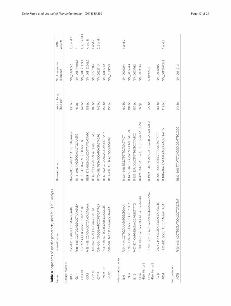
2.4 mg/ml vs 1.5 mg/ml dissolved in the ATCC®EMEMmedium. MEM was complemented also with 1 mM so-dium pyruvate and 1X non-essential amino acids (Bio-chrom). When maintained in these media, cells dividedmore rapidly retaining their morphological features (notshown). For the phenotypical analysis, we plated thecells at 30,000 cells/cm2 in T25 flasks and let them growfor 3 days until almost confluent. IFNγ (10 ng/ml), whenused, was added in the last 36 h. The expression of spe-cific lineage markers was tested by immunocytochem-istry and flow cytometry. The primary antibodies usedfor these evaluations are reported in Table 3. The ex-pression of inflammatory genes was analyzed at themRNA level by reverse transcription followed by real-time (Q)-PCR analysis, performed according to ourstandard protocol [109]. In brief, total cytoplasmic RNAwas prepared from cells using TRIZOL reagent (Invitro-gen) and 1 μg aliquots were converted to cDNA usingrandom hexamer primers and the ImProm-II ReverseTranscriptase kit (Promega). Q-PCRs were performedusing the following cycling conditions: 35 cycles of de-naturation at 95 °C for 20 s; annealing at 60 °C for 30 s;and extension at 72 °C for 30 s; the Brilliant SYBR GreenQPCR Master Mix 2X (Agilent); and the specific primersreported in Table 4. All the primer sets, except the one
used for iNOS and COX2 expression, aligned only hu-man sequences. Q-PCR reactions were carried out in a20 μl reaction volume in a MX3000P real time PCR ma-chine (Stratagene). Relative mRNA concentrations werecalculated from the take-off point of reactions (thresholdcycle, Ct) using the comparative quantitation method[109]. Ct values for β-actin (ACTB) expression served asa normalizing signal. ACTB was selected consideringthat it is a constitutively highly expressed gene in humanmicroglia [2]. Q-PCR efficiency ranged between 94 and106%. At the end of Q-PCR, the products were sepa-rated by electrophoresis through 2% agarose gels con-taining 0.1 μg/ml ethidium bromide to ensure theproduct was the correct size. For functional experi-ments, cells were plated at 30,000 cells/cm2 in 96 wellplates (100 μl/well), let recover overnight and stimu-lated the day after in complete growth medium, i.e.,EMEM containing with 10% FBS and antibiotics. IL-6release was measured with a specific ELISA assay(R&D Systems).The morphology of a viable culture is shown in Fig. 3,
i.e., 1 day after plating (Fig. 3a) and when cells reachedconfluency (day 3 post plating, Fig. 3b). In line with Ete-mad and colleagues [29], the HMC3 (ATCC®CRL-3304)cells display a complex morphology, with both globular,
A B
C D E
Fig. 3 HMC3 cell morphology and labeling of cytoskeletal F-actin filaments. a–b The human microglial cell line HMC3 as it was observed byphase-contrast microscopy at in vitro day 1 (a), and when cells reached the confluency (b). × 10 magnification, scale bar 100 μM. c–e Arepresentative example of confocal images (1024 × 1024 pixels) acquired at × 20 magnification with a confocal laser scanning system (A1+,Nikon). Cells were grown on glass coverslips for 24 h, and their morphology was evaluated by labeling the cytoskeletal F-actin filaments withtetramethylrhodamine (TRITC)-conjugated phalloidin (red fluorescence). Cells were counterstained with the nuclear probe, 4′,6-damidino-2-phenylindole dihydrochloride (DAPI, blue fluorescence). The merged image is shown in (e). Scale bar 50 μM
Dello Russo et al. Journal of Neuroinflammation (2018) 15:259 Page 15 of 24

bipolar, and elongated cells detected in culture byrhodamine-phalloidin staining (Fig. 3c–e). We confirmedthat cells are IBA1 positive (Fig. 4d–g). Moreover, we de-tected at the mRNA level the expression of CX3CR1 andCSF1-R (Fig. 4g–h), together with other specific markers,comprised in the recently characterized microglial signa-ture [2], i.e., P2RY12 and TMEM119 (Fig. 4h). The CCR2transcript was undetectable (Fig. 4g), in line with previousobservations [29, 38]. Consistently with the literature andthe ATCC®‘s data sheet, the HMC3 cells were GFAP nega-tive (Fig. 5a, i.e., the dark gray histogram completely over-laps the background histogram). The glioblastoma U373cell line, kindly provided by Prof. Grassi (Institute ofPhysiology, Catholic University Medical School, Rome,Italy), was used as a positive control for the anti-GFAPantibody (Fig. 3a). The activation marker MHCII(HLA-DR in the figure) was negative under basal condi-tion and upregulated in the 28% of the HMC3 cell popula-tion by IFNγ treatment (36 h, 10 ng/ml) (Fig. 5b). Inaddition, resting HMC3 cells were CD14 (Fig. 5c) andCD11b (Fig. 5d) negative, and these markers were not in-duced by IFNγ. With respect to CD14 expression, our re-sults are in agreement with the original characterization[17] and the evaluation performed in flow cytometry byEtemad and colleagues [29]. Finally, the HMC3(ATCC®CRL-3304) cells expressed CD68 under basal con-ditions (Fig. 5e), in agreement with previous observations[17, 29]. However, CD68 was expressed at low level indi-cated by a mean fluorescence intensity (MFI) of 3.3, andno further induction with IFNγ was detected (MFI = 3.6).As shown in Fig. 6a, the HMC3 (ATCC®CRL-3304) cellswere found entirely and homogenously positive for humanleukocyte antigens A, B, and C (MHCI antigens). Theantibody used for this analysis is specific for the human
antigens and does not cross react with rat antigens. Inaddition, a HLA-B-specific amplification between the firstand the third intron was carried out by PCR on genomicDNA samples, extracted by the HMC3 (ATCC®CRL-3304)cells using the QIAamp® DNA Blood mini kit (Qiagen,Hilden, Germany) [110]. The HLA-B locus-specific ampli-fication was performed using the following validatedprimers: 5BIn1–57 forward primer (forward 5´-GGGAGGAGCGAGGGGACCG/CCA G-3′; intron 1 36–57)and 3BIn3–37 reverse primer (reverse 5´-GGAGGCATCCCCGGCGACCTAT-3′; intron 3 37–59), yielding a922 bp product [111]. The PCR reaction contained 100 ngtotal DNA, 1X PCR buffer, 300 nM of each primer, 1.25 UAccuPrime™ PfX DNA Polymerase (Invitrogen Corpor-ation) in 50 μl final volume. After initial denaturation(10 min at 95 °C), a total of 40 PCR cycles were con-ducted, using the following two-step PCR conditions: de-naturation at 95 °C for 20 s and annealing/extension at68 °C for 1 min, in a MasterCycler Ep thermocycler®(Eppendorf, Hamburg, Germany). The amplicons wereseparated by electrophoresis through 1.5% agarose gelscontaining 0.1 μg/ml ethidium bromide. A human gen-omic DNA sample was provided by ViiV HealthcareLdt. and used as positive Control for the amplification[112]. Data are presented in Fig. 6b. Taken together,this evidence further confirm the human origin of theHMC3 (ATCC®CRL-3304) cell line.Consistently with the literature, the HMC3 (ATCC®CRL-
3304) cells produced sizable amounts of ROS. Reactive freeradicals were measured using H2DCF-DA [2,7- dichlorodi-hydrofluorescein diacetate (Invitrogen)]. At the end of theexperiment, the incubation medium was replaced by Hank’sbalanced salt solution (HBSS) containing 20 μM ofH2DCF-DA [20]. Cells were incubated at 37 °C, in a
Table 3 Antibodies and reagents used for immunocytochemistry and flow cytometry
Immunocytochemistry
mAbs or immunostaining reagents Concentration Final dilution used
DAPI (4′,6-diamidino-2-phenylindole, dihydrochloride)(ThermoFisher Cat. N. D1306)
5 mg/ml 1/2000
Rhodamine phalloidin (ThermoFisher Cat. N. R415) 200 U/ml 1/500
IBA1(Abcam Cat. N. ab15690)
3.5 mg/ml 1/100
Flow Cytometry
mAbs Optimal Ab volume per test (1 × 10⁶) Final dilution used
PE-conjugated anti-GFAP (BD Pharmingen Cat. N. 561,483) 5 μl 1/40
PE-conjugated anti-HLA-DR (BD Bioscience Cat. N. 562,304) 5 μl 1/50
FITC-conjugated anti-CD14 (BD Pharmingen Cat. n 555,397) 20 μl 1/10
PE-conjugated anti-CD11b (eBioscience™ Cat. N. 12–0118-42) 5 μl 1/20
PE-conjugated anti CD68 (BD Horizon Cat. N. 564,944) 5 μl 1/40
FITC-conjugated anti-HLA-ABC (BD Biosciences Cat. N. 555,552) 20 μl 1/10
Dello Russo et al. Journal of Neuroinflammation (2018) 15:259 Page 16 of 24
Table
4Sequ
encesof
specificprim
ersets,usedforQ-PCRanalysis
Gen
esForw
ardprim
erReverseprim
erProd
uctleng
th(Basepair)
NCBI
Reference
sequ
ence
mRN
Avariants
Line
agemarkers
IBA1
F150–170:TCA
TGTCCC
TGAAAC
GAATG
R265–285:C
CAGCATC
ATC
CTGAGAAAG
136bp
NM_032955.2
1,3and4
CD14
F636–655:C
GCTC
GAGGACCTA
AAGATA
R711–693:A
AGCTG
GAAAGTG
CAAGTC
76bp
NM_001174105.1
4
CX3
CR1
F187-207:G
GCTG
AGGCCT
GTTATA
TTG
R315–333:TGACA
CTC
TTGGGCT
TCT
147bp
NM_001171174.1
1,2,3and4
CCR2
F425–445:C
CACAAGCT
GAACAGAGAAA
R558–579:G
GGAGCA
CCGTA
ATCATA
ATC
155bp
NM_001123041.2
AandB
P2RY12
F674–692:A
GACCA
CCA
GGCC
ATTTA
R837–858:CAGACTAGACC
GAACTCTGAT
185bp
NM_022788.4
1and2
CSF1R
F344–364:C
AGGGAATC
CCAGTG
ATA
GA
R470–489:TGGAGCCATC
AGAGTA
CAG
146bp
NM_005211.3
2,3and4
TMEM
119
F408–428:A
CTTCCTG
GATG
GGATA
GTG
R542–562:G
GGAAGGACGATG
GGTA
ATA
155bp
NM_181724.2
TREM
2F588–607:AGCC
TCTTGGAAGGAGAAA
R719–737:A
GTTCACTG
GGTG
GATG
T150bp
NM_018965.3
Inflammatoryge
nes
IL-6
F394–414:C
CTTCCAAAGATG
GCTG
AAA
R524–543:TG
GCTTGTTCCTC
ACT
ACT
150bp
NM_000600.4
1and2
TNFα
F1305–1324:G
AGCCAGCTC
CCTC
TATTTA
R1485–1466:GGGAACAGCCTA
TTGTTCA
G181bp
NM_000594.3
IL-1β
F407-427:C
ATG
GGATA
ACGAGGCTTATG
R556–537:CCACTTGTTGCTC
CATA
TCC
150bp
NM_000576.2
COX2
(Rat/Hum
an)
F1384–1407:TTG
CTGGCAGGGTTGCTG
GTG
GTA
R1469–1449:C
ATC
TGCC
TGCTC
TGGTC
AATC
GAA
86bp
NM_000963.4
NOS2
(Rat/Hum
an)
F1701–1725:CTG
CATG
GAACAGTA
TAAGGCAAAC
R1929–1905:AGACAGTTTC
TGGTC
GATG
TCATG
A229bp
AF049656.1
TGFβ:
F2423-2442:C
AGTC
ACCA
TAGCAACACTC
R2583–2567:CC
TGGCC
TGAACTAC
TATCT
161bp
NM_000660.5
ARG
1F905–925:GGGCTA
CTC
TCAGGATTAGAT
R1016–996:C
GAAACA
AGCCA
AGGTTATTG
112bp
NM_001244438.1
1and2
Normalization
ACTB
F590–613:A
CGTTGCTA
TCCAGGCTG
TGCTA
TR830–807:TTA
ATG
TCACGCACG
ATTTC
CCGC
241bp
NM_001101.3
Dello Russo et al. Journal of Neuroinflammation (2018) 15:259 Page 17 of 24

humidified atmosphere with 5% CO2, for 45 min. Thefluorescence signal due to H2DCF-DA oxidation within thecells was quantified using a microplate fluorescence reader(PerkinElmer Inc.), using 485 nm as excitation and 535 nmas emission wavelength [113]. Pro-inflammatory cytokinesper se, at 10 ng/ml, did not modify basal levels of ROS pro-duction, whereas IL-1β and TNFα each used in combin-ation with IFNγ slightly increased ROS production byHMC3 cells (0.1–0.2-fold increases), in line with previousobservations [21]. However, this effect was not consistentamong different experiments, carried out between passage6 and 13. Interestingly, the production of ROS by HMC3cells growth in either MEM or DMEMF12 was significantlylower, thus suggesting a less activated phenotype underbasal conditions when kept in these media (not shown). Inaddition, we assessed the production of NO indirectly bymeasuring nitrite accumulation in the incubation media
[108]. Nitrite levels were undetectable under basal condi-tions and remained so after a 24-h treatment with differentpro-inflammatory stimuli, including LPS, LPS/IFNγ, andIL-1β, and TNFα used alone or in combination with IFNγ(data not shown). Consistently, resting HMC3(ATCC®CRL-3304) cells did not express iNOS, neither itsexpression was induced by pro-inflammatory activation(Table 5). Conversely, we observed a basal expression ofIL-6 (Table 5) in resting HMC3 cells, together with a siz-able release of the cytokine in the incubation mediumunder basal conditions, i.e., 150 pg/ml. The expression levelof IL-6 increased in response to pro-inflammatory cyto-kines, particularly IL-1β in combination with IFNγ (II,Table 5). In addition, the cytokine mixture (II) stimulatedbasal IL-6 secretion by 20-folds (data not shown). Otherpro- and anti-inflammatory genes were studied at transcrip-tional levels under basal conditions and after 24-h
A C E
B D F
G
H
Fig. 4 HMC3 cell expression of microglial lineage markers. a–f Representative example of confocal images (1024 × 1024 pixels) acquired at × 20magnification with a confocal laser scanning system (A1+, Nikon). HMC-3 cells immuno-labeled for IBA1 (green fluorescence, d, f) andDAPI stained (blue fluorescence, a, b) are shown. Merged images are shown in (e, f). Control experiments performed by omitting theprimary antibody are shown in (a, c, e). No green fluorescence was present (c, e), indicating neither spontaneous fluorescence nor non-specificity of the secondary antibody. Scale bar 50 μM. g–h Total RNA was prepared from human microglial HMC3 cells 24 h afterincubation in complete growth medium and retrotranscribed using random hexamers. Real time (Q)-PCR analysis for the mRNA levels ofIBA1, CX3CR1, CCR2, P2RY12, TMEM119, and CSF1-R was carried out according to our standard protocols [109]. g, h panels show arepresentative gel image of PCR products obtained at the end of the analysis from two different RNA samples for each condition. In thelast line is shown the actin amplification, as positive control
Dello Russo et al. Journal of Neuroinflammation (2018) 15:259 Page 18 of 24

treatments with II (Table 5). The cytokine mixture pro-moted a significant up-regulation of IL-1β, TNFα, andCOX2, leaving unmodified the expression of TGFβ andARG1. In line with our data, it has been recently shownthat the HMC3 (ATCC®CRL-3304) cells express at themRNA level several pro-inflammatory genes, includingIL-1β, TNFα, IL-6, COX2, and in addition hemeoxygenase-1 [31]. Interestingly, lumirubin, a photoisomerof bilirubin (generated during phototherapic treatment ofneonatal hyperbilirubinemia), promoted microglial inflam-matory activation as indicated by the upregulation of allthese genes except COX2 [31]. Moreover, thepro-inflammatory effects of bilirubin photoisomers weremore prominent on rat hippocampal slices, furtherhighlighting relevant differences between human and ro-dent experimental models. Finally, increased expressionand release of both IL-1β and IL-18 has been observed inthe HMC3 (ATCC®CRL-3304) cells by treatment with
100 ng/ml LPS for 24 h followed by additional 30 min with5 mM adenosine triphosphate (ATP) [32]. Exposure ofHMC3 (ATCC®CRL-3304) cells to LPS/ATP was associatedto increased expression of the intracellular NOD-like recep-tor 3 and activation of caspase 1, together with a significantincrease of cFos nuclear protein levels. Pro-inflammatoryeffects of LPS/ATP were indeed reversed by knockdown ofcFos. [32]. The treatment with LPS/ATP significantly af-fected microglial viability. All the effects of the LPS/ATPcombination were reduced by the α-2 adrenergic agonist,dexmedetomidine [32]. In these research papers, theHMC3 cells were maintained in MEM [31] or EMEM [32]with 10% FCS and antibiotics.
ConclusionsEvidence reviewed in the present paper suggest that thehuman microglial cell line clone 3 generated in thelaboratory of Prof. Tardieu has been extensively
A
B C D E
Fig. 5 Phenotypical characterization of HMC3 cells under basal conditions and in response to IFNγ. HMC3 cells were plated at the density of30,000 cells/cm2 in T25 flasks, and grown for 3 days when cells were almost confluent. In the experiments in which INFγ was used, cells werestimulated for the last 36 h. Controls did not receive any stimulus for the same time period (“resting” HMC3 cells). For detection of intracellularantigens, aliquots of 1 × 10⁶ cells in 100 μl were fixed using BD cytofix (BD Pharmingen) at 4 °C for 30 min, and permeabilized with BD FACSpermeabilizing solution at 4 °C for 30 min. Cells were then stained using the following antibodies: PE-conjugated anti Human GFAP mouse Mab,BD Bioscience (a), and PE-CF594-conjugated anti Human CD68 mouse Mab, BD Bioscience (e), according to the manufacturer’s instructions. Forthe evaluation of surface antigens, aliquots of 5 × 10⁵ cells in 100 μl were directly incubated in PBS buffer containing the following antibodies:PE-CF594-conjugated anti Human HLA-DR mouse Mab, BD Bioscience (b); FITC-conjugated anti Human CD14 mouse Mab, BD Pharmingen (c);and PE-conjugated anti Human CD11b mouse Mab, E-Bioscience (d). Cells were analyzed by the 6-parameter (2 scatter and 4 fluorescencesignals) Coulter Epics XL flow cytometer (Beckman-Coulter). Control histograms (white histograms) indicate level of cell autofluorescence in theemission wavelength that pertains the fluorochrome-conjugated Mab. Panel (a) shows expression of GFAP on HMC3 cells at passage 4 (darkgray), and on the human glioblastoma U373 cell line (light gray) that constitutively expresses GFAP. As autofluorescence signal in the two celllines was similar, only one representative histogram is plotted in panel (a) (white histogram). As shown in this panel, the HMC3 cells stainnegatively for GFAP (the dark gray histogram completely overlaps the background histogram), whereas the U373 cells express the targetantigen. Two different populations expressing GFAP at different levels were identified (light gray plot): 68% of the U373 cells express GFAP at lowlevel (GFAP-dim), whereas 32% of the cells express GFAP at higher level (GFAP-high). Panels (b–e) show results from HMC3 cells obtained atpassage 7. In these panels, white histograms indicate cell autofluorescence, light gray histograms and dark gray histograms are representative ofcontrol (“resting”) HMC3 cells and IFNγ-treated HMC3 cells, respectively. Gating strategy: all analyses were obtained after gating according tomorphological characteristics (not shown). As levels of autofluorescence did not change according to cell treatment, one representative exampleis plotted for each emission wavelength. Linear regions are used for calculating percentage of positive cells (plots A–B). Central tendency of CD68expression on HMC3 cells in the different experimental conditions is represented by the mean fluorescence intensity (MFI, see text)
Dello Russo et al. Journal of Neuroinflammation (2018) 15:259 Page 19 of 24

characterized, with respect to cell morphology, antigenicprofile, and cell functions. However, cells were usedunder different denominations, i.e., CHME3, HMC3,C13-NJ, and CHME-5, which contributed to data frag-mentation. In this regard, the experimental datareviewed in this article show that the cells, regardlessthe name adopted, retain a similar antigenic profile to-gether with similar functional properties. The cells arecapable of responding to a pattern of chemokines andinflammatory stimuli, regulating the expression of typicalactivation markers of microglia. Therefore, these celllines should be regarded as a unique experimentalmodel. Caution should be taken with data obtained
using microglial cells named CHME-5, since it has beenreported that several aliquots in use in different labora-tories were cross contaminated by rat glioma cells [19].Laboratories using the CHME-5 cells should perform anauthentication procedure to confirm the human origin.In this regard, the HMC3 (ATCC®CRL-3304) cellspresent the advantage to have been authenticated byATCC® according to a complex procedure, includingmorphology evaluation, karyotyping, and PCR based ap-proaches to confirm the identity of human cell lines andto rule out both intra- and inter-species contamination.In addition, we showed that the HMC3(ATCC®CRL-3304) cells retain most of the original anti-genic properties and express also more specific micro-glial markers (recently identified) [2]. Resting cellsproduce significant amounts of ROS and IL-6. The lattercan be further induced by pro-inflammatory cytokines(particularly IL-1β in combination with IFNγ at 10 ng/ml). Evidence from the literature together with our ori-ginal data may represent an important starting point forresearchers that are planning to use this experimentalmodel.
AbbreviationsACTB: β-actin; ADAMTS: A disintegrin and metalloproteinase withthrombospondin motifs; APP: Amyloid beta precursor protein;ARG: Arginase; ATCC®: American Type Culture Collection; ATP:Adenosine triphosphate; Aβ: Amyloid beta peptide; BDNF: Brainderived neurotrophic factor; CCL: C-C motif chemokine ligand; CCR:C-C chemokine receptor; CD: Cluster of differentiation; CHME3: Humanmicroglial clone 3 cell line; CM: Conditioned medium; CNS: Centralnervous system; coMTb: Human peripheral blood monocytes infectedwith Mycobacterium tuberculosis; COX: Cyclooxygenase; CSF1-R: Colony-stimulating factor 1 receptor; CXCR: C-X-C chemokine receptor; DAPI: 4′,6-Diamidino-2-phenylindole dihydrochloride; DHA: Docosahexaenoic acid;DMEM: Dulbecco’s modified minimal essential medium; ELISA: Enzyme-linkedimmunosorbent assay; EMEM: Eagle’s minimum essential medium;EPA: Eicosapentenoic acid; ERK: Extracellular signal–regulated kinase;FCS: Fetal calf serum; FITC-conjugated: Fluorescein isothiocyanateconjugated; GFAP: Glial fibrillary acidic protein; GM-CSF: Granulocyte-macrophage colony-stimulating factor; H2DCF-DA: 2,7-Dichlorodihydrofluoresceindiacetate; HBSS: Hank’s balanced salt solution; HCV: Hepatitis virus C; HIV: Humanimmunodeficiency virus; HLA-DR: Human leukocyte Antigen D Related;
A
B
Fig. 6 Expression of the human class I MHC antigens on HMC3 cells.a HMC3 were plated at the density 30,000 cells/cm2 in T25 flasks,and grown for 3 days when cells were almost confluent. For thedetection of class I MHC surface antigens, aliquots of 5 × 10⁵ cells in100 μl were directly incubated with FITC-conjugated anti humanHLA-ABC mouse Mab, BD Bioscience. This antibody is specific forhuman MHCI antigens, and does not cross react with rat MHCIantigens. Cells were analyzed by the 6-parameter (2 scatter and 4fluorescence signals) Coulter Epics XL flow cytometer (Beckman-Coulter). Results from HMC3 cells at passage 8 are shown. Gatingstrategy: the analysis was obtained after gating according tomorphological characteristics (not shown). Background histogram(white) indicates level of cell autofluorescence. A linear region wasused for calculating percentage of positive cells. b Gel image of PCRproducts obtained at the end of a HLA Locus B specific amplificationprotocol. The 922 bp amplicons were separated by electrophoresisthrough 1.5% agarose gels containing 0.1 μg/ml ethidium bromide.Lines 2–3 amplification products from genomic DNA extracted byHMC3 (ATCC®CRL-3304) cells; lines 4–5 amplification products from apositive control, i.e., an anonymous human genomic DNA sampleprovided by ViiV Healthcare Ldt
Table 5 Gene expression analysis of the HMC3 (ATCC®CRL-3304) cells
Gene Control (mean thresholdcycle, Ct)
II (Fold variation relativeto control)
Pro-inflammatory genes
IL-6 27.97 ± 0.17 (n = 12) 43.75 ± 2.59 (n = 11)
TNFα 32.50 ± 0.20 (n = 12) 5.37 ± 0.45 (n = 11)
IL1β 29.02 ± 0.10 (n = 12) 4.71 ± 0.32 (n = 12)
COX2 33.00 ± 0.50 (n = 9) 2.51 ± 0.46 (n = 9)
NOS2 No Ct (n = 9) No Ct (n = 9)
Anti-inflammatory genes
TGFβ 28.50 ± 0.16 (n = 12) 1.02 ± 0.06 (n = 12)
ARG1 29.51 ± 0.10 (n = 10) 1.07 ± 0.07 (n = 11)
Dello Russo et al. Journal of Neuroinflammation (2018) 15:259 Page 20 of 24

HMC3: Human microglial clone 3 cell line; hTERT: Human telomerasereverse transcriptase; IBA1: Ionized calcium binding adaptor molecule 1;IFNγ: Interferon-γ; II: Interferon-γ/interleukin-1β; IL: Interleukin; iNOS: Induciblenitric oxide synthase; IP-10: Interferon-γ inducible protein 10; IRES: Internalribosome entry site; IRF: Interferon regulatory factor; JAK: Janus kinases;JEV: Japanese encephalitis virus; LOX: Lipoxygenase; LPS: Lipopolysaccharide;LXA4: Lipoxin A4; Mab: Monoclonal antibody; MAPK: Mitogen-activated proteinkinase; MaR: Maresin; MCP-1: Monocyte chemoattractant protein 1; M-CSF: Macrophage colony-stimulating factor; M-CSF-R: Macrophagecolony-stimulating factor receptor; MFI: Mean fluorescence intensity;MHCII: Class II major histocompatibility complex; miR- or miRNA: MicroRNA;MMF: Monomethylfumarate; MMP: Matrix metalloprotease; NF70KD: Neuronalneurofilament staining; NFκB: Nuclear factor kappa-light-chain-enhancer of acti-vated B cells; NGF : Nerve growth factor; NO: Nitric oxide; NOS2: Inducible nitricoxide synthase; NOX: NADPH oxidase; NPCs: Neuronal precursor cells;NS3: Nonstructural protein 3, also known as p-70; NSE: Nonspecific esteraseactivity; P2RY12 : Purinergic receptor 12; PE-conjugated: Phycoerythrin-conjugated; PSA-NCAM: Polysialylated neural cell adhesion molecule;RANTES: Regulated on activation, normal T cell expressed and secreted;ROS: Free oxygen radicals; RvD1: Resolving D1; sAPPα: Secreted amyloidprecursor protein-α; SHswe: Human neuroblastoma SH-SY5Y cells overex-pressing APP695 Swedish mutation protein; STAT: Signal transducer andactivator of transcription; SV40: Simian vacuolating virus 40;TGF: Transforming growth factor; TIMP: Tissue inhibitor ofmetalloproteinases; TLRs: Toll-like receptors; TMEM119: Transmembraneprotein 119; TNFα: Tumor necrosis factor α; TRAF: TNF receptor-associated factor; TREM2: Triggering receptor expressed on myeloid cells2; TRIM: Tripartite motif-containing protein; TRITC: Tetramethylrhodamine;UTR: Untranslated regions; VEGF: Vascular endothelial growth factor;α7nAcR: α7 nicotinic acetylcholine receptor; αMSH: α-melanocytestimulating hormone
AcknowledgementsThe authors would like to thank Prof. M. Tardieu, Pediatric Neurology Department,Université Paris Sud, Paris, for the clarification of the cell line nomenclature. Theauthors are grateful to Prof. G. Pani, Institute of General Pathology, UniversitàCattolica del S. Cuore, Rome, for the stimulating discussion and helpfulsuggestions on the cell line, and for the critical reading of this manuscript.Finally, the authors would like to acknowledge the support and assistancewith flow cytometry received by Dr. Alexia Buzzonetti and Dr. MarcoFossati, Immunology Laboratory, Department of Oncological Gynecology,Università Cattolica del S. Cuore, Rome.
FundingThe present research is funded by Angelini S.p.A. and by Fondi di Ateneo2017, Università Cattolica del S. Cuore, both grants awarded to CDR. Datawere timely shared with Angelini S.p.A through progress and final reports,and during one internal meeting.
Availability of data and materialsThe datasets used and/or analyzed during the current study are available fromthe corresponding author on reasonable request.
Authors’ contributionsCDR supervised the experiments, collected and analyzed the data, conceivedthe paper, and wrote the primary draft. NC handled the HMC3 (ATCC®CRL-3304) cell line and performed the experiments and most of the phenotypicaland molecular assays. IC supervised the project, contributed to design theexperimental plan, and helped with data analysis. DM and FP performed theimmunocytochemistry and confocal imaging. GP shared his cell culture protocolsand helped in the optimization of culture conditions. PN contributed to studydesign and manuscript editing. AB performed the flow cytometric analysis. NCand AB significantly contributed to the literature revision and manuscript editing.All authors read and approved the final manuscript.
Ethics approval and consent to participateAll the experiments were carried out on the human microglial cell line,HMC3 (ATCC®CRL-3304) cells authenticated and distributed by ATCC®. Thecell line has been used under the term and conditions indicated in theapproved material transfer agreement. The use of a single anonymizedhuman genomic DNA sample was authorized by ViiV Healthcare Ldt.,
under the terms and conditions signed by the Italian HLA-B*57:01Virological-Clinical Network. Ethics approval was not required since thesample derived from a previous study is approved by the local EthicalCommittee which comprised future use for methodological studies [112].
Consent for publicationWe used a single anonymized human genomic DNA sample provided byViiV Healthcare Ltd., for which consent for publication is not required [112].
Competing interestsThe authors declare that they have no competing interests.
Publisher’s NoteSpringer Nature remains neutral with regard to jurisdictional claims in publishedmaps and institutional affiliations.
Author details1Institute of Pharmacology, Università Cattolica del S. Cuore, L.go F Vito 1,00168 Rome, Italy. 2Pharmacology Unit, Fondazione Policlinico UniversitarioA. Gemelli IRCCS, Rome, Italy. 3Angelini RR&D (Research, Regulatory &Development) - Angelini S.p.A., Rome, Italy. 4Institute of Human Physiology,Università Cattolica del S. Cuore, Rome, Italy. 5Institute of Otolaryngology,Università Cattolica del S. Cuore, Rome, Italy. 6Immunology Laboratory,Department of Oncological Gynecology, Università Cattolica del S. Cuore,Rome, Italy.
Received: 12 June 2018 Accepted: 21 August 2018
References1. Butovsky O, Jedrychowski MP, Moore CS, Cialic R, Lanser AJ, Gabriely G,
Koeglsperger T, Dake B, Wu PM, Doykan CE, Fanek Z, Liu L, Chen Z,Rothstein JD, Ransohoff RM, Gygi SP, Antel JP, Weiner HL. Identification of aunique TGF-β-dependent molecular and functional signature in microglia.Nat Neurosci. 2014;17:131–43.
2. Gosselin D, Skola D, Coufal NG, Holtman IR, Schlachetzki JCM, Sajti E, JaegerBN, O'Connor C, Fitzpatrick C, Pasillas MP, Pena M, Adair A, Gonda DD, LevyML, Ransohoff RM, Gage FH, Glass CK. An environment-dependent transcriptionalnetwork specifies human microglia identity. Science. 2017;356 https://doi.org/10.1126/science.aal3222.
3. Sousa C, Biber K, Michelucci A. Cellular and molecular characterization ofmicroglia: a unique immune cell population. Front Immunol. 2017;8:198.https://doi.org/10.3389/fimmu.2017.00198.
4. Salter MW, Stevens B. Microglia emerge as central players in brain disease.Nat Med. 2017;23:1018–27.
5. Wake H, Moorhouse AJ, Jinno S, Kohsaka S, Nabekura J. Resting microgliadirectly monitor the functional state of synapses in vivo and determine thefate of ischemic terminals. J Neurosci. 2009;29:3974–80.
6. Tremblay ME, Lowery RL, Majewska AK. Microglial interactions withsynapses are modulated by visual experience. PLoS Biol. 2010;8:e1000527.
7. Paolicelli RC, Bolasco G, Pagani F, Maggi L, Scianni M, Panzanelli P, GiustettoM, Ferreira TA, Guiducci E, Dumas L, Ragozzino D, Gross CT. Synapticpruning by microglia is necessary for normal brain development.Science. 2011;333:1456–8.
8. Chen Z, Jalabi W, Hu W, Park HJ, Gale JT, Kidd GJ, Bernatowicz R, GossmanZC, Chen JT, Dutta R, Trapp BD. Microglial displacement of inhibitorysynapses provides neuroprotection in the adult brain. Nat Commun.2014;5:4486.
9. Sierra A, Encinas JM, Deudero JJ, Chancey JH, Enikolopov G, Overstreet-Wadiche LS, Tsirka SE, Maletic-Savatic M. Microglia shape adult hippocampalneurogenesis through apoptosis-coupled phagocytosis. Cell Stem Cell. 2010;7:483–95.
10. Ueno M, Fujita Y, Tanaka T, Nakamura Y, Kikuta J, Ishii M, Yamashita T. LayerV cortical neurons require microglial support for survival during postnataldevelopment. Nat Neurosci. 2013;16:543–51.
11. Shigemoto-Mogami Y, Hoshikawa K, Goldman JE, Sekino Y, Sato K. Microgliaenhance neurogenesis and oligodendrogenesis in the early postnatalsubventricular zone. J Neurosci. 2014;34:2231–43.
Dello Russo et al. Journal of Neuroinflammation (2018) 15:259 Page 21 of 24

12. Du L, Zhang Y, Chen Y, Zhu J, Yang Y, Zhang HL. Role of microglia inneurological disorders and their potentials as a therapeutic target. MolNeurobiol. 2017;54:7567–84.
13. Dello Russo C, Lisi L, Tentori L, Navarra P, Graziani G, Combs CK. Exploitingmicroglial functions for the treatment of glioblastoma. Curr Cancer Drug Targets.2017;17:267–81.
14. Lin L, Desai R, Wang X, Lo EH, Xing C. Characteristics of primary ratmicroglia isolated from mixed cultures using two different methods. JNeuroinflammation. 2017;14:101.
15. Figuera-Losada M, Rojas C, Slusher BS. Inhibition of microglia activation as aphenotypic assay in early drug discovery. J Biomol Screen. 2014;19:17–31.
16. Smith AM, Dragunov M. The human side of microglia. Trends Neurosci.2014;37:125–35.
17. Janabi N, Peudenier S, Héron B, Ng KH, Tardieu M. Establishment of humanmicroglial cell lines after transfection of primary cultures of embryonicmicroglial cells with the SV40 large T antigen. Neurosci Lett. 1995;195:105–8.
18. Nagai A, Nakagawa E, Hatori K, Choi HB, McLarnon JG, Lee MA, Kim SU.Generation and characterization of immortalized human microglial cell lines:expression of cytokines and chemokines. Neurobiol Dis. 2001;8:1057–68.
19. Garcia-Mesa Y, Jay TR, Checkley MA, Luttge B, Dobrowolski C, Valadkhan S,Landreth GE, Karn J, Alvarez-Carbonell D. Immortalization of primarymicroglia: a new platform to study HIV regulation in the central nervoussystem. J Neuro-Oncol. 2017;23:47–66.
20. Li B, Bedard K, Sorce S, Hinz B, Dubois-Dauphin M, Krause KH. NOX4 expressionin human microglia leads to constitutive generation of reactive oxygen speciesand to constitutive IL-6 expression. J Innate Immun. 2009;1:570–81.
21. Jadhav VS, Krause KH, Singh SK. HIV-1 Tat C modulates NOX2 and NOX4expressions through miR-17 in a human microglial cell line. J Neurochem.2014;131:803–15.
22. Nakagawa Y, Chiba K. Diversity and plasticity of microglial cells in psychiatricand neurological disorders. Pharmacol Ther. 2015;154:21–35.
23. Reiner A, Heldt SA, Presley CS, Guley NH, Elberger AJ, Deng Y, D'Surney L,Rogers JT, Ferrell J, Bu W, Del Mar N, Honig MG, Gurley SN, Moore BM.Motor, visual and emotional deficits in mice after closed-head mild traumaticbrain injury are alleviated by the novel CB2 inverse agonist SMM-189. Int J MolSci. 2014;16:758–87.
24. Zhu W, Carney KE, Pigott VM, Falgoust LM, Clark PA, Kuo JS, Sun D. Glioma-mediated microglial activation promotes glioma proliferation and migration:roles of Na+/H+ exchanger isoform 1. Carcinogenesis. 2016;37:839–51.
25. Patel AB, Tsilioni I, Leeman SE, Theoharides TC. Neurotensin stimulatessortilin and mTOR in human microglia inhibitable by methoxyluteolin, apotential therapeutic target for autism. PNAS. 2016;113:E7049–58.
26. Roy S, Gumulec J, Kumar A, Raudenska M, Baig MH, Polanska H, Balvan J,Gupta M, Babula P, Odstrčilík J, Choi I, Provaznik I, Masarik M. The effect ofBenzothiazolone-2 on the expression of Metallothionein-3 in modulatingAlzheimer's disease. Brain Behav. 2017;7:e00799.
27. Harris AR, Yuan JX, Munson JM. Assessing multiparametric drug response intissue engineered tumor microenvironment models. Methods. 2018;134-135:20–31.
28. Rees GF, Lago SG, Cox DA, Tomasik J, Rustogi N, Weigelt K, Ozcan S, Cooper J,Drexhage H, Leweke FM, Bahn S. Evidence of microglial activation followingexposure to serum from first-onset drug-naïve schizophrenia patients. BrainBehav Immun. 2018;67:364–73. https://doi.org/10.1016/j.bbi.2017.10.003.
29. Etemad S, Zamin RM, Ruitenberg MJ, Filgueira L. A novel in vitro humanmicroglia model: characterization of human monocyte-derived microglia. JNeurosci Methods. 2012;209:79–89.
30. Ambrosius B, Faissner S, Guse K, von Lehe M, Grunwald T, Gold R, Grewe B,Chan A. Teriflunomide and monomethylfumarate target HIV-inducedneuroinflammation and neurotoxicity. J Neuroinflammation. 2017;14:51.
31. Jašprová J, Dal Ben M, Hurný D, Hwang S, Žížalová K, Kotek J, Wong RJ,Stevenson DK, Gazzin S, Tiribelli C, Vítek L. Neuro-inflammatory effects ofphotodegradative products of bilirubin. Sci Rep. 2018;8:7444.
32. Li H, Zhang X, Chen M, Chen J, Gao T, Yao S. Dexmedetomidine inhibitsinflammation in microglia cells under stimulation of LPS and ATP by c-Fos/NLRP3/caspase-1 cascades. EXCLI J. 2018;17:302–11.
33. Peudenier S, Hery C, Montagnier L, Tardieu M. Human microglial cells:characterization in cerebral tissue and in primary culture, and study of theirsusceptibility to HIV-1 infection. Ann Neurol. 1991;29:152–61.
34. Sébire G, Emilie D, Wallon C, Héry C, Devergne O, Delfraissy JF, Galanaud P,Tardieu M. In vitro production of IL-6, IL-1 beta, and tumor necrosis factor-alphaby human embryonic microglial and neural cells. J Immunol. 1993;150:1517–23.
35. Cross AK, Woodroofe MN. Chemokines induce migration and changes inactin polymerization in adult rat brain microglia and a human fetalmicroglial cell line in vitro. J Neurosci Res. 1999;55:17–23.
36. Hjorth E, Frenkel D, Weiner H, Schultzberg M. Effects of immunomodulatorysubstances on phagocytosis of a beta(1-42) by human microglia. Int JAlzheimers Dis. 2010; https://doi.org/10.4061/2010/798424.
37. Liu J, Hjorth E, Zhu M, Calzarossa C, Samuelsson EB, Schultzberg M, ÅkessonE. Interplay between human microglia and neural stem/progenitor cells inan allogeneic co-culture model. J Cell Mol Med. 2013;17:1434–43.
38. Flynn G, Maru S, Loughlin J, Romero IA, Male D. Regulation of chemokinereceptor expression in human microglia and astrocytes. J Neuroimmunol.2003;136:84–93.
39. Zhu M, Wang X, Hjorth E, Colas RA, Schroeder L, Granholm AC, Serhan CN,Schultzberg M. Pro-resolving lipid mediators improve neuronal survival andincrease Aβ42 phagocytosis. Mol Neurobiol. 2016;53:2733–49.
40. Rajalakshmy AR, Malathi J, Madhavan HN. Hepatitis C virus NS3 mediatedmicroglial inflammation via TLR2/TLR6 MyD88/NF-κB pathway and toll likereceptor ligand treatment furnished immune tolerance. PLoS One. 2015;10:e0125419.
41. Nijaguna MB, Patil V, Urbach S, Shwetha SD, Sravani K, Hegde AS,Chandramouli BA, Arivazhagan A, Marin P, Santosh V, Somasundaram K.Glioblastoma-derived macrophage colony-stimulating factor (MCSF) inducesmicroglial release of insulin-like growth factor-binding protein 1 (IGFBP1) topromote angiogenesis. J Biol Chem. 2015;290:23401–15.
42. Ransohoff RM, Schafer D, Vincent A, Blachère NE, Bar-Or A.Neuroinflammation: ways in which the immune system affects the brain.Neurotherapeutics. 2015;12:896–909.
43. Hjorth E, Zhu M, Toro VC, Vedin I, Palmblad J, Cederholm T, Freund-Levi Y,Faxen-Irving G, Wahlund LO, Basun H, Eriksdotter M, Schultzberg M. Omega-3 fatty acids enhance phagocytosis of Alzheimer's disease-related amyloid-β42 by human microglia and decrease inflammatory markers. J AlzheimersDis. 2013;35:697–713.
44. Zhu M, Wang X, Schultzberg M, Hjorth E. Differential regulation ofresolution in inflammation induced by amyloid-β42 and lipopolysaccharidesin human microglia. J Alzheimers Dis. 2015;43:1237–50.
45. Sedgwick JD, Schwender S, Imrich H, Dörries R, Butcher GW, ter Meulen V.Isolation and direct characterization of resident microglial cells from thenormal and inflamed central nervous system. Proc Natl Acad Sci U S A.1991;88:7438–42.
46. Martin S, Vincent JP, Mazella J. Involvement of the neurotensin receptor-3 inthe neurotensin-induced migration of human microglia. J Neurosci. 2003;23:1198–205.
47. Lindberg C, Hjorth E, Post C, Winblad B, Schultzberg M. Cytokine productionby a human microglial cell line: effects of beta-amyloid and alpha-melanocyte-stimulating hormone. Neurotox Res. 2005;8:267–76.
48. Lindberg C, Crisby M, Winblad B, Schultzberg M. Effects of statins on microglia.J Neurosci Res. 2005;82:10–9.
49. Baldassarre M, Baronio CM, Morozova-Roche LA, Barth A. Amyloid β-peptides 1-40 and 1-42 form oligomers with mixed β-sheets. Chem Sci.2017;8:8247–54.
50. Fernandes A, Ribeiro AR, Monteiro M, Garcia G, Vaz AR, Brites D. Secretomefrom SH-SY5Y APP(Swe) cells trigger time-dependent CHME3 microgliaactivation phenotypes, ultimately leading to miR-21 exosome shuttling.Biochimie. 2018; https://doi.org/10.1016/j.biochi.2018.05.015.
51. Gasic-Milenkovic J, Dukic-Stefanovic S, Deuther-Conrad W, Gärtner U,Münch G. β-Amyloid peptide potentiates inflammatory responses inducedby lipopolysaccharide, interferon -γ and ‘advanced glycation endproducts’in a murine microglia cell line. Eur J Neurosci. 2003;17:813–21.
52. Shahbazi S, Kaur J, Singh S, Achary KG, Wani S, Jema S, Akhtar J, Sobti RC.Impact of novel N-aryl piperamide NO donors on NF-κB translocation inneuroinflammation: rational drug-designing synthesis and biologicalevaluation. Innate Immun. 2018;24:24–39.
53. Pareek S, Roy S, Kumari B, Jain P, Banerjee A, Vrati S. MiR-155 induction inmicroglial cells suppresses Japanese encephalitis virus replication and negativelymodulates innate immune responses. J Neuroinflammation. 2014;11:97.
54. Kumari B, Jain P, Das S, Ghosal S, Hazra B, Trivedi AC, Basu A, Chakrabarti J,Vrati S, Banerjee A. Dynamic changes in global microRNAome andtranscriptome reveal complex miRNA-mRNA regulated host response toJapanese encephalitis virus in microglial cells. Sci Rep. 2016;6:20263.
55. Manocha GD, Mishra R, Sharma N, Kumawat KL, Basu A, Singh SK.Regulatory role of TRIM21 in the type-I interferon pathway in Japanese
Dello Russo et al. Journal of Neuroinflammation (2018) 15:259 Page 22 of 24

encephalitis virus-infected human microglial cells. J Neuroinflammation.2014;11:24.
56. Sharma N, Verma R, Kumawat KL, Basu A, Singh SK. miR-146a suppressescellular immune response during Japanese encephalitis virus JaOArS982strain infection in human microglial cells. J Neuroinflammation. 2015;12:30.
57. Sharma N, Kumawat KL, Rastogi M, Basu A, Singh SK. Japanese encephalitisvirus exploits the microRNA-432 to regulate the expression of suppressor ofcytokine signaling (SOCS) 5. Sci Rep. 2016;6:27685.
58. Gupta MK, Behera SK, Dehury B, Mahapatra N. Identification and characterizationof differentially expressed genes from human microglial cell samples infectedwith Japanese encephalitis virus. J Vector Borne Dis. 2017;54:131–8.
59. Rawat P, Spector SA. Development and characterization of a humanmicroglia cell model of HIV-1 infection. J Neuro-Oncol. 2017;23:33–46.
60. Forton DM, Karayiannis P, Mahmud N, Taylor-Robinson SD, Thomas HC.Identification of unique hepatitis C virus quasispecies in the central nervoussystem and comparative analysis of internal translational efficiency of brain,liver, and serum variants. J Virol. 2004;78:5170–83.
61. Wang JS, Wu D, Huang DY, Lin WW. TAK1 inhibition-induced RIP1-dependent apoptosis in murine macrophages relies on constitutive TNF-αsignaling and ROS production. J Biomed Sci. 2015;22:76.
62. Meertens L, Labeau A, Dejarnac O, Cipriani S, Sinigaglia L, Bonnet-Madin L,Le Charpentier T, Hafirassou ML, Zamborlini A, Cao-Lormeau VM, CoulpierM, Missé D, Jouvenet N, Tabibiazar R, Gressens P, Schwartz O, Amara A. Axlmediates Zika virus entry in human glial cells and modulates innateimmune responses. Cell Rep. 2017;18:324–33.
63. Mishra R, Chhatbar C, Singh SK. HIV-1 tat C-mediated regulation of tumornecrosis factor receptor-associated factor-3 by microRNA 32 in humanmicroglia. J Neuroinflammation. 2012;9:131.
64. Hughes CE, Nibbs RJB. A guide to chemokines and their receptors. FEBS J.2018; https://doi.org/10.1111/febs.14466.
65. Le Thuc O, Blondeau N, Nahon JL, Rovère C. The complex contribution ofchemokines to neuroinflammation: switching from beneficial to detrimentaleffects. Ann N Y Acad Sci. 2015;1351:127–40.
66. Chou J, Chan MF, Werb Z. Metalloproteinases: a functional pathway formyeloid cells. Microbiol Spectr. 2016;4(2). https://doi.org/10.1128/microbiolspec.MCHD-0002-2015.
67. Cross AK, Woodroofe MN. Chemokine modulation of matrixmetalloproteinase and TIMP production in adult rat brain microglia and ahuman microglial cell line in vitro. Glia. 1999;28:183–9.
68. Green JA, Rand L, Moores R, Dholakia S, Pezas T, Elkington PT, Friedland JS.In an in vitro model of human tuberculosis, monocyte-microglial networksregulate matrix metalloproteinase-1 and -3 gene expression and secretionvia a p38 mitogen activated protein kinase-dependent pathway. JNeuroinflammation. 2013;10:107.
69. Frentzou GA, Bradford C, Harkness KA, Haddock G, Woodroofe MN, CrossAK. IL-1β down-regulates ADAMTS-13 mRNA expression in cells of thecentral nervous system. J Mol Neurosci. 2012;46:343–51.
70. Nicholson E, Randall AD. Na(v)1.5 sodium channels in a human microglialcell line. J Neuroimmunol. 2009;215:25–30.
71. Sessa G, Podini P, Mariani M, Meroni A, Spreafico R, Sinigaglia F, Colonna M,Panina P, Meldolesi J. Distribution and signaling of TREM2/DAP12, thereceptor system mutated in human polycystic lipomembraneousosteodysplasia with sclerosing leukoencephalopathy dementia. Eur JNeurosci. 2004;20:2617–28.
72. Macouillard-Poulletier de Gannes F, Merle M, Canioni P, Voisin PJ. Metabolicand cellular characterization of immortalized human microglial cells underheat stress. Neurochem Int. 1998;33:61–73.
73. Voisin P, Bouchaud V, Merle M, Diolez P, Duffy L, Flint K, Franconi JM, Bouzier-Sore AK. Microglia in close vicinity of glioma cells: correlation betweenphenotype and metabolic alterations. Front Neuroenerg. 2010;2:131.
74. Atanassov CL, Muller CD, Dumont S, Rebel G, Poindron P, Seiler N. Effect ofammonia on endocytosis and cytokine production by immortalized humanmicroglia and astroglia cells. Neurochem Int. 1995;27:417–24.
75. Macouillard-Poulletier de Gannes F, Belaud-Rotureau MA, Voisin P, LeducqN, Belloc F, Canioni P, Diolez P. Flow cytometric analysis of mitochondrialactivity in situ: application to acetylceramide-induced mitochondrialswelling and apoptosis. Cytometry. 1998;33:333–9.
76. Shaikh SB, Nicholson LF. Effects of chronic low dose rotenone treatment onhuman microglial cells. Mol Neurodegener. 2009;4:55.
77. Macouillard-Poulletier de Gannes F, Leducq N, Diolez P, Belloc F, Merle M,Canioni P, Voisin PJ. Mitochondrial impairment and recovery after heat
shock treatment in a human microglial cell line. Neurochem Int. 2000;36:233–41.
78. Shaikh SB, Uy B, Perera A, Nicholson LF. AGEs-RAGE mediated up-regulationof connexin43 in activated human microglial CHME-5 cells. Neurochem Int.2012;60:640–51.
79. Bigl K, Gaunitz F, Schmitt A, Rothemund S, Schliebs R, Münch G, Arendt T.Cytotoxicity of advanced glycation endproducts in human micro- andastroglial cell lines depends on the degree of protein glycation. J NeuralTransm. 2008;115:1545–56.
80. Remondini D, Nylund R, Reivinen J, Poulletier de Gannes F, Veyret B, Lagroye I,Haro E, Trillo MA, Capri M, Franceschi C, Schlatterer K, Gminski R, Fitzner R, TauberR, Schuderer J, Kuster N, Leszczynski D, Bersani F, Maercker C. Gene expressionchanges in human cells after exposure to mobile phone microwaves.Proteomics. 2006;6:4745–54.
81. Chen H, Chong ZZ, De Toledo SM, Azzam EI, Elkabes S, Souayah N. Delayedactivation of human microglial cells by high dose ionizing radiation. BrainRes. 2016;1646:193–8.
82. Chugh P, Fan S, Planelles V, Maggirwar SB, Dewhurst S, Kim B. Infection ofhuman immunodeficiency virus and intracellular viral Tat protein exert apro-survival effect in a human microglial cell line. J Mol Biol. 2007;366:67–81.
83. Chugh P, Bradel-Tretheway B, Monteiro-Filho CM, Planelles V, Maggirwar SB,Dewhurst S, Kim B. Akt inhibitors as an HIV-1 infected macrophage-specificanti-viral therapy. Retrovirology. 2008;5:11.
84. Lucas A, Kim Y, Rivera-Pabon O, Chae S, Kim DH, Kim B. Targeting the PI3K/Akt cell survival pathway to induce cell death of HIV-1 infectedmacrophages with alkylphospholipid compounds. PLoS One. 2010;5:e13121.
85. Kim Y, Hollenbaugh JA, Kim DH, Kim B. Novel PI3K/Akt inhibitors screenedby the cytoprotective function of human immunodeficiency virus type 1 tat.PLoS One. 2011;6:e21781.
86. Aalinkeel R, Mangum CS, Abou-Jaoude E, Reynolds JL, Liu M, Sundquist K,Parikh NU, Chaves LD, Mammen MJ, Schwartz SA, Mahajan SD. Galectin-1reduces neuroinflammation via modulation of nitric oxide-arginasesignaling in HIV-1 transfected microglia: a gold nanoparticle-Galectin-1“Nanoplex” a possible neurotherapeutic? J NeuroImmune Pharmacol. 2017;12:133–51.
87. Alvarez-Carbonell D, Garcia-Mesa Y, Milne S, Das B, Dobrowolski C, Rojas R,Karn J. Toll-like receptor 3 activation selectively reverses HIV latency inmicroglial cells. Retrovirology. 2017;14:9.
88. Xiao R, Li S, Cao Q, Wang X, Yan Q, Tu X, Zhu Y, Zhu F. Human endogenousretrovirus W env increases nitric oxide production and enhances themigration ability of microglia by regulating the expression of induciblenitric oxide synthase. Virol Sin. 2017;32:216–25.
89. Campbell LA, Richie CT, Zhang Y, Heathward EJ, Coke LM, Park EY, HarveyBK. In vitro modeling of HIV proviral activity in microglia. FEBS J. 2017;284:4096–114.
90. Samikkannu T, Atluri VS, Nair MP. HIV and cocaine impact glial metabolism:energy sensor AMP-activated protein kinase role in mitochondrial biogenesis andepigenetic remodeling. Sci Rep. 2016;6:31784.
91. Zhang Y, Yin C, Zhang T, Li F, Yang W, Kaminski R, Fagan PR, Putatunda R,Young WB, Khalili K, Hu W. CRISPR/gRNA-directed synergistic activationmediator (SAM) induces specific, persistent and robust reactivation of theHIV-1 latent reservoirs. Sci Rep. 2015;5:16277.
92. Jeong JJ, Kim DH. 5,7-Dihydroxy-6-Methoxy-flavonoids eliminate HIV-1 D3-transfected cytoprotective macrophages by inhibiting the PI3K/Akt signalingpathway. Phytother Res. 2015;29:1355–65.
93. Zenón F, Cantres-Rosario Y, Adiga R, Gonzalez M, Rodriguez-Franco E,Langford D, Melendez LM. HIV-infected microglia mediate cathepsin B-induced neurotoxicity. J Neuro-Oncol. 2015;21:544–58.
94. Jeong JJ, Kim B, Kim DH. Ginsenoside Rb1 eliminates HIV-1 (D3)-transducedcytoprotective human macrophages by inhibiting the AKT pathway. J MedFood. 2014;17:849–54.
95. Yoo SY, Le TK, Jeong JJ, Kim DH. Poligapolide, a PI3K/Akt inhibitor inimmunodeficiency virus type 1 TAT-transduced CHME5 cells, isolated fromthe rhizome of Polygala tenuifolia. Chem Pharm Bull. 2014;62:467–71.
96. Jeong JJ, Kim B, Kim DH. Ginsenoside Rh1 eliminates the cytoprotectivephenotype of human immunodeficiency virus type 1-transduced humanmacrophages by inhibiting the phosphorylation of pyruvate dehydrogenaselipoamide kinase isozyme 1. Biol Pharm Bull. 2013;36:1088–94.
97. Le TK, Jeong JJ, Kim DH. Clionosterol and ethyl cholestan-22-enol isolatedfrom the rhizome of Polygala tenuifolia inhibit phosphatidylinositol 3-kinase/Akt pathway. Biol Pharm Bull. 2012;35:1379–83.
Dello Russo et al. Journal of Neuroinflammation (2018) 15:259 Page 23 of 24

98. Wires ES, Alvarez D, Dobrowolski C, Wang Y, Morales M, Karn J, Harvey BK.Methamphetamine activates nuclear factor kappa-light-chain-enhancer ofactivated B cells (NF-κB) and induces human immunodeficiency virus (HIV)transcription in human microglial cells. J Neuro-Oncol. 2012;18:400–10.
99. Cox C, Cao S, Lu Y. Enhanced detection and study of murine norovirus-1using a more efficient microglial cell line. Virol J. 2009;6:196. https://doi.org/10.1186/1743-422X-6-196.
100. Dreses-Werringloer U, Bhuiyan M, Zhao Y, Gérard HC, Whittum-Hudson JA,Hudson AP. Initial characterization of Chlamydophila (chlamydia)pneumoniae cultured from the late-onset Alzheimer brain. Int J MedMicrobiol. 2009;299:187–201.
101. Abere B, Wikan N, Ubol S, Auewarakul P, Paemanee A, Kittisenachai S,Roytrakul S, Smith DR. Proteomic analysis of chikungunya virus infectedmicrogial cells. PLoS One. 2012;7:e34800. https://doi.org/10.1371/journal.pone.0034800.
102. Arbour N, Côté G, Lachance C, Tardieu M, Cashman NR, Talbot PJ. Acuteand persistent infection of human neural cell lines by human coronavirusOC43. J Virol. 1999;73:3338–50.
103. Arbour N, Ekandé S, Côté G, Lachance C, Chagnon F, Tardieu M, CashmanNR, Talbot PJ. Persistent infection of human oligodendrocytic and neuroglialcell lines by human coronavirus 229E. J Virol. 1999;73:3326–37.
104. Wolf B, Diop F, Ferraris P, Wichit S, Busso C, Missé D, Gönczy P. Zika viruscauses supernumerary foci with centriolar proteins and impaired spindlepositioning. Open Biol. 2017;7 https://doi.org/10.1098/rsob.160231.
105. Edwards JA, Denis F, Talbot PJ. Activation of glial cells by humancoronavirus OC43 infection. J Neuroimmunol. 2000;108:73–81.
106. Lisi L, Laudati E, Miscioscia TF, Dello Russo C, Topai A, Navarra P.Antiretrovirals inhibit arginase in human microglia. J Neurochem. 2016;136:363–72.
107. Kaur G, Dufour JM. Cell lines: valuable tools or useless artifacts.Spermatogenesis. 2012;2:1–5.
108. Corral-Vázquez C, Aguilar-Quesada R, Catalina P, Lucena-Aguilar G, Ligero G,Miranda B, Carrillo-Ávila JA. Cell lines authentication and mycoplasmadetection as minimun quality control of cell lines in biobanking. Cell TissueBank. 2017;18:271–80.
109. Dello Russo C, Lisi L, Tringali G, Navarra P. Involvement of mTOR kinase incytokine-dependent microglial activation and cell proliferation. BiochemPharmacol. 2009;78:1242–51.
110. Dello Russo C, Lisi L, Lofaro A, Di Giambenedetto S, Federico B, Madeddu G,Salerno M, Mura MS, Pirazzoli A, de Luca A, Cauda R, Navarra P. Novelsensitive, specific and rapid pharmacogenomic test for the prediction ofabacavir hypersensitivity reaction: HLA-B*57:01 detection by real-time PCR.Pharmacogenomics. 2011;12(4):567–76.
111. Cereb N, Maye P, Lee S, Kong Y, Yang SY. Locus-specific amplification ofHLA class I genes from genomic DNA: locus-specific sequences in the firstand third introns of HLA-A, -B, and - C alleles. Tissue Antigens. 1995;45(1):1–11.
112. Meini G, Dello Russo C, Allice T, Barresi R, D'Arrigo R, Falasca F, Lipsi MR,Paolucci S, Zanussi S, Antonetti R, Baldanti F, Basaglia G, Bruzzone B, Polilli E,Ghisetti V, Pucillo LP, Turriziani O, Pirazzoli A, Navarra P, Zazzi M. Firstexternal quality assurance program of the Italian HLA-B*57:01 networkassessing the performance of clinical virology laboratories in HLA-B*57:01testing. J Clin Virol. 2016;78:1–3.
113. Lisi L, Tramutola A, Navarra P, Dello Russo C. Antiretroviral agents increaseNO production in gp120/IFNγ-stimulated cultures of rat microglia via anarginase-dependent mechanism. J Neuroimmunol. 2014;266:24–32.
Dello Russo et al. Journal of Neuroinflammation (2018) 15:259 Page 24 of 24