the future of c-breath tests - united nations...
TRANSCRIPT

Food and Nutrition Bulletin, vol. 23, no. 3 (supplement) © 2002, The United Nations University. 53
The future of 13C-breath tests
The authors are affiliated with Fischer ANalysen Instru-mente GmbH, in Leipzig, Federal Republic of Germany.
Mention of the names of firms and commercial products does not imply endorsement by the United Nations University.
Abstract
13C-breath tests are widely applied as a tool to investigate metabolic processes and infectious diseases, but most of them have not yet entered into clinical practice. In order to promote the introduction of more 13C-breath tests into clinical routine application we evaluated the tests so far known with respect to some reasonable criteria, which are valued by one of three marks. The sum ∑ of these marks is assumed as a measure of the total value of the respective 13C-breath test. In this way we arrived at 10 tests with a ∑ value of 10 or more points which seem to have an especially high potential to successfully enter into clinical practice. This is particularly true for the 13C-breath tests with urea, aminopyrine, acetate, galactose, caffeine, octanoic acid, and phenylalanine as substrates. Measures are proposed for promoting the clinical applica-tion of such tests.
Key words: 13C-breath test, medical diagnosis, disease, digestive system
Introduction
Breath tests have proven to be good tools to investigate metabolic processes. Usually such tests are carried out as follows [1–3]. The person to be studied takes in a certain amount of a carbon compound or a product containing carbon labeled with its stable isotope 13C. If this carbon is transformed into carbon dioxide and if this transformation is the rate-determining step of the metabolic degradation of the substance taken in, we can characterize the kinetics of this metabolic process by measuring the 13C-content of the exhaled
carbon dioxide in dependence on time. Radioactive labeling of carbon using 14C can be easily followed by scintillation counting, but it cannot be used for chil-dren and pregnant women and it is scorned for use in others because of its radiation risks. On the other hand labeling with the stable isotope 13C does not imply any radiation risk. The isotope analysis can be performed either mass-spectrometrically or by optical spectrometry. In the form of nondispersive infrared spectrometry the latter method becomes increasingly important, because it implies simple devices which can easily be operated.
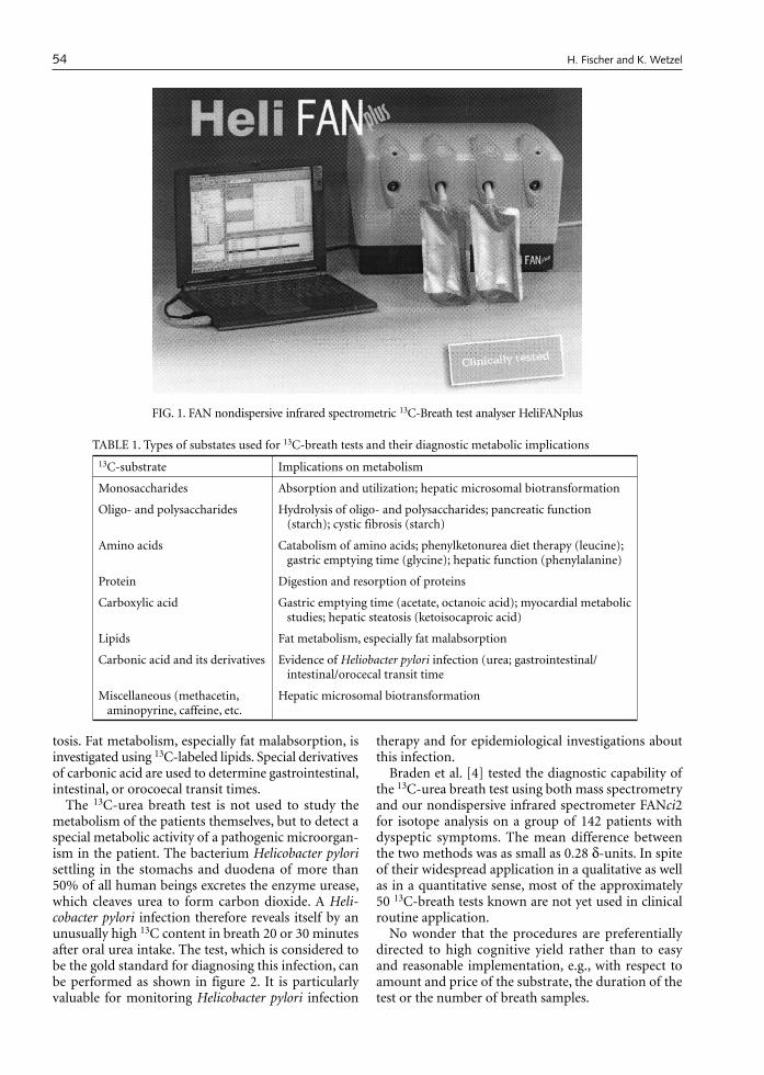
Figure 1 demonstrates one of the two nondispersive infrared spectrometric devices we have developed and now produce for carrying out 13C-breath tests. The compact device is particularly attractive for medical and laboratory practices and small hospitals. The white box on the right hand side contains the optical system with its cuvettes, infrared sources and infrared detec-tors, and the gas management system. The bags with the breath samples are attached to one of four connect-ing pieces in front of the box. The computer on the left side is connected to the analyzer.
13C-breath tests are good tools to use to investigate metabolic processes and for diagnosing metabolic and infectious diseases (table 1). The left column specifies the types of substrates used for 13C-breath tests and the right one the metabolic implications, which can be achieved by such tests.
Monosaccharides labeled with 13C can be used to investigate absorption and utilization of monosaccha-rides and hepatic microsomal biotransformation. Some oligo- and polysaccharides are used for exploring their hydrolysis and for studying pancreatic function and cystic fibrosis. Certain amino acids labeled with 13C are valid for investigating their catabolism, for controlling phenylketonuria diet-therapy, for measuring gastric emptying, and for studying hepatic function. Diges-tion and absorption of proteins is explored using 13C-labeled proteins. Certain carboxylic acids labeled with 13C are can be used to measure gastric emptying and to study myocardial metabolic processes and hepatic stea-
Heinz Fischer and Klaus Wetzel
54 55H. Fischer and K. Wetzel The future of 13C-breath tests
tosis. Fat metabolism, especially fat malabsorption, is investigated using 13C-labeled lipids. Special derivatives of carbonic acid are used to determine gastrointestinal, intestinal, or orocoecal transit times.
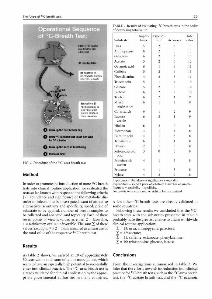
The 13C-urea breath test is not used to study the metabolism of the patients themselves, but to detect a special metabolic activity of a pathogenic microorgan-ism in the patient. The bacterium Helicobacter pylori settling in the stomachs and duodena of more than 50% of all human beings excretes the enzyme urease, which cleaves urea to form carbon dioxide. A Heli-cobacter pylori infection therefore reveals itself by an unusually high 13C content in breath 20 or 30 minutes after oral urea intake. The test, which is considered to be the gold standard for diagnosing this infection, can be performed as shown in figure 2. It is particularly valuable for monitoring Helicobacter pylori infection
therapy and for epidemiological investigations about this infection.
Braden et al. [4] tested the diagnostic capability of the 13C-urea breath test using both mass spectrometry and our nondispersive infrared spectrometer FANci2 for isotope analysis on a group of 142 patients with dyspeptic symptoms. The mean difference between the two methods was as small as 0.28 δ-units. In spite of their widespread application in a qualitative as well as in a quantitative sense, most of the approximately 50 13C-breath tests known are not yet used in clinical routine application.
No wonder that the procedures are preferentially directed to high cognitive yield rather than to easy and reasonable implementation, e.g., with respect to amount and price of the substrate, the duration of the test or the number of breath samples.
TABLE 1. Types of substates used for 13C-breath tests and their diagnostic metabolic implications
13C-substrate Implications on metabolism
Monosaccharides Absorption and utilization; hepatic microsomal biotransformation
Oligo- and polysaccharides Hydrolysis of oligo- and polysaccharides; pancreatic function (starch); cystic fibrosis (starch)
Amino acids Catabolism of amino acids; phenylketonurea diet therapy (leucine); gastric emptying time (glycine); hepatic function (phenylalanine)
Protein Digestion and resorption of proteins
Carboxylic acid Gastric emptying time (acetate, octanoic acid); myocardial metabolic studies; hepatic steatosis (ketoisocaproic acid)
Lipids Fat metabolism, especially fat malabsorption
Carbonic acid and its derivatives Evidence of Heliobacter pylori infection (urea; gastrointestinal/ intestinal/orocecal transit time
Miscellaneous (methacetin, aminopyrine, caffeine, etc.
Hepatic microsomal biotransformation
FIG. 1. FAN nondispersive infrared spectrometric 13C-Breath test analyser HeliFANplus
54 55H. Fischer and K. Wetzel The future of 13C-breath tests
Method
In order to promote the introduction of more 13C-breath tests into clinical routine application we evaluated the tests so far known with respect to the following criteria [5]: abundance and significance of the metabolic dis-order or infection to be investigated, want of attractive alternatives, sensitivity and specificity, speed, price of substrate to be applied, number of breath samples to be collected and analyzed, and topicality. Each of these seven points of view is valued as either 2 = favorable, 1 = satisfactory, or 0 = unfavorable. The sum ∑ of these values, i.e., up to 7 × 2 = 14, is assumed as a measure of the total value of the respective 13C-breath test.
Results
As table 2 shows, we arrived at 10 of approximately 50 tests with a total sum of ten or more points, which seem to have an especially high potential to successfully enter into clinical practice. The 13C-urea breath test is already validated for clinical application by the appro-priate governmental authorities in many countries.
A few other 13C-breath tests are already validated in some countries.
Following these results we concluded that the 13C-breath tests with the substrates presented in table 3 probably have the greatest chance to attain worldwide clinical routine application:
∑ = 13: urea, aminopyrine, galactose;∑ = 12: acetate;∑ = 11: caffeine, octanoate, phenylalanine;∑ = 10: trioctanoine, glucose, lactose.
Conclusions
From the investigations summarized in table 3. We infer that the efforts towards introduction into clinical practice for 13C-breath tests, such as the 13C-urea breath test, the 13C-acetate breath test, and the 13C-octanoic
TABLE 2. Results of evaluating 13C-breath tests in the order of decreasing total value
SubstrateImpor-tance
Expendi-ture Accuracy
Total value
Urea 5 2 6 13
Aminopyrine 6 2 5 13
Galactose 6 2 5 13
Acetate 5 2 5 12
Octanoic acid 6 1 4 11
Caffeine 5 2 4 11
Phenylalanine 4 2 5 11
Trioctanoin 5 1 4 10
Glucose 5 2 3 10
Lactose 6 2 2 10
Triolein 6 2 1 9
Mixed triglyceride
6 1 2 9
Corn starch 6 1 2 9
Lactose ureide
6 2 1 9
Hiolein 6 2 0 8
Bicarbonate 3 1 4 8
Palmitic acid 4 1 3 8
Tripalmitin 6 1 1 8
Ethanol 2 1 5 8
Ketoisocaproic acid
3 0 5 8
Protein-rich matter
4 1 3 8
Fructose 4 1 3 8
Xylose 4 2 2 8
Importance = abundance + significance + topicality.Expenditure = speed + price of substrate + number of samples.Accuracy = sensibility + specificity.For brevity tests with a sum or eight or less are omitted.
FIG. 2. Procedure of the 13C-urea breath test
56 H. Fischer and K. Wetzel
acid breath test should be continued and extended at least to the other above mentioned tests, particularly to the 13C-aminopyrine and the 13C-galactose breath test (∑ = 13).
Such efforts should be preferentially directed towards:» Simplifying the procedure, especially diminishing the
number of breath samples without noticeable loss of sensitivity and specificity.
» Enhancing of sensitivity and specificity, especially by improving the attending circumstances of the test procedure (e.g., suitable diet before and during the test). According to our considerations this could be helpful for introducing 13C-breath tests with ami-nopyrine, galactose, caffeine, glucose, phenylalanine, octanoic acid, lactose, and cholesteryloctanoate into clinical practice.
» Minimizing the tracer amount to be applied without noticeable loss of sensitivity and specificity.
» Shortening the duration of the test. This could be preferentially advantageous for introducing 13C-ami-nopyrine, 13C-lactose, and 13C-cholesteryloctanoate breath test into clinical routine application.Combination of any 13C-breath test with tests
for measuring gastric emptying times, in order to eliminate the effect of gastric emptying on substrate metabolism.
Many other substrates for 13C-breath tests will be found which make use of such a fascinating and simple way to obtain and measure samples for char-acterizing metabolic processes in humans. Foods, their constituents, like amino acids, carbohydrates, fatty acids, or triglycerides as well as xenobiotics, especially pharmaceuticals, will be used as substrates of 13C-breath tests.
TABLE 3. Substrates of 13C-breath tests for potential use in routine clinical applications and their diagnostic and meta-bolic implications
Substrate Diagnostic capability
Gastric and duodenic diseases
Octanoic acid Gastric emptying of solids
Acetate Gastric emptying of liquids
Urea Helicobacter pylori infection
Pancreatic diseases
Trioctanoin Fat metabolism; cystic fibrosis; steatorrhea
Liver diseases
Aminopyrine Activity of microsomal monoxygenases
Caffeine Hepatic microsomal biotransformation
Galactose Liver fibrosis in chronic hepatitis B
Glucose Glucose absorption and utilization, especially in children and diabetes mellitus patients
Phenylalanine Hepatocyte functional capacity; cytosolic enzyme activity
Intestinal diseases
Lactose Lactase deficiency; lactose assimilation
References
1. Krumbiegel P. Stable isotope pharmaceuticals for clini-cal research and diagnosis. Jena: Verlag Gustav Fischer, 1991:57–72.
2. Graham KS, Graham DY. Contemporary diagnosis and management of Helicobacter pylori associated gastroin-testinal diseases. Newtown, Penn., USA: Handbooks in Health Care Co., 1998.
3. Wetzel K, Fischer H. Recent results of the development and application of 13C-breath tests. Leipzig: Fischer ANalysen Instrumente GmbH, 1999.
4. Braden B, Caspary WF, Lemcke B. Nondispersive Infra-red Spectrometry for 13CO2/12CO2-measurements: A clinically feasible analyser for stable isotope breath tests in gastroenterology. Zeitschrift für Gastroenterologie 1999;37:477–81.
5. Wetzel K, Fischer H. 13C-breath tests in medical research and clinical diagnosis. Leipzig: Fischer ANalysen Instru-mente GmbH, 2001.
Food and Nutrition Bulletin, vol. 23, no. 3 (supplement) © 2002, The United Nations University. 57
Activable enriched stable isotope iron-58 for monitoring absorption rate of juvenile athletes for iron: a case study
Qinfang Qian, Zhifang Chai, Weiyu Feng, and Peiqun Zhang are affiliated with the Laboratory of Nuclear Analytical Techniques in the Institute of High Energy Physics, Chinese Academy of Sciences in Beijing, China. Jidi Chen and Jianx-iang Pan are affiliated with the Institute of Sports Medicine at Peking University in Beijing.
Qinfang Qian, Zhifang Chai, Weiyu Feng, Jidi Chen, Peiqun Zhang, and Jianxiang Pan
Abstract
Activable enriched stable isotopes can play a unique role in studies of nutritional status, metabolism, absorption rates, and bioavailability of minerals. As a practical example, eight juvenile athletes were selected to test the absorption rates of iron during training and non-training periods by enriched stable isotope of Fe-58 (enriched degree: 51.1%) via activation analysis Fe-58 (n,γ) Fe-59 of the collected feces samples. The results indicated that the average iron absorption rates of the juvenile athletes with and without training are 9.1 ± 2.8 and 11.9 ± 4.7%, respectively, which implies that the long-term endurance training with high intensity makes the iron absorption rate of athletes lower. In the meantime, the comparison of the activable enriched isotope technique with atomic absorption spectrometry was performed, which showed that the former was better than the latter in reliability and sensitivity. It is because this nuclear method can distinguish the exogenous and endogenous iron in the samples, but not for non-nuclear methods.
Key words: activable stable isotope Fe-58, absorption rate of iron, juvenile athlete, neutron activation analysis, atomic absorption spectroscopy
Introduction
Activable enriched stable isotope technique (AESIT) is one of the isotopic tracer techniques. Compared with a relatively common radioactive isotope tracing, it pos-sesses the following merits and drawbacks [1–3]. First, there is no radiation damage. Thus, it is especially suit-
able for the radiation-sensitive population, e.g., chil-dren, pregnant women, elderly, and immuno-deficient subgroups. Second, there is no radiolysis effect, which makes it possible to label the metalloorganic macro-molecules, e.g., metalloproteins and metalloenzymes, with long-term stability. However, the determination of stable isotopes is more difficult than radioactivity counting. Thus, the cost for AESIT is higher than for radioactive tracers.
AESIT has been widely used to study the nutritional status, metabolism, absorption rate, and bioavailability of minerals in organisms [4–8]. In many cases it is not replaceable by other techniques, even by radioactive isotope tracing.
Anemia is one of the common epidemic diseases caused by nutrient deficiency and other factors. The proportion of this disease is 10% to 30% in industrial-ized countries and 30% to 90% in developing coun-tries [9]. Many athletes suffer this disease [10], which severely affects their sport levels. Long-term endur-ance training with high intensity leads to numerous changes in the normal physiological and biochemical functions of athletes’ bodies. One of the symptoms is iron-deficiency anemia. In the study of anemia, the absorption rate of iron is one of the important indica-tors. Although some non-nuclear methods (like atomic absorption spectroscopy, AAS) have been developed to determine the absorption rate of iron, their reliability is skeptical. These methods cannot distinguish the endogenous or exogenous iron in the human body. We used the enriched stable isotope Fe-58 to determine the absorption rate of juvenile athletes for iron via the activation reaction of Fe-58 (n,γ) Fe-59, which is easily detected by gamma counting of the radioactivity of the activation product Fe-59.
Experimental
In AESIT some general guidelines have to be followed. The natural abundance of the selected stable isotope should be as low as possible, at least below 10 %. The

58 59Q. Qian et al. Activable enriched stable isotope iron-58
enriched stable isotope should be easily prepared and at low cost, and the enriched abundance should exceed its natural one at least by a factor of 10. The cross-section of neutron activation reaction of this isotope should be high; and the activation product should be easily detected with proper half-life and gamma radiation energy. The stable isotope Fe-58 was selected to moni-tor the absorption rate of iron because of its proper nuclear properties—its natural abundance –0.31%; the cross-section of activation reaction Fe-58 (n,γ) Fe-59 –0.98 barn; the half-life of Fe-59–45 days and its main gamma ray energy—1099 and 1291 keV.
Subjects
Eight 15 to 18 year-old normal juvenile athletes were selected from the Beijing Si-Sa-Hai Sport School. There iron absorption rates were determined twice, once during the training period and another time during a non-training period. Each period lasted two weeks. Their diets were strictly controlled and recorded during the whole study. The subjects were not allowed to con-sume any extra food. This experiment was approved by the Ministry of Hygiene and all the subjects signed a consent form.
Determination method of absorption rate of iron by AESIT of Fe-58
The enriched stable isotopes of Fe-58 (enriched degree 51.1%) were purchased from Chinese Institute of Atomic Energy, Beijing, as a chemical species of Fe2O3. A bit of hydrochloric acid was used to dissolve the Fe2O3, which was then diluted with deionized water.
On the 8th or 9th day of the experiment, the diluted Fe-58 solution was orally administered to the subjets. In order to define the excretion time of the feces labeled by Fe-58 for five days, coccinellin capsules were taken by the subjects with supper on the first day and with breakfast on the sixth day after administration of Fe-58.
Sample diets were collected for five days after admin-istration of Fe-58 and homogenized in an agitator with an appropriate amount of deionized water. The iron content in the diets was analyzed by atomic absorption spectroscopy (AAS) and the iron intake for each subject was then estimated.
After administration of Fe-58 the feces samples were collected when the coccinellin first appeared and continued until the coccinellin again appeared. The feces samples were homogenized with an appropriate amount of a solution of ethanol and sulfuric acid. A big portion of the samples was weighed and ashed at 480°C for 24 hours in a muffle furnace; 100 mg of the ashed feces samples were packaged in high pure aluminum
foil for irradiation in a nuclear reactor.Four portions of 0.1 ml oral Fe-58 solution each were
pipetted into clean polyethylene film, dried under an infrared lamp, and finally packaged using filter paper and aluminum foil for irradiation.
The above samples (feces and oral Fe-58 solution) were irradiated at neutron flux of 2.3 × 10¹³ n/cm2.s for 80 hours. After cooling for 30 days, the radioactiv-ity of Fe-59 in the samples was counted by high pure germanium detector connected to Ortec multichannel analyzer (USA). The peak areas at 1099 and 1291 keV were taken for calculation of Fe-58 content in the samples.
Calculation of absorption rate of iron
The absorption rate of iron in the athletes’ bodies was calculated by the following formula :
Absorption rate of iron (%) = (total intake amount of Fe-58 – total excretion amount of Fe-58 in feces)/
total intake amount of Fe-58 × 100%.
The total intake amount of Fe-58 includes that from the added Fe-58 solution and that from the diet, which contains natural Fe-58. The latter can be calcu-lated by the iron content in the diet multiplied by its natural abundance 0.3 %. The total amount of Fe-58 excreted in the feces included that from the fraction of Fe-58 non-absorbed by the gastrointestinal tract after its administration and that from the natural Fe-58 excreted. The latter can be neglected in comparison with the former. Thus, the total Fe-58 amount in feces represents the non-absorbed Fe-58 amount.
In addition to AESIT, AAS was also used to deter-mine the absorption rate of iron in these athletes by a Type GGX-5 atomic absorption spectrometer. It used a 5 mA lamp currency, 2483 nm wavelength, 0.1 nm slit; combustion-supporting gas 6 L/minute; and acetylene 1.6 L/minute. The absorption rate of iron was estimated on the basis of the balance method.
Results and discussion
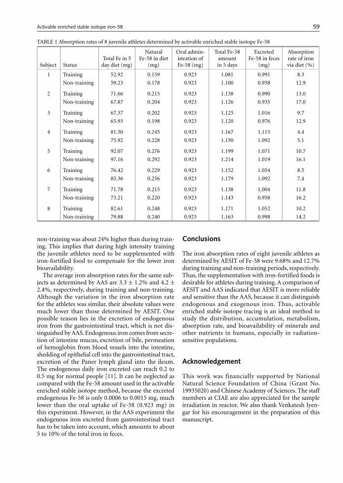
The iron absorption rates for 8 juvenile athletes during training and non-training periods as determined by the enriched stable isotope Fe-58 are listed in table 1. The iron absorption rates during training varied from 4.4T to 13.0 % with an average of 9.58 ± 2.90%, and were much lower than those during the non-training period which ranged from 5.1% to 17.0% with an average of 12.7 ± 4.7%. All subjects, except one, showed a higher absorption rate of iron during training than during non-training. The statistical t-test indicates a signifi-cant difference (p < .05) between them. On average the iron absorption rate of the juvenile athletes during
58 59
TABLE 1 Absorption rates of 8 juvenile athletes determined by activable enriched stable isotope Fe-58
Subject StatusTotal Fe in 5
day diet (mg)
Natural Fe-58 in diet
(mg)
Oral admin-istration of Fe-58 (mg)
Total Fe-58 amount in 5 days
Excreted Fe-58 in feces
(mg)
Absorption rate of iron via diet (%)
1 Training 52.92 0.159 0.923 1.081 0.991 8.3
Non-training 59.23 0.178 0.923 1.100 0.958 12.9
2 Training 71.66 0.215 0.923 1.138 0.990 13.0
Non-training 67.87 0.204 0.923 1.126 0.935 17.0
3 Training 67.37 0.202 0.923 1.125 1.016 9.7
Non-training 65.93 0.198 0.923 1.120 0.976 12.9
4 Training 81.50 0.245 0.923 1.167 1.115 4.4
Non-training 75.92 0.228 0.923 1.150 1.092 5.1
5 Training 92.07 0.276 0.923 1.199 1.071 10.7
Non-training 97.16 0.292 0.923 1.214 1.019 16.1
6 Training 76.42 0.229 0.923 1.152 1.054 8.5
Non-training 85.36 0.256 0.923 1.179 1.092 7.4
7 Training 71.78 0.215 0.923 1.138 1.004 11.8
Non-training 73.21 0.220 0.923 1.143 0.958 16.2
8 Training 82.61 0.248 0.923 1.171 1.052 10.2
Non-training 79.88 0.240 0.923 1.163 0.998 14.2
Q. Qian et al. Activable enriched stable isotope iron-58
non-training was about 24% higher than during train-ing. This implies that during high intensity training the juvenile athletes need to be supplemented with iron-fortified food to compensate for the lower iron bioavailability.
The average iron absorption rates for the same sub-jects as determined by AAS are 3.3 ± 1.2% and 4.2 ± 2.4%, respectively, during training and non-training. Although the variation in the iron absorption rate for the athletes was similar, their absolute values were much lower than those determined by AESIT. One possible reason lies in the excretion of endogenous iron from the gastrointestinal tract, which is not dis-tinguished by AAS. Endogenous iron comes from secre-tion of intestine mucus, excretion of bile, permeation of hemoglobin from blood vessels into the intestine, shedding of epithelial cell into the gastrointestinal tract, excretion of the Paner lymph gland into the ileum. The endogenous daily iron excreted can reach 0.2 to 0.5 mg for normal people [11]. It can be neglected as compared with the Fe-58 amount used in the activable enriched stable isotope method, because the excreted endogenous Fe-58 is only 0.0006 to 0.0015 mg, much lower than the oral uptake of Fe-58 (0.923 mg) in this experiment. However, in the AAS experiment the endogenous iron excreted from gastrointestinal tract has to be taken into account, which amounts to about 5 to 10% of the total iron in feces.
Conclusions
The iron absorption rates of eight juvenile athletes as determined by AESIT of Fe-58 were 9.68% and 12.7% during training and non-training periods, respectively. Thus, the supplementation with iron-fortified foods is desirable for athletes during training. A comparison of AESIT and AAS indicated that AESIT is more reliable and sensitive than the AAS, because it can distinguish endogenous and exogenous iron. Thus, activable enriched stable isotope tracing is an ideal method to study the distribution, accumulation, metabolism, absorption rate, and bioavailability of minerals and other nutrients in humans, especially in radiation-sensitive populations.
Acknowledgement
This work was financially supported by National Natural Science Foundation of China (Grant No. 19935020) and Chinese Academy of Sciences. The staff members at CIAE are also appreciated for the sample irradiation in reactor. We also thank Venkatesh Iyen-gar for his encouragement in the preparation of this manuscript.
60 Q. Qian et al.
References
1. Roth E. Critical evaluation of the use and analysis of stable isotopes. Pure Appl Chem 1997;69:1753–1828.
2. Chai ZF. Introduction to activation analysis. (in Chi-nese) Beijing: Atomic Energy Press, 1980.
3. Chai ZF. Applications of neutron activation analysis in environmental science, biology and geoscience. Beijing: Atomic Energy Press, 1992.
4. Feng WY, Ding WJ, Qian GF, Chai ZF. Comparison of the chromium distribution in organs and subcellular fractions of normal and diabetic rats by using enriched stable isotope Cr-50 trace technique. Biol Trace Elem Res 1999;71-72:121–9.
5. Yu SY, Zhang Z, Yang FQ. Determination of absorption rate of children with anemia for iron in diets by stable isotope Fe-58. Chin J Nutr 1986; 8:193–6.
6. Feng WY, Ding WJ, Qian QF, Chai ZF. Study on the metabolism of physiological amounts of Cr(III) intra-gastrical administration in normal rats using activable enriched stable isotope Cr-50 compound as a tracer. J Radioanal Nucl Chem 1998;247:15–19.
7. Feng WY, Qian QF, Ding WJ, Chai ZF. Study of chro-mium speciation in normal and diabetic rats by activa-ble enriched stable isotope technique. J Radioanal Nucl Chem 2000;244:321–5.
8. Chai ZF. Modern nuclear analytical techniques and their applications in China. J Nucl Radiochem Sci 2000;1:19–22.
9. Liao QG. Research progress of iron deficiency-related symptoms. J Intern Med 1988;11:97–101
10. Clement DB, Sawchuk LL. Iron status and sport per-formance. Sports Med 1984;1:65–70.
11. Kong XR. Nutritional, physiological and clinical sig-nificance of essential trace elements. Hefei: Science & Technological Press, 1982.
12. Qian QF, Feng WY, Chen JD, Zhang PQ, Chai ZF, Pan JX, Wu YY, Zheng SQ, Liu XP, Chao ZY. Study of the effect of supplementation of iron-fortified food to Chinese juvenile athletes by nuclear analysis techniques and blood analysis. J Radioanal Nucl Chem Letters 1996; 212: 51–9.