the frail patient with heart disease: care! · the frail patient with heart disease: ... n....
TRANSCRIPT

19 V
A 0
347
BA
The frail patient with heart disease: please handle with care!
76
Number 76 - July 2018

Heart and Metabolism
Heart and Metabolism
Editor in Chief
Editorial Board
Published by
Publication Director
Scientific coordinator
Correspondence to
Aim and Scope
Website
Heart and Metabolism
DesignLayout
Printed
© 2018 by
ISSN
a Servier publication
Mario Marzilli, MD, PhD, Italy
Luis Henrique W. Gowdak, MD, PhD, BrazilDerek J. Hausenloy, PhD, UKGary D. Lopaschuk, PhD, CanadaMichael Marber, MB (BS), PhD, UK
Les Laboratoires Servier
Philippe Gonnard, MD
Sophie [email protected]
Sherri [email protected]
Heart and Metabolism is a journal published three times a year, focusing on the management of cardiovascular diseases. Its aim is to inform cardiologists and other specialists about the newest findings on the role of metabolism in cardiac disease and to explore their potential clinical implications. Each issue includes an editorial, followed by articles on a key topic. Experts in the field explain the metabolic consequences of cardiac disease and the multiple potential targets for phar-macotherapy in ischemic and nonischemic heart disease.
www.heartandmetabolism.com
is indexed in EMBASE, SCOPUS, and PASCAL/INIST-CNRS.
Studio DTC - ServierBleu Banquise
in France
Les Laboratoires Servier
1566-0338
All rights reserved throughout the world and in all languages. No part of this publication may be reproduced, transmitted, or stored in any form or by any means either mechanical or electronic, including photocopying, recording, or through an information storage and retrieval system, without the written permission of the copyright holder.Opinions expressed do not necessarily reflect the views of the publishers, editors, or editorial board. The authors, edi-tors, and publishers cannot be held responsible for errors or for any consequences arising from the use of the information contained in this journal.
00-00-0000 / PEFC Certified / This publication is from sustainably managed forests and controlled sources. / www.pefc.org

Glossary Gary d. lopaschuk
Mini-Mental Status Examination (MMSE) The MMSE is a screening instrument/questionnaire used to assess mental status by testing five areas of cognitive function: orientation, registration, attention and calculation, recall, and language. The maximum score is 30, while a score of 23 or less is indicative of cognitive impairment.
Montreal Cognitive Assessment (MoCA)The MoCA is a screening instrument designed to detect mild cognitive impairment in patients scoring in the normal range of the MMSE. Multiple cogni-tive domains are assessed with the MoCA, including short-term memory; visuospatial abilities; executive function; attention, concentration, and working me-mory; language; and orientation to time and place. A clinical cut-off score of 26 is indicative of mild cogni-tive impairment.
SarcopeniaSarcopenia is a condition involving a degenerative loss of skeletal muscle mass of ≈0.5% to 1% per year (once an individual reaches 50 years of age). It is therefore characterized by muscle atrophy in addition to a reduction in muscle tissue quality. Sarcopenia is frequently associated with both cachexia and frailty syndrome.
Society of Thoracic Surgeons scoreThe STS score is an American risk score toolset (the European equivalent is the EuroSCORE) used to pre-dict operative mortality of adult cardiac surgery within 30 days of the operation or later if the patient remains hospitalized.
Transcatheter aortic valve implantationTAVI is a surgical procedure that involves a small inci-sion in the chest, following which a catheter is inser-ted through the groin into a large blood vessel for im-planting an aortic valve (usually made of natural tissue from either a cow or a pig) over an individual’s existing aortic valve. The catheter is removed once the new valve is implanted and will start working immediately.
Heart Metab. (2018) 76:40-41

Contents
EDITORIALThe frail patient with heart disease: please handle with care! . . . . . . . . . . . . . . . . . . . . . . . . . . . . . . . . . . . . . . . . . . 2D. Hausenloy
ORIGINAL ARTICLESThe frail patient with heart disease: an emerging and challenging issue . . . . . . . . . . . . . . . . . . . . . . . . . 4N. Veronese
Frailty, heart failure, and cognitive impairment: a triangle in elderly people . . . . . . . . . . . . . . . . . . . . . 8K. Shinmura
Aortic stenosis in the frail patient: maximizing the benefit of TAVI . . . . . . . . . . . . . . . . . . . . . . . . . . . . . . . . . . .13A. Anand, N. L. Mills
Cardiac surgery in the frail patient: managing the increased risks . . . . . . . . . . . . . . . . . . . . . . . . . . . . . . . . 18L. Y. Koh, N. C. Hwang
Trimetazidine in the frail patient . . . . . . . . . . . . . . . . . . . . . . . . . . . . . . . . . . . . . . . . . . . . . . . . . . . . . . . . . . . . . . . . . . . . . . . . . . . . . . . . . . . . . 23C. Vitale, M. Fini, G. M. C. Rosano
CASE REPORT Managing the frail patient undergoing a percutaneous coronary intervention . . . . . . . . . . . . . . . . 27L. Candilio
REFRESHER CORNEREnergy metabolism in the aged heart . . . . . . . . . . . . . . . . . . . . . . . . . . . . . . . . . . . . . . . . . . . . . . . . . . . . . . . . . . . . . . . . . . . . . . . . . . . . . . 32G. D. Lopaschuk
HOT TOPICSExercise and diet for heart disease in the frail patient: fact or fiction? . . . . . . . . . . . . . . . . . . . . . . . . . . 36S. Nanayakkara, D. M. Kaye
GLOSSARY . . . . . . . . . . . . . . . . . . . . . . . . . . . . . . . . . . . . . . . . . . . . . . . . . . . . . . . . . . . . . . . . . . . . . . . . . . . . . . . . . . . . . . . . . . . . . . . . . . . . . . . . . . . . . . . . . . . . . 40G. D. Lopaschuk
1

Heart Metab. (2018) 76:2-3Editorial - derek Hausenloy
Improvements in life expectancy in our aging society have resulted in an increased prevalence of frailty in patients with ischemic heart disease (IHD), heart
failure (HF), and cardiac valve disease. Frailty, which has been defined as a state of increased vulnerability to external stressors and decreased physiological reserve, has important implications on the manage-ment of these conditions in elderly patients – in particular, its’ presence is known to be predictive of worse clinical outcomes. In this issue of Heart and Metabolism, strategies for improving the detection and management of frail patients with cardiovascular disease (CVD) are discussed. In the opening article, Dr Nicola Veronese explores the challenges facing the detection and management of the frail patient with CVD. The close interplay be-tween frailty and CVD is highlighted, with frailty be-ing a risk factor for developing CVD and vice versa. This can be explained in part by the shared etiological factors between these two conditions, including low-grade inflammation, cellular senescence, and endo-crine dysregulation, among others. In the next article, Dr Ken Shinmura explores the triangular relationship between frailty, HF, and cognitive impairment. In at least one-quarter of elderly patients, HF is complicat-ed by both physical frailty and cognitive impairment. The pathophysiology underlying HF directly contrib-utes to frailty by reducing exercise capacity and skel-etal muscle function. Furthermore, patients with HF are more susceptible to cognitive impairment, which
accelerates the development of physical frailty and HF, resulting in a vicious cycle. Crucially, there are no standardized screening tools for cognitive impair-ment in patients with HF, and there is an incomplete understanding of the complex relationships between frailty, HF, and cognitive impairment. Strategies that increase cardiac output, such as exercise have been shown to increase cognitive function and may be used as a therapeutic intervention in frail patients with HF. Transcatheter aortic valve implantation (TAVI) is an increasingly common intervention for older patients with aortic stenosis who are at risk of complications from major cardiac surgery. The challenge has been to have objective and reproducible physical frailty measures that can be used to identify patients at the very highest risk of early mortality or worsening dis-ability after TAVI. In their article, Drs Atul Anand and Nicholas L. Mills, discuss several frailty measures that may be used to help assess risk in older patients with aortic stenosis and guide patient selection for TAVI in order to maximize the benefit of treatment. In the fol-lowing article, Drs Li Ying Koh and Nian Chih Hwang provide an anesthetists’ perspective on managing frail patients with coronary artery disease undergo-ing cardiac surgery who are at risk of experiencing worse clinical outcomes postsurgery. Conventional preoperative risk scores do not take into account the increased physiological vulnerability of the frail pa-tient; therefore, frailty-specific risk scores are needed
The frail patient with heart disease: please handle with care!Derek Hausenloy, MD, PhD, FACC, FESCCardiovascular and Metabolic Disorders Program, Duke-NUS Graduate Medical School, Singapore; National Heart Research Institute Singapore, National Heart Centre Singapore; The Hatter Cardiovascular Institute, Institute of Cardiovascular Science, University College London, UK; National Institute of Health Research University Col-lege London Hospitals Biomedical Research Centre, London, UK
Correspondence: Professor Derek Hausenloy, Cardiovascular and Metabolic Disorders Program, Duke-NUS Graduate Medical School Singapore, 8 College Road, Singapore 169857E-mail: [email protected]
2

3
Heart Metab. (2018) 76:2-3 editorial - Hausenloy
The frail patient with heart disease: please handle with care!
to improve preoperative risk assessment in this set-ting. The issue of cardiovascular pharmacotherapy in frail patients with IHD is addressed in the article by Drs Cristiana Vitale, Massimo Fini, and Giuseppe Rosano. Certain cardiovascular drugs are known to impair quality of life and functional capacity in frail pa-tients with CVD. In this regard, β-blockers are associ-ated with an increased risk of cognitive decline and a reduced ability to independently perform activities of daily living, and ivabradine, which is well tolerated in frail elderly patients, may provide an alternative thera-py. Conversely, drugs known to have a positive effect on functional capacity and quality of life in frails pa-tients can be considered. For example, the antiangi-nal agent trimetazidine has been shown to improve myocardial ischemia, exercise capacity, quality of life, and prognosis in elderly patients. Frailty has been associated with worse clinical out-comes following acute coronary syndrome and PCI. Therefore, frailty status should be taken into careful consideration when treatment strategies are planned. In this issue’s case report, Dr Luciano Candilio de-scribes the case of an 87-year-old patient undergo-ing PCI for non–ST-segment elevation myocardial in-farction and evidence of multivessel coronary artery disease, who had significant comorbidities, including chronic kidney impairment, peripheral artery disease, and chronic obstructive pulmonary disease. His frailty status was carefully evaluated and the risks and ben-efits of potential management strategies were taken into account by his heart team. He underwent a suc-cessful staged PCI to his left main stem and right
coronary artery with chronic total occlusion, and he reported no symptoms on a subsequent follow-up and a significantly improved quality of life. In the refresher corner, Dr Gary D. Lopaschuk reviews the changes in cardiac metabolism in the aged heart, and, subsequently, how these alterations in energy production can compromise the ability of the heart to adapt to stresses requiring an increase in energy demand. Therefore, improving both cardiac energy production and the efficiency of energy pro-duction may be a novel therapeutic strategy to lessen cardiac disease in the elderly. Finally, in the hot topics article, Drs Shane Nanay-akkara and David M. Kaye review the role of diet and exercise as potential strategies for improving health outcomes in frail patients with CVD. In HF patients with frailty, it has been shown that exercise reduces rehospitalization rates, improves quality of life, and is a cost-effective intervention. Although frailty and nutritional status are closely linked, the evidence for dietary modification, such as increased protein intake or vitamin supplementation, has produced mixed re-sults. Further studies are needed to investigate the efficacy of therapeutic strategies for improving health outcomes in frail patients with CVD. In summary, this issue of Heart and Metabolism highlights the challenges in detecting and managing frail patients with IHD, HF, and cardiac valve disease. Much more work is needed to improve frailty-specific risk assessment so management can be personal-ized to aged patients in order to improve health out-comes in this increasingly important patient group. L

4
Introduction
Frailty has been traditionally defined as “reduced physiological reserve and increased vulner-ability for poor resolution of homeostasis after
a stressor event.”1 It is a common condition in older people, affecting about one person in ten.2 The prev-alence of frailty in people affected by cardiovascular disease (CVD) is higher than in those without CVD,3 but increasing numbers of studies suggest that the relationship between frailty and CVD is closely inter-twined, ie, frail people are at an increased risk of CVD
and vice versa.4 Finally, the presence of frailty in pa-tients with CVD (and vice versa) appears to have im-portant prognostic implications.5 This article provides an overview of the current evidence regarding frailty, CVD, and their coexistence in terms of the underly-ing pathophysiology and their impact on clinical out-comes.
Epidemiological research regarding frailty and CVD
From an epidemiological point of view, frailty and CVD are strongly associated. Traditional CVD risk
The frail patient with heart disease: an emerging and challenging issue
Nicola Veronese, MDNational Research Council, Neuroscience Institute, Aging Branch, Padua, Italy
Correspondence: Nicola Veronese, MD; National Research Council, Neuroscience Institute, Aging Branch, Via Giustiniani, 2 - 35128 Padua, Italy
E-mail: [email protected]
AbstractFrailty and cardiovascular disease (CVD) are two common conditions that may affect clinical outcomes in older people. Epidemiological studies suggest that CVD is one of the most important contributor to the development of frailty in the aged patient, and the latter can therefore be considered a potential CVD risk factor. Moreover, traditional CVD risk factors are also known to be important for developing frailty. This close relationship between CVD and frailty is due, in part, to the shared etiological factors, which include low-grade inflammation, cellular senescence, and endocrine dysregulation. Therefore, the early detection of frailty is important in the management of patients with CVD or in those who are at a high risk of developing CVD. In this regard, the use of a comprehensive geriatric assessment (CGA) may be considered in these patients. Unfortunately, the literature available for the use of CGA is only based on observational data, which may be biased; therefore, future studies are needed to understand the true role of CGA for detecting frailty in patients with CVD. In this article, an overview is provided of the current evidence regarding frailty, CVD, and their coexistence in terms of the underlying pathophysiology and their impact on clinical outcomes. L Heart Metab. 2018;76:4-7
Keywords: cardiovascular disease; comprehensive geriatric assessment; frailty
Heart Metab. (2018) 76:4-7Original Article

5
Heart Metab. (2018) 76:4-7 Veronese
Frailty and cardiovascular disease
factors, namely diabetes, obesity, and a sedentary lifestyle, appear to be risk factors for developing frail-ty.6-8 Moreover, it has been reported that subclinical CVD parameters (eg, the presence of atherosclerotic plaques and higher carotid intima media thickness) are more commonly observed in frail subjects when compared with nonfrail patients.9 Finally, recent litera-ture has shown that CVD is one of the strongest risk factors for developing frailty4 and that the presence of frailty can increase the risk of developing CVD.10,11 When frailty coexists with CVD, the clinical implica-tions for older people are more impactful with clini-cal importance. Frailty has been associated with in-creased morbidity, mortality, and disability in patients affected by CVD undergoing cardiac or noncardiac procedures, although most of the research data have been limited by studies using small sample sizes and by a lack of randomized trials.12
Common pathophysiology underlying CVD and frailty
From a pathophysiological point of view, it has been reported that there are many molecular and cellular pathways in common between frailty and CVD (Fig-ure 1). First, low-grade inflammation is more common
in frail older patients when compared with less frail older patients,13 and low-grade inflammation is well-established to play a major role in the development of CVD.14 Second, frail patients are known to have cellular and intracellular alterations typical of cellular senescence (eg, marked DNA damage15 and shorter telomere length),16 the presence of which may con-
tribute to the development of CVD. Third, endocrine dysregulation that is present in frail patients (eg, lower insulin-like growth factor (IGF)-1 levels)17 can further increase the risk of CVD in patients affected by frailty. An important role may be played by insulin resistance, a key factor for developing CVD, which is more preva-lent in frail patients than in less frail patients.18
Importance of frailty in the management of older people with CVD
The topic of frailty in the context of CVD is of increas-ing importance to both geriatric medicine and cardiol-ogy. Appropriate and early intervention may help pre-vent the development of frailty in patients with CVD.19 Examples of these interventions include physical ex-ercise (particularly resistance training), nutrition, cog-nitive training, and medication review.20 Since frailty is traditionally defined by physical performance items, particular importance is given to physical exercise. Exercise seems to have a positive effect on various measures used to determine frailty (eg, cognition, physical functioning, and psycho-logical well-being) and some studies revealed that exercise may prevent or, at least, delay the onset of frailty.21 However, we do not know which type of physical exercise is best for preventing and treating frailty, since it is estimated that aerobic endurance training can improve peak oxygen consumption, but resistance training is the best way to increase muscle strength and mass.22 Probably, a combined interven-tion (both aerobic and resistance training) is the best way to treat frailty successfully.22
Diet is the other intervention for treating frailty. Most studies have shown that dietary supplements or improvements in dietary intake can improve fac-tors related to frailty, such as muscle strength, walk-ing speed in frail or prefrail older adults.23 However, nutritional interventions are probably not sufficient for treating/preventing frailty without physical exer-cise.24 Several interventions used for treating frailty are useful for reducing CVD risk25 and specific interven-tions, such as a heart transplant, are useful for re-versing frailty.26 Finally, frailty, particularly in its initial stages, can represent a window for appropriate inter-ventions, specifically lifestyle interventions, that may delay the onset of CVD and consequently reduce dis-ability, hospitalization, and mortality.27
AbbreviationsCGA: comprehensive geriatric assessment; CVD: car-diovascular disease
Frailty
In�ammation
Other CVDrisk factors
Cellularsenescence
Endocrinedysregulations
Fig. 1 Pathophysiological associations between frailty and cardio-vascular disease.

6
Veronese Heart Metab. (2018) 76:4-7Frailty and cardiovascular disease
Comprehensive geriatric assessment in patients with CVD
An increasing body of literature supports the impor-tance of a comprehensive geriatric assessment (CGA) in the management of medical conditions common in older individuals, eg, hip fractures.28 Therefore, it is likely that older patients with CVD may also ben-efit from a global and multidimensional geriatric ap-proach. The literature regarding this topic, which is limited to a few studies,29 has shown that older adults with low CGA scores had worse short- and long-term prognoses.29 To the best of our knowledge, only ob-servational studies are available, and, although they can provide important information, they may suffer from some biases. Moreover, these studies explored only mortality as an outcome, whereas other parame-ters of clinical importance, such as quality of life, were not included.29 CGA could be useful in the manage-ment of CVD for several reasons, particularly because interventions to prevent frailty may break the vicious cycle between frailty and CVD, which would improve global physiological reserve and consequently out-comes.30 Recent guidelines recommend the early recogni-tion of frailty in older patients to provide an estimate of prognosis and to avoid potentially ineffective and expensive medical interventions.31 For example, the European Society of Hypertension and the European Union Geriatric Medicine Society Working Group on the management of hypertension have suggested that, for frail subjects, therapeutic decisions should be preceded by: (i) obtaining accurate information on functional capacity and cognitive status; (ii) paying attention to multiple drug administration; (iii) stratify-ing the frailty status using one of the available rapid methods; and (iv) identifying and correcting factors or conditions that predispose patients to common and possibly severe adverse treatment effects.32
Therefore, the clinical approach to older patients affected by CVD cannot be limited to a traditional, purely cardiological paradigm, but should also con-sider the peculiarities of these syndromes, which also include common issues in the physical, psychosocial, and cognitive domains. Complex clinical pictures and highly unstable health trajectories distinguish older ill adults, for whom a traditional clinical approach that is only based on disease-specific guidelines can be misleading with regard to prognosis, resulting in poor
quality of care and negative outcomes.33 In this re-gard, physical performance assessment contributes to functional evaluation and provides important prog-nostic information in older patients affected by CVD.
Future directions
The research of the potential relationship between frailty and CVD is intriguing, but unfortunately, it is only based on observational data. A summary of the most important concepts are provide here:• FrailpatientswithnoclinicalevidenceofCVD:life-
style interventions (eg, physical exercise, nutrition-al interventions) might reduce the onset of CVD compared with standard care. The use of other common primary prevention interventions, such as low-dose aspirin, is still debated.34
• FrailpatientswithCVD:manyoftheinterventionsthat can be used for reversing frailty are probably useful for improving CVD outcomes, but more in-terventional research is needed.
• RoleofCGA:anintegratedmodelofcare,ashasbeen developed for orthogeriatrics, is probably the best approach to understand the role of a geriatri-cian in the treatment of frailty in patients affected by CVD and vice versa.
Conclusions
Frailty and CVD are two common conditions in older people. Increasing literature reports demonstrate an important interdependence between the presence of frailty and CVD. Interventional studies are needed to obtain a better understanding of the role of treating frailty to prevent CVD. L
REFERENCES
1. Clegg A, Young J, Iliffe S, Rikkert MO, Rockwood K. Frailty in elderly people. Lancet. 2013;381(9868):752-762.
2. Collard RM, Boter H, Schoevers RA, Oude Voshaar RC. Preva-lence of frailty in community-dwelling older persons: a system-atic review. J Am Geriatr Soc. 2012;60(8):1487-1492.
3. Afilalo J. Frailty in patients with cardiovascular disease: why, when, and how to measure. Curr Cardiovasc Risk Rep. 2011;5(5):467-472.
4. Afilalo J, Alexander KP, Mack MJ, et al. Frailty assessment in the cardiovascular care of older adults. J Am Coll Cardiol. 2014;63(8):747-762.
5. von Haehling S, Anker SD, Doehner W, Morley JE, Vellas B. Frailty and heart disease. Int J Cardiol. 2013;168(3):1745-1747.
6. Savela SL, Koistinen P, Stenholm S, et al. Leisure-time physical activity in midlife is related to old age frailty. J Gerontol A Biol

Sci Med Sci. 2013;68(11):1433-1438.7. Stenholm S, Strandberg TE, Pitkälä K, Sainio P, Heliövaara M,
Koskinen S. Midlife obesity and risk of frailty in old age during a 22-year follow-up in men and women: the mini-Finland follow-up survey. J Gerontol A Biol Sci Med Sci. 2014;69(1):73-78.
8. Veronese N, Stubbs B, Fontana L, et al. Frailty is associated with an increased risk of incident type 2 diabetes in the elderly. J Am Med Dir Assoc. 2016;17(10):902-907.
9. Veronese N, Sigeirsdottir K, Eiriksdottir G, et al. Frailty and risk of cardiovascular diseases in older persons: the age, gene/environment susceptibility-Reykjavik study. Rejuvenation Res. 2017;20(6):517-524.
10. Veronese N, Cereda E, Stubbs B, et al. Risk of cardiovascular disease morbidity and mortality in frail and pre-frail older adults: results from a meta-analysis and exploratory meta-regression analysis. Ageing Res Rev. 2017;35:63-73.
11. Sergi G, Veronese N, Fontana L, et al. Pre-frailty and risk of cardiovascular disease in elderly men and women: the Pro.V.A. study. J Am Coll Cardiol. 2015;65(10):976-983.
12. Finn M, Green P. The influence of frailty on outcomes in cardio-vascular disease. Rev Esp Cardiol. 2015;68(8):653-656.
13. Soysal P, Stubbs B, Lucato P, et al. Inflammation and frailty in the elderly: a systematic review and meta-analysis. Ageing Res Rev. 2016;31:1-8.
14. Libby P. Inflammation and cardiovascular disease mecha-nisms. Am J Clin Nutr. 2006;83(2):456S-460S.
15. Ashar FN, Moes A, Moore AZ, et al. Association of mitochon-drial DNA levels with frailty and all-cause mortality. J Mol Med. 2015;93(2):177-186.
16. Zaslavsky O, Cochrane BB, Thompson HJ, Woods NF, Herting JR, LaCroix A. Frailty: a review of the first decade of research. Biol Res Nurs. 2013;15(4):422-432.
17. Cappola AR, Xue QL, Fried LP. Multiple hormonal deficien-cies in anabolic hormones are found in frail older women: the Women’s Health and Aging studies. J Gerontol A Biol Sci Med Sci. 2009;64(2):243-248.
18. Fontana L, Addante F, Copetti M, et al. Identification of a meta-bolic signature for multidimensional impairment and mortality risk in hospitalized older patients. Aging Cell. 2013;12(3):459-66.
19. Lorenzo-López L, Maseda A, de Labra C, Regueiro-Folgueira L, Rodríguez-Villamil JL, Millán-Calenti JC. Nutritional determi-nants of frailty in older adults: a systematic review. BMC Geri-atr. 2017;17(1):108.
20. Santos-Eggimann B, Sirven N. Screening for frailty: older pop-ulations and older individuals. Public Health Rev. 2016;37(1):7.
21. Silva RB, Aldoradin-Cabeza H, Eslick GD, Phu S, Duque G. The effect of physical exercise on frail older persons: a system-atic review. J Frailty Aging. 2017;6(2):91-96.
22. Aguirre LE, Villareal DT. Physical exercise as therapy for frailty. Nestle Nutr Inst Workshop Ser. 2015;83:83-92.
23. Manal B, Suzana S, Singh DK. Nutrition and frailty: a review of clinical intervention studies. J Frailty Aging. 2015;4(2):100-106.
24. Puts MTE, Toubasi S, Andrew MK, et al. Interventions to pre-vent or reduce the level of frailty in community-dwelling older adults: a scoping review of the literature and international poli-cies. Age Ageing. 2017;46(3):383-392.
25. de Labra C, Guimaraes-Pinheiro C, Maseda A, Lorenzo T, Millán-Calenti JC. Effects of physical exercise interventions in frail older adults: a systematic review of randomized controlled trials. BMC Geriatr. 2015;15(1):154.
26. Jha SR, Hannu MK, Wilhelm K, et al. Reversibility of frailty in advanced heart failure patients listed for transplantation. J Heart Lung Transplant. 2016;35(4):S29.
27. Gary R. Evaluation of frailty in older adults with cardiovascular disease: incorporating physical performance measures. J Car-diovasc Nurs. 2012;27(2):120-131.
28. Pilotto A, Cella A, Pilotto A, et al. Three decades of compre-hensive geriatric assessment: evidence coming from different healthcare settings and specific clinical conditions. J Am Med Dir Assoc. 2017;18(2):192.e1-192.e11.
29. Carraro S, Veronese N, De Rui M, Manzato E, Sergi G. Acute decompensated heart failure: decision pathways for older peo-ple. Eur Geriatric Med. 2015;6(5):456-461.
30. Flint K. Which came first, the frailty or the heart disease?: ex-ploring the vicious cycle. J Am Coll Cardiol. 2015;65(10):984-986.
31. Yourman LC, Lee SJ, Schonberg MA, Widera EW, Smith AK. Prognostic indices for older adults: a systematic review. JAMA. 2012;307(2):182-192.
32. Benetos A, Bulpitt CJ, Petrovic M, et al. An expert opinion from the European Society of Hypertension-European Union Geriatric Medicine Society working group on the manage-ment of hypertension in very old, frail subjects. Hypertension. 2016;67(5):820-825.
33. Tinetti ME, McAvay G, Trentalange M, Cohen AB, Allore HG. Association between guideline recommended drugs and death in older adults with multiple chronic conditions: popula-tion based cohort study. BMJ. 2015;351:h4984.
34. Veronese N, Stubbs B, Noale M, et al. Polypharmacy is associ-ated with higher frailty risk in older people: an 8-year longitudinal cohort study. J Am Med Dir Association. 2017;18(7):624-628.
7
Heart Metab. (2018) 76:4-7 Veronese
Frailty and cardiovascular disease

8
Heart Metab. (2018) 76:8-12Original Article
Frailty, heart failure, and cognitive impairment: a triangle in elderly people
Ken Shinmura, MD, PhD, FAHAProfessor and Chairman, Division of General Medicine, Department of Internal Medicine,
Hyogo College of Medicine, Nishinomiya, Hyogo, Japan
Correspondence: Ken Shinmura, Department of Internal Medicine, Hyogo College of Medicine, 1-1, Mukogawa-cho, Nishinomiya city, Hyogo, 663-8501, Japan
E-mail: [email protected]
AbstractConsidering the high incidence of heart failure (HF) in elderly individuals, more attention should be given to geriatric conditions, especially frailty and cognitive impairment. These conditions significantly affect the course of HF, its management, and its prognosis in the elderly. The recently developed concept of frailty includes both the decline in physical function and cognition. The prevalence of physical frailty and cogni-tive impairment ranges between 15% and 74% and between 25% and 80%, respectively, depending on the criteria used for the diagnosis and on the study population. It is estimated that, in at least one-quarter of elderly patients, HF is complicated with both physical frailty and cognitive impairment. To date, there are no standardized screening tools for cognitive impairment in patients with HF, but the Montreal Cognitive Assessment seems to be better than the Mini-Mental Status Examination. The mechanistic relationships between HF and cognitive impairment are complex and have not been fully elucidated. One of the most important factors is cerebral perfusion abnormalities in patients with HF; therefore, specific interventions that can increase cardiac output may improve cognitive impairment in patients with HF. Increasing evidence demonstrates that cognitive function significantly improves following, among other possible treatments, exercise in patients with HF. Further investigations regarding the pathophysiological interaction among physical frailty, HF, and cognitive impairment are needed to implement strategies to treat or prevent frailty in elderly patients with HF. L Heart Metab. 2018;76:8-12
Keywords: cognitive impairment; elderly; physical frailty
Elderly patients with heart failure: high risk for frailty and cognitive impairment
A long with the robust increase in the elderly population, the increasing incidence of heart failure (HF) in older people has become the most
challenging problem in developed countries due to the
associated high mortality rates and economic costs.1,2 Considering the advanced age of individuals with HF, we should pay more attention to geriatric conditions, including multiple morbidities, polypharmacy, disability, malnutrition, frailty, and cognitive impairment.1,3-5 Each of these conditions significantly affects the course of HF, its management, and its prognosis in the elderly.

9
Heart Metab. (2018) 76:8-12 sHinmura
Frailty, heart failure, and cognitive impairment
Frailty represents a complex clinical syndrome characterized by decreased physiological reserve, increased vulnerability to stressors, and most im-portantly, reversibility by appropriate interventions.2-5 Frailty was mainly considered from the perspective of decline in physical function, the so-called physi-cal frailty. Recently, neuropsychiatric status, including cognitive impairment and depression, as well as so-cial conditions, such as solitude, have been shown to contribute to frailty.5,6 A consensus group consisting of the International Academy on Nutrition and Aging and the International Association of Gerontology and Geriatrics defined cognitive frailty as “a syndrome in older adults with evidence of both physical frailty and cognitive impairment without a clinical diagnosis of Alzheimer’s disease or another dementia.”7
Among patients with HF, the prevalence of frailty ranged from 15% to 74%, depending on the criteria used for diagnosis and on the study population.3,5 The pathophysiology of HF directly contributes to frailty by reducing exercise capacity and skeletal muscle func-tion. Furthermore, patients with HF are more suscep-tible to cognitive impairment, which accelerates the development of physical frailty and HF, resulting in a vicious cycle.1-4
This review discusses the pathophysiology and clinical implications of and therapeutic strategies for cognitive impairment in elderly patients with HF. Definitions, assessment, and epidemiology
Cognition is a superior cortical function involving multiple brain processes that allow an individual to perceive information, learn, and remember specific knowledge and use this to solve problems and plan actions in daily life.8,9 Cognitive function covers differ-ent specific aspects, known as cognitive domains, in-cluding memory, attention/working memory, psycho-motor speed, executive function, language/speech, and visuospatial/constructional function.8,9 Cognitive impairment in elderly patients with HF indicates impairment of one or more of the above-
mentioned cognitive domains, and presents acutely as delirium or chronically as dementia or mild cog-nitive impairment.2,3,10 Dementia is a chronic condi-tion characterized by severe cognitive impairment that interferes with an individual’s ability to perform basic activities of daily living (ADL) and instrumental ADL (IADL), social activities, and occupational re-sponsibilities. Dementia is progressive and generally irreversible. In contrast, mild cognitive impairment is defined as chronic cognitive deficits that make any performance of IADL more difficult than usual, but which are not severe enough to impair the ability to perform most IADL and basic ADL. Despite the ob-served constant rate of mild cognitive impairment progressing to dementia, mild cognitive impairment is thought to be a reversible condition, similar to physical frailty.2,3,6,10
The prevalence of cognitive impairment in patients with HF ranged from 25% to 80%, depending on the measures used and the characteristics of the HF sample studied.4,9-15 Patients with HF have a higher risk for cognitive impairment than people without HF, after controlling for other factors, such as age, sex, and comorbidities.16 In patients of similar age with or without HF, patients with HF had worse cognition in the domains of memory, attention, psychomotor speed, and executive function.8,9 In contrast, lan-guage and visuospatial ability are less affected in pa-tients with HF, although only a few studies assessed them in patients with HF. Interestingly, Athilingam et al showed that the pattern of impaired cognitive do-mains was different between patients with HF with reduced ejection fraction and patients with HF with preserved ejection fraction.17 This finding might be associated with the pathophysiology of cognitive im-pairment in patients with HF. Despite the higher prevalence of cognitive impair-ment, there are no standardized tools recommended to screen for cognitive impairment in patients with HF. The Mini-Mental State Examination (MMSE) is a widely used instrument for cognitive testing in older people, with or without HF10,11,13,18; however, it seems to lack sensitivity for detecting mild cognitive impair-ment.10,11,18 Patients with HF and mild cognitive im-pairment will often score within the normal range on the MMSE, meaning that mild cognitive impairment may be underestimated. A recent study demon-strated that the observed prevalence of cognitive im-pairment by the MMSE score corrected by age and
AbbreviationsADL: activities of daily living; CBF: cerebral blood flow; HF: heart failure; IADL: instrumental ADL; LVAD: left ventricular assist device; MMSE: Mini-Mental Status Examination; MoCA: Montreal Cognitive Assessment

education were 27.6% in patients with HF (mean age, 71±11 years).13 The Montreal Cognitive Assessment (MoCA) is being increasingly used in patients with HF because the MoCA covers numerous cognitive do-mains and is sensitive for detecting cognitive deficits in older patients with HF.9-12,18 A recent study demon-strated that physical frailty was identified in 49% of patients with HF and that 58% of them had cogni-tive impairment detected by the MoCA.12 In contrast, the complication of cognitive impairment in nonfrail patients with HF was only 8%, although the mean age of the sample in this study was 57±10 years. Therefore, it is expected that at least one-quarter of patients with HF are suffering from cognitive frailty. A recent systematic review and meta-analysis indi-cated that the odds ratio for cognitive impairment in the HF population was 1.67 (95% CI, 1.15-2.42) in case control studies involving those with and without HF (1414 participants).16 This study also revealed that the prevalence of cognitive impairment in HF cohorts (4175 participants) was 43% (95% CI, 30-55).
Pathophysiology
The mechanistic relationships between HF and cogni-tive impairment are complex and not fully elucidated; however, there are several emerging themes within the literature that provide mechanistic insight into this relationship (Figure 1).2,3,6,9,11,19 Risk factors are inde-pendently associated with both HF and cognitive im-pairment.2,3,6 For example, coronary artery disease, hypertension, and diabetes mellitus are independent risk factors for cognitive impairment, which is also frequently observed in patients with HF. In addition, depres-sion, atrial fibrillation, and sleep apnea are more common in patients with HF than in the general population, and each of these conditions is indepen-dently associated with cogni-tive impairment.2,3,6 One of the most important factors for cognitive impair-ment is hemodynamic stress in patients with HF.2,9,19 Reduction in cerebral blood flow (CBF) is often considered a determi-nant in brain changes affecting
patients with HF. CBF depends on several variables, such as cardiac output, blood pressure, and cerebro-vascular reactivity.9 Cerebral microvascular architec-ture in older patients is disrupted by a combination of age-associated changes and vascular risk factors.2 Therefore, it is difficult to maintain adequate CBF in response to hemodynamic disturbances. The capac-ity of cerebral vascular autoregulation is further re-duced in older patients with HF. In addition, disruption in cerebral perfusion may result from abnormal blood viscosity that contributes to the development of mi-croemboli in patients with HF.2,19 Cardiac output is an important determinant of CBF.9,19 Evidence from large observational studies showed that reduced cardiac output is linked to cognitive impairment.9,19 A recent study by Suzuki et al demonstrated that reduced CBF in the posterior hippocampus was significantly asso-ciated with the severity of cognitive impairment in pa-tients with HF.20 The posterior hippocampus plays a major role in cognitive function and its hypoxic vulner-ability has been confirmed in patients being resusci-tated after cardiac arrest.20 In addition to hemodynamic stress and hyperco-agulation in patients with HF, systemic inflammation may contribute to the development of cognitive im-pairment by inducing neuroinflammation and disrupt-ing neurovascular coupling in the blood-brain bar-rier.6,11,19 The coexistence of systemic inflammation is also associated with the development of physical frailty in patients with HF.5 Furthermore, the changes in the neurohormonal axis of patients with HF may have a role in the relationship between HF, cognitive impairment, and structural brain changes,6,11 includ-
10
Reduced skeletal muscle mass
Hemodynamic stress
Systemic in�ammation
Neurohumoral imbalance
Covert comorbidity
Malnutrition
Shared vascular risk factors
Lower self-care management
Increased vulnerability to stressors
Small vessel disease
Heartfailure
Cognitiveimpairment
Frailty in the broad sense
Physicalfrailty
Cognitivefrailty
Fig. 1 Pathophysiology and the impact between heart failure, cognitive impairment, and frailty.
sHinmura Heart Metab. (2018) 76:8-12Frailty, heart failure, and cognitive impairment

11
Heart Metab. (2018) 76:8-12 sHinmura
Frailty, heart failure, and cognitive impairment
ing elevated serum levels of cortisol and catechol-amines and activation of the renin-angiotensin-aldo-sterone system.
Clinical impact and therapeutic strategies
Cognitive impairment can affect the ability of elderly patients with HF to manage their disease, recognize worsening of symptoms, make appropriate decisions about their health, and adhere to specific and com-plex therapeutic regimens, meaning that they have a significantly lower self-care management.9,10 The co-existence of cognitive impairment in patients with HF is very important in determining mortality, hospital ad-mission, poor quality of life, and functional decline.1-3 At worst, patients with HF and cognitive impairment exhibited almost a five-fold increase in mortality.15
The course of cognitive changes in patients with HF was examined in the context of HF treatments and the length of follow-up periods vs a control group.14 Hajduk et al reported that a significant de-cline in cognitive function was observed in patients with HF followed-up after more than 1 year.14 In con-trast, cognitive function in patients with HF improved over a short time period (<1 year) when they under-went interventions to ameliorate cardiac function. In the studies using a comparison group without HF, cognitive function in patients with HF decreased or stabilized over time, suggesting that patients with HF are at risk for cognitive decline, but this risk seems to be modified by appropriate cardiac treatment. While cognitive function improved after cardiac transplantation and following left ventricular assist de-vice (LVAD) implantation,21,22 recent studies showed that LVAD implantation did not improve cognitive function significantly, although it improved the frailty status.23,24 Cardiac resynchronization therapy is re-ported to not only improve cardiac function, but also cognitive function in selected patients with symptom-atic HF.25,26 These interventions might improve cardi-ac output and reduce cerebral hypoperfusion, but are not applicable in all patients with HF. Other possible treatments include exercise, in-creasing physical activity, and treatment for comor-bidities, such as hypothyroidism, vitamin B12 defi-ciency, sleep apnea, anticholinergic medication use, depression, infections, and visual and hearing distur-bances.2,9-11 Taking measures to minimize polyphar-macy and malnutrition in patients with HF are useful
to prevent cognitive decline.1,2 Treatment with an-giotensin-converting enzyme inhibitors27 or digoxin28
may improve neuropsychological functions. Increas-ing evidence demonstrates that cognitive function significantly improves following exercise in patients with HF.2,11,29,30 Exercise and cardiac rehabilitation are also effective to prevent the development of physical frailty.1,5
Unfortunately, no definitive consensus on the opti-mal method to avoid changes in cognitive function in patients with HF has been achieved. Further investi-gations regarding the pathophysiological interactions among physical frailty, HF, and cognitive impairment are needed to identify strategies to treat or prevent cognitive impairment in elderly patients with HF. L
Funding sources: This study was supported by the JSPS KAKENHI (Grant Number 16KT0012) (2016-2018) and by the Vehicle Racing Commemorative Foundation (2017-2018). Disclosures: The author declares no conflicts of interest.
REFERENCES
1. Butrous H, Hummel SL. Heart failure in older adults. Can J Cardiol. 2016;32(9):1140-1147.
2. Harkness K, Heckman GA, McKelvie RS. The older patient with heart failure: high risk for frailty and cognitive impairment. Expert Rev Cardiovasc Ther. 2012;10(6):779-795.
3. Heckman GA, McKelvie RS, Rockwood K. Individualizing the care of older heart failure patients. Curr Opin Cardiol. 2018;33(2):208-216.
4. Hill E, Taylor J. Chronic heart failure care planning: consider-ations in older patients. Card Fail Rev. 2017;3(1):46-51.
5. Shinmura K. Cardiac senescence, heart failure, and frailty: a triangle in elderly people. Keio J Med. 2016;65(2):25-32.
6. Fougére B, Delrieu J, Del Campo N, Soriano G, Sourdet S, Vel-las B. Cognitive frailty: mechanisms, tools to measure, preven-tion and controversy. Clin Geriatr Med. 2017;33(3):339-355.
7. Kelaiditi E, Cesari M, Canevelli M, et al. Cognitive frailty: rational and definition from an (I.A.N.A./I.A.G.G.) international consen-sus group. J Nutr Health Aging. 2013;17(9):726-734.
8. Bauer LC, Johnson JK, Pozehl BJ. Cognition in heart failure: an overview of the concepts and their measures. J Am Acad Nurse Pract. 2011;23(11):577-585.
9. Leto L, Feola M. Cognitive impairment in heart failure patients. J Geriatr Cardiol. 2014;11(4):316-328.
10. Cameron J, Gallagher R, Pressler SJ. Detecting and managing cognitive impairment to improve engagement in heart failure self-care. Curr Heart Fail Rep. 2017;14(1):13-22.
11. Ampadu J, Morley JE. Heart failure and cognitive dysfunction. Int J Cardiol. 2015;178:12-23.
12. Denfeld QE, Winters-Stone K, Mudd JO, Hiatt SO, Chien CV, Lee CS. Frequency of and significance of physical frailty in patients with heart failure. Am J Cardiol. 2017;119(8):1243-1249.
13. Gonzalez-Moneo MJ, Sanchez-Benavides G, Verdu-Rotellar JM, et al. Ischemic aetiology, self-reported frailty, and gender with respect to cognitive impairment in chronic heart failure pa-tients. BMC Cardiovasc Disord. 2016;16(1):163.
14. Hajduk AM, Kiefe CI, Person SD, Gore JG, Saczynski JS. Cog-nitive change in heart failure: a systematic review. Circ Cardio-vasc Qual Outcomes. 2013;6(4):451-460.

12
sHinmura Heart Metab. (2018) 76:8-12Frailty, heart failure, and cognitive impairment
15. Zuccala G, Pedone C, Cesari M, et al. The effects of cognitive impairment on mortality among hospitalized patients with heart failure. Am J Med. 2003;115(2):97-103.
16. Cannon JA, Moffitt P, Perez-Moreno AC, et al. Cognitive im-pairment and heart failure: systematic review and meta-analy-sis. J Card Fail. 2017;23(6):464-475.
17. Athilingam P, D’Aoust RF, Miller L, Chen L. Cognitive profile in persons with systolic and diastolic heart failure. Congest Heart Fail. 2013;19(1):44-50.
18. Davis KK, Allen JK. Identifying cognitive impairment in heart failure: a review of screening measures. Heart Lung. 2013;42(2):92-97.
19. van der Velpen IF, Yancy CW, Sorond FA, Sabayan B. Impaired cardiac function and cognitive brain aging. Can J Cardiol. 2017;33(12):1587-1596.
20. Suzuki H, Matsumoto Y, Ota H, et al. Hippocampal blood flow abnormality associated with depressive symptoms and cogni-tive impairment in patients with chronic heart failure. Circ J. 2016;80(8):1773-1780.
21. Bhat G, Yost G, Mahoney E. Cognitive function and left ven-tricular assist device implantation. J Heart Lung Transplant. 2015;34(11):1398-1405.
22. Roman DD, Kubo SH, Ormaza S, Francis GS, Bank AJ, Shum-way SJ. Memory improvement following cardiac transplanta-tion. J Clin Exp Neuropsychol. 1997;19(5):692-697.
23. Jha SR, Hannu MK, Newton PJ, et al. Reversibility of frailty after bridge-to-transplant ventricular assist device
implantation or heart transplantation. Transplant Direct. 2017;3(7):e167.
24. Maurer MS, Horn E, Reyentovich A, et al. Can a left ventricular assist device in individuals with advanced systolic heart failure improve or reverse frailty? J Am Geriatr Soc. 2017;65(11):2383-2390.
25. Fumagalli S, Pieragnoli P, Ricciardi G, et al. Cardiac resynchro-nization therapy improves functional status and cognition. Int J Cardiol. 2016;219:212-217.
26. Proietti R, Manzoni GM, Cravello L, Castelnuovo G, Bernier ML, Essebag V. Can cardiac resynchronization therapy im-prove cognitive function? A systematic review. Pacing Clin Electrophysiol. 2014;37(4):520-530.
27. Zuccala G, Onder G, Marzetti E, et al; GIFA Study Group. Use of angiotensin-converting enzyme inhibitors and variations in cognitive performance among patients with heart failure. Eur Heart J. 2005;26(3):226-233.
28. Laudisio A, Marzetti E, Pagano F, Cocchi A, Bernabei R, Zuc-cala G. Digoxin and cognitive performance in patients with heart failure: a cohort, pharmacoepidemiological survey. Drugs Aging. 2009;26(2):103-112.
29. Galioto R, Fedor AF, Gunstad J. Possible neurocognitive ben-efits of exercise in persons with heart failure. Eur Rev Aging Phys Act. 2015;12:6.
30. Gary RA, Brunn K. Aerobic exercise as an adjunct therapy for improving cognitive function in heart failure. Cardiol Res Pract. 2014;2014:157508.

13
Introduction
Transcatheter aortic valve implantation (TAVI) has opened the possibility of definitive treatment of aortic stenosis to a wider population of increas-
ingly older and frailer patients. As the most common valvular disease in the Western World, rates of severe symptomatic aortic stenosis are rising as the popula-tion ages.1 Increasingly, these patients have greater comorbidities and are considered at an excessive risk of death or complications from conventional surgi-cal valve replacement. The PARTNER randomized controlled trial (Placement of AoRtic TraNscathetER valves) on TAVI vs medical management demon-strated a 45% reduction in the 12-month mortality
with an intervention.2 However, medical complexity continues to challenge decision-making in this area; among those who received TAVI in the PARTNER trial, nearly one-third had died within 12 months and a small, but significant, number experienced periprocedural strokes or major vascular complica-tions. Uncertainty remains for many older individuals presenting with symptomatic severe aortic stenosis and multimorbidity. Increasingly, this risk calculation is framed by frailty, in an acknowledgement that cur-rent surgical tools, such as the Society of Thoracic Surgeons (STS) score, do not accurately represent the risk from TAVI in this aging population.3,4 Frailty describes the loss of strength, endurance, and physiological reserve across multiple body sys-
Aortic stenosis in the frail patient: maximizing the benefit of TAVI
Atul Anand, MBChB, MRCP; Nicholas L. Mills, MBChB, PhD, FRCP, FESCBHF Centre for Cardiovascular Science, University of Edinburgh, UK
Correspondence: Dr Atul Anand, BHF Centre for Cardiovascular Science, Room SU.305 Chancellor’s Building, University of Edinburgh, Edinburgh, EH16 4SB, UK
E-mail: [email protected]
AbstractTranscatheter aortic valve implantation (TAVI) is an increasingly common intervention for older patients with aortic stenosis deemed at high risk of complications from major cardiac surgery, but identifying those who will benefit can be challenging. Frailty, as a measure of physiological reserve, may be a useful prognostic marker in this population. In this brief review, we summarize the frailty tools that have been studied in TAVI cohorts and the reported outcomes for these patients. Frailty is associated with poorer outcomes after TAVI and assessment provides information beyond conventional surgical risk calculators, such as from the Society of Thoracic Surgeons (STS) and EuroSCORE. Of more use to the clinician is understanding that objective and reproducible physical frailty measures can identify patients at the very highest risk of early mortality or worsening disability after TAVI. Using these tools to help assess risk in older patients with aortic stenosis and guide patient selection for TAVI has great potential to maximize the benefit of treatment. L Heart Metab. 2018;76:13-17
Keywords: aortic stenosis; frailty; TAVI
Heart Metab. (2018) 76:13-17Original Article

14
tems that increases the risk of dependency or death.5 Proponents of frailty assessment in TAVI argue that such a holistic evaluation may maximize the ben-efits by targeting interventions to those most likely to gain functional benefit, while protecting others at excessively high risk from potential harm. However, applying such theories to individualized patient man-agement is not simple. For example, there are over
60 frailty tools in the literature6 and decompensated heart failure is a well-recognized driver of the frailty state, which may therefore be responsive to TAVI.7,8 In this review, we summarize the current evidence for the use of frailty assessment to guide the manage-ment of older patients with an aortic stenosis.
Studies describing frailty in TAVI patients
The European Society of Cardiology (ESC) guidelines covering patient selection for TAVI have been unable to recommend an optimum tool for frailty, instead suggesting a “heart team” assessment, including car-diologists, cardiac surgeons, and imaging specialists, with the potential to include general practitioners, geriatricians, and intensive care doctors.9 The lack of consensus around an optimum frailty measure is reflected across the eleven key cohort studies sum-marized in Table I.10-21 While the mean age of patients
AbbreviationsCFS: Clinical Frailty Scale; EFT: Essential Frailty Tool-set; FRAILTY-AVR: FRAILTY in older adults undergoing Aortic Valve Replacement study; OCEAN-TAVI: Opti-mized transCathEter vAlvular interventioN-Trans-catheter Aortic Valve Implantation registry; PARTNER: Placement of AoRtic TraNscathetER valves trial; STS: Society of Thoracic Surgeons; TAVI: transcatheter aortic valve implantation
anand and mills Heart Metab. (2018) 76:13-17TAVI in frail patients
Author, year
Country Definition of frailty nMean age
(years)
Proportion frail (%)
Mortality at 1 year
(%)
Relative risk for frail patients(compared with nonfrail
patients)
Ewe et al, 201013
Netherlands/Italy
Fried criteria based on gait speed, grip strength, weight loss, physical activity, and exhaustion
147 80 33 15 MACCE* at 9 months (RR, 4.20; 95% CI, 2.00-8.84) adjusted for logistic EuroSCORE, PVD, previous CABG, and baseline LVEF
Stortecky et al, 201214
Switzerland Frailty index based on geriatric assessment of cognition, nutrition, timed get-up-and-go, ADLs, and disability. Scored 0-7 with ≥3 considered frail
100 84 49 19 MACCE* at 30 days (RR, 4.78; 95% CI, 0.96-23.77)All-cause mortality at 30 days (RR, 8.33; 95% CI, 0.99-70.48) MACCE* at 1 year (RR, 4.17; 95% CI, 1.37-12.72) adjusted for STS scoreAll-cause mortality at 1 year (RR, 2.93; 95% CI, 0.93-9.24) adjusted for STS score
Rodes-Cabau et al, 201215
Canada Subjective assessment by a multidisciplinary team
339 81 25 – All-cause mortality at 42 months (RR, 1.41; 95% CI, 1.02-1.96) adjusted for AF, CVD, COPD, eGFR, and pulmonary hypertension
Kamga et al, 201316
Belgium SHERPA score (age, ADLs, cognitive decline, falls, and self-perceived health)
30 86 73 (moderate / high risk)
27 All-cause mortality at 1 year (RR, 2.74; 95% CI, 1.39-5.39) per point increase in SHERPA adjusted for sex, BMI, pulmonary hypertension, and diabetes
Zahn et al, 201317
Germany Presumed subjective assessment (limited detail)
1318 82 18 20 All-cause mortality at 1 year (RR, 1.50; 95% CI, 1.19-1.89)
Table I Selected studies and outcomes in TAVI cohorts measuring frailty. *MACCE defined as composite of death, nonfatal stroke, heart failure, or nonfatal myocardial infarction. **Poor quality of life defined as Kansas City Cardiomyopathy Questionnaire Overall Summary score <45 or a decrease of ≥10 points on serial testing before and after TAVI. +Only the Bonn subgroup that received frailty assessment was considered from this multicenter study. †Only the development cohort of this study was included. The validation data set does not contain frailty related outcome data.Abbreviations: ADLs, activities of daily living; AF, atrial fibrillation; BMI, body mass index; CABG, coronary artery bypass grafting; COPD, chronic obstruc-tive pulmonary disease; CSHA, Canadian Study on Health and Ageing; CVD, cardiovascular disease; eGFR, estimated glomerular filtration rate; LVEF, left ventricular ejection fraction; PVD, peripheral vascular disease; STS, Society of Thoracic Surgeons.

15
Heart Metab. (2018) 76:13-17 anand and mills
TAVI in frail patients
undergoing TAVI within these studies is consistent (80 to 86 years old), the prevalence of frailty shows significant heterogeneity (5% to 73%), reflecting the nine different tools employed. Subjective or “end-of-the-bed” assessment is the most frequently reported measure, but this is clearly open to variable interpre-tations by clinicians. In community cohorts, such as-sessments of frailty demonstrate low sensitivity and specificity for the gold-standard Fried frailty pheno-type,22 which is defined by displaying at least three of five measurable frailty markers: weakness, slowness, low physical activity, weight loss, or exhaustion.23 We have previously performed a systematic re-view and meta-analysis of studies in which frailty measures have been reported in relation to outcomes
following TAVI.24 Across 10 cohort studies and 4592 patients undergoing TAVI, frailty was associated with increased short-term mortality at 30 days (HR, 2.35; 95% CI, 1.78-3.09; P<0.001) and later mortality at 1 year (HR, 1.63; 95% CI, 1.34-1.97; P<0.001). For this latter outcome, objective frailty tools appeared to identify TAVI patients at the highest risk compared with those classified as nonfrail (HR, 2.63; 95% CI, 1.87-3.70; P<0.001). Since this meta-analysis, two major studies including a further 1861 TAVI patients have been reported. The recent FRAILTY-AVR study (FRAILTY in older adults undergoing Aortic Valve Replacement) provided a comprehensive direct comparison of seven different frailty tools in a large multicenter cohort including 646 TAVI and 374 con-
Puls et al, 201418
Germany Katz index of ADLs (score <6 frail)
300 82 48 28 All-cause mortality at 30 days (RR, 3.05; 95% CI, 1.40-5.70)Minor bleeding at 30 days (RR, 1.50; 95% CI, 1.05-2.16)Renal failure requiring dialysis at 30 days (RR, 2.01; 95% CI, 1.09-3.70)All-cause mortality at 18 months (RR, 2.67; 95% CI, 1.70-4.30) adjusted for age and sex
Seiffert et al, 201419
Germany CSHA Clinical Frailty Scale25 (frailty scored at ≥6)
347+ 81 5 24 All-cause mortality at 1 year (RR, 1.41; 95% CI, 1.23-1.63) adjusted for age and sex
Capodanno et al, 201420
Italy Geriatric Status Scale based on ADLs, cognition, continence, and mobility. Scored 0-3 with ≥2 labelled frail
1256† 82 24 – All-cause mortality at 30 days (RR, 2.09; 95% CI, 1.30-3.37)
Debonnaire et al, 201521
Netherlands/Italy
Presumed subjective assessment
511 82 19 16 All-cause mortality at 1 year (RR, 1.29; 95% CI, 0.80-2.06)
Green et al, 201512
USA Frailty score composed of serum albumin, grip strength, gait speed, and ADLs. Scored between 0-12 with ≥6 considered frail
244 86 45 24 All-cause mortality at 30 days (RR, 1.34; 95% CI, 0.59-3.04)All-cause mortality at 1 year (RR, 2.50; 95% CI, 1.40-4.35) adjusted for baseline variables with univariate significanceAll-cause mortality or poor quality of life** at 1 year (RR, 2.40; 95% CI, 1.14-5.05) adjusted for baseline variables with univariate significance
Afilalo et al, 201710
Canada, USA, France
Essential Frailty Toolset composed of timed chair rises, cognitive impairment, hemoglobin, and albumin levels (frailty scored at ≥3 points)
646 82 37 14 All-cause mortality at 1 year (RR, 3.36; 95% CI, 2.20-5.13) adjusted for STS score
Shimura et al, 201711
Japan CSHA Clinical Frailty Scale25 (frailty scored at ≥5 points)
1215 84 29 9 All-cause mortality at 1 year (RR, 1.62; 95% CI, 1.12-2.34) adjusted for logistic EuroSCORE and multiple baseline variables
Table I Continued.

ventional surgical valve replacement patients.10 An even larger cohort of 1215 Japanese TAVI registry patients were assessed using the Clinical Frailty Scale (CFS)25 in the OCEAN-TAVI study (Optimized trans-CathEter vAlvular interventioN-Transcatheter Aortic Valve Implantation).11 In both studies, a frail state was independently associated with mortality beyond con-ventional surgical risk assessments. The FRAILTY-AVR study compared the frailty phe-notype with a further five tools: (i) the CFS; (ii) the Short Physical Performance Battery; (iii) the Bern scale; (iv) the Columbia scale; and (v) the Essential Frailty Toolset (EFT).10 This latter measure comprises a score between 0 and 5 for completion of 5 timed chair rises (up to 2 points if unable), cognitive impair-ment measured by a mini-mental state examination (1 point), anemia (1 point), and low serum albumin (1 point). The EFT proved the strongest predictor of 1-year mortality and improved discrimination of the STS score. After adjustment, patients identified as frail by EFT had a 3-fold greater 1-year mortality than nonfrail patients (OR, 3.36; 95% CI, 2.20-5.13). The CFS provides a brief semiquantitative en-hancement to the subjective frailty assessment and was an independent predictor of mortality in the OCEAN-TAVI study beyond the logistic EuroSCORE and standard baseline variables.11 While it does not require specialist equipment, such as grip strength dynamometers, the CFS necessitates knowledge of symptoms, functional status, and disability. Shimura et al11 demonstrated that this tool correlated with measurable physical frailty markers, such as gait speed and grip strength.
Outcomes beyond mortality
Despite the variation in frailty tools employed across these studies, there is a clear and consistent link between frailty and poor outcomes after TAVI. The ESC guidelines for TAVI state that patients should have a life expectancy of at least 1 year and be an-ticipated to gain improvement in quality of life after the procedure.26 As shown in Table I, the majority of patients did survive to 1 year, although 12-month mortality rates varied from 9% to 28% across these studies. For an elderly patient approaching the end of life with symptomatic aortic stenosis, quality rather than quantity of life may take precedence. However, study outcomes to date have focused on mortality
after TAVI rather than functional changes in survivors, with the exception of two studies. In the subgroup of patients within the PARTNER trial who underwent frailty assessments, a strong association between frailty and adverse outcomes was observed when a poor or worsening Kansas City Cardiomyopathy Questionnaire score was added to mortality at 1 year as a composite outcome (RR, 2.40; 95% CI, 1.14-5.05).12 The frailty tool used was an index consist-ing of physical measures, functional ability, and se-rum albumin. In the FRAILTY-AVR study cohort, 35% were either dead or more disabled 1 year after TAVI, and preprocedure frailty, measured by EFT, was an independent predictor of this outcome.10 The Valve Academic Research Consortium also defined impor-tant and reportable procedural outcomes after TAVI, including stroke, major bleeding, and the requirement for renal replacement therapy. Unfortunately, data on these outcomes in relation to frailty status is limited.27
Choice of frailty tool
The weight of evidence is therefore converging on the use of a specific tool rather than subjective assess-ments of frailty. The inclusion of physical measure-ments, such as timed chair rises or gait speed, have demonstrated incremental value in the prediction of functional recovery after TAVI, rather than simply a risk of death. The aim of frailty assessments should not be to deny patients the potential symptomatic benefit achieved through a successful TAVI procedure,2 but to help make informed discussions using an individual-ized risk assessment. Frail patients undergoing TAVI could be targeted for additional periprocedural sup-port, including involvement of specialist geriatric ser-vices. It is arguable that clinicians seeking the tool with the best evidence to predict the likelihood of a “good outcome” should add the EFT to their routine assess-ment of patients under consideration for TAVI. The CFS is attractive as a more rapid tool without requiring specific physical or cognitive testing; its inclusion in a consecutive Japanese TAVI patient registry suggests feasibility for adoption into routine clinical care. How-ever, the CFS has not yet been shown to help predict important nonmortality outcomes after TAVI. Crucially, an objective frailty evaluation also identifies a popula-tion of patients without frailty, who may otherwise be denied intervention based on a subjective assessment that may bias against the oldest, but fittest individuals.
16
anand and mills Heart Metab. (2018) 76:13-17TAVI in frail patients

17
Heart Metab. (2018) 76:13-17 anand and mills
TAVI in frail patients
Conclusions
Frailty assessment in the workup of patients for TAVI provides important information on prognosis that is independent of conventional cardiac surgical risk as-sessment. The use of objective measurement tools has the potential to improve individualized decision-making in an older population at risk of harm or lim-ited benefit from invasive interventions. L
Funding sources: Dr Anand is supported by a Clinical Research Fellowship from Chest, Heart and Stroke Scotland (RES/Fell/A163) and Professor Mills by the Butler Senior Clinical Research Fellowship from the British Heart Foundation (FS/16/14/32023). Disclosures: The author declares no conflicts of interest.
REFERENCES
1. Osnabrugge RLJ, Mylotte D, MS SJH, et al. Aortic stenosis in the elderly. J Am Coll Cardiol. 2013;62(11):1002-1012.
2. Leon MB, Smith CR, Mack M, et al; PARTNER Trial Investiga-tors. Transcatheter aortic-valve implantation for aortic steno-sis in patients who cannot undergo surgery. N Engl J Med. 2010;363(17):1597-1607.
3. Rosenhek R, Iung B, Tornos P, et al. ESC Working Group on Valvular Heart Disease Position Paper: assessing the risk of interventions in patients with valvular heart disease. Eur Heart J. 2012;33(7):822-828.
4. Osswald BR, Gegouskov V, Badowski-Zyla D, et al. Overes-timation of aortic valve replacement risk by EuroSCORE: im-plications for percutaneous valve replacement. Eur Heart J. 2009;30(1):74-80.
5. Morley JE, MD BV, van Kan MD GA, et al. Frailty consensus: a call to action. J Am Med Dir Assoc. 2013;14(6):392-397.
6. Buta BJ, Walston JD, Godino JG, et al. Frailty assessment in-struments: systematic characterization of the uses and con-texts of highly-cited instruments. Ageing Res Rev. 2016;26:53-61.
7. Uchmanowicz I, Łoboz-Rudnicka M, Szeląg P, Jankowska-Polańska B, Łoboz-Grudzień K. Frailty in Heart Failure. Curr Heart Fail Rep. 2014;11(3):266-273.
8. McNallan SM, Chamberlain AM, Gerber Y, et al. Measuring frailty in heart failure: a community perspective. Am Heart J. 2013;166(4):768-774.
9. Vahanian A, Alfieri OR, Al-Attar N, et al. Transcatheter valve implantation for patients with aortic stenosis. Eur J Cardio-Thoracic Surg. 2008;34(1):1-8.
10. Afilalo J, Lauck S, Kim DH, et al. Frailty in older adults under-going aortic valve replacement: the FRAILTY-AVR study. J Am Coll Cardiol. 2017;70(6):689-700.
11. Shimura T, Yamamoto M, Kano S, et al; OCEAN-TAVI In-vestigators. Impact of the clinical frailty scale on outcomes
after transcatheter aortic valve replacement. Circulation. 2017;135(21):2013-2024.
12. Green P, Arnold SV, Cohen DJ, et al. Relation of frailty to out-comes after transcatheter aortic valve replacement (from the PARTNER trial). Am J Cardiol. 2015;116(2):264-269.
13. Ewe SH, Ajmone Marsan N, Pepi M, et al. Impact of left ven-tricular systolic function on clinical and echocardiographic outcomes following transcatheter aortic valve implantation for severe aortic stenosis. Am Heart J. 2010;160(6):1113-1120.
14. Stortecky S, Schoenenberger AW, Moser A, et al. Evaluation of multidimensional geriatric assessment as a predictor of mortal-ity and cardiovascular events after transcatheter aortic valve implantation. JACC Cardiovasc Interv. 2012;5(5):489-496.
15. Rodés-Cabau J, Webb JG, Cheung A, et al. Long-term out-comes after transcatheter aortic valve implantation: insights on prognostic factors and valve durability from the Canadian multicenter experience. J Am Coll Cardiol. 2012;60(19):1864-1875.
16. Kamga M, Boland B, Cornette P, et al. Impact of frailty scores on outcome of octogenarian patients undergoing transcath-eter aortic valve implantation. Acta Cardiol. 2013;68(6):599-606.
17. Zahn R, Gerckens U, Linke A, et al. Predictors of one-year mortality after transcatheter aortic valve implantation for severe symptomatic aortic stenosis. Am J Cardiol. 2013;112(2):272-279.
18. Puls M, Sobisiak B, Bleckmann A, et al. Impact of frailty on short- and long-term morbidity and mortality after transcath-eter aortic valve implantation: risk assessment by Katz Index of activities of daily living. EuroIntervention. 2014;10(5):609-619.
19. Seiffert M, Sinning JM, Meyer A, et al. Development of a risk score for outcome after transcatheter aortic valve implantation. Clin Res Cardiol. 2014;103(8):631-640.
20. Capodanno D, Barbanti M, Tamburino C, et al; OBSERVANT Research Group. A simple risk tool (the OBSERVANT score) for prediction of 30-day mortality after transcatheter aortic valve replacement. Am J Cardiol. 2014;113(11):1851-1858.
21. Debonnaire P, Fusini L, Wolterbeek R, et al. Value of the “TA-VI2-SCORe” versus surgical risk scores for prediction of one year mortality in 511 patients who underwent transcatheter aortic valve implantation. Am J Cardiol. 2015;115(2):234-242.
22. Clegg A, Rogers L, Young J. Diagnostic test accuracy of simple instruments for identifying frailty in community-dwelling older people: a systematic review. Age Ageing. 2015;44(1):148-152.
23. Fried LP, Tangen CM, Walston J, et al. Frailty in older adults: evidence for a phenotype. J Gerontol A Biol Sci Med Sci. 2001;56(3):M146-M156.
24. Anand A, Harley C, Visvanathan A, et al. The relationship be-tween preoperative frailty and outcomes following transcatheter aortic valve implantation: a systematic review and meta-analy-sis. Eur Heart J Qual Care Clin Outcomes. 2017;3(2):123-132.
25. Rockwood K, Song X, MacKnight C, et al. A global clini-cal measure of fitness and frailty in elderly people. CMAJ. 2005;173(5):489-495.
26. Vahanian A, Alfieri O, Andreotti F, et al. Guidelines on the man-agement of valvular heart disease (version 2012). Eur J Cardio-thorac Surg. 2012;42(4):S1-S44.

Heart Metab. (2018) 76:18-22Original Article
Introduction
Frailty is a concept that describes an impaired capability of an individual to recover from patho-logical or iatrogenic stressors.1 Frailty is different
from disability, as disability is the impaired ability to carry out functional tasks.2 While the elderly may be frail, the frail individual may not be elderly. The pathophysiology of frailty is believed to be attributed to immune, endo-crine, and metabolic dysfunction.3 Increased inflam-matory cytokines coupled with dysregulation of energy metabolism lead to a catabolic state, giving rise to “sarcopenia.” Age-related decline and chronic diseases further potentiate this catabolic state and contribute to
multiorgan dysfunction, impairing the homeostatic abil-ity of the frail patient during a period of stress.3 Figure 1 describes the pathophysiology of frailty. In cardiac surgery, frail patients were found to be at an increased risk of in-hospital mortality (odds ra-tio, 1.8; 95% CI, 1.1-3.0) and institutional discharge (odds ratio, 6.3; 95% CI, 4.2-9.4), as well as having reduced mid-term survival (hazard ratio, 1.5; 95% CI, 1.1 2.2).4,5 These effects of frailty were independent of age. The use of 5-meter gait speed as a single measure of frailty, independently predicted operative mortality (odds ratio, 1.11 per 0.1 m/sec decrease in gait speed; 95% CI, 1.07-1.16) as well as the com-posite outcome of mortality or major morbidity.6
Cardiac surgery in the frail patient: managing the increased risks
Li Ying Koh, M.B.B.S, M.Med1; Nian Chih Hwang, M.B.B.S, FFARCSI, GDAcu1,2
1Department of Anaesthesiology, Division of Anaesthesiology, Singapore General Hospital, Singapore2Department of Cardiothoracic Anaesthesia, National Heart Centre, Singapore
Correspondence: Nian Chih Hwang, Department of Anaesthesiology, Singapore General Hospital, 1 Hospital Drive, Singapore, 169608; Department of Cardiothoracic Anaesthesia, National Heart Centre, Singapore, 5 Hospital Drive,
Singapore 169609 E-mail: [email protected]
AbstractFrailty, as a reflection of biological age rather than chronological age, has been shown to predispose car-diac surgery patients to higher in-hospital mortality, major morbidity, institutional discharge, and reduced mid-term survival. With an increasing cardiovascular risk burden and the growing adoption of minimally invasive techniques for high-risk elderly patients deemed unsuitable for open surgery, the number of frail patients presenting for cardiac interventions is set to rise over the next few years. Identifying this vulnerable group of patients using comprehensive risk scoring systems, including frailty assessment tools, as well as disability and comorbidity assessments, helps individualize management to the physiological capacity of each patient and optimize the use of limited health care resources. Perioperative nutritional supplementa-tion, physical rehabilitation, and pharmacological agents, together with a balanced anesthetic technique, may benefit the frail patient presenting for cardiac interventions. L Heart Metab. 2018;76:18-22
Keywords: frailty; pathophysiology; pre-habilitation; sarcopenia
18

19
Heart Metab. (2018) 76:18-22 koH and Hwang
Cardiac surgery in the frail patient
In transcatheter aortic valve implantation (TAVI) procedures, frailty was an independent predictive fac-tor of increased late cumulative mortality risk (hazard ratio, 1.28; 95% CI, 1.10-1.49) and it was associated with poorer quality of life at 1 year.7,8 Similar results were reported for aortic surgery, where the unadjust-ed 30-day/in-hospital and 1-year composite major morbidity and mortality outcomes were significantly worse for frail vs nonfrail patients.9 In addition to poor-er outcomes, the median cost for hospitalization for cardiac surgery was higher in frail patients compared
with nonfrail patients, thereby increasing the econom-ic burden on both families and societies.10
Risk scoring
Traditional cardiosurgical operative risk scoring, such as the European System of Cardiac Operative Risk Evaluation (EuroSCORE) and Society of Thoracic Surgeons (STS) scores are predominantly based on a comorbidity assessment.11-15 These scores do not consider frailty, which contribute to the physiologi-cal vulnerability of the patient.11-15 The Comprehen-sive Assessment of Frailty (CAF) score developed by Sündermann et al has been found to correlate sig-nificantly with observed 30-day mortality.16 At 1 year, a condensed version of this CAF score performed better than the STS and EuroSCORE in estimating mortality risk.17 Scoring systems that have been de-scribed for use in the cardiac surgery population can be categorized into the domains of frailty, disability, and comorbidity.18 In the domain of frailty, various methods have been used for assessment, including:• Physicalphenotype,usingtheFriedCriteria19;• Physical performance, using5-meter gait speed
and handgrip strength;• Sarcopenia,usingpsoasareaandvolume;• Expert judgment-based tools, using the Clinical
Frailty Score,20 which is based on functional per-formance and independence: ranges from 1 (ro-bust health) to 7 (complete functional dependence on others); and
• Multidimensionaltools(includingphysical,cognitive,performance, self-rated health, psychological, so-cial aspects), using the CAF score16 (Fried criteria,19 physical performance test, Clinical Frailty Score,20 laboratory values of albumin, creatinine, and forced expiratory volume in 1 second, body mass index score), and the Edmonton Frail Scale21 (9 domains: cognition, continence, functional independence, functional performance, general health status, mood, nutrition, social support, use of medication).
In the domain of disability, assessment tools that have been used include:• Nagi Scale22 (including difficulty pulling or push-
ing large objects, bending, crouching, kneeling, extending arms above the head, handling small objects with fingers, lifting more than 5 kg weight); and
AbbreviationsCAF: Comprehensive Assessment of Frailty; EuroSCORE: European System of Cardiac Operative Risk Evaluation; PREHAB study: PRE-operative rehabilitation for reduction of Hospitalization After coronary Bypass and valvular surgery; PROMM: Predicted Risk of Mortality or Major Morbidity; STS: Society of Thoracic Sugeons; TAVI: transcatheter aortic valve implantation
Cellular agingGenetics
Chronic disease states
Development of sarcopeniaand multiorgan dysfunction
Potentiates PotentiatesLeads to Leads to
Contributes to Contributes to
Manifested by
Physical phenotype of frailty• Weakness• Slowness• Weight loss• Exhaustion• Low level of physical activity
Progressive proin�ammatory state, with an increase in
interleukin-6 and C-reactive protein, forming a
catabolic milieu
Progressive endocrine and metabolic dysfunction with
decreased anabolichormones, increased cortisol,
and increased insulin resistance
Fig. 1 Flow diagram describing the pathophysiology of frailty. Cellular aging, chronic disease states, and genetic variation contribute to the development of a progressive proinflammatory state and endo-crine and metabolic dysregulation. A catabolic state ensues, which further potentiates their root causes, leading to a vicious cycle and giving rise to multiorgan dysfunction and sarcopenia. The physical phenotype of frailty subsequently manifests in the form of weakness, slowness, weight loss, exhaustion, and low levels of physical activity.

20
koH and Hwang Heart Metab. (2018) 76:18-22Cardiac surgery in the frail patient
• ActivitiesofDailyLiving23 (bathing, dressing, toilet-ing, transferring, continence, feeding).
In the domain of comorbidity, scoring systems in-clude:• ParsonnetScore24;• EuroSCOREII13; and• STS-PROMM(PredictedRiskOfMortalityorMa-
jor Morbidity).14,15
While there is significant heterogeneity in the cri-teria used for frailty and postoperative outcomes in the referenced studies, strong evidence exists for an association between frailty and adverse outcomes in cardiac surgery. Hence, incorporating standard-ized and validated scoring systems that include clini-cal, frailty, and disability assessments, as part of the preoperative evaluation process, will help identify pa-tients at high risk of poor postoperative outcomes. With valid outcome estimates, the surgeons can for-mulate the best treatment options for their patients and help patients make informed decisions regarding their care.
Perioperative intervention
Pre-habilitation, nutritional supplementation, and pharmacological therapies have been described,25-27 some showing an improvement in outcomes, but evi-dence in the frail patient is lacking.28,29 The length of stay in the intensive care unit and hospital were re-duced after coronary artery bypass grafting surgery in low-risk patients who had been subjected to a pre-operative 10-week exercise regimen; in addition, pre-operative inspiratory muscle training has the potential to decrease the risk of respiratory complications.28,29 Ongoing studies may give us clearer answers to these questions. The PREHAB study (PRE-operative rehabilitation for reduction of Hospitalization After coronary Bypass and valvular surgery), a multicenter randomized controlled trial, compared an additional 8-week exercise and education program at a certi-fied medical fitness facility to standard of care for frail patients to determine if pre-habilitation improves 3- and 12-month clinical outcomes in elective cardiac surgery.30
An international multidisciplinary expert group on nutrition in cardiac surgery recommends determining nutritional risk as part of the preoperative assessment and commencing nutritional supplementation in mal-nourished patients at least 2 to 7 days before car-
diac surgery. Postoperative nutrition should also be restarted within 24 hours of surgery and at least 80% of the target requirement achieved by the third post-operative day, with a view of adding immunomodu-lating components in complex or prolonged surgical procedures.31 Current pharmacological interventions include protein and vitamin D supplementation for sarcopenia, or anabolic steroids and growth hor-mones to improve strength, but evidence supporting these interventions is poor.32
A multidisciplinary and multidimensional approach to the care of elderly patients involving geriatricians and using the Comprehensive Geriatric Assess-ment to evaluate functional ability, as well as physi-cal, cognitive, and mental health has been reported.33 Although this approach may improve postoperative outcomes and reduce postoperative length of stay in elective surgery, evidence in the cardiac surgical set-ting is lacking.34,35
Anesthesia for the frail patient presenting for a cardiac intervention
While there are no published recommendations on anesthesia for elderly frail patients presenting specifi-cally for cardiac surgery, it is reasonable to assume that the same principles for noncardiac surgery will apply. The recommendations from The American Ge-riatric Society Best Practice Report that are relevant to cardiac surgery include avoiding benzodiazepines as much as possible, selecting glycopyrrolate as the anticholinergic agent of choice, providing adequate pain control, and titrating the depth of anesthesia ac-cording to processed electroencephalogram moni-toring to minimize the risk of postoperative delirium.35 Elderly patients are also more prone to postoperative respiratory failure due to inspiratory muscle atrophy and change in lung mechanics. Preoperative coun-seling should include the risk of potential prolonged postoperative mechanical ventilation, especially af-ter complex cardiac surgery.36 In addition, altered baroreceptor and adrenoceptor responsiveness lead to increased intraoperative hemodynamic instabil-ity and unpredictable sensitivity to vasoconstrictors and inotropes commonly used in cardiac anesthetic practice. Hyperglycemia has been linked with poorer neurocognitive outcomes, among other problems, especially in predisposed elderly individuals, and the latest guidelines by the Society of Thoracic Surgeons

21
Heart Metab. (2018) 76:18-22 koH and Hwang
Cardiac surgery in the frail patient
recommend a perioperative glucose target below 10 mmol/L.37,38 Prudent temperature management on cardiopulmonary bypass may decrease the risk of postoperative cognitive impairment.39 Additional care must also be taken with intraoperative patient positioning and padding of pressure points, paying special attention to skeletal deformities, such as ky-phoscoliosis, especially in cardiac surgeries that are typically longer than other types of surgeries.
Delirium in the frail patient
Delirium is one of the most common complications after cardiac surgery, occurring in up to one-fifth of the patients after cardiac surgery.40 Postoperative delirium accounts for significant morbidity, increased rates of delayed institutional discharge, cognitive impairment, and mortality.41,42 There is no effective treatment for delirium, hence it is important to identify at-risk patients early and have resources directed at preventive measures.43 The Clinical Practice Guide-lines that were recommended by the American Ge-riatric Society for managing postoperative delirium in the general surgical patient, recommend a number of pharmacological and nonpharmacological interven-tions.44 The society strongly recommends that a mul-tidisciplinary team administer nonpharmacological interventions to at-risk patients to prevent delirium. These interventions include improving sleep quality by using nonpharmacological sleep protocols and better sleep hygiene, promoting early mobility and physical rehabilitation, providing visual and hearing aids, helping with cognitive reorientation, and ensur-ing adequate fluid and caloric intake, bowel move-ment, and medication use.
Conclusions
Going forward, there are two main challenges in man-aging the frail patient presenting for cardiac interven-tions. First, a standardized and validated method to assess frailty that is practical and reproducible by both specialists and nonspecialists needs to be created to identify at-risk patients better to facilitate appropriate risk counseling and management. Second, the types and timing of perioperative nutritional, physical, and pharmacological interventions that will improve out-comes for the frail patient need to be determined. The use of a robust risk scoring system, together with a
multidisciplinary approach to nonpharmacological and pharmacological interventions, will allow for greater pa-tient-centric care and improve medical and functional outcomes in this high-risk group of patients after car-diac intervention. L
REFERENCES
1. Afilalo J, Lauck S, Kim DH et al. Frailty in older adults undergo-ing aortic valve replacement: the FRAILTY-AVR study. J Am Coll Cardiol. 2017;70(6):689-700.
2. Afilalo J, Mottillo S, Eisenberg MJ, et al. Addition of frailty and disability to cardiac surgery risk scores identifies elderly pa-tients at high risk of mortality or major morbidity. Circ Cardio-vasc Qual Outcomes. 2012;5(2):222-228.
3. Afilalo J, Alexander KP, Mack MJ, et al. Frailty assessment in the cardiovascular care of older adults. J Am Coll Cardiol. 2014;63(8):747-762.
4. Sepehri A, Beggs T, Hassan A, et al. The impact of frailty on outcomes after cardiac surgery: a systematic review. J Thorac Cardiovasc Surg. 2014;148(6):3110-3117.
5. Lee DH, Buth KH, Martin BJ, Yip AM, Hirsch GM. Frail patients are at increased risk for mortality and pro-longed institutional care after cardiac surgery. Circulation. 2010;121(8):973-978.
6. Afilalo J, Kim S, O’Brien S, et al. Gait speed and operative mor-tality in older adults following cardiac surgery. JAMA Cardiol. 2016;1(3):314-321.
7. Shimura T, Yamamoto M, Kano S, et al. OCEAN-TAVI In-vestigators. Impact of the clinical frailty scale on outcomes after transcatheter aortic valve replacement. Circulation. 2017;135(21):2013-2024.
8. Osnabrugge RL, Arnold SV, Reynolds MR, et al; CoreValve U.S. Trial Investigators. Health Status after transcatheter aor-tic valve replacement in patients at extreme surgical risk re-sults from the CoreValve U.S. trial. JACC Cardiovasc Interv. 2015;8(2):315-323.
9. Ganapathi AM, Englum BR, Hanna JM, et al. Frailty and risk in proximal aortic surgery. J Thorac Cardiovasc Surg. 2014;147(1):186-191.
10. Goldfarb M, Bendayan M, Rudski LG, et al. Cost of cardiac surgery in frail compared with nonfrail older adults. Can J Car-diol. 2017;33(8):1020-1026.
11. Gogbashian A, Sedrakyan A, Treasure T. EuroSCORE: a sys-tematic review of international performance. Eur J Cardiotho-rac Surg. 2004;25(5):695-700.
12. Nashef SAM, Roques F, Hammill BG, et al; EuroSCORE Proj-ect Group. Validation of European System for Cardiac Opera-tive Risk Evaluation (EuroSCORE) in North American cardiac surgery. Eur J Cardiothorac Surg. 2002;22:101-105.
13. Lebreton G, Merle S, Inamo J, et al. Limitations in the inter-observer reliability of EuroSCORE: what should change in Eu-roSCORE II? Eur J Cardiothorac Surg. 2011;40(6):1304-1308.
14. Shahian DM, O’Brien SM, Filardo G, et al. The Society of Thoracic Surgeons 2008 cardiac surgery risk models: part 1—coronary artery bypass grafting surgery. Ann Thorac Surg. 2009;88(1):S2-S22.
15. O’Brien SM, Shahian DM, Filardo G, et al. The Society of Tho-racic Surgeons 2008 cardiac surgery risk models: part 2--iso-lated valve surgery. Ann Thorac Surg. 2009;88(suppl 1):S23-S42.
16. Sündermann S, Dademasch A, Praetorius J, et al. Comprehen-sive assessment of frailty for elderly high-risk patients undergo-ing cardiac surgery. Eur J Cardiothorac Surg. 2011;39(1):33-37.
17. Sündermann S, Dademasch A, Rastan A, et al. One-year

22
koH and Hwang Heart Metab. (2018) 76:18-22Cardiac surgery in the frail patient
follow-up of patients undergoing elective cardiac surgery as-sessed with the Comprehensive Assessment of Frailty test and its simplified form. Interact Cardiovasc Thorac Surg. 2011;13(2):119-123.
18. Rajabali N, Rolfson D, Bagshaw SM. Assessment and utility of frailty measures in critical illness, cardiology, and cardiac sur-gery. Can J Cardiol. 2016;32(9):1157-1165.
19. Fried LP, Tangen CM, Walston J, et al; Cardiovascular Health Study Collaborative Research Group. Frailty in older adults: evidence for a phenotype. J Gerontol A Biol Sci Med Sci. 2001;56(3):146-156.
20. Rockwood K, Song X, MacKnight C, et al. A global clini-cal measure of fitness and frailty in elderly people. CMAJ. 2005;173(5):489-495.
21. Rolfson DB, Majumdar SR, Tsuyuki RT, Tahir A, Rockwood K. Validity and reliability of the Edmonton Frail Scale. Age Ageing. 2006;35(5):526-529.
22. Nagi SZ. An epidemiology of disability among adults in the Unit-ed States. Milbank Mem Fund Q Health Soc. 1976;54(4):439-467.
23. Katz S. Assessing self-maintenance: activities of daily living, mobility, and instrumental activities of daily living. J Am Geriatr Soc. 1983;31(12):721-727.
24. Parsonnet V, Dean D, Bernstein AD. A method of uniform strat-ification of risk for evaluating the results of surgery in acquired adult heart disease. Circulation. 1989;79(6 Pt 2):1.3-1.12.
25. Borst SE. Interventions for sarcopenia and muscle weakness in older people. Age Ageing. 2004;33(6):548-555.
26. Breen L, Phillips SM. Interactions between exercise and nutri-tion to prevent muscle waste during ageing. Br J Clin Pharma-col. 2013;75(3):708-715.
27. Graham A, Brown CH. Frailty, aging, and cardiovascular sur-gery. Anesth Analg. 2017;124(4):1053-1060.
28. Arthur HM, Daniels C, McKelvie R, Hirsh J, Rush B. Effect of a preoperative intervention on preoperative and postoperative outcomes in low-risk patients awaiting elective coronary artery bypass graft surgery. A randomized, controlled trial. Ann Intern Med. 2000;133(4):253-262.
29. Hulzebos EH, Smit Y, Helders PP, van Meeteren NL. Preop-erative physical therapy for elective cardiac surgery patients. Cochrane Database Syst Rev. 2012;11:CD010118.
30. Stammers AN, Kehler DS, Afilalo J, et al. Protocol for the PREHAB study—pre-operative rehabilitation for reduction of hospitalization after coronary bypass and valvular surgery: a randomised controlled trial. BMJ Open. 2015;5(3):e007250.
31. Stoppe C, Goetzenich A, Whitman G, et al. Role of nutrition support in adult cardiac surgery: a consensus statement from
the International Multidisciplinary Expert Group on Nutrition in Cardiac Surgery. Crit Care. 2017;21(1):131.
32. Griffiths R, Mehta M. Frailty and anaesthesia: what we need to know. Contin Educ Anaesth Crit Care Pain. 2014;14(6):273-277.
33. Stuck AE, Siu AL, Wieland GD, Adams J, Rubenstein LZ. Comprehensive geriatric assessment: a meta-analysis of con-trolled trials. Lancet. 1993;342(8878):1032-1036.
34. Partridge JS, Harari D, Martin FC, Dhesi JK. The impact of pre-operative comprehensive geriatric assessment on postopera-tive outcomes in older patients undergoing scheduled surgery: a systematic review. Anaesthesia. 2014;69(suppl 1):8-16.
35. American Geriatrics Society Expert Panel on Postoperative Delirium in Older Adults. Postoperative delirium in older adults: best practice statement from the American Geriatrics Society. J Am Coll Surg. 2015;220(2):136-148.e1.
36. Chambers DJ, Allan MWB. Anaesthesia in the elderly. Anaesth Intensive Care Med. 2017;18(1):22-26.
37. Zhang XP, Yan XW, Gorman J, Hoffman SN, Zhang L, Bos-carino JA. Perioperative hyperglycemia is associated with postoperative neurocognitive disorders after cardiac surgery. Neuropsychiatr Dis Treat. 2014;10:361-370.
38. Lazar HL, McDonnell M, Chipkin SR, et al. Society of Thoracic Surgeons Blood Glucose Guideline Task Force. The Society of Thoracic Surgeons practice guideline series: blood glucose management during adult cardiac surgery. Ann Thorac Surg. 2009;87(2):663-669.
39. Grigore AM, Grocott HP, Mathew JP, et al. Neurologic Out-come Research Group of the Duke Heart Center. The rewarm-ing rate and increased peak temperature alter neurocognitive outcome after cardiac surgery. Anesth Analg. 2002;94(1):4-10.
40. Martin BJ, Buth KJ, Arora RC, Baskett RJ. Delirium as a predic-tor of sepsis in post-coronary artery bypass grafting patients: a retrospective cohort study. Crit Care. 2010;14(5):R171-R176.
41. Koster S, Oosterveld FGJ, Hensens AG, Wijma A, van der Palen J. Delirium after cardiac surgery and predictive validity of a risk checklist. Ann Thorac Surg. 2008;86:1883-1887.
42. Gottesman RF, Grega MA, Bailey MM, et al. Delirium after coronary artery bypass graft surgery and late mortality. Ann Neurol. 2010;67(3):338-344.
43. Inouye SK, Westendorp RG, Saczynski JS. Delirium in elderly people. Lancet. 2014;383(9920):911-922.
44. American Geriatrics Society Expert Panel on Postoperative Delirium in Older Adults. American Geriatrics Society abstract-ed clinical practice guideline for postoperative delirium in older adults. J Am Geriatr Soc. 2014;63(1):142-150.

23
Heart Metab. (2018) 76:23-26Original Article
Introduction
F railty refers to older adults who are at an increased risk of poor clinical outcomes, such as increased mortality, hospitalization or
rehospitalization, disability, cognitive decline, and falls. The term comprises a compendium of medical factors that negatively influence the physiological state (physical capability, mobility, cognitive function) and reduces resistance to endogenous, exogenous, acute, and chronic stressors, thereby facilitating
increased clinical exposure to adverse outcomes. The term frail, from the Latin fragilis (easily broken), is no longer confined to geriatrics and gerontology, but now extends to all domains of medicine, particularly cardiovascular medicine. Epidemiological studies indicate that the preva-lence of frailty increases with age, resulting in with nearly 7% of people >65 years old and more than 45% of people >85 years old being frail. In addition, the prevalence of frailty is greater in females and in residents of long-term care facilities.1 As patients with
Trimetazidine in the frail patientCristiana Vitale, MD, PhD; Massimo Fini, MD; Giuseppe M. C. Rosano, MD, PhD, FESC, FHFA
Department of Medical Sciences, IRCCS San Raffaele, Rome, Italy
Correspondence: Cristiana Vitale, MD, PhD, Department of Medical Sciences, IRCCS San Raffaele, Rome, Italy, Via di val Cannuta 249, Rome, Italy E-mail: [email protected]
AbstractFrailty encompasses a coexistence of medical factors that reduce resistance to endogenous and exogenous stressors and increase morbidity and mortality. The prevalence of frailty increases with age and in females. Frailty is an important factor to consider in the management of cardiovascular disease (CVD). Patients with CVD often have several overlapping chronic conditions that often require medical therapies, which may negatively affect concurrent diseases. To prioritize treatment strategies, it is necessary to recognize frailty and unnecessary polypharmacy early. Some cardiac medications should be used cautiously in frail patients with CVD as their use may increase the risk of serious adverse events. β-Blockers are associated with an increased risk of cognitive decline and a reduction in the ability to independently perform activities of daily living. Therefore, in frail and highly vulner-able elderly patients, it is best to avoid prescribing β-blockers, especially when they are not strictly needed. Ivabradine is well tolerated in frail elderly patients with comorbidities and is a sound alternative to β-blockers. ACE inhibitors and indapamide can also be used in frail patients and patients with multimorbidities. Trimetazidine is effective and safe in elderly patients with CVDs and multimorbidities; it also significantly reduces the frequency of angina attacks and contributes to the positive dynamics of the indicators of quality of life in elderly frail patients. Therefore, for treating frail patients with CVD, priority should be given to drugs that have a positive effect on functional capacity and quality of life. L Heart Metab. 2018;76:23-26
Keywords: cardiovascular disease; frailty

24
cardiovascular disease (CVD) age, it is easy to under-stand why frailty has become an important factor to consider in the management of CVD. Indeed, the in-creased life expectancy of the general population and the reduced mortality from acute cardiac events have changed the epidemiology of CVD, with an increasing number of patients with chronic heart diseases. Since the prevalence of most chronic diseases increases with age, many older patients now suffer from a high-er number of overlapping chronic conditions, which is also known as multimorbidity. Multimorbidity is not the only contributor to frailty, with age-related loss of muscle mass (sarcopenia), reduced nutritional intake, low physical activity and disability (defined as difficulty or dependency in carrying out activities necessary for independent living), and cognitive impairment all play-ing key roles in determining the frailty phenotype.2-3
Although the concept of frailty is well accepted, there is still a lack of standardized instruments for its assessment. In addition, many older adults are not frail, meaning that chronological age, in most cases, neither provides a reliable estimate of biological age nor of how the prevailing underlying disease may af-fect frailty. Therefore, different frailty models may dif-ferently identify frailty as multimorbidity, polypharma-cy, and nutritional status. In the frail cardiac patient, the underlying CVD is often the main disease leading to unfavorable outcomes and disability, but, in some instances, other diseases may be more prevalent; therefore, identifying the disease associated with an unfavorable outcome is the key to prioritizing treat-ments in frail patients.4-5
Frailty influences the effect of drugs, as drugs used to treat a given disease may negatively affect other concurrent diseases. Therefore, frail patients are also vulnerable to clinically important drug-to-drug interactions and adverse drug reactions. Frail cardiac patients are often hospitalized due to adverse drug reactions or interactions between cardiac and noncardiac medications. Polypharmacy is associated with a higher incidence of frailty and with increased rates of mortality, incident disability, hospitalization, and emergency department visits in frail and prefrail older adults, but not in nonfrail adults.6 Since most of
these events are often preventable,7-10 early recogni-tion of frailty and unnecessary polypharmacy is war-ranted. However, if the cardiac disease is prevalent, the potential negative effects of cardiac medications on other metabolic or neurologic diseases should be taken into account without discontinuing the cardiac medications because they are, in many instances, the only drugs favorably influencing prognosis. It is, there-fore, important to include the assessment of frailty in the clinical evaluation of older adults in order to guide their management and coordinate better care.
Cardiovascular medications in frail patients with CVD
Caution must be taken with the use of certain cardiac medications in frail patients as they may lead to nega-tive effects on cognitive or functional decline and may result in an increased risk of serious adverse events (Table I). Indeed, recent studies have shown that, among older patients in nursing homes, β-blocker
AbbreviationsACE: angiotensin-converting enzyme; CVD: cardiovas-cular disease; NYHA: New York Heart Association
Drug Adverse events
ACE inhibitors / ARBs
HyperkalemiaHyponatremiaRenal Failure
Antiplatelets / anticoagulants
Increased bleeding risk
β-Blockers ConfusionDecrease in functional capacityDecrease in mental functionLethargyPostural hypotensionAutonomic dysfunctionDepression
Calcium channel blockers
Postural hypotensionFlushingHeadacheEdemaConstipation
Digoxin ConfusionToxicity
Diuretics GoutHypokalemia (thiazide and loop)
Nitrates Postural hypotensionDecreased baroreflex functionHeadacheDizzinessWeakness or faintingNausea and vomiting
Ivabradine / ranolazine / trimetazidine
No significant adverse events in multi-morbid elderly. Adjust the dose of ranolazine and trimetazidine in impaired renal function
Table I Possible negative effects of certain types of medications on cognitive or functional decline in frail patients. Abbreviations: ACE, angiotensin-converting enzyme; ARB, angiotensin receptor blocker.
Vitale and otHers Heart Metab. (2018) 76:23-26Trimetazidine in the frail patient

25
Heart Metab. (2018) 76:23-26 Vitale and otHers Trimetazidine in the frail patient
therapy was associated with a 33% increased risk of cognitive decline and of having a major decline in the ability to independently perform activities of daily liv-ing.11 Steinman et al studied 15 720 patients aged 65 and older (mean age, 83 years; 71% women) who lived in nursing homes and had a previous myocardial infarction. Nearly one-third (30%) of the patients had intact cognition, 52% had mild-to-moderate cognitive impairment, and 18% had dementia. Of these, 8953 patients (60%) were initiated on a β-blocker and 6767 patients (40%) did not receive a β-blocker. Patients on β-blockers were more likely to experience a major functional decline and they had similar rates of rehos-pitalization, as did patients not on β-blockers. In the subgroup of patients with moderate-to-severe cog-nitive impairment, patients who received β-blockers were more likely to experience functional decline (OR, 1.34; 95% CI, 1.11-1.61), with a number needed to harm of 36. Similarly, patients with severe function-al dependence were also more likely to experience functional decline after receiving β-blockers (OR, 1.32; 95% CI, 1.10-1.59), with a number needed to harm of 25. Therefore, although β-blockers improve outcomes in patients with a previous myocardial infarction and reduced ejection fraction, it is best to avoid prescrib-ing β-blockers to frail and highly vulnerable elderly pa-tients due to the negative effects on cognitive func-tion. Another potential problem associated with the use of β-blockers is the deterioration of the autonom-ic responses, favoring the occurrence of orthostatic hypotension and associated falls, despite not having chronic hypotension or fulfilling orthostatic hypoten-sion criteria during clinical examination. Therefore, to preserve functional status, indepen-dence, and quality of life, therapeutic alternatives to β-blockers should be used. Ivabradine is well toler-ated in the elderly with comorbidities and is a sound alternative to β-blockers. The recent UK multicenter LIVE:LIFE prospective cohort study showed that iv-abradine improved quality of life, functional status, and New York Heart Association (NYHA) class in typical older patients with heart failure, comorbidities, and polypharmacy.12 Angiotensin-converting enzyme (ACE) inhibitors and indapamide can also be used in frail patients and in patients with multimorbidities.13-18 The PROGRESS study (Perindopril pROtection aGainst REcurrent Stroke Study) showed that per-indopril improves cognition in patients with a previ-
ous ischemic event. Indapamide is the only diuretic that has been prospectively studied in the elderly. The HYVET study (HYpertension in the Very Elderly Trial), although not primarily aimed at frail patients, reported a significant reduction in mortality, stroke, and occur-rence of heart failure in elderly hypertensive patients (again not frail).
Trimetazidine in elderly and frail patients
Among antianginal medications, trimetazidine is effec-tive and safe in elderly patients with CVDs and multi-morbidities. Early reports of an increased risk of falls with trimetazidine have not been confirmed. Several studies have reported an improvement in left ventric-ular function, exercise capacity, and muscle strength in elderly patients with ischemic heart failure, most of whom had multimorbidities and were frail. More re-cently, our group reported that trimetazidine improves muscle performance and reverses the negative effect of aging in animals and humans.19-24 Our group has also shown that trimetazidine improved left ventricular function and exercise capacity in elderly patients, es-pecially in frail patients with heart failure. We have also shown that, in these patients, trimetazidine improved quality of life and functional capacity.23-24 The effect of trimetazidine on quality of life parameters seems to be related to the improvement in left ventricular func-tion and to an increase in skeletal muscle strength. The effects seen in patients receiving trimetazidine are most probably related to an improved efficiency of myocardial cells that often suffer from chronic hy-poperfusion due to anatomic and metabolic derange-ments present in elderly patients. Trimetazidine is an effective antianginal drug in el-derly frail patients. In the elderly frail patients included in the TRIMPOL-I study (TRIMetazidine in POLand), trimetazidine significantly reduced the frequency of angina attacks. In the TRIMER study (TRIMEtazidine in eldeRly people), a 3-month treatment with trimeta-zidine reduced the frequency of angina attacks, re-duced the frequency of ST-segment depression on ECG, and contributed to the positive dynamics of the indicators of quality of life. Trimetazidine is well toler-ated and improves angina and myocardial ischemia in elderly and frail patients with coronary artery disease. Since trimetazidine is devoid of any significant effect on heart rate and blood pressure, it is extremely well tolerated in most subsets of patients with multimor-
26
Vitale and otHers Heart Metab. (2018) 76:23-26Trimetazidine in the frail patient
bidities. Therefore, trimetazidine represents an ideal treatment for elderly and frail cardiovascular patients.
Conclusion
Although the role of frailty in determining the thera-peutic decision and patient outcomes is clear, the assessment of frailty is not included in the manage-ment of patients with CVD or in most contempo-rary models of outcome assessment. The reasons for noninclusion are not certain, but could relate to concerns about the complexity of measurements or to the lack of widely accepted and standardized ap-proaches. Cardiovascular drugs may impair quality of life and functional capacity in frail patients with CVD. Therefore, drugs that have a positive effect on functional capacity and quality of life in elderly and frail patients are warranted. Trimetazidine is an ef-fective antianginal drug that has been shown to im-prove myocardial ischemia, exercise capacity, qual-ity of life, and prognosis in elderly patients, most of whom are either frail or multimorbid and either with or without heart failure. L
REFERENCES
1. Kanwar A, Singh M, Lennon R, Ghanta K, McNallan SM, Roger VL. Frailty and health-related quality of life among residents of long-term care facilities. J Aging Health. 2013;25(5):792-802.
2. Fried LP, Ferrucci L, Darer J, Williamson JD, Anderson G. Un-tangling the concepts of disability, frailty, and comorbidity: im-plications for improved targeting and care. J Gerontol A Biol Sci Med Sci. 2004;59(3):255-263.
3. Marengoni A, Angleman S, Melis R, et al. Aging with multimor-bidity: a systematic review of the literature. Ageing Res Rev. 2011;10(4):430-439.
4. Onder G, Vetrano DL, Cherubini A, et al. Prescription drug use among older adults in Italy: a country-wide perspective. J Am Med Dir Assoc. 2014;15(7):531.e11-531.e15.
5. Onder G, Bonassi S, Abbatecola AM, et al. High prevalence of poor quality drug prescribing in older individuals: a nationwide report from the Italian Medicines Agency (AIFA). J Gerontol A Biol Sci Med Sci. 2014;69(4):430-437.
6. Bonaga B, Sánchez-Jurado PM, Martínez-Reig M, et al. Frailty, polypharmacy, and health outcomes in older adults: the frail-ty and dependence in Albacete study. J Am Med Dir Assoc. 2018;19(1):46-52
7. Pirmohamed M, James S, Meakin S, et al. Adverse drug reac-tions as cause of admission to hospital: prospective analysis of 18 820 patients. BMJ. 2004;329(7456):15-19.
8. Leendertse AJ, Van Den Bemt PM, Poolman JB, Stoker LJ, Egberts AC, Postma MJ. Preventable hospital admissions re-lated to medication (HARM): cost analysis of the HARM study. Value Health. 2011;14(1):34-40.
9. Rogers S, Wilson D, Wan S, Griffin M, Rai G, Farrell J. Medi-cation-related admissions in older people: a cross-sectional, observational study. Drugs Aging. 2009;26(11):951-961.
10. Veronese N, Stubbs B, Noale M, et al. Polypharmacy is associ-
ated with higher frailty risk in older people: an 8-year longitudi-nal cohort study. J Am Med Dir Assoc. 2017;18(7):624-628
11. Steinman MA, Zullo AR, Lee Y, et al. Association of β-blockers with functional outcomes, death, and rehospitalization in old-er nursing home residents after acute myocardial infarction. JAMA Intern Med. 2017;177(2):254-262.
12. Zachariah D, Stevens D, Sidorowicz G, et al; LIVE:LIFE Study Investigators. Quality of life improvement in older patients with heart failure initiated on ivabradine: results from the UK multi-centre LIVE:LIFE prospective cohort study. Int J Cardiol. 2017;249:313-318.
13. Dong YF, Kataoka K, Tokutomi Y, et al. Perindopril, a centrally active angiotensin-converting enzyme inhibitor, prevents cog-nitive impairment in mouse models of Alzheimer’s disease. FASEB J. 2011;25(9):2911-2920.
14. Yamada K, Horita T, Takayama M, et al. Effect of a centrally active angiotensin converting enzyme inhibitor, perindopril, on cognitive performance in chronic cerebral hypo-perfusion rats. Brain Res. 2011;1421:110-120.
15. de Galan BE, Zoungas S, Chalmers J, et al; ADVANCE Col-laborative group. Cognitive function and risks of cardiovascular disease and hypoglycaemia in patients with type 2 diabetes: the Action in Diabetes and Vascular Disease: Preterax and Diamicron Modified Release Controlled Evaluation (ADVANCE) trial. Diabetologia. 2009;52(11):2328-2336.
16. Peters R, Beckett N, Forette F, et al. Vascular risk factors and cognitive function among 3763 participants in the Hyperten-sion in the Very Elderly Trial (HYVET): a cross-sectional analy-sis. Int Psychogeriatr. 2009;21(2):359-368.
17. Peters R, Beckett N, Forette F, et al; HYVET Investigators. Incident dementia and blood pressure lowering in the Hyper-tension in the Very Elderly Trial cognitive function assessment (HYVET-COG): a double-blind, placebo controlled trial. Lancet Neurol. 2008;7(8):683-689.
18. Tzourio C, Anderson C, Chapman N, et al; PROGRESS Col-laborative Group. Effects of blood pressure lowering with per-indopril and indapamide therapy on dementia and cognitive decline in patients with cerebro-vascular disease. Arch Intern Med. 2003;163(9):1069-1075.
19. Ferraro E, Pin F, Gorini S, et al. Improvement of skeletal muscle performance in ageing by the metabolic modulator trimetazi-dine. J Cachexia Sarcopenia Muscle. 2016;7(4):449-457.
20. Lopatin YM, Rosano GM, Fragasso G, et al. Rationale and benefits of trimetazidine by acting on cardiac metabolism in heart failure. Int J Cardiol. 2016;203:909-915.
21. Ferraro E, Giammarioli AM, Caldarola S, et al. The metabolic modulator trimetazidine triggers autophagy and counteracts stress-induced atrophy in skeletal muscle myotubes. FEBS J. 2013;280(20):5094-5108.
22. Fragasso G, Rosano G, Baek SH, et al. Effect of partial fatty acid oxidation inhibition with trimetazidine on mortality and morbidity in heart failure: results from an international multicen-tre retrospective cohort study. Int J Cardiol. 2013;163(3):320-325.
23. Marazzi G, Gebara O, Vitale C, et al. Effect of trimetazidine on quality of life in elderly patients with ischemic dilated cardiomy-opathy. Adv Ther. 2009;26(4):455-461.
24. Vitale C, Wajngaten M, Sposato B, et al. Trimetazidine improves left ventricular function and quality of life in elderly patients with coronary artery disease. Eur Heart J. 2004;25(20):1814-1821.
25. Szwed H, Pachocki R, Domzal-Bochenska M, et al. Efficacy and tolerance of trimetazidine, a metabolic antianginal, in com-bination with a hemodynamic antianginal in stable exertion an-gina. TRIMPOL I, a multicenter study [article in French]. Presse Med. 2000;29(10):533-538.

27
Heart Metab. (2018) 76:27-31case report
Introduction
A n 87-year-old man presented to the emergency department of his local district general hospital with severe chest pain, on a background of
crescendo angina despite optimal medical therapy (OMT) over the preceding 4 months. He denied any history of breathlessness, palpitations, syncope,
or other cardiac symptoms. His cardiovascular risk factors included sex, age, dyslipidemia, hypertension, and significant peripheral arterial disease (PAD), with noncritical carotid artery disease, previous abdominal aortic aneurysmectomy, and a residual infrarenal aneurysm, for which he was undergoing regular follow-up visits. Furthermore, he also had a medi-cal history of Hashimoto thyroiditis, iron deficiency
Managing the frail patient undergoing a percutaneous coronary intervention
Luciano Candilio, MDSan Raffaele Hospital, Milan, Italy
Hammersmith Hospital, Imperial College, London, UK
Correspondence: Dr Luciano Candilio, Hammersmith Hospital, Du Cane Rd, London W12 0HS, UK E-mail: [email protected]
AbstractFrailty describes an enhanced vulnerability to stressors due to a multisystem impairment, leading to a progressive decline in homeostatic reserve and resiliency. It has been associated with worse clinical outcomes following acute coronary syndrome (ACS) and coronary revascularization. Therefore, frailty status should be taken into careful consideration when treatment strategies are planned. Over the last few decades, the average life expectancy has progressively increased, which has resulted in an increasing number of elderly, frail patients presenting with coronary artery disease and requiring a percutaneous coronary intervention (PCI). Although traditional cardiac risk scores can help risk-stratify patients according to clinical end points, they might not help identify frail patients who may benefit from invasive or noninvasive therapy. Here, we describe a case of an 87-year-old man who was admitted to the emergency department with a non–ST-segment elevation myocardial infarction and evidence of multivessel coronary artery disease, with significant comorbidities, including chronic kidney impairment, peripheral artery disease, and chronic obstructive pulmonary disease. His frailty status was carefully evaluated and the risks and benefits of potential management strategies were taken into account by the heart team. He underwent successful staged PCI to his left main stem and right coronary artery with chronic total occlusion, and reported no symptoms on a subsequent follow-up and a significantly improved quality of life. L Heart Metab. 2018;76:27-31
Keywords: elderly; frailty; heart team; multivessel coronary artery disease; percutaneous coronary intervention
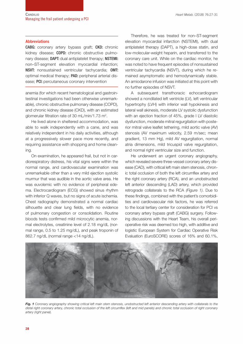
28
anemia (for which recent hematological and gastroin-testinal investigations had been otherwise unremark-able), chronic obstructive pulmonary disease (COPD), and chronic kidney disease (CKD), with an estimated glomerular filtration rate of 30 mL/min/1.73 m2. He lived alone in sheltered accommodation, was able to walk independently with a cane, and was relatively independent in his daily activities, although at a progressively slower pace more recently, and requiring assistance with shopping and home clean-ing. On examination, he appeared frail, but not in car-diorespiratory distress, his vital signs were within the normal range, and cardiovascular examination was unremarkable other than a very mild ejection systolic murmur that was audible in the aortic valve area. He was euvolemic with no evidence of peripheral ede-ma. Electrocardiogram (ECG) showed sinus rhythm with inferior Q waves, but no signs of acute ischemia. Chest radiography demonstrated a normal cardiac silhouette and clear lung fields, with no evidence of pulmonary congestion or consolidation. Routine bloods tests confirmed mild microcytic anemia, nor-mal electrolytes, creatinine level of 2.18 mg/dL (nor-mal range, 0.5 to 1.25 mg/dL), and peak troponin of 862.7 ng/dL (normal range <14 ng/dL).
Therefore, he was treated for non–ST-segment elevation myocardial infarction (NSTEMI), with dual antiplatelet therapy (DAPT), a high-dose statin, and low-molecular-weight heparin, and transferred to the coronary care unit. While on the cardiac monitor, he was noted to have frequent episodes of nonsustained ventricular tachycardia (NSVT), during which he re-mained asymptomatic and hemodynamically stable. An amiodarone infusion was initiated at this point with no further episodes of NSVT. A subsequent transthoracic echocardiogram showed a nondilated left ventricle (LV), left ventricular hypertrophy (LVH) with inferior wall hypokinesis and lateral wall akinesis, moderate LV systolic dysfunction with an ejection fraction of 45%, grade I LV diastolic dysfunction, moderate mitral regurgitation with poste-rior mitral valve leaflet tethering, mild aortic valve (AV) stenosis (AV maximum velocity, 2.59 m/sec; mean gradient, 13 mm Hg), mild AV regurgitation, normal atria dimensions, mild tricuspid valve regurgitation, and normal right ventricular size and function. He underwent an urgent coronary angiography, which revealed severe three-vessel coronary artery dis-ease (CAD), with critical left main stem stenosis, chron-ic total occlusion of both the left circumflex artery and the right coronary artery (RCA), and an unobstructed left anterior descending (LAD) artery, which provided retrograde collaterals to the RCA (Figure 1). Due to these findings, combined with the patient’s comorbidi-ties and cardiovascular risk factors, he was referred to the local tertiary center for consideration for PCI vs coronary artery bypass graft (CABG) surgery. Follow-ing discussions with the Heart Team, his overall peri-operative risk was deemed too high, with additive and logistic European System for Cardiac Operative Risk Evaluation (EuroSCORE) scores of 16% and 60.1%,
candilio Heart Metab. (2018) 76:27-31Managing the frail patient undergoing a PCI
Fig. 1 Coronary angiography showing critical left main stem stenosis, unobstructed left anterior descending artery with collaterals to the distal right coronary artery, chronic total occlusion of the left circumflex (left and mid panels) and chronic total occlusion of right coronary artery (right panel).
AbbreviationsCABG: coronary artery bypass graft; CKD: chronic kidney disease; COPD: chronic obstructive pulmo-nary disease; DAPT: dual antiplatelet therapy; NSTEMI: non–ST-segment elevation myocardial infarction; NSVT: nonsustained ventricular tachycardia; OMT: optimal medical therapy; PAD: peripheral arterial dis-ease; PCI: percutaneous coronary intervention

29
Heart Metab. (2018) 76:27-31 candilio
Managing the frail patient undergoing a PCI
respectively, a EuroSCORE II of 10.1%, a Society of Thoracic Surgery risk score of 45.1% for morbidity and mortality and 7.7% for mortality. He underwent urgent PCI to his left main stem using the left radial artery approach, with predila-tation using a 3.0 x 12 mm noncompliant balloon, implantation of a 3.5 x 24 mm drug-eluting stent, followed by postdilatation with a 4.5 x 12 mm non-compliant balloon, with excellent final angiographic results (Figure 2, left panel). The patient was then discharged 2 days later on DAPT, with stable renal function and a plan for an outpatient review 4 to 6 weeks later following a dobutamine stress echo-cardiogram for further assessment of his viable LV inferior wall. This exam showed reversible inferior wall ischemia, for which a staged PCI for the chronic total occlusion of the RCA was advised, following a careful evaluation of the potential risks and ben-efits and a discussion with the patient. The RCA PCI took place with a dual right femoral and left radial approach. Successful retrograde recanalization of the RCA via the septal collaterals was achieved via a reverse controlled antegrade and retrograde sub-intimal tracking (CART) technique following a failed attempt with an antegrade approach. After predila-tation of the proximal and mid–RCA with a 3.0 x 15 mm noncompliant balloon, 4 overlapping drug-elut-ing stents were implanted from the RCA ostium to the distal segment, with good angiographic results (Figure 2, right panel). Periprocedural nephropro-
tection was ensured with adequate intravenous hy-dration, with maintenance of stable renal function. Medical management was suggested for the chron-ic total occlusion of the left circumflex artery. The patient was discharged successfully and has remained asymptomatic at subsequent follow-up vis-its, reporting a significantly improved quality of life.
Discussion
We report a complex case of a frail 87-year-old man, initially presenting acutely with an NSTEMI and evi-dence of severe multivessel CAD that also involved the LMS, in the context of significant comorbidities, including moderate LVSD, CKD, COPD, and signifi-cant PAD, who then underwent a successful staged PCI. Current guidelines recommend primary or urgent PCI for patients admitted acutely with an ST-segment elevation myocardial infarction (STEMI) or an NSTEMI, respectively.1,2 Over the last few decades, with an ag-ing population and a progressive increase in cardio-vascular risk factors, an increasing number of elderly patients are receiving invasive revascularization.3 How-ever, these patients are at an increased risk of ACS-related complications, such as bleeding, infections, heart failure, renal failure, and stroke, and the evidence for potential benefits of invasive treatment is limited.4 A recent meta-analysis3 that investigated the association between frailty, ACS treatment, and clinical outcomes showed that frailty was independently associated with increased mortality following ACS (adjusted all-cause mortality hazard ratios [HR] for patients with frailty of 1.54-5.39). More importantly, older people with frailty were significantly less likely to receive guideline-indicat-ed ACS care, including PCI, with rates ranging from 6.7% to 43.7% vs 30.4% to 69.5%.3
Importantly, recent studies comparing OMT with percutaneous or surgical coronary revascularization for NSTEMI patients >75 years old demonstrated a reduced risk of death and major cardiac events with invasive therapy.5-7 Crucially, in subjects admitted with STEMI, mortality and morbidity from heart failure, dysrhythmia, and postmyocardial infarction, the com-plications are significantly reduced with invasive treat-ment, regardless of the patient’s age and despite the increased risk of bleeding in this category.8 Therefore, this result contradicts the historical clinical practice of a more conservative approach for this patient subset due to the combination of increased risk factors and a relatively short life expectancy. In addition, a relevant heterogeneity exists within the elderly population, with particular regards to clini-cal presentation, coronary anatomy, frailty, comor-bidities, cognitive impairment, and estimated quality of life, all of which may have a significant impact on procedural and clinical outcomes and should there-
Fig. 2 Angiographic results following a staged percutaneous coronary intervention to the left main stem (left panel) and right coronary artery (right panel).

30
candilio Heart Metab. (2018) 76:27-31Managing the frail patient undergoing a PCI
fore be taken into account when evaluating the risks and benefits of OMT vs coronary revascularization in these patients.3 There is increasing evidence that the potential clinical outcomes in the elderly undergoing invasive coronary treatment following ACS are more related to physiological age than chronological age and that patient frailty constitutes one of the most sig-nificant determinants in this regard.9
Frailty is a complex syndrome characterized by reduced resilience to stressors and increased physio-logical vulnerability, with a progressive loss of reserve and physiological function,10 which has been asso-ciated with a significantly higher risk of hospitaliza-tion, morbidity, and mortality post–PCI, including a prolonged recovery period, more frequent and severe postoperative complications, such as bleeding, stent restenosis and thrombosis, stroke, and heart failure.11 This is most likely attributable to the increased inflam-matory activation and impaired coagulation cascades observed in this patient subset.11
In current practice, the phenotypic frailty models12 and the frailty index of multiple deficits by Rockwood et al9 are the most commonly used approaches for assessing patient frailty. The former approaches in-clude the Fried model and the International Associa-tion of Nutrition and Aging frailty scale (FRAIL), which are similarly both based on 5 physical indicators; the Fried model is based on grip strength, exhaustion, unintended weight loss, slow gait speed, and low physical activity, while the FRAIL model is based on fatigue, resistance, ambulation, illnesses, and loss of weight.12 They are favored by physicians caring for the elderly because of their relatively simple bedside use. The Rockwood Clinical Frailty Scale,11 derived from the Canadian Study of Health and Aging Clinical Frailty Scale (CSHA-CFS), consists of a 9-point scale, from 0 (very fit) to 9 (terminally ill), and it is based on impaired mobility, function, and self-rated health. A number of studies have been conducted to evaluate the association between frailty, as assessed by the above-mentioned frailty scores, with mortality in patients undergoing PCI, presenting with or without ACS. In a prospective cohort study13 of 628 patients of at least 65 years of age undergoing PCI, participants were assessed for frailty (Fried criteria), comorbidities, and quality of life: at least 60% of these subjects were deemed frail or intermediately frail, and importantly, the 3-year mortality was 28% for frail patients and 6% for nonfrail patients, with a strong association be-
tween frailty and mortality / myocardial infarction (HR, 2.61; 95% CI, 1.52-4.50). Similarly, in another study14 that included 745 patients undergoing PCI, frail pa-tients required longer hospitalizations after PCI and presented increased rates of 30-day mortality (HR, 4.8; 95% CI, 1.4-16.3; P=0.013) and 1-year mortality (HR, 5.9; 95% CI, 2.5-13.8; P<0.001), and frailty, as assessed by the CSHA-CFS score, was a predictor of length of hospital stay and mortality, independently of age, sex, and comorbidities. Interestingly, other studies have often led to con-flicting results on the role of frailty in clinical outcomes post–PCI, demonstrating no significant association between frailty and mortality. A recent meta-analysis3 that included a total of 8 studies and 2332 patients (mean age, 69 years; male sex, 68%; follow-up dura-tion, 30±28 months) concluded that frailty was a sig-nificant predictor of all-cause mortality following PCI, with a 2.97-fold increased risk of all-cause mortality (95% CI, 1.56-5.66; P=0.001). Importantly, a signifi-cant heterogeneity in the pooled HRs was identified, which was mainly due to the different frailty scores used and clinical presentations. Subsequent subgroup analyses demonstrated that both the Fried score and CSHA-CFS were significant predictors of mortality with pooled HRs of 2.78 and 5.99, respectively. Our patient presented with a FRAIL score of 3, based on fatigue, resistance, and illness, which put him in the frail category of the scoring system. Sim-ilarly, based on his CSHA-CFS score of 6, he was deemed moderately frail, as he required help with outside activities and with cleaning his house, as well as minimal assistance with indoor activities. However, the patient was extremely keen to maintain his rela-tive independence and therefore very determined to undergo coronary revascularization. The decision on staged vs “one-time” multivessel PCI was dictated based on the patient’s age, frailty, comorbidities, and relevant angiographic findings. In a recent meta-anal-ysis15 on 1090 patients of at least 60 years of age presenting with NSTEMI and evidence of multivessel CAD, the primary composite end point of myocardial infarction and cardiac death during a 3-year follow-up was not significantly different between the staged and the “one-time” revascularization strategies (7% vs 9.5%; P=0.110), and multivariate analysis showed the benefit of staged PCI on the primary events in the elderly (HR, 0.638; 95% CI, 0.408-0.998; P=0.049), with a propensity score matched cohort analy-

31
Heart Metab. (2018) 76:27-31 candilio
Managing the frail patient undergoing a PCI
sis demonstrating that staged PCI was associated with lower rates of primary events (6.1% vs 10.4%; P=0.046) and myocardial infarction (3.4% vs 7.4%; P=0.037) at 3 years.
Conclusions
The current case report demonstrated successful staged multivessel PCI in a frail patient presenting with NSTEMI, for whom careful consideration was given to multiple factors with a potential impact on clinical out-comes, including age, frailty, comorbidities, and angio-graphic findings. Older patients referred for PCI should be systematically assessed for frailty status, which should play a crucial role on the final decision of inva-sive vs noninvasive strategies in this patient subset. LREFERENCES
1. Amsterdam EA, Wenger NK, Brindis RG, et al. 2014 AHA/ACC guideline for the management of patients with non-ST-elevation acute coronary syndromes. Circulation. 2014;130(25):2354-2394.
2. Nauta ST, Gaspersz M, Deckers JW. The new European So-ciety of Cardiology guidelines on myocardial revascularisation: an appraisal. Heart. 2012;98(1):11-14.
3. Tse G, Gong M, Nunez J, et al. Frailty and mortality outcomes after percutaneous coronary intervention: a systematic review and meta-analysis. J Am Med Dir Assoc. 2017;18(12):1097.
4. Fox KA, Dabbous OH, Goldberg RJ, et al. Prediction of risk of death and myocardial infarction in the six months after presen-tation with acute coronary syndrome: prospective multinational observational study (GRACE). BMJ. 2006;333(7578):1091.
5. Cannon CP, Weintraub WS, Demopoulos LA, et al; TACTICS Investigators. Comparison of early invasive and conservative strategies in patients with unstable coronary syndromes treat-ed with the glycoprotein IIb/IIIa inhibitor tirofiban. N Engl J Med. 2001;344(25):1879-1887.
6. Tegn N, Abdelnoor M, Aaberge L, et al; After Eighty Study Inves-tigators. Invasive versus conservative strategy in patients aged 80 years or older with non-ST-elevation myocardial infarction or unstable angina pectoris (After Eighty study): an open-label randomised controlled trial. Lancet. 2016;387(10023):1057-1065.
7. Bach RG, Cannon CP, Weintraub WS, et al. The effect of routine, early invasive management on outcome for elderly patients with non-ST-segment elevation acute coronary syn-dromes. Ann Intern Med. 2004;141(3):186-195.
8. Cockburn J, Hildick-Smith D, Trivedi U, de Belder A. Coronary revascularisation in the elderly. Heart. 2017;103(4):316-324.
9. Rockwood K, Song X, MacKnight C, et al. A global clini-cal measure of fitness and frailty in elderly people. CMAJ. 2005;173(5):489-495.
10. Fried LP, Hadley EC, Walston JD, et al. From bedside to bench: research agenda for frailty. Sci Aging Knowledge Envi-ron. 2005;2005(31):pe24.
11. Fried TR, Mor V. Frailty and hospitalization of long-term stay nursing home residents. J Am Geriatr Soc. 1997;45(3):265-269.
12. Fried LP, Tangen CM, Walston J, et al. Frailty in older adults: evidence for a phenotype. J Gerontol A Biol Sci Med Sci. 2001;56(3):M146-M156.
13. Singh M, Rihal CS, Lennon RJ, Spertus JA, Nair KS, Roger VL. Influence of frailty and health status on outcomes in patients with coronary disease undergoing percutaneous revasculariza-tion. Circ Cardiovasc Qual Outcomes. 2011;4(5):496-502.
14. Murali-Krishnan R, Iqbal J, Rowe R, et al. Impact of frailty on outcomes after percutaneous coronary intervention: a pro-spective cohort study. Open Heart. 2015;2(1):e000294.
15. Yu XF, Li Y, Wang QC, Wang XZ, Liang M, Zhao X, et al. Staged versus “one-time” multivessel intervention in elderly patients with non-ST-elevation acute coronary syndrome. J Geriatr Car-diol. 2016;13(9):760-767.

Heart Metab. (2018) 76:32-35RefresHer Corner
Introduction
It has been well established that aging is a major contributor to the probability of developing car-diovascular diseases, which includes a marked
increase in the incidence and severity of heart fail-ure.1,2 Aging also results in a number of pathophysi-ological alterations in the heart, both at a cellular and tissue level, that contribute to an increased risk of developing cardiovascular disease,1-3 which includes the development of cardiac hypertrophy, arrhythmias, and left ventricular dilatation, as well as alterations in the extracellular matrix, increased cardiomyocyte loss, alterations in calcium homeostasis, apoptotic signaling, autophagy, reactive oxygen species (ROS) generation, and many other key metabolic pathways in the heart.4 In addition, aging results in dramatic changes in energy homeostasis in the heart, which includes the development of mitochondrial
dysfunction.5 These alterations in energy metabo-lism are likely to be important contributors to the development of cardiovascular diseases in the aging population. Despite this, our understanding of what effect aging has on cardiac energy metabolism is incomplete and still contradictory. This paper reviews what is known with regard to the effects of aging on cardiac energy metabolism.
Aging effects on mitochondrial function
The heart has a very high energy demand and must produce large amounts of energy, in the form of ade-nosine triphosphate (ATP), to sustain contractile func-tion.6 The majority of this ATP production originates from mitochondrial oxidative phosphorylation.6 In ad-dition to this central role in energy production in the heart, mitochondria also perform a number of other essential functions in the heart, including roles in cal-
Energy metabolism in the aged heartGary D. Lopaschuk, PhD
Alberta Heart Institute, Departments of Pediatrics, University of Alberta, Edmonton, Alberta, Canada
Correspondence: Dr. Gary D. Lopaschuk, 423 Heritage Medical Research Centre, Departments of Pediatrics, Faculty of Medicine and Dentistry, University of Alberta, Edmonton, Alberta, T6G 2S2, Canada
E-mail: [email protected]
AbstractAging is associated with a decline in energy production in the heart that can compromise the ability of the heart to adapt to stresses requiring an increase in energy demand. The main source of energy for the heart arises from mitochondrial oxidative phosphorylation. With aging, mitochondrial function becomes compromised, which can lead to a decrease in energy production and an increase in the production of reactive oxygen species. Impaired energetics in the aging heart can result not only from impaired mitochondrial ATP production, but also from a decrease in metabolic flexibility in the type of fuel used by the heart for energy production, as well as a decreased efficiency of ATP transfer from the site of mitochondrial production to the site of use. Improving both cardiac energy production and the efficiency of energy production may be a novel therapeutic approach to lessen cardiac disease in the elderly. L Heart Metab. 2018;76:32-35
Keywords: fatty acid oxidation; glucose oxidation; glycolysis; mitochondria
32

33
Heart Metab. (2018) 76:32-35 lopascHuk Energy metabolism in the aged heart
cium homeostasis, ROS generation, and apoptotic signaling. As a result, situations where mitochondrial dysfunction occurs can have severe consequences on cardiac function, cardiac electrical activity, cardio-myocyte integrity, and cardiomyocyte survival. Aging results in defects in cardiac mitochondrial function, which are the major reasons for the cellular and organ dysfunction that occurs with aging.7 These defects include a decrease in cardiomyocyte mito-chondrial volume, morphology, and respiration.8 A key site of these mitochondrial defects is the electron transport chain, which is responsible for mitochon-drial oxidative phosphorylation (Figure 1). Mitochon-drial defects in the electron transport chain occurs, in part, due to an age-related decrease in transcriptional and functional activity of the mitochondrial oxidative phosphorylation complexes.7-10 This decrease in mi-tochondrial respiratory chain enzymes occurs to a larger extent in interfibrillar mitochondria vs subsar-colemmal mitochondria,7 suggesting that aging com-promises ATP supply to the cardiomyocyte contrac-tile proteins. The expression and activity of complex III (cytochrome oxidase), complex IV, and complex V (ATP synthase) are particularly susceptible to aging, while complex I is relatively unaltered.7,11 A decrease in cardiolipin, the key mitochondrial lipid, contributes to the decreased activity in complexes II-V.12 The mitochondrial electron transport chain is also a major site of ROS production in the heart. While the complexes I and III are major sites of ROS production, it is an aging-induced impairment of complex III that appears to be responsible for the increased cardiac ROS production seen with aging.7,13 Impaired flux through the electron transport chain increases the direct interaction of the reduced redox centers with molecular oxygen to produce the free radical O2
-, par-ticularly in complex III. The increased ROS production in the aging heart can lead to oxidative damage in the mitochondria, including protein sulfhydryl oxidation,
lipid peroxidation, and mitochondrial DNA damage. ROS also acts in mitochondrial metabolism–based stress signaling pathways, including the apoptotic pathway and the mitochondrial permeability transi-tion pore (MPTP) opening. Increased mitochondrial ROS production increases apoptosis observed in the heart during aging.7 Aging is also associated with an increase in the susceptibility of MPTP opening.14 An increased ROS production with aging may contribute to this increased opening of the MPTP.7 The opening of the MPTP can compromise mitochondrial function, increase mitochondrial calcium content, and alter membrane potential, and compromise cardiomyo-cyte function and survival.
Aging effects on myocardial energy production Three potential effects of aging on myocardial en-ergy production include: (i) a decreased capacity for energy production; (ii) alterations in fuel selection by the heart; and (iii) a decrease in energy transfer in the heart. These alterations in energy production, energy fuel selection, and energy transfer can negatively af-fect heart function and contribute to the development of heart failure in the aging heart.
Energy production capacity in the aging heart
The compromised cardiac mitochondrial integrity and the impairments in electron transport chain activity seen with aging are two important contributing fac-tors to a decreased capacity for ATP production in the aging heart, which is associated with a decrease in high energy phosphate levels in the aging human heart.15 In addition, the tricarboxylic acid cycle (TCA), which is critical for producing reduced equivalents necessary for the electron transport chain, also de-creases with aging.9 This decrease is partly due to a decrease in the expression of a number of genes of the TCA cycle.10 As will be discussed, acetyl CoA supply to the TCA cycle is also compromised in the aging heart. Combined with the changes in the elec-tron transport chain, mitochondrial oxidative capacity can drop by 50% in the human heart with aging.16
Energy substrate selection in the aging heart
Mitochondrial oxidative metabolism can use a variety of carbon substrates to produce the large amount of
AbbreviationsCK/PCr: creatine kinase/phosphocreatine; CPT: car-nitine palmitoyltransferase; ETC: electron transport chain; GLUT: glucose transporter; MCT: monocarbox-ylate transporter; MPC: mitochondrial pyruvate carri-er; MPTP: mitochondrial permeability transition pore; PDH: pyruvate dehydrogenase; ROS: reactive oxygen species; TCA: tricarboxylic acid

energy necessary to sustain contractile function, in-cluding the oxidation of fatty acids, pyruvate derived from glucose and lactate, ketones, and amino ac-ids (Figure 1).6 The majority of cardiac mitochondrial ATP production originates from fatty acid oxidation, although pyruvate from glucose (glucose oxidation)
can also be a significant source.6 Normally, the heart maintains a high degree of metabolic flexibility, switch-ing back and forth between different energy substrates to ensure an adequate energy production by the heart to meet the high energy demand of the heart. How-ever, during aging, this metabolic flexibility is compro-
mised. Unfortunately, there is no good consensus as to what actual switches occur in energy substrate metabolism. While it is generally thought that there is an impaired myo-cardial fatty acid oxidation and a compensatory increase in glucose metabolism (see ref-erence 7 for a review of this subject), this is not supported by direct measurements of energy substrate flux. Direct measurements of energy sub-strate metabolism in the heart showed an increase in fatty acid oxidation in aged rats compared with young rats, with a parallel decrease in carbohydrate oxidation.17 This finding is supported by mea-surements of gene profiles in hearts of aging humans, in which an increase in fatty acid oxidation genes and a de-crease in pyruvate dehydro-genase (the rate-limiting en-zyme for glucose and lactate oxidation) was observed.10 It should be noted, however, that aging-induced increases in pyruvate dehydrogenase have also been observed in the rat heart.18 As a result, further studies are needed to have a better understanding of what changes in fuel selec-tion occur in the aging heart. It should be noted that inhibi-tion of fatty acid oxidation in the skeletal muscle of aging mice improves glucose and insulin tolerance and protects
34
TCA cycle
I
II
III
IV
V
NADH
PDH Fatty acyl CoA
2
Pyruvate
FADH
MPC
CPT2
CPT1
Fatty acyl CoA
Fatty acyl carnitine
GLUT4
GLUT1
Glucose Glucose
Pyruvate
Lactate Lactate MCT
Fatty acids
ADP
ATP
ADP
ATP
Glucose oxidation
Fatty acid oxidation
Glycolysis
ETC
Ketones Ketones
Amino acids
Amino acids
Ketone oxidation
Amino acid oxidation
?
?
O 2
O2-
H2O
MPTP
CK/PCr shuttle
ATP
Indicates changes with aging
?
Mitochondria Cytoplasm
ROS
Acetyl CoA
Fig. 1 Alterations in myocardial energy metabolism in the aged heart. In the aerobic heart, mito-chondrial fatty acid oxidation and glucose oxidation are the major sources of energy production in the heart. In the aging heart, both mitochondrial ETC activity and TCA cycle activity are compro-mised, leading to an impaired production of ATP from oxidative metabolism. Impaired ETC activity can also increase ROS production in the aging heart. The aging heart also becomes “metabolically inflexible” with fatty acid oxidation increasing at the expense of glucose oxidation, a situation that can decrease cardiac efficiency. Glycolysis increases in the aging heart in an attempt to increase ATP production, although this increase in glycolytic ATP production cannot compensate for the loss of mitochondrial ATP production, leaving the heart in a potentially “energy starved” situation.Abbreviations: CK/PCr, creatine kinase/phosphocreatine; CPT, carnitine palmitoyltransferase; ETC, electron transport chain; GLUT, glucose transporter; MCT, monocarboxylate transporter; MPC, mitochondrial pyruvate carrier; MPTP, mitochondrial permeability transition pore; PDH, pyruvate dehydrogenase; ROS, reactive oxygen species; TCA, tricarboxylic acid.
lopascHuk Heart Metab. (2018) 76:32-35Energy metabolism in the aged heart

35
Heart Metab. (2018) 76:32-35 lopascHuk Energy metabolism in the aged heart
against age-related metabolic dysfunction.19 Whether a similar approach may also be beneficial in the aging human heart has yet to be determined. With a decrease in mitochondrial oxidative capac-ity and possible decreases in fatty acid and carbo-hydrate oxidation, the aging heart can become “en-ergy starved,” particularly during times of stress or increased energy demand. One attempt to counter this decrease in mitochondrial ATP production is to increase glycolytic ATP production. An increase in the reliance on glycolysis as a source of ATP produc-tion has been shown in aging rats and humans.20,21 However, the amount of ATP produced from glycoly-sis is small compared with the amount of mitochon-drial ATP produced, leaving the potential for the aging heart to remain in an energy compromised state.
Energy transfer in the aging heart
Transfer of mitochondrial produced ATP to its site of use in the cytoplasm requires a creatine kinase (CK)/phosphocreatine (PCr) shuttle pathway (Figure 1). To date, there is no clear consensus as to whether this energy transfer is compromised with aging. Studies have shown no change, an increase, or a decrease in the CK/PCr pathway (see reference 5 for a review of this topic). While decreases in PCr have been ob-served in aging humans,22 it is not clear if this is due to alterations in the actual energy transfer process or a decrease in the actual mitochondrial energy produc-tion, resulting in a decreased transfer of high energy phosphates from ATP to PCr. While studies have at-tempted to improve cardiac energetics in the aging heart by altering the CK/PCr shuttle, the results of these studies have not been encouraging.
Conclusions
Aging leads to defects in mitochondrial energy produc-tion in the heart, due, in large part, to decreased mito-chondrial integrity and decreased activity of the electron transport chain and TCA cycle. Alterations in energy substrate selection contribute to a metabolic inflexibil-ity in the aging heart that may decrease the ability of the heart to deal with pathological stress and increased energy demands, which raises the possibility that thera-peutic strategies to improve mitochondrial energy pro-duction and metabolic flexibility may be an approach to improve cardiac function in the elderly population. L
REFERENCES
1. Olivetti G, Melissari M, Capasso JM, Anversa P. Cardiomyopa-thy of the aging human heart. Myocyte loss and reactive cel-lular hypertrophy. Circ Res. 1991;68(6):1560-1568.
2. Shih H, Lee B, Lee RJ, Boyle AJ. The aging heart and post-infarc-tion left ventricular remodeling. J Am Coll Cardiol. 2011;57(1):9-17.
3. Papp Z, Czuriga D, Balogh L, Balogh Á, Borbély A. How cardiomyocytes make the heart old. Curr Pharm Biotechnol. 2012;13(13):2515-2521.
4. Lakatta EG. So! What’s aging? Is cardiovascular aging a dis-ease? J Mol Cell Cardiol. 2015:83:1-13.
5. Tepp K, Timohhina N, Puurand M, et al. Bioenergetics of the aging heart and skeletal muscles: modern concepts and con-troversies. Ageing Res Rev. 2016;28:1-14.
6. Lopaschuk GD, Ussher JR, Folmes CD, Jaswal JS, Stanley WC. Myocardial fatty acid metabolism in health and disease. Physiol Rev. 2010;90(1):207-258.
7. Lesnefsky EJ, Chen Q, Hoppel CL. Mitochondrial metabolism in aging heart. Circ Res. 2016;118(10):1593-1611.
8. Preston CC, Oberlin AS, Holmuhamedov EL, et al. Aging-induced alterations in gene transcripts and functional activity of mitochondrial oxidative phosphorylation complexes in the heart. Mech Ageing Dev. 2008;129(6):304-312.
9. Tepp K, Puurand M, Timohhina N, et al. Changes in the mi-tochondrial function and in the efficiency of energy transfer pathways during cardiomyocyte aging. Mol Cell Biochem. 2017;432(1-2):141-158.
10. Emelyanova L, Preston C, Gupta A, et al. Effect of aging on mitochondrial energetics in the human atria. J Gerontol A Biol Sci Med Sci. 2018;73(5):608-616.
11. Paradies G, Ruggiero FM, Petrosillo G, Gadaleta MN, Qua-gliariello E. Effect of aging and acetyl-L-carnitine on the activity of cytochrome oxidase and adenine nucleotide translocase in rat heart mitochondria. FEBS Lett. 1994;350(2-3):213-215.
12. Paradies G, Ruggiero FM. Age-related changes in the activity of the pyruvate carrier and in the lipid composition in rat-heart mitochondria. Biochim Biophys Acta. 1990;1016(2):207-212.
13. Barja G. Mitochondrial free radical production and aging in mammals and birds. Ann N Y Acad Sci. 1998;854:224-238.
14. Hafner AV, Dai J, Gomes AP, et al. Regulation of the mPTP by SIRT3-mediated deacetylation of CypD at lysine 166 suppress-es age-related cardiac hypertrophy. Aging. 2010;2(12):914-923.
15. Köstler H, Landschütz W, Koeppe S, et al. Age and gender dependence of human cardiac phosphorus metabolites deter-mined by SLOOP 31P MR spectroscopy. Magn Reson Med. 2006;56(4):907-911.
16. Conley KE, Jubrias SA, Esselman PC. Oxidative capacity and ageing in human muscle. J Physiol. 2000;526(Pt 1):203-210.
17. Sample J, Cleland JG, Seymour AM. Metabolic remodeling in the aging heart. J Mol Cell Cardiol. 2006;40(1):56-63.
18. Moreau R, Heath SH, Doneanu CE, Harris RA, Hagen TM. Age-re-lated compensatory activation of pyruvate dehydrogenase complex in rat heart. Biochem Biophys Res Commun. 2004;325(1):48-58.
19. Ussher JR, Fillmore N, Keung W, et al. Genetic and pharma-cological inhibition of malonyl coa decarboxylase does not exacerbate age-related insulin resistance in mice. Diabetes. 2016;65(7):1883-1891.
20. McMillin JB, Taffet GE, Taegtmeyer H, Hudson EK, Tate CA. Mi-tochondrial metabolism and substrate competition in the aging Fischer rat heart. Cardiovasc Res. 1993;27(12):2222-2228.
21. Kates AM, Herrero P, Dence C, et al. Impact of aging on sub-strate metabolism by the human heart. J Am Coll Cardiol. 2003;41(2):293-299.
22. Esterhammer R, Klug G, Wolf C, et al. Cardiac high-energy phosphate metabolism alters with age as studied in 196 healthy males with the help of 31-phosphorus 2-dimensional chemical shift imaging. PLoS One. 2014;9(6):e97368.

Heart Metab. (2018) 76:36-39Hot Topics
Introduction
Over the past decade or so, we have observed a major shift in the nature of the challenges fac-ing cardiovascular clinicians and researchers.
Advances in the treatment and prevention of coro-nary disease have resulted in a diminution in deaths attributable to coronary heart disease. We now live in an aging society, where hypertension remains highly prevalent,1 heart failure with preserved ejection fraction (HFPEF) is emerging as the most common HF phenotype,2 and there is a significant increase in nonsurgical percutaneous valve interventions for aor-tic stenosis. Although age alone cannot be implicated for each of these, it is clear that future developments in the management of cardiovascular disease will be closely intertwined with our understanding of the pathophysiology of cardiovascular aging and its interactions with comorbidities, such as frailty.
The definition of frailty remains challenging; however, at its core, it represents a state of increased vulner-ability to external stressors and a decreased physi-ological reserve. Although the components of frailty are diverse, emerging paradigms include a cycle of immune dysregulation with chronic inflammation, a catabolic state, and progressive sarcopenia, which further contributes to physical inactivity.3 Frailty is an independent prognostic factor for both coronary artery disease and heart failure. Within the cardiovascular system,4 frailty is associated with increased atherosclerosis and coronary calcification, with a reduced vasodilatory capacity, impaired mi-crovascular function, increased arterial stiffness, and increased myocardial fibrosis. Together, these play a major physiological role in the pathogenesis of heart failure. Finally, degenerative diseases of the conduct-ing system and left atrial fibrosis contribute to the
Exercise and diet for heart disease in the frail patient: fact or fiction?
Shane Nanayakkara, MBBS, FRACP1,2,3; David M. Kaye, MBBS, FRACP, FACC, FESC, PhD1,2,3
1Alfred Hospital, Department of Cardiovascular Medicine, Melbourne, Victoria, Australia2Baker Heart and Diabetes Institute, Heart Failure Research Group, Melbourne, Victoria, Australia
3Monash University, Faculty of Medicine, Nursing and Health Sciences, Melbourne, Victoria, Australia
Correspondence: Professor David M. Kaye, Department of Cardiology, Alfred Hospital, Commercial Rd, Melbourne 3004, Victoria, Australia E-mail: [email protected]
AbstractFrailty represents a state of increased physiological vulnerability and diminished reserve, as well as a complex interaction between age, comorbidities, demographics, and lifestyle. Frailty has repeatedly been demonstrated to be an independent prognostic factor across the spectrum of cardiovascular disease; however, frail patients are often excluded from major clinical trials, leading to a significant gap in the evidence. In this review, we discuss the impact of nutrition and exercise interventions on frailty and, consequently, on cardiovascular disease. L Heart Metab. 2018;76:36-39
Keywords: frailty; heart disease; lifestyle intervention
36

37
Heart Metab. (2018) 76:36-39 nanayakkara and kaye Exercise, diet, frailty, and cardiovascular disease
increased prevalence of both bradyarrhythmias and tachyarrhythmias.
Assessing frailty
Frailty has consistently been demonstrated to be a major independent prognostic factor for patients with a variety of cardiovascular diseases, both in the acute and chronic setting and in the periprocedural period. Multiple different scoring systems exist to quantify frailty, with the Fried index5 being the system most cit-ed in short- and long-term outcome studies, including within cardiovascular disease. The Fried index analyz-es five domains (gait speed, weakness, exhaustion, weight loss, and physical inactivity); however, it does not examine cognition or mood. It remains unclear if cognition and mood are part of the pathobiological diagnosis of frailty or if they simply act as a modifier for its impact on quality of life. Of these domains, ex-ercise and dietary counseling, as modifiable factors, can have a significant impact on outcomes.
Treating cardiovascular disease in the elderly
Coronary artery disease
The elderly represent the fastest growing group of pa-tients being referred for cardiac surgery; however, newer interventional and structural techniques have reduced the need for major operative care. Traditionally, pre-dicting outcomes following both percutaneous coro-nary intervention (PCI) and cardiac surgery have been limited in accuracy in elderly patients, with a tendency to overestimate mortality. In this setting, frailty assess-ments have demonstrated an independent, incremen-tal benefit in predicting adverse outcomes, particularly concerning gait speed, disability, and quantification of comorbidities.6 In patients undergoing PCI, frailty was again markedly predictive of mortality, and improved the discrimination of predictive models.7 Most importantly, the involvement of patients in a dedicated cardiac re-habilitation program improves morbidity and mortality; however, there is a paucity of studies examining frailty that are specifically tailored toward cardiac rehabilitation.
Heart failure
The primary limiting symptom for patients with heart failure is exercise intolerance, measured objectively
as peak oxygen consumption at maximal exercise or peak Vo2. Both central and peripheral limitations re-sult in an approximate 35% reduction in peak Vo2, re-gardless of ejection fraction.4 It has become increas-ingly apparent that frailty is a common comorbidity in patients with heart failure, with reduced physical activity leading to sarcopenia, as well as the systemic nature of the disease itself, with direct consequences on the pulmonary and renal systems in particular. Ex-ercise training has demonstrated significant benefit across the spectrum of symptoms and ejection frac-tions, with a well-established safety profile, although a greater benefit has been seen in patients with heart failure with reduced ejection fraction (HFREF) versus patients with HFPEF. Overall, however, there is a lack of data on guideline-based medical therapy in elderly patients for a multitude of reasons, primarily due to the exclusion of these patients from large-scale clini-cal trials. The HF-ACTION trial (Heart Failure and A Con-trolled Trial Investigating Outcomes of exercise traiN-ing),8 an international, multicenter, controlled trial, randomized 2331 patients to 36 supervised exercise sessions followed by home-based training or usual care. The median age was 59 and 20% of the pa-tients were over the age of 70. These patients had a significantly higher comorbidity score, a lower body mass index, and severe systolic dysfunction (median ejection fraction, 26%). Overall, there was a nonsig-nificant reduction in the primary end point (a combi-nation of mortality or hospitalization), which became significant after multivariate adjustment. Subgroup analysis revealed no significant difference with age concerning the response to exercise training; impor-tantly, age was the strongest predictor of peak Vo2,
9 independent of peak heart rate, which is another significant predictor. This noted, age alone only ex-plained 12% of the variance in peak Vo2, meaning that other peripheral factors that contribute to frailty may be explanatory. The overall impact of exercise-based rehabilitation on heart failure was elegantly summarized in a 2014 Cochrane review10 that reviewed 33 randomized con-trolled trials (n=4740 patients). Both patients with HFREF and HFPEF were included, although data on patients with HFPEF were only present in a portion of the four trials. Overall, there was no difference in the 12-month mortality, even though there was a trend toward a reduction at the long-term (10-year) follow-

38
nanayakkara and kaye Heart Metab. (2018) 76:36-39Exercise, diet, frailty, and cardiovascular disease
up. At the 12-month follow-up, admission rates were significantly reduced, quality of life was improved, and support for the cost-effectiveness of exercise-based rehabilitation was demonstrated.
Nutrition, frailty, and CVD
Frailty and nutritional status are closely linked. Unin-tentional weight loss is a key domain of the original Fried criteria, which may occur due to inadequate en-ergy intake, an important modifiable risk factor. Low-ered energy intake is commonly seen with increasing age, and it has been associated with the develop-ment of frailty, especially when under the threshold of 25 kcal/kg/day.11 Protein is a critical factor to maintain muscle mass, with up to 15% of patients over the age of 60 demonstrating intake below the recommended dietary allowance.12 A careful balance must be struck between adequate intake and avoidance of exces-sive protein load for a delicate renal system. Although patients able to receive at least 25 to 30 grams of protein have evidence of slowed sarcopenia,13 large randomized trials have not demonstrated the benefit of higher protein intake in frail patients, which may be due, in part, to impaired synthesis of muscle protein in the context of frail muscle, rather than through low-ered protein intake alone. High sodium intake is also strongly correlated with elevated blood pressure, particularly in older adults.1 In addition to the vitamins B6 and B12, vitamins C14 and D15 have both been independently linked with frailty, although a U-shaped curve is noted with the latter. Vitamin D supplementation improves muscle strength and reduces frailty; however, there is con-flicting evidence regarding cardiovascular outcomes, with the most recent data suggesting no significant benefit.16 Other micronutrients have also been im-plicated, but this requires further evaluation in large-scale randomized trials. The Mediterranean diet is considered beneficial for patients with cardiovascular disease. A recent systematic review and meta-analysis demonstrated that greater adherence to the diet reduced the risk of frailty, and the consequent impact on cardiovascular disease will require further investigation. The gut microbiome has recently become the focus of attention, with significant evidence demon-strating a link with hypertension,17 heart failure, and other forms of cardiovascular disease. Frail older
adults have a less diverse microbiome than younger adults,18 which may potentially provide a key link be-tween aging, frailty, systemic inflammation, and the development of cardiovascular disease.
Conclusions
Frailty is increasingly prevalent in older patients with cardiovascular disease, remains an independent prognostic indicator of outcomes, and it is modifiable through prescriptive exercise and nutritional counsel-ing. Efforts should be made to quantify frailty using validated scales and target the frail patient, regardless of age, with appropriate lifestyle-modifying therapies to reduce cardiovascular morbidity and mortality. It is well recognized that frail patients are often excluded from major research studies; consequently, there is a drive to encourage trialists to include patients of increasing age, to carefully and objectively measure frailty, and to determine the impact of interventions on slowing the progression of frailty or reversing it. L
REFERENCES
1. Benjamin EJ, Virani SS, Callaway CW, et al. Heart disease and stroke statistics—2018 update: a report from the American Heart Association. Circulation. 2018;137(12):e67-e492.
2. van Riet EE, Hoes AW, Wagenaar KP, Limburg A, Landman MA, Rutten FH. Epidemiology of heart failure: the prevalence of heart failure and ventricular dysfunction in older adults over time. A systematic review. Eur J Heart Fail. 2016;18(3):242-252.
3. Afilalo J, Alexander KP, Mack MJ, et al. Frailty assessment in the cardiovascular care of older adults. J Am Coll Cardiol. 2014;63(8):747-762.
4. Nanayakkara S, Marwick TH, Kaye DM. The ageing heart: the systemic and coronary circulation. Heart. 2018;104(5):370-376.
5. Fried LP, Tangen CM, Walston J, et al; CHS Collaborative Re-search Group. Frailty in older adults: evidence for a phenotype. J Gerontol A Biol Sci Med Sci. 2001;56(3):M146-M156.
6. Afilalo J, Mottillo S, Eisenberg MJ, et al. Addition of frailty and disability to cardiac surgery risk scores identifies elderly pa-tients at high risk of mortality or major morbidity. Circ Cardio-vasc Qual Outcomes. 2012;5(2):222-228.
7. Singh M, Rihal CS, Lennon RJ, Spertus JA, Nair KS, Roger VL. Influence of frailty and health status on outcomes in patients with coronary disease undergoing percutaneous revasculariza-tion. Circ Cardiovasc Qual Outcomes. 2011;4(5):496-502.
8. O’Connor CM, Whellan DJ, Lee KL, et al; HF-ACTION Investi-gators. Efficacy and safety of exercise training in patients with chronic heart failure: HF-ACTION randomized controlled trial. JAMA. 2009;301(14):1439-1450.
9. Forman DE, Clare R, Kitzman DW, et al; HF-ACTION Investi-gators. Relationship of age and exercise performance in pa-tients with heart failure: the HF-ACTION study. Am Heart J. 2009;158(suppl 4):S6-S15.
10. Taylor RS, Sagar VA, Davies EJ, et al. Exercise-based re-habilitation for heart failure. Cochrane Database Syst Rev.

39
Heart Metab. (2018) 76:36-39 nanayakkara and kaye Exercise, diet, frailty, and cardiovascular disease
2014;(4):CD003331. 11. Vellas BJ, Hunt WC, Romero LJ, Koehler KM, Baumgartner
RN, Garry PJ. Changes in nutritional status and patterns of morbidity among free-living elderly persons: a 10-year longitu-dinal study. Nutrition. 1997;13(6):515-519.
12. Roubenoff R. Sarcopenia: a major modifiable cause of frailty in the elderly. J Nutr Health Aging. 2000;4(3):140-142.
13. Singh M, Stewart R, White H. Importance of frailty in patients with cardiovascular disease. Eur Heart J. 2017;35(26):1726-1731.
14. Bartali B, Semba RD, Frongillo EA, et al. Low micronutrient levels as a predictor of incident disability in older women. Arch Intern Med. 2006;166(21):2335-2340.
15. Ensrud KE, Ewing SK, Fredman L, et al; Study of Osteopo-rotic Fractures Research Group. Circulating 25-hydroxyvitamin D levels and frailty status in older women. J Clin Endocrinol Metab. 2010;95(12):5266-5273.
16. Jenkins DJA, Spence JD, Giovannucci EL, et al. Supplemental vitamins and minerals for CVD prevention and treatment. J Am Coll Cardiol. 2018;71(22): 2570-2584.
17. Marques FZ, Mackay CR, Kaye DM. Beyond gut feelings: how the gut microbiota regulates blood pressure. Nat Rev Cardiol. 2017;15(1):20-32.
18. Biagi E, Nylund L, Candela M, et al. Through ageing, and be-yond: gut microbiota and inflammatory status in seniors and centenarians. PLoS One. 2010;5(5):e10667.

40
Heart Metab. (2018) 76:40-41Glossary gary d. lopascHuk
Activities of Daily LivingActivities of daily living are basic tasks that must be accomplished over a daily timescale for an individual to thrive, including personal hygiene, management of continence, dressing, feeding, and ambulation. Ac-tivities of daily living are used as a measurement of an individual’s functional status.
Cellular Senescence Cellular senescence is a cellular stress response characterized essentially by irreversible arrest of cellu-lar proliferation that can occur in response to a poten-tially oncogenic stress (persistent genomic damage, oncogene activation, epigenomic perturbations), and it involves the activation of tumor suppressor genes. The physiological consequences of cellular senes-cence include tumor suppression and optimal tissue repair, while the pathophysiological consequences in-clude age-related tumor progression and age-related degenerative alterations.
Clinical Frailty Scale (CFS)The CFS is a practical and efficient tool for evaluating frailty, which is a condition characterized by increased vulnerability to external stressors. It involves the use of clinical descriptors and pictographs. The scale was designed to aid clinicians in stratifying older adults ac-cording to their level of vulnerability.
Comprehensive Assessment of Frailty (CAF) scoreThe CAF score is another toolset available to clinicians to help assess the risk for elderly patients undergoing cardiac surgery, but unlike other scoring tools (eg, the Society of Thoracic Surgeons [STS] score), the CAF score also includes measures of frailty (eg, weakness, self-reported exhaustion, standing balance, etc) in addition to laboratory data and a patient’s clinical fea-tures.
Comprehensive Geriatric Assessment (CGA)The CGA is a multidisciplinary diagnostic and thera-peutic intervention pathway that identifies medical, psychosocial, and functional limitations in frail pa-tients. The CGA is intended to help develop a coordi-nated plan to maximize health with aging.
Endocrine Dysregulation Endocrine dysregulation refers to disturbances in the regulatory processes (negative feedback and positive feedback control circuits) that govern the release of hormones. As hormones regulate a myriad of physi-ological functions, endocrine dysregulation can con-tribute to a broad array of pathophysiological states.
Essential Frailty Toolset (EFT)The EFT is a simple, but highly predictive composite score of 4 indicators (time to stand five times, cogni-tive impairment, hemoglobin levels, and serum albu-min) used for predicting mortality after transcatheter aortic valve implantation TAVI or surgical aortic valve replacement.
European System of Cardiac Operative Risk Evaluation (EuroSCORE)EuroSCORE is a European risk score toolset (the American equivalent is the STS score) used to predict operative mortality of adult cardiac surgery within 30 days of the operation or later if the patient remains hospitalized.
Frailty Frailty is a common clinical syndrome in older adults that increases the risk of poor health outcomes, in-cluding falls, incident disability, hospitalization, and mortality. Increased vulnerability results from aging-associated declines in reserve and function across multiple physiological systems. Frailty is operationally defined as the presence of three (of five) phenotypic criteria indicative of compromised energetics: low handgrip strength, low energy, decreased walking speed, low physical activity, and unintentional weight loss (10 lbs/4.54 kg over the course of 1 year).
Left Ventricular Assist Device (LVAD)An LVAD is a mechanical pump (transcutaneous or implanted in the left ventricle) used to support left ventricular and circulatory function in the setting of heart failure. LVADs are typically used as a bridge-to-transplant therapy or a destination therapy (long-term treatment with an LVAD to prolong and improve patient life).