the establishment of two paclitaxel-resistant prostate cancer cell
TRANSCRIPT

The Prostate 67:955 ^ 967 (2007)
The Establishmentof TwoPaclitaxel-ResistantProstateCancerCell Lines andtheMechanisms
of Paclitaxel ResistancewithTwoCell Lines
Masashi Takeda,1 Atsushi Mizokami,1* Kiminori Mamiya,1 You Qiang Li,1
Jian Zhang,2 Evan T. Keller,3 and Mikio Namiki1
1Departmentof Integrative CancerTherapyandUrology,KanazawaUniversityGraduate SchoolofMedical Sciences,Kanazawa, Ishikawa, Japan
2DepartmentofMedicine,DivisionofHematology/Oncology,UniversityDrive, Pittsburgh3Unit for Laboratory AnimalMedicineandDepartmentof Pathology,UniversityofMichigan,
AnnArbor,Michigan
BACKGROUND. Although paclitaxel is used for hormone-resistant prostate cancer, relapsedefinitely occurs later. Details of themolecularmechanism responsible for paclitaxel- resistanceremain unclear.METHODS. We established paclitaxel-resistant cells, DU145-TxR and PC-3-TxR from parentDU145 and PC-3. To characterize these cells, we examined cross-resistance to other anticancerdrugs. Expression of several potential genes that had been related to drug-resistance wascompared with parent cells by RT-PCR andWestern blotting. Methylation analysis of multipledrug resistance (MDR1) promoterwas carried out using bisulfite-modifiedDNA fromcell lines.Knockdown experiments using small interfering RNA (siRNA)were also performed to confirmresponsibility of drug-resistance. Finally, cDNA microarray was performed to quantify geneexpression in PC-3 and PC-3-TxR cells.RESULTS. The IC50 for paclitaxel in DU145-TxR and PC-3-TxR was 34.0- and 43.4-fold higherthan that in bothparent cells, respectively. Both cells showed cross-resistance to somedrugs, butnot to VP-16 and cisplatin. Methylation analysis revealed that methylated CpG sites of MDR1promoter in DU145 and PC-3 cells were demethylated in DU145-TxR cells, but not in PC-3-TxRcells. KnockdownofP-glycoprotein (P-gp),whichwasup-regulated in resistant cells, byMDR-1siRNA restored paclitaxel sensitivity in DU145-TxR but not in PC-3-TxR, indicating that up-regulation of P-gp was not always main cause of paclitaxel-resistance. Microarray analysisidentified 201 (1.34%) up-regulated genes and 218 (1.45%) out of screened genes in PC-3-TxR.CONCLUSIONS. Our data will provide molecular mechanisms of paclitaxel-resistance andbe useful for screening target genes to diagnose paclitaxel sensitivity. Prostate 67: 955–967,2007. # 2007 Wiley-Liss, Inc.
KEY WORDS: prostate cancer; paclitaxel resistance; MDR-1; cDNA microarray
INTRODUCTION
Prostate cancer (PCa) is the most common malig-nancy and the second most frequent cause of cancer-related death ofmen in the United States [1]. Androgendeprivation treatment is very effective for more than80% of advanced PCa. More than half of those cases ofadvanced PCa become resistant to deprivation treat-ment after several years and then several other
*Correspondence to: Atsushi Mizokami, MD, PhD, Department ofIntegrative Cancer Therapy and Urology, Kanazawa UniversityGraduate School of Medical Sciences, 13-1 Takara-machi, Kanazawa,Ishikawa 920-8640, Japan. E-mail: [email protected] 24 November 2006; Accepted 14 February 2007DOI 10.1002/pros.20581Published online 17 April 2007 in Wiley InterScience(www.interscience.wiley.com).
� 2007Wiley-Liss, Inc.

palliative treatments, such as estramustine phosphate(EMP), steroids, are employed for these patients.However, the results are very disappointing becausea half of those cases lead to death within a year or2 years.
Recently, the taxanes [paclitaxel or docetaxel (DTX)]with other agents, such as EMP or predonisone havebeen used for hormone-resistant prostate cancer(HRPC) and have shown good response [2–5]. Pacli-taxel, which is purified from Taxus brevifolia, stabilizemicrotubule and causes apoptosis [6]. The responserates of taxane-based combination therapies are betterthan combination therapies with other anticanceragents. However, even HRPC treated with paclitaxel-based chemotherapy also relapses as occurred usingother anticancer agents. Then the prognosis of thepatients after the relapse is extremely poor.
In order to investigate themechanisms of paclitaxel-resistance, several paclitaxel-resistance cell lines havebeen generated in ovarian cancer, breast cancer, andlung cancer [7,8]. Some ofmajormechanisms of taxane-resistance are overexpression of multiple drug resis-tance (MDR1), andmultidrug resistance protein (MRP)family [9]. Especially accumulation of P-glycoprotein(P-gp) encoded from MDR1 might cause resistance ofseveral drugs in some cancers. The microtubuledynamics may also be important for paclitaxel-resis-tance because the target of paclitaxel is themicrotubule[10]. As for the role of bcl-2 as a modulator of paclitaxelsensitivity remains controversial. In human paclitaxel-resistant hepatocellular carcinoma cells bcl-2 wasoverexpressed [11]. Whereas bcl-2 expression wasconsistently down-regulated in T47-D breast cancercells [12]. In PCa, although Bcl-2/Bcl-xL bispecificantisense oligonucleotide also enhanced paclitaxelchemosensitivity in PC-3 and LNCaP cells [13,14],involvement to paclitaxel-resistance of Bcl-2/Bcl-xL inPCa is not clear. Recently, cDNA microarray analyseswere performed in order to reveal the key genes that arerelated with paclitaxel resistance. Not only MDR-1gene but also Rho guanine dinucleotide phosphatedissociation inhibitor beta (RhoGDI) and insulin-likegrowth factor-binding protein 3 (IGFBP-3) were up-regulated in paclitaxel-resistant ovarian cancer celllines [15]. Villeneuve et al.[16]described that 1.9% of1,728 geneswere regulated in paclitaxel-resistantMCF-7 breast cancer cells. Thus it is very important to knowthe mechanisms of paclitaxel-resistance in PCa.
In the present study, we established two paclitaxel-resistant cell lines from androgen-independent DU145and PC-3 PCa cell lines by increasing concentration ofpaclitaxel gradually. Although both cell lines showedresistance to paclitaxel over 30 timesmore than parentscells and cross-resistance to other anticancer drugs, themechanism of resistance was different.
MATERIALSANDMETHODS
Cell Culture andCell ProliferationAssay
DU145 and PC-3 cells purchased from Americantype culture collection were cultured in Dulbecco’smodified Eagle medium (DMEM) and RPMI1640containing 5% fetal calf serum (FCS) and penicillin/streptomycin (Invitrogen, CA, USA). Cell growthinhibition assay was preformed by plating 1� 105 cellson 6-well plates. Twenty-four hours later, cells weretreated with the indicated concentration of anticanceragents, and cultured for an additional 48 hr. At the endof the culture period, the cells were trypsinized andcounted with a hemocytometer.
Establishmentof Paclitaxel-resistant DU145 andPC-3 Cell Lines
Paclitaxel-resistant cancer cells were obtained bystepwise increased concentrations of paclitaxel. DU145and PC-3 cells maintained as described above wereincubated with 10 nM paclitaxel for 2 days. Then themedium was changed to fresh one without paclitaxeland cells were cultured cells grow well. Whenever wesubcultured, the cells were incubated with gradualincreasing concentration of paclitaxel for 2 days andcultured without paclitaxel until cells growwell. Somealiquots of the cells were stored whenever we sub-cultured it. When cells were killed by increasedpaclitaxel, the aliquots were subcultured again andlower concentration of paclitaxel was used for treat-ment. Cells that grew at themaximum concentration ofpaclitaxel were stored for further analyses. For main-tenance of paclitaxel-resistant cells, 10 nM paclitaxelwas added into the normal medium every time.
RNA extraction and RT-PCR. Twenty-four hoursafter plating of 1� 106 DU145 or PC-3 cells, total RNAwas purified with RNeasy mini kit (Qiagen, Maryland,USA). Complementary DNA (cDNA) was made byreverse-transcription (RT) of 1 mg each total RNAusingThermoScript RT-PCR system (Invitrogen). EachcDNA sample was amplified with ExTaq (Takara,Japan). PCR reactions for indicated genes were carriedout using the following forward (F) and reverse (R) inTable I. Each of the amplified PCR products wasdetermined by electrophoresis on an 1.5% agarose gel.
Western blot analysis. Twenty-four hours after plat-ing 1� 106 DU145, DU145-TxR or PC-3, and PC-3-TxRcells on 6 cm dishes in DMEM-5% FBS, the cells werelysed with 200 ml hypotonic buffer (20 mM Tris-HCl(pH 7.6), 10 mM NaCl, 1 mM MgCl2, and 0.5% NP-40)and themembrane and cytosol fractions were collectedby centrifugation as described previously [17]. To
The Prostate DOI 10.1002/pros
956 Takeda et al.
extract nuclear protein, the centrifuged pellet afterseparating cytosol fraction was lysed with 50 mlhypertonic buffer (20 mM Tris-HCl (pH 7.6), 0.42 MNaCl, 1 mM EDTA, and 0.5% NP-40) and nuclearfraction were collected by centrifugation. To extractwhole cell protein, cells were lysed with hypertonicbuffer directly. Fiftymicrograms of cytosol protein, 50 mgof whole cell protein, or 10 mg of nuclear protein wasloaded in each lane of 7.5 or 12.5%ReadyGel J (Bio-Rad,NY), subjected to electrophoresis, then electrotrans-ferred to a PVDF membrane (Bio-Rad). The immobi-lized proteins were incubated with primary antibody,P-gp (rabbit polyclonal IgG, 200-fold dilution; SantaCruz, CA), YB-1 (goat polyclonal IgG, 200-folddilution;Santa Cruz), or GAPDH (rabbit polyclonal IgG, 1,000-fold dilution; Trevigen, MD). The presence of primaryantibody was visualized by Super signal west picoluminol/enhancer solution (Pearce, IL).
Methylation analysis of MDR1 promoter. GenomicDNA from PC-3, PC-3-TxR, DU145, and DU145-TxRwas purified using Blood and cell cultureDNAmini kit(Quiagen) 24 hr after 5� 105 cells were plated on 6 cmdish. Onemicrogram of DNAwas subjected to sodiumbisulfite modification kit (BisulFast DNAModificationKit, Toyobo, Osaka, Japan). MDR-1 (223 bp) promoterregion (�183 toþ40 of transcription initiation site) wasamplified from bisulfite-modified DNA as describedby Enokida et al. [18,19]. The amplified DNA wasfurther amplified using methylation-specific primer(MSP) or unmethylation-specific primer (USP) after100-fold dilution of the amplified DNA [19]. PCRreaction was modified to 948C 15 s, 708C 30 s, 728C and20 cycles forMSP primers and 948C 15 s, 688C 30 s, 728Cand 20 cycles for USP primers. Then DNA sequence
analysiswas also carried out using the amplified 223 bpPCR products.
Small interfering RNA transfection. MDR-1 smallinterfering RNA (siRNA), lamina/C siRNA, non-targeting siRNA were purchased from Dharmacon(Lafayette, CO). After 3� 104 DU145-TxR and PC-3-TxR cells or 3� 105 those cells were cultured on 24-wellplates or in 6-well plates for total RNA purification orfor protein extraction, respectively, cells were trans-fected with 0, 10, 20, or 30 nM MDR-1 siRNA, 30 nMlamina/C siRNA, and 30 nM non-targeting siRNA byX-treme GENE siRNA Transfection Reagent (Roche).Forty-eight hours after transfection, total RNA andproteinwas extracted. In order to see the effect of siRNAon drug resistance, cells were transfected with 30 nMMDR-1 siRNA or non-targeting siRNA 24 hr afterplating on 24-well plates. Twenty-four hours later cellswere treated with 0, 1, 3, 10, 30, 100, 300, and 1,000 nMpaclitaxel and cultured for 48 hr. Then the cells weretrypsinized and counted with a hemocytometer.
cDNAMicroarrayAnalysis
Twenty-four hours after plating of 5� 105 PC-3 cells,total RNAwaspurifiedwithRNeasymini kit (Qiagen,).RNA samples were sent to Hokkaido system science(Sapporo, Japan) and analyzed byAgilent technologies(human 1A microarray kit).
RESULTS
Establishmentof Paclitaxel-resistant Cell Lines
When we examined the sensitivity for paclitaxelof parent DU145 and PC-3 cells, IC50 values ofthese cells were 11.3 and 5.0 nM, respectively
The Prostate DOI 10.1002/pros
TABLE I. The PrimersUsed for RT-PCRAnalysis
Gene Forward Reverse
GAPDH 50-GACCACAGTCCATGCCATCA-30 50-TCCACCACCCTGTTGCTGTA-30
MDR-1 50-ATGCTCTGGCCTTCTGG ATG GGA-30 50-ATGGCGATCCTCTGCTTCTGCCCA C-30
MRP-1 50-GCATGA TCCCTGAAGACGA-30 50-TAGAGCTGG CCCTTGTACTC-30
MRP2 50-TAGAGCTGGCCCTTGTACTC-30 50-TCAACTTCCCAGACATCCTC-30
MRP-3 50-CGCCTGTTTTTCTGGTGGTT-30 50-TCCCCCAGTCACAAAGATG -30
MRP-4 50-GCTGAGAATGACGCACAGAA-30 50-TCCCAGCAAGGCACGATATT-30
MRP-5 50-GTCCTGGGTATAGAAGTGTG-30 50-CAGAAGATCCACACAACCCT-30
MRP-6 50-TTGGATTCGCCCTCATAGTC-30 50-TCTTTTGGTCTCAGTGGCCT-30
MRP-7 50-CTCCCACTGGATCTCTCAGC-30 50-TCGCATACACGGTGAGGTAG-30
Fas 50-CAGGCTAACCCCACTCTATG-30 50-TGGGGGTGCATTAGGCCATT-30
Caspase-8 50-ACTTCAGACACCAGGCAGGGC T-30 50-GCCCCTGCATCCAAGTGTGTTC-30
Bcl-2 50-ATGTCCAGCCAGCTGCACCTGAC-30 50-GCAGAGTCTTCAGAGACAGCCAGG-30
Bax 50- GCTTCAGGGTTTCATCCAGG-30 50-AAAGTAGGAGAGGAGGCCGT-30
c-jun 50- GGAAA GACCTTCTATGACGATGC -30 50-GAACCCCTCCTGCTCATCTGT CAC-30
YB-1 50-GACTGCCATAGAGAATAACCCCAG-30 50-CTCTCTAGGCTGTTTTGGGCGAGGA-30
Sp-1 50-GCTGCCGCTCCCAACTTACA-30 50-ATCGTGACTGCCTGAGAGCT-30
The Establishmentof TwoPaclitaxel-Resistant Prostate CancerCell Lines 957
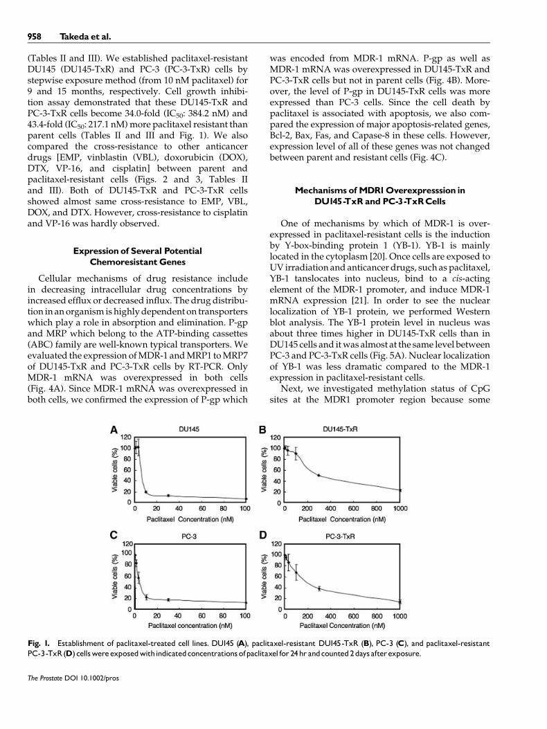
(Tables II and III). We established paclitaxel-resistantDU145 (DU145-TxR) and PC-3 (PC-3-TxR) cells bystepwise exposure method (from 10 nM paclitaxel) for9 and 15 months, respectively. Cell growth inhibi-tion assay demonstrated that these DU145-TxR andPC-3-TxR cells become 34.0-fold (IC50: 384.2 nM) and43.4-fold (IC50: 217.1 nM)more paclitaxel resistant thanparent cells (Tables II and III and Fig. 1). We alsocompared the cross-resistance to other anticancerdrugs [EMP, vinblastin (VBL), doxorubicin (DOX),DTX, VP-16, and cisplatin] between parent andpaclitaxel-resistant cells (Figs. 2 and 3, Tables IIand III). Both of DU145-TxR and PC-3-TxR cellsshowed almost same cross-resistance to EMP, VBL,DOX, and DTX. However, cross-resistance to cisplatinand VP-16 was hardly observed.
Expression of Several PotentialChemoresistantGenes
Cellular mechanisms of drug resistance includein decreasing intracellular drug concentrations byincreased efflux or decreased influx. The drug distribu-tion in anorganism ishighlydependent on transporterswhich play a role in absorption and elimination. P-gpand MRP which belong to the ATP-binding cassettes(ABC) family are well-known typical transporters. Weevaluated the expression ofMDR-1 andMRP1 toMRP7of DU145-TxR and PC-3-TxR cells by RT-PCR. OnlyMDR-1 mRNA was overexpressed in both cells(Fig. 4A). Since MDR-1 mRNA was overexpressed inboth cells, we confirmed the expression of P-gp which
was encoded from MDR-1 mRNA. P-gp as well asMDR-1 mRNA was overexpressed in DU145-TxR andPC-3-TxR cells but not in parent cells (Fig. 4B). More-over, the level of P-gp in DU145-TxR cells was moreexpressed than PC-3 cells. Since the cell death bypaclitaxel is associated with apoptosis, we also com-pared the expression of major apoptosis-related genes,Bcl-2, Bax, Fas, and Capase-8 in these cells. However,expression level of all of these genes was not changedbetween parent and resistant cells (Fig. 4C).
MechanismsofMDR1Overexpresssion inDU145-TxR and PC-3-TxRCells
One of mechanisms by which of MDR-1 is over-expressed in paclitaxel-resistant cells is the inductionby Y-box-binding protein 1 (YB-1). YB-1 is mainlylocated in the cytoplasm [20]. Once cells are exposed toUV irradiation and anticancer drugs, such as paclitaxel,YB-1 tanslocates into nucleus, bind to a cis-actingelement of the MDR-1 promoter, and induce MDR-1mRNA expression [21]. In order to see the nuclearlocalization of YB-1 protein, we performed Westernblot analysis. The YB-1 protein level in nucleus wasabout three times higher in DU145-TxR cells than inDU145 cells and itwas almost at the same level betweenPC-3 and PC-3-TxR cells (Fig. 5A). Nuclear localizationof YB-1 was less dramatic compared to the MDR-1expression in paclitaxel-resistant cells.
Next, we investigated methylation status of CpGsites at the MDR1 promoter region because some
The Prostate DOI 10.1002/pros
Fig. 1. Establishment of paclitaxel-treated cell lines. DU145 (A), paclitaxel-resistant DU145-TxR (B), PC-3 (C), and paclitaxel-resistantPC-3-TxR(D) cellswereexposedwithindicatedconcentrationsofpaclitaxel for24hr andcounted2days afterexposure.
958 Takeda et al.

groups reported inverse correlation between methyla-tion and MDR1 expression in [19,22,23]. Since DU145-TxR and PC-3-TxR cells overexpressed MDR1 mRNAcompared to parent cells, we expected that paclitaxel-resistance might cause demethylation of CpG sites atMDR1 promoter. AlthoughMSP published by Enokidaet al. detected PCR products from bisulfite-modifiedDNA in both parent cells and paclitaxel-resistant cells,USP detected stronger PCR band in DU145-TxR cellsthan inDU145 cells, suggesting thatMDR1 promoter in
DU145-TxR cells is lessmethylated than inDU145 cells.However, USP did not detect PCR band in PC-3-TxRcells compared to PC-3 (Fig. 5B). To further confirm themethylated CpG site at the MDR1 promoter, weperformed DNA sequence analysis using bisulfite-modified DNA. The MDR1 promoter region of DU145cells was methylated at the CpG sites of �134, �105,�59, �56, �51, �34, and �29 of the transcriptioninitiation site. The MDR1 promoter region of DU145-TxR cells was methylated only at the CpG site of -105
The Prostate DOI 10.1002/pros
Fig. 2. Cross-resistance of DU145 and DU145-TxR cells. DU145and DU145-TxR cells were exposedwith indicated concentrationsofEMP, docetaxel (DTX), vinblastin (VBL), doxorubicin (DOX), cis-platin (CDDP), and etoposide (VP-16) for 24 hr and counted 2 daysafterexposure.
Fig. 3. Cross-resistanceofPC-3andPC-3-TxRcells.PC-3andPC-3-TxRcellswereexposedwithindicatedconcentrationsofEMP,doc-etaxel(DTX),vinblastin(VBL),doxorubicin(DOX),cisplatin(CDDP),andetoposide(VP-16) for24hrandcounted2daysafterexposure.
TABLE III. IC50 Value of PC-3 and PC-3-TxRCells
Drug PC-3 PC-3-TxR Fold difference
PTX (nM) 5.00 217.1 43.4EMP (mM) 8.57 33.0 3.85DTX (nM) 3.67 28.2 7.68VBL (nM) 8.00 27.4 2.43DOX (nM) 121.3 1,218.2 10.0VP-16 (mM) 4.40 5.95 1.35CDDP (mM) 1.47 1.66 1.13
TABLE II. IC50 Value ofDU145 andDU145-TxRCells
Drug DU145 DU145-TxR Fold difference
PTX (nM) 11.3 384.2 34.0EMP (mM) 15.1 49.6 3.28DTX (nM) 8.30 55.6 6.70VBL (nM) 14.1 40.8 2.89DOX (nM) 17.5 61.1 3.49VP-16 (mM) 0.83 1.10 1.33CDDP (mM) 1.32 1.97 1.49
The Establishmentof TwoPaclitaxel-Resistant Prostate Cancer Cell Lines 959

(data not sown). Especially, the important region forMDR1 transcriptional regulation that included a G-box(�59, �56, and �51) [24] was demethylated in DU145-TxR cells (Fig. 5C). This demethylation of MDR1promoter in DU145-TxR cells was coincident with theenhanced MDR1 expression. Whereas DNA sequenceanalysis of the amplified PCR product showed that theMDR1 promoter regions of PC-3 and PC-3-TxR cellswere methylated at the CpG sites of �134, �110, �59,�51,�34, and�29 and at the CpG sites of,�110,�105,�59,�56,�51, and�29, respectively. Much differencewas not observed in the methylated sites and thenumber between PC-3 and PC-3-TxR promoter region.
Recoveryof Paclitaxel SensitivitybyMDR-1Knockdown
In order to investigate if MDR-1 mRNA overexpres-sion in TxR cells is the main cause of paclitaxelresistance, we knocked-down the MDR-1 mRNA byMDR-1 siRNA. Ten to thirty nanometer MDR-1 siRNAdown-regulatedMDR-1mRNA inDU145-TxR andPC-3-TxR cells 48 h after transfection (Fig. 5A and C). Non-targeting siRNA and laminin siRNA failed to inhibitMDR-1 mRNA expression. MDR-1 mRNA down-regulation by MDR-1 siRNA treatment also inhibitedthe expression of P-gp protein.
Since MDR-1 siRNA down-regulated P-gp, weconfirmed if MDR-1 down-regulation could restore
paclitaxel sensitivity. As shown in Table IV andFigure 5B and D, IC50 of in parent DU145 and PC-3cells was not changed when non-target (NT) siRNA orMDR-1 siRNA was transfected. Transfection withMDR-1 siRNAintoDU145-TxR cells after 48hr restoredpaclitaxel sensitivity compared to transfectionwithNTsiRNA (Fig. 6B). IC50 of paclitaxel of DU145-TxR wasreduced from 537.9 nM to 60.8 nM and recovery ratiobecame 88.7% 48 hr after transfection (Table IV).Whereas transfection with MDR-1 siRNA into PC-3-TxR cells hardly changed paclitaxel sensitivity. IC50 ofpaclitaxel of PC-3-TxRwas reducedonly from198.4 nMto 140.6 nMand recovery ratio became229.1% (Table IVand Fig. 6D) sensitivity.
Mechanismsof Paclitaxel Resistance inPC-3-TxRCells
Although P-gp overexpression played importantrole on paclitaxel resistance in DU145-TxR cells, thiswas not an important factor in PC-3-TxR cells. Thereshould be P-gp-independent pathway to becomepaclitaxel-resistance. In order to identify the genes thatare associatedwith onpaclitaxel resistance inPC-3-TxRcells, we performed cDNA microarray using mRNAfrom parent PC-3 and PC-3-TxR cells and compareddifferentially expressed genes as described inMaterialsand Methods. Approximately 15,000 genes werescreened by microarray analysis. two hundred andone (1.34%) of screened genes were inducedmore than
The Prostate DOI 10.1002/pros
Fig. 4. Expressionofvariousdrug-resistance-relatedgenesinparentandpaclitaxel-resistantcells. (A)RT-PCRofMDRandMRP1-7mRNAinDU145,DU145-TxR,PC-3,andPC-3-TxRcells.AftermRNAwaspurifiedfromthesecells,RT-PCRwasperformedusingprimersasdescribedinTable1. (B)ExpressionofP-gp.Cellswereculturedfor12hin thepresenceofindicatedconcentrationofDHTorAdiolandharvested.Membraneand cytosol proteinwere extracted as described in Materials and Methods and loaded on an 7.5% SDS-polyacrylamide gel for Western blotanalysis. After proteinwas transferred to PVDF-membrane, anti-P-gp antibody and anti-GAPDH antibody were employed for detection of170kDaP-gp and 37kDaGAPDHprotein, respectively. (C) RT-PCRofbcl-2, Bax,Fas, andcapase-8mRNAinDU145,DU145-TxR,PC-3, andPC-3-TxRcells.
960 Takeda et al.

two-fold and 218 (1.45%) of genes were reduced morethan two-fold in PC-3-TxR cell line compared withparent PC-3 cell line. TablesVandVIdescribe themajor30 genes that showed up-regulated and down-regu-lated expression in PC-3-TxR cells comparedwith PC-3cells. As we confirmed in Figure 4, MDR-1 genes was
up-regulated to 6.0-fold in PC-3-TxR cells. Somemicrotubule-related genes, tubuline b6, b2, and b4,wereup-regulated to 3.5-, 2.2-, and2.1-fold inPC-3-TxRcells, respectively. Calcium is an important factorthat is associated with microtubule polymerization.Calcium-binding protein, S100A9 and S100A8 were
The Prostate DOI 10.1002/pros
Fig. 5. Expression ofYB-1protein andmethylation status ofMDR1promoter. (A)Westernblottingof YB-1protein.Whole cell protein andnuclearproteinwereextractedasdescribedinMaterialsandMethodsandloadedona12.5%SDS-polyacrylamidegelforWesternblotting.Afterproteinwas transferred to PVDFmembrane, anti-YB-1or GAPDH antibody was employed for detection of 35.4 or 37 kDaYB-1or GAPDHprotein, respectively.B: Detection of methylated and unmethylated promoter of MDR1genes.USP and MSP were employed for detectionof unmethylated and methylated MDR1 promoter after the 223 bp MDR1 promoter region was amplified from bisulfite-modified DNA.C: Bisulfite-modified DNA sequence of MDR1promoter.The sequences of bisulfite-modified MDR1promoter regions fromDU145,DU145-TxR,PC-3, andPC-3-TxRcellswere shownfrom�65to�21of transcriptioninitiation site.Underlines anddoubleunderline showmethylatedCpGsites andG-box,respectively.
TABLE IV. IC50 Value of Paclitaxel in iMDR-1-Transfected TxRCells
Transfected cells IC50 (nM) Relative resistant ratio Recovery ratio
DU145 (NT siRNA) 9.74 1.0DU145 (iMDR-1) 9.11 0.94 6%DU145-TxR (NT siRNA) 537.9 55.2DU145-TxR (iMDR-1) 60.8 6.24 88.7%PC-3 (NT siRNA) 10.5 1.0PC-3 (iMDR-1) 10.0 0.95 5%PC-3-TxR (NT siRNA) 198.4 18.9PC-3-TxR (iMDR-1) 140.6 13.4 29.1%
The Establishmentof Two Paclitaxel-Resistant Prostate CancerCell Lines 961

down-regulated to 4.34- and 2.56-fold in PC-3-TxRcells, respectively. Other calcium-related genes, tumor-associated calcium signal transducer 1 (TACSTD1),S100P, and S100A2 mRNA were also down-regulatedin PC-3-TxR cells. MMP-1 that is related with cancerinvasion is overexpressed in multiple drug-resistantcell lines [25]. We also observed overexpression ofMMP-1 in PC-3-TxR cells (4.77-fold).
DISCUSSION
In order to elucidate the mechanisms of paclitaxel -resistant in hormone refractory PCa, we establishedtwo paclitaxel-resistant cell lines from androgen-independent cell lines. Several potential mechanismshave been proposed for resistance to taxans. The resultthat cross-resistance to cisplatin and VP-16 was notobserved in both paclitaxel-resistant cell lines indicatesthat resistance to paclitaxel is resulted from differentpathways from resistance to cisplatin and VP-16.Although paclitaxel induces apoptosis, we could notdetect differences of expression in apoptosis-relatedgenes, such as bcl-2, bax, caspase 8 between parent cells
and TxR cells. One of major mechanisms of paclitaxel-resistance is overexpression of P-gp [9]. The MDR-1overexpression was the important factor as a respon-sible gene when DU145 cells became paclitaxel resis-tance. Since MDR-1 siRNA almost restored paclitaxelsensitivity in DU145-TxR cells, P-gp overexpression isthe main reason of paclitaxel resistance in this cell line.
Our results showed that one ofmainmechanisms bywhich of MDR-1 was overexpressed in paclitaxel-resistant DU145 cells was the demethylation of CpGsites at the MDR1 promoter region. Originally CpGsites at the MDR1 promoter region in parent DU145cells were hypermethylated [19]. Because it is rare, asfor the necessity of MDR1, expression of MDR1 isinhibited for cancer cell by methylation of MDR1promoter. However, when cells can leave damage bypaclitaxel, demethylation of MDR1 promoter, espe-cially G-box that includes Sp1-binding site and EGR-1-binding site and is very important for transcription [24],is promoted and induces expression of MDR1 so thatcell themselves survives it, then cells may be going toremove paclitaxel from intracellular. However, itremains unclear why PC-3-TxR cells overexpressed
The Prostate DOI 10.1002/pros
Fig. 6. PaclitaxeliniMDR-1transfectedTxRcells.AandC:Forty-eighthoursafter transfectionwith0,10,20,or30nMMDR-1siRNA,30nMLaminA/C siRNA (La), and 30 nM non-targeting siRNA (La), total RNA andproteinwas extracted according to theMaterials andMethods.BandD: Inorder to see theeffectof siRNAondrugresistance,cellswere transfectedwith30nMMDR-1siRNAornon-targeting siRNA24hrafterplatingon24-wellplates.Twenty-fourhoursafter transfectionwith30nMnon-targetingiRNAoriMDR-1,cellsweretreatedwith0,1,3,10,30,100, 300, and1,000 nM paclitaxel and cultured for 48 hr.Then the cells were countedwith a hemocytometer.The data representmean oftriplicateexperiments andthebars showSD.ThedataweredescribedinTable1V.
962 Takeda et al.

The Prostate DOI 10.1002/pros
TABLE V. List of GeneswhichwereOverexpressedin PC-3-TxRCells
Genename
Systematicname
PC-3-TxRsignal
PC-3signal
Foldchange Description
TNS NM_022648 704 97 7.23 TensinABCB1 NM_000927 5,902 980 6.02 ATP-binding cassette, subfamily B (MDR/TAP)LAMA4 NM_002290 10,609 1,991 5.33 Laminin, alpha 4 (LAMA4)IGSF4 NM_014333 2,586 486 5.32 Immunoglobulin superfamily, member 4CD33L3 AK092746 22,410 4,403 5.09 cDNA FLJ35427 fis, clone SMINT2001731MMP1 NM_002421 10,946 2,293 4.77 Matrix metalloproteinase 1 (interstitial collagenase)TIMP4 NM_003256 2,786 644 4.33 Tissue inhibitor of metalloproteinase 4AUTS2 NM_015570 821 199 4.12 Autism susceptibility candidate 2PLA2G7 NM_005084 4,353 1,068 4.08 Phospholipase A2, group VII (platelet-activating
factor acetylhydrolase, plasma)ROBO4 NM_019055 2,058 508 4.05 Roundabout homolog 4, magic roundabout (Drosophila)IL1RL1 NM_016232 1,121 286 3.92 Interleukin 1 receptor-like 1 (IL1RL1), transcript
variant 1POU4F3 NM_002700 557 148 3.76 POU domain, class 4, transcription factor 3SLC35F2 NM_017515 31,443 8,611 3.65 Solute carrier family 35, member F2FZD4 NM_012193 9,899 2,791 3.55 Frizzled homolog 4 (Drosophila) (FZD4)MGC4083 NM_032525 20,402 5,787 3.53 Tubulin beta MGC4083TFPI2 AK092499 21,336 6,056 3.52 cDNA FLJ35180 fis, clone PLACE6014882, similar to
Tissue Factor pathway inhibitor 2CHKA NM_001277 893 262 3.41 Choline kinasePFTK1 NM_012395 2,295 705 3.26 PFTAIRE protein kinase 1BNIP3 NM_004052 26,713 8,310 3.21 BCL2/adenovirus E1B 19 kDa interacting protein 3C14orf149 NM_144581 25,746 8,094 3.18 Hypothetical protein FLJ25436H19 AK056774 2,557 815 3.14 cDNA FLJ32212 fis, clone PLACE6003399, weakly
similar to SPIDROIN 1MGC2574 NM_024098 24,317 7,812 3.11 Hypothetical protein MGC2574LOC114990 NM_138440 1,953 647 3.02 Hypothetical protein BC013767MGC10981 BC004397 776 259 2.99 Hypothetical protein MGC10981PRSS11 NM_002775 14,523 5,115 2.84 Protease, serine, 11 (IGF binding)DPYSL4 NM_006426 776 274 2.83 Dihydropyrimidinase-like 4GPR56 NM_005682 32,764 11,683 2.80 G protein-coupled receptor 56BAG3 NM_004281 3,952 1,425 2.77 BCL2-associated athanogene 3SC5DL BC012333 4,652 1,685 2.76 Sterol-C5-desaturase (ERG3 delta-5-desaturase
homolog, fungal)-likeC10orf125 NM_198472 3,036 1,101 2.76 FLJ26016 protein (FLJ26016)MTVR1 NM_152832 8,897 3,302 2.69 Mouse mammary turmor virus receptor homolog 1GPR4 NM_005282 1,507 565 2.66 G protein-coupled receptor 4FAHD1 NM_031208 6,677 2,517 2.65 Hypothetical protein DKFZp566J2046FLJ37078 NM_153043 395 149 2.64 Hypothetical protein FLJ37078POLA2 NM_002689 10,368 3,935 2.64 Polymerase (DNA-directed), alpha (70kD)LOC201194 AK022617 540 207 2.61 cDNA FLJ12555 fisMGC16291 NM_032770 3,167 1,228 2.58 Hypothetical protein MGC16291ESM1 NM_007036 7,643 2,966 2.58 Endothelial cell-specific molecule 1CDCA5 NM_080668 15,949 6,219 2.56 Cell division cycle associated 5DGAT2 NM_032564 530 207 2.56 Diacylglycerol O-acyltransferase homolog 2SLC35F2 AK128062 2,548 998 2.55 cDNA FLJ46182 fisLOC201194 AK022617 488 192 2.54 cDNA FLJ12555 fis, clone NT2RM4000764ESM1 NM_007036 6,500 2,560 2.54 Endothelial cell-specific molecule 1LOC201194 AK022617 553 218 2.53 cDNA FLJ12555 fis, clone NT2RM4000764EHBP1L1 AL834433 2,554 1,008 2.53 cDNA DKFZp762C186FANCA NM_000135 874 348 2.51 Fanconi anemia, complementation group AESM1 NM_007036 10,124 4,042 2.50 Endothelial cell-specific molecule 1 (ESM1), mRNA
The Establishmentof Two Paclitaxel-Resistant Prostate CancerCell Lines 963

The Prostate DOI 10.1002/pros
TABLE VI. Listof Geneswhichwere Repressed in PC-3-TxR
Genename
Systematicname
PC-3-TxRsignal
PC-3signal
Foldchange Description
IL23A NM_016584 988 16,873 17.07 Interleukin 23, alpha subunit p19CALB1 NM_004929 482 7,509 15.57 Calbindin 1, 28 kDaCTEN NM_032865 591 6,318 10.69 C-terminal tensin-likePLAC8 NM_016619 290 2,886 9.95 Placenta-specific 8LXN NM_020169 214 1,957 9.13 Latexin proteinCDH1 NM_004360 626 4,957 7.92 Cadherin 1, type 1, E-cadherin (epithelial)S100A2 NM_005978 2,579 17,878 6.93 S100 calcium-binding protein A2KLK6 NM_002774 140 930 6.66 Kallikrein 6 (neurosin, zyme)IL6 NM_000600 1,592 10,288 6.46 Interleukin 6 (interferon, beta 2)LCN2 NM_005564 1,277 7,457 5.84 Lipocalin 2 (oncogene 24p3)IL13RA2 NM_000640 143 752 5.26 Interleukin 13 receptor, alpha 2CSF2 NM_000758 1,595 8,099 5.08 Colony stimulating factor 2 (granulocyte-macrophage)CD33 NM_001772 205 1,037 5.07 CD33 antigen (gp67)SERPINB4 NM_002974 708 3,545 5.01 Serine (or cysteine) proteinase inhibitor, clade B
(ovalbumin), member 4CYP1B1 NM_000104 3,090 15,173 4.91 Cytochrome P450, family 1, subfamily B, polypeptide 1NOX5 NM_024505 1,707 7,840 4.59 NADPH oxidase, EF hand calcium-binding domain 5CRB3 NM_174882 390 1,774 4.55 Crumbs homolog 3 (Drosophila) (CRB3)NMES1 NM_032413 3,567 16,181 4.54 Normal mucosa of esophagus specific 1PTAFR NM_000952 297 1,344 4.53 Platelet-activating factor receptorTHBD NM_000361 165 749 4.53 ThrombomodulinSAT NM_002970 2,227 10,029 4.50 Spermidine/spermine N1-acetyltransferaseFXYD6 NM_022003 594 2,674 4.50 FXYD domain containing ion transport regulator 6SAA1 NM_000331 410 1,834 4.47 Serum amyloid A1 (SAA1)TACSTD1 NM_002354 4,722 21,027 4.45 Tumor-associated calcium signal transducer 1IL1F7 NM_014439 1,532 6,811 4.45 Interleukin 1 family, member 7 (zeta)ADMP NM_145035 504 2,213 4.39 ADMPIFI27 NM_005532 211 915 4.35 Interferon, alpha-inducible protein 27S100A9 NM_002965 2,678 11,624 4.34 S100 calcium-binding protein A9 (calgranulin B)AY358920 AY358920 216 931 4.31 Clone DNA129549 ALGV3072 (UNQ3072)IFI27 NM_005532 139 594 4.27 Interferon, alpha-inducible protein 27ALOX5AP NM_001629 2,028 8,503 4.19 Arachidonate 5-lipoxygenase-activating proteinAREG NM_001657 1,461 6,122 4.19 Amphiregulin (schwannoma-derived growth factor)ANKRD1 NM_014391 131 545 4.16 Ankyrin repeat domain 1 (cardiac muscle)SGNE1 NM_003020 748 3,076 4.11 Secretory granule, neuroendocrine protein 1 (7B2 protein)IFITM1 NM_003641 698 2,783 3.99 Interferon induced transmembrane protein 1 (9–27)FCGR2C NM_201563 198 785 3.96 Fc fragment of IgG, low affinity IIc, receptor for (CD32)FLJ31204 NM_174912 150 584 3.89 Hypothetical protein FLJ31204S100P NM_005980 2,465 9,570 3.88 S100 calcium-binding protein PHIST1H1C NM_005319 3,943 14,847 3.77 Histone 1, H1cCD33 AY162464 500 1,875 3.75 Sialic acid-binding immunoglobulin-like lectin 3CXCL2 NM_002089 4,477 16,726 3.74 Chemokine (CXC motif) ligand 2CXCL3 NM_002090 4,728 17,552 3.71 Chemokine (CXC motif) ligand 3SERPINB3 NM_006919 1,206 4,461 3.70 Serine (or cysteine) proteinase inhibitor, clade B
(ovalbumin), member 3CCL20 NM_004591 844 3,109 3.68 Chemokine (CC motif) ligand 20KLF5 NM_001730 2,148 7,908 3.68 Kruppel-like factor 5 (intestinal)AMIGO2 NM_181847 852 3,108 3.65 Homo sapiens amphoterin induced gene 2AIM2 NM_004833 232 845 3.64 Absent in melanoma 2THC1991570 THC1991570 1,416 5,074 3.58 AY140952 G-protein coupled receptor GPR110SAA2 NM_030754 450 1,591 3.54 Serum amyloid A2TGFA NM_003236 1,294 4,544 3.51 Transforming growth factor, alphaFGFBP1 NM_005130 1,619 5,680 3.51 Heparin-binding growth factor-binding protein
964 Takeda et al.

MDR1 mRNA compared to PC-3 cells although theexpression level in PC-3-TxR cells was lower than inDU145-TxR cells. It will be very interesting to studywhy paclitaxel exposure causes demethylation of theMDR1 promoter region of DU145 cells.
Inhibition of MDR-1 hardly restored resistance inPC-3-TxR although PC-3-TxR cells overexpressed P-gpcompared to parent PC-3 cells. Only by overexpressionof p-gp, there is not explanation of the mechanismthat PC -3 cells become paclitaxel resistance. Othermechanisms should be involved in paclitaxel-resis-tance in PC-3-TxR cells. Lin et al.[26] demonstratedthatDOX resistance rat PCa cell line expressedmore Id-1, MIF, and GSTpi mRNA than parent cell line . Theyalso showed that overexpression of Id-1 causedpaclitaxel-resistance in the cell line.However,we couldnot detect thedifference of Id-1 expression betweenPC-3 and PC-3-TxR cells although Id-1 mRNA wastemporally down-regulated by paclitaxel treatment inPC-3 cells.
In order to investigate what genes are involved inpaclitaxel resistance, we compared gene expressionprofile between PC-3 and PC-3-TxR cells. To the best ofour knowledge, this is the first report that comparedgene expression profile about paclitaxel-resistance inhormone refractory PCa cell line. Expressions of manygenes were also altered in paclitaxel-resistant breastcancer cells [16]. Expression patterns were similar insome of these genes, such as MDR1 and S100P.However, those in PC-3-TxR cells were different frombreast cancer cells, suggesting that different mechan-isms are involved in becoming paclitaxel-resistance indifferent cancers.
Paclitaxel shows the effect as an anticancer drug bystabilizing polymer of microtubule [27]. Alterations ofmicrotubule formation in resistant cells is also impor-tant factors [10,28]. Li et al.[29] demonstrated bymicroarray analysis that taxotere regulatedmanygenesincluding microtubule, apoptosis, and cell cycle-related genes in PCa cell lines, PC-3 and LNCaP cells.Especially, microtubule-related genes are down-regu-lated in those cells. They treated cells with taxoteretransiently and compare the regulated genes beforeand after treatment. Down-regulated genes after treat-mentmay be the geneswhich, as a result of having beenimpaired, were inhibited by taxotere. Or up-regulatedgenes may be the genes which, as a result, are elevatedwhen apoptosis by taxotere is induced. Ranganathanet al.[30] demonstrated that increase in tubulin bIII(nine-fold) and bIVa (five-fold) were observed inDU145 cells that became paclitaxel-resistance. Orret al.[10] also reviewed that alterations in tubulincomposition expression were associated with pacli-taxel resistance.Wealso confirmed theup-regulation ofsome tubulin b-6 (3.53-fold), �2 (2.22-fold), and �4
(2.13-fold) in PC-3-TxR cells by cDNA microarrayanalysis. However, overexpression of bIII isotype inhuman prostate carcinoma cells by stable transfectionfailed to confer antimicrotubule drug resistance tothese cells [31]. Interestingly, overexpression of tubulinb are relatedwithpoor prognosis and resistance [32].Atleast overexpression of tubulin bs may be thought witha good marker predicting with reactivity for paclitaxeland prognosis. Wewill investigate if overexpression oftubulin bs causes paclitaxel resistance and progressionin PC-3 cells.
Paclitaxel is known to repress influx of calcium intocytoplasm [33,34]. Reduction of calcium-associatedproteins expression may be a cause of repression ofcalcium influx by paclitaxel and may not be amechanism of paclitaxel resistance. However, calciumdynamics which is associated with microtubule poly-merization is important factor for paclitaxel-resistance.Moreover, altered intracellular calcium homeostasismay contribute to the paclitaxel-resistant phenotype[35]. Microarray analysis in this study revealed adecline of S100A8/S100A9 expression in PC-3-TxRcells compared with parent PC-3 cells. Calcium-induced complexes of S100A8 and S100A9 have beenshown to colocalize with microtubules during activa-tion of monocytes. They directly bind to tubulin andpromote tubulin polymerization in a calcium-depen-dent manner [36]. Then failure of tetramer formationwas associated with a lack of functional activity ofS100A8/S100A9 complexes in promoting the forma-tion of microtubules [37]. A decline of S100A8/S100A9expression would also inhibit the formation of micro-tubules. Therefore, since paclitaxel cannot stabilize theformation of microtubules due to a decline of S100A8/S100A9 expression in PC-3-TxR cells, paclitaxel mightnot be able to show effect as an anticancer drug inresistant cells.
In conclusions, after we established paclitaxel-resistant hormone refractory PCa cell lines, we com-pared resistant cells with parent cells. This comparisonwill make it more possible to identify the genes whichcause paclitaxel resistance except MDR-1. Not onlyMDR-1 gene but also many genes were up-regulatedand down-regulated. We have to still distinguish thegenes that are responsible for resistance from the genesthat are regulated as a result one by one. Nevertheless,identification of these genes will be useful for thinkingstrategies using taxanes to individual hormone refrac-tory PCa.
ACKNOWLEDGMENTS
We thank Saeko Fujii, Yukari Kawabuchi, andChiharu Shimoda for assistance. This work wassupported in part by Japan Society for the Promotionof Science Grant 17591669 (A. Mizokami).
The Prostate DOI 10.1002/pros
The Establishmentof Two Paclitaxel-Resistant Prostate Cancer Cell Lines 965

REFERENCES
1. Jemal A, Siegel R, Ward E, Murray T, Xu J, Smigal C, Thun MJ.Cancer statistics, 2006. CA Cancer J Clin 2006;56(2) 106–130.
2. Obasaju C, Hudes GR. Paclitaxel and docetaxel in prostatecancer. Hematol Oncol Clin North Am 2001;15(3):525–545.
3. Petrylak DP, Tangen CM, Hussain MH, Lara PN Jr, Jones JA,Taplin ME, Burch PA, Berry D, Moinpour C, Kohli M, BensonMC, Small EJ, Raghavan D, Crawford ED. Docetaxel andestramustine compared with mitoxantrone and prednisone foradvanced refractory prostate cancer. N Engl J Med 2004;351(15):1513–1520.
4. Tannock IF, de Wit R, Berry WR, Horti J, Pluzanska A, Chi KN,Oudard S, Theodore C, James ND, Turesson I, Rosenthal MA,Eisenberger MA. Docetaxel plus prednisone or mitoxantroneplus prednisone for advanced prostate cancer. N Engl J Med2004;351(15):1502–1512.
5. Oudard S, Banu E, Beuzeboc P, Voog E, Dourthe LM, Hardy-Bessard AC, Linassier C, Scotte F, Banu A, Coscas Y, Guinet F,Poupon MF, Andrieu JM. Multicenter randomized phase IIstudy of two schedules of docetaxel, estramustine, and pre-dnisone versus mitoxantrone plus prednisone in patients withmetastatic hormone-refractory prostate cancer. J Clin Oncol2005;23(15):3343–3351.
6. Fulton B, Spencer CM. Docetaxel. A review of its pharmacody-namic and pharmacokinetic properties and therapeutic efficacyin themanagement ofmetastatic breast cancer.Drugs 1996;51(6):1075–1092.
7. Duan Z, Lamendola DE, Duan Y, Yusuf RZ, Seiden MV.Description of paclitaxel resistance-associated genes in ovarianand breast cancer cell lines. Cancer Chemother Pharmacol2005;55(3):277–285.
8. Teraishi F, Wu S, Sasaki J, Zhang L, ZhuHB, Davis JJ, Fang B. P-glycoprotein-independent apoptosis induction by a novelsynthetic compound, MMPT [5-[(4-methylphenyl)methylene]-2-(phenylamino)-4(5H)-thiazolone]. J Pharmacol Exp Ther2005;314(1):355–362.
9. Hopper-Borge E, Chen ZS, Shchaveleva I, Belinsky MG, KruhGD. Analysis of the drug resistance profile of multidrugresistance protein 7 (ABCC10): resistance to docetaxel. CancerRes 2004;64(14):4927–4930.
10. Orr GA, Verdier-Pinard P, McDaid H, Horwitz SB. Mechanismsof Taxol resistance related to microtubules. Oncogene2003;22(47):7280–7295.
11. ChunE, LeeKY. Bcl-2 andBcl-xL are important for the inductionof paclitaxel resistance in human hepatocellular carcinoma cells.Biochemical and Biophysical Research Communications 2004;315(3):771–779.
12. Ferlini C, Raspaglio G, Mozzetti S, Distefano M, Filippetti F,Martinelli E, Ferrandina G, Gallo D, Ranelletti FO, Scambia G.Bcl-2 down-regulation is a novel mechanism of paclitaxelresistance. Mol Pharmacol 2003;64(1):51–58.
13. Yamanaka K, Rocchi P, Miyake H, Fazli L, So A, Zangemeister-Wittke U, Gleave ME. Induction of apoptosis and enhancementof chemosensitivity in human prostate cancer LNCaP cells usingbispecific antisense oligonucleotide targeting Bcl-2 and Bcl-xLgenes. BJU Int 2006;97(6):1300–1308.
14. Yamanaka K, Rocchi P, Miyake H, Fazli L, Vessella B,Zangemeister-Wittke U, Gleave ME. A novel antisense oligonu-cleotide inhibiting several antiapoptotic Bcl-2 family membersinduces apoptosis and enhances chemosensitivity in androgen-independent human prostate cancer PC3 cells. Mol Cancer Ther2005;4(11):1689–1698.
15. GotoT, TakanoM, SakamotoM,KondoA,Hirata J, KitaT, TsudaH, Tenjin Y, Kikuchi Y. Gene expression profiles with cDNAmicroarray reveal RhoGDI as a predictive marker for paclitaxelresistance in ovarian cancers. Oncol Rep 2006;15(5):1265–1271.
16. Villeneuve DJ, Hembruff SL, Veitch Z, Cecchetto M, Dew WA,Parissenti AM. cDNAmicroarray analysis of isogenic paclitaxel-and doxorubicin-resistant breast tumor cell lines reveals distinctdrug-specific genetic signatures of resistance. Breast Cancer ResTreat 2006;96(1):17–39.
17. Mizokami A, Koh E, Fujita H, Maeda Y, Egawa M, Koshida K,Honma S, Keller ET, Namiki M. The adrenal androgenandrostenediol is present in prostate cancer tissue after andro-gen deprivation therapy and activates mutated androgenreceptor. Cancer Res 2004;64(2):765–771.
18. Ueda K, Pastan I, GottesmanMM. Isolation and sequence of thepromoter region of the human multidrug-resistance (P-glyco-protein) gene. J Biol Chem 1987;262(36):17432–17436.
19. Enokida H, Shiina H, Igawa M, Ogishima T, Kawakami T,Bassett WW, Anast JW, Li LC, Urakami S, Terashima M, VermaM, Kawahara M, Nakagawa M, Kane CJ, Carroll PR, Dahiya R.CpG hypermethylation of MDR1 gene contributes to thepathogenesis and progression of human prostate cancer. CancerRes 2004;64(17):5956–5962.
20. Bargou RC, Jurchott K, Wagener C, Bergmann S, Metzner S,Bommert K, Mapara MY, Winzer KJ, Dietel M, Dorken B,Royer HD. Nuclear localization and increased levels oftranscription factor YB-1 in primary human breast cancers areassociated with intrinsic MDR1 gene expression. Nat Med1997;3(4):447–450.
21. Ohga T, Uchiumi T, Makino Y, Koike K, Wada M, Kuwano M,Kohno K. Direct involvement of the Y-box binding protein YB-1in genotoxic stress-induced activation of the human multidrugresistance 1 gene. J Biol Chem 1998;273(11):5997–6000.
22. Nakayama M, Wada M, Harada T, Nagayama J, Kusaba H,Ohshima K, Kozuru M, Komatsu H, Ueda R, Kuwano M.Hypomethylation status of CpG sites at the promoter region andoverexpression of the human MDR1 gene in acute myeloidleukemias. Blood 1998;92(11):4296–4307.
23. Tada Y, Wada M, Kuroiwa K, Kinugawa N, Harada T,Nagayama J, Nakagawa M, Naito S, Kuwano M. MDR1 geneoverexpression and altered degree of methylation at thepromoter region in bladder cancer during chemotherapeutictreatment. Clin Cancer Res 2000;6(12):4618–4627.
24. Cornwell MM, Smith DE. SP1 activates the MDR1 promoterthrough one of two distinct G-rich regions that modulatepromoter activity. J Biol Chem 1993;268(26):19505–19511.
25. Yang JM, Xu Z, Wu H, Zhu H, Wu X, Hait WN. Overexpressionof extracellular matrix metalloproteinase inducer in multidrugresistant cancer cells. Mol Cancer Res 2003;1(6):420–427.
26. Lin JC,ChangSY,HsiehDS,LeeCF,YuDS. Theassociation of Id-1, MIF and GSTpi with acquired drug resistance in hormoneindependent prostate cancer cells. Oncol Rep 2005;13(5):983–988.
27. Dumontet C, Sikic BI. Mechanisms of action of and resistance toantitubulin agents: microtubule dynamics, drug transport, andcell death. J Clin Oncol 1999;17(3):1061–1070.
28. Drukman S, Kavallaris M. Microtubule alterations and resis-tance to tubulin-binding agents (review). Int J Oncol 2002;21(3):621–628.
29. Li Y, Li X, Hussain M, Sarkar FH. Regulation of microtubule,apoptosis, and cell cycle-related genes by taxotere in prostatecancer cells analyzed by microarray. Neoplasia 2004;6(2):158–167.
The Prostate DOI 10.1002/pros
966 Takeda et al.

30. Ranganathan S, Benetatos CA, Colarusso PJ, Dexter DW, HudesGR. Altered beta-tubulin isotype expression in paclitaxel-resistant human prostate carcinoma cells. Br J Cancer1998;77(4):562–566.
31. Ranganathan S, McCauley RA, Dexter DW, Hudes GR.Modulation of endogenous beta-tubulin isotype expression asa result of human beta(III)cDNA transfection into prostatecarcinoma cells. Br J Cancer 2001;85(5):735–740.
32. Song JH,Choi CH, YeomHJ,Hwang SY, KimTS.Monitoring thegene expression profiles of doxorubicin-resistant acute myelo-cytic leukemia cells by DNA microarray analysis. Life Sci2006;79(2):193–202.
33. Burke WJ, Raghu G, Strong R. Taxol protects against calcium-mediated death of differentiated rat pheochromocytoma cells.Life Sci 1994;55(16):313–319.
34. Furukawa K, MattsonMP. Taxol stabilizes [Ca2þ]i and protectshippocampal neurons against excitotoxicity. Brain Res 1995;689(1):141–146.
35. Padar S, van Breemen C, Thomas DW, Uchizono JA, Livesey JC,Rahimian R. Differential regulation of calcium homeostasis inadenocarcinoma cell line A549 and its Taxol-resistant subclone.Br J Pharmacol 2004;142(2):305–316.
36. Vogl T, Ludwig S, Goebeler M, Strey A, Thorey IS, Reichelt R,Foell D, Gerke V,ManitzMP, NackenW,Werner S, Sorg C, RothJ. MRP8 and MRP14 control microtubule reorganization duringtransendothelial migration of phagocytes. Blood 2004;104(13):4260–4268.
37. LeukertN,Vogl T, StrupatK, Reichelt R, SorgC, Roth J. Calcium-dependent tetramer formationof S100A8 andS100A9 is essentialfor biological activity. J Mol Biol 2006;359(4):961–972.
The Prostate DOI 10.1002/pros
The Establishmentof TwoPaclitaxel-Resistant Prostate Cancer Cell Lines 967