the epidemiology and pathophysiology of caseous ... · reported to be widely prevalent in the...
TRANSCRIPT

CentralBringing Excellence in Open Access
Cite this article: Osman AY, Nordin ML, Kadir AA, Saharee AA (2018) The Epidemiology and Pathophysiology of Caseous Lymphadenitis: A Review. J Vet Med Res 5(3): 1129.
Journal of Veterinary Medicine and Research
*Corresponding authorAbdinasir Yusuf Osman, Department of Veterinary Clinical Studies, Faculty of Veterinary Medicine, Universiti Malaysia Kelantan, Pengkalan Chepa 16100 Kota, Bharu, Kelantan, Malaysia, E-mail:
Submitted: 04 April 2018
Accepted: 19 April 2018
Published: 02 April 2018
ISSN: 2378-931X
Copyright© 2018 Dey et al.
OPEN ACCESS
Keywords•C.pseudotuberculosis;Epidemiology;
Pathophysiology; Small ruminant
Abstract
Caseous lymphadenitis is one of the most significant zoonotic diseases caused by C. pseudotuberculosis with enormous economic losses in animal industry worldwide. The global burden of its incidence in animal populations remains at an alarming rate. The impact of the disease is multidimensional in nature and not always well understood, therefore, significantly complicating effective policy response. The pathogenesis is complex and governed by several factors working together in synergistic manner. Information related to the epidemiology and pathophysiology is still scarce in the database and control programmes are rarely implemented. Therapy is based on wide spectrum antibiotics with mysterious outcome as pre-existing vaccines appear not promising. Thus, understanding the biological behaviour of the disease becomes a fundamental issue. In this review, we highlight various key aspects of the disease with special reference to the epidemiology and the pathophysiology of the disease in sheep and goat populations.
INTRODUCTIONCorynebacterium pseudotuberculosis is a causative agent of
chronic infections in a number of different mammalian species, the most significant of which is caseous lymphadenitis (CLA) or cheesy gland, a chronic granulomatous infectious disease of sheep and goats that is characterized by abscessation of one or more lymph nodes. The organism was first isolated from a case of lymphangitis in a cow in 1888 by French bacteriologist Edward Nocard. Years later, however, another bacteriologist, Hugo von Preisz, isolated analogous organism from a different case. CLA is distributed globally and causes important economic losses for ovine and caprine breeders due to body wasting, subsequently reduced meat, wool, milk yields and segregation of affected animals, condemnation of downgraded of affected carcasses and skins in abattoirs [1]. C. pseudotuberculosis commonly causes CLA in sheep and goats and, less commonly, pneumonia, hepatitis, mastitis, arthritis, orchitis, subcutaneous abscess, abortion, still birth and prenatal mortalities and mastitis in cattle and buffaloes [2]. In Malaysian small ruminants, the average prevalence of CLA was found to be 30% using a combined diagnostics tests [3] and 11.1% using conventional methods [4].
Numbers of serological modalities have been suggested to detect the evidence of CLA infection in animals [5-7]. Even though these tests might be considered as a fundamental point for revealing of sub-clinically infected animals, most of them suffer from low sensitivity, poor specificity and lack of ability to
differentiate between previously exposed animals and those still harboring the pathogen.
The critical factors influencing the transmission dynamics and the coarse of infections consist of microbial adaptation and change; host susceptibility; climate alteration; changing ecosystem, demographic, and population including issues of economic development and land use; international trade; technology and industry; reduction in animal and public health services. Therefore, this review is conducted to provide more comprehensive and stimulating overview regarding the epidemiology and pathophysiology of caseous lymphadenitis, and discus the importance these aspects in controlling of the disease.
THE ORGANISM AND DISEASE HISTORYAs a result of various discoveries related to this
microbe, the organism has come to be recognized as Bacillus pseudotuberculosis. The latter name has lasted until the end of 19th century and was given in the assumption that there was a clinical similarity of lesions between caseous nodules and Mycobacterium pseudotuberculosis. Through different names in different editions, the organism was placed in the genus of Corynebacterium and at the same time renamed to become recognized as Corynebacterium ovis. Since, the latter name indicated for specification and missed entirely the general ability that may imply for other mammalian species which might also be a host for the same agent, the species name, therefore,
Review Article
The Epidemiology and Pathophysiology of Caseous Lymphadenitis: A ReviewAbdinasir Yusuf Osman1,2*, Muhammad Luqman Nordin1,3, Arifah Abdul Kadir3 and Abdul Aziz Saharee2 1Department of Veterinary Clinical Studies, Faculty of Veterinary Medicine, Universiti Malaysia Kelantan, Malaysia2Department of Veterinary Clinical Studies, Faculty of Veterinary Medicine, Universiti Putra Malaysia, Malaysia 3Department of Preclinical Studies, Faculty of Veterinary Medicine, Universiti Putra Malaysia, Malaysia

CentralBringing Excellence in Open Access
Dey et al. (2018)Email:
J Vet Med Res 5(3): 1128 (2018) 2/7
had been changed back from ovis to the earlier designation of pseudotuberculosis. From that time, the C. pseudotuberculosis has formally become known as the officially recognized disagnation that cause caseous lymphadenitis [8].
GEOGRAPHICAL DISTRIBUTIONThe global distribution of caseous lymphadenitis is almost a
mirror image of farmed small ruminants. The disease is generally multicontinent and it has been reported to be widely prevalent in Europe, Africa, Australia, South and North America, and the Middle East [9]. CLA, in many of these countries, has been generally considered as a devastating disease which is characterized by economic losses and other health issues associated with animal welfare in livestock. The infection of the disease, however, had been observed predominantly in sheep and goats.
The presence of close genotypic relationship, however, among C. pseudotuberculosis isolates ovine/goat origin from a various countries in the world [2], might be sufficient evidence that the infections had a common source, and subsequently supporting the assumption that the spread of the disease followed the exportation of sheep by colonial powers in 18th century [10]. New strict regulations, related to the presence of lesions in imported carcasses were applied worldwide. Large scale studies on different aspects of the disease including pathogenesis and epidemiological - led to further series studies of control strategies designated to reduce the remarkable CLA prevalence of that time and, therefore offered preventive ways for future studies.
PATHOGENESIS VIRULENCE FACTORSA recent study in general has mainly focused on only two known
virulence factors identified as phospholipase D (PLD) and mycolic acids. Neither virulent strain nor plasmids of C. pseudotuberculosis have yet been described. Unlike other pathogenic organisms, the genome of this pathogen has yet to be fully sequenced. The organism’s virulence mechanisms, therefore, remain poorly understood. The major virulence factor of C. pseudotuberculosis, however, is so far considered to be phospholipase D. The virulence factors believed to promote bacterial dissemination by increasing vascular permeability following infection leading to leakage of plasma from blood vessels and into the surrounding tissues, and from there into the lymphatic drainage [11]. This effect may assist pathogenesis by favoring the lymphatic drainage of C. pseudotuberculosis in tissue fluid. Studies have shown that the exotoxin (PLD) is necessary for establishment of the disease [12] while mutant strains are unable to cause abscessation of the lymph nodes. Further evidence for the importance of PLD in establishment of CLA might come from the observation that the current vaccines are produced from formalin-inactivated PLD-rich C. pseudotuberculosis culture supernatants in which PLD is considered the major virulence factor of CLA.
Mycolic acid of C. pseudotuberculosis does not produce a protective capsule but has instead a waxy mycolic acid coat on the cell wall surface. This coat has well established cytotoxic properties, which play a major role in pathogenicity [13]. The subcutaneous injection into mice of mycolic acid extracted from C. pseudotuberculosis results in the production of a localized swelling, with congestion and a central area of haemorrhagic
necrosis. In addition, mycolic acid induces degenerative changes and death in phagocytizing leucocytes (Batey, 1986) [14]. However, unlike the lethal effect of injection of similar molecules extracted from mycobacteria, the cytotoxic effect of C. pseudotuberculosis mycolic acid is confined to the site of injection [15]. Some authors have suggested that the mycolic acid coat enables C. pseudotuberculosis to survive for extended periods within the environment, a feature common to other members of the actinomycete family. Corynebacterium pseudotuberculosis is indeed relatively resistant to environmental conditions [16].
In natural infections, the waxy mycolic acid coat of C. pseudotuberculosis provides the organism with mechanical, and possibly biochemical, protection from the hydrolytic enzymes present within lysosomes which in turn enables the bacterium to survive phagocytosis and to exist within the host as a facultative intracellular parasite [17]. This capacity is likely to be essential for the migration of the organism from the point of initial entry to the eventual site of lesion development. In addition, the toxic nature of mycolic acid seems to contribute to abscess formation. In artificial infections of mice, a direct relationship was demonstrated between the quantity of cell wall lipid produced by different isolates of C. pseudotuberculosis and their ability to produce chronic abscessation.
HOST RESPONSEThe host cellular response to infection with organism
(C. pseudotuberculosis) has not been described in detail. In pathological studies involving small ruminants particularly sheep challenged subcutaneously; vast numbers of neutrophils were found to infiltrate the site of inoculation within the first few hours following infection. And within 24 h they began to move to the local drainage lymph nodes. It was notably reported that number of neutrophils began to decline after 3 days, while the numbers of macrophages at the inoculation site increased significantly [18]. Thereafter, a period of generalized inflammation of the lymph node was shown, micro abscesses developed within the cortical region of the node within 24 h of inoculation these became more numerous after approximately 6 days, from which point they began to enlarge and coalesce to form more significant purulent foci. These early pyogranulomas contained clumps of bacteria and cellular debris and a high proportion of eosinophils, giving a characteristic green colour to the purulent material. In addition and concurrently with the cellular response at the site of inoculation, the infiltration of neutrophils declined while monocytes/macrophages became the predominant cell type within the infected node [9]. The lesion became encapsulated shortly afterwards, resulting finally in a reduction in the inflammatory response within the parenchyma of the lymph node. Once infection has become established, gradual expansion of the lymph node may occur, depending on its location and whether or not it ruptures to discharge its contents. Lymph node enlargement is brought about through a process of repeated necrosis of the lesion capsule, followed by its reformation. Initially, pus within the abscesses is soft and semi fluid. However, over time this purulent material becomes more solid and scattered clumps of bacteria may be observed within it. Small nodules of mineralization form within the purulent material, which cause it to become paler in color. Additionally,
CentralBringing Excellence in Open Access
Dey et al. (2018)Email:
J Vet Med Res 5(3): 1128 (2018) 3/7
these calcified foci tend to form in concentric layers, giving lesions a lamellated appearance, which is often described as being similar to the cross-sectional view of an onion. This “onion ring” presentation is characteristic in countries where the disease has been endemic for a significant period of time.
EPIDEMIOLOGY OF CLA GLOBAL PREVALENCE OF CLA
The disease is generally multicontinental and it has been reported to be widely prevalent in the majority of the sheep rearing areas. However, only a few countries in this world has relatively conducted epidemiological studies or research aimed at establishing disease prevalence rates where most of those studies were farm and abattoir based research. Among flocks surveyed in Australia, the average prevalence of CLA in adult sheep was reported 58% in 1973 and 53% in1984 [14]. In an abattoir statistics was 54% of adult ewes and 3.4% of lambs showed evidence of infection at meat inspection [14] whereas surveys showed, particularly in western parts of Australia, that prevalence levels within the adult population to be as high as 61% [20]. Subsequent surveys reported steady declined prevalence rate. This generally could be reasonably related to the introduction of a CLA vaccine in 1983 [21] and its increasing acceptance within the farming community. In 1986-1987, an abattoir survey conducted by meat inspectors in New Zealand identified lesions of CLA in 7.1% of the adult sheep slaughtered and 0.64% of lambs [21]. Surveys conducted in USA, particularly western parts, suggested that the average disease prevalence amongst adult ewes was as great as 42.5% [22]. Similarly, other studies conducted in the Canadian province of Quebec, showed that the prevalence of clinical CLA ranged from 21% to 36% amongst culled adult sheep [23].
Abattoir surveys from Alberta indicated that up to 5% of mutton carcasses and 0.03% of lamb carcasses were condemned due to CLA, and that a further 8% of all carcasses were trimmed to remove CLA lesions [24]. In Brazil, surveys conducted on sheep and goat farms in Southeastern parties particularly the state of Minas Gerais reportedly indicated high prevalence of the disease where it was estimated to be 70.9% in sheep [25] and 78.9% in goats [23]. CLA was also identified as the leading cause of sheep carcass condemnation in South African abattoirs [8] where losses of between 0.24% and 0.3% of all sheep carcasses were attributed to CLA and substantial additional losses were incurred due to carcass trimming [8]. In Malaysia, a survey of Caseous Lymphadenitis (CLA), conducted on small ruminant farms reported that average disease prevalence to be around 30% using a two (AGPT and Elisa) combined diagnostics tests [3,4].
SURVIVALIt was reported that the organism is capable of surviving in
the soil for several weeks, and therefore this might give a possible explanation that the period of infection might last for a period of time. According to previously published data in 19th century, the microbe may be recovered from the faces of infected animals [26], and then it has been documented as potential significance for mode of transmission in the subsequent reviews. Until recently, there are few research studies about the resistance
of C. pseudotuberculosis to chemical disinfectants that include calcium hypochlorite, formalin and cresol solution. Moreover, the organism is also capable of surviving in commercial sheep dip solutions for 24h or more, a point of relevance to disease control [27].
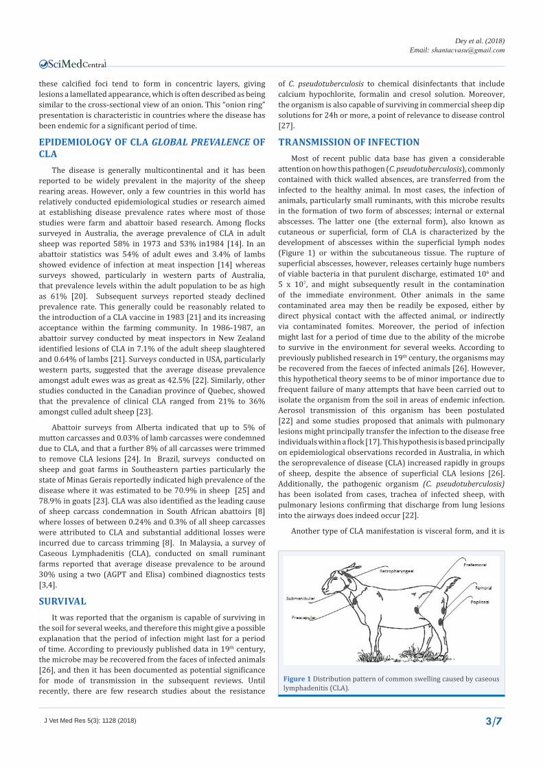
TRANSMISSION OF INFECTIONMost of recent public data base has given a considerable
attention on how this pathogen (C. pseudotuberculosis), commonly contained with thick walled absences, are transferred from the infected to the healthy animal. In most cases, the infection of animals, particularly small ruminants, with this microbe results in the formation of two form of abscesses; internal or external abscesses. The latter one (the external form), also known as cutaneous or superficial, form of CLA is characterized by the development of abscesses within the superficial lymph nodes (Figure 1) or within the subcutaneous tissue. The rupture of superficial abscesses, however, releases certainly huge numbers of viable bacteria in that purulent discharge, estimated 106 and 5 x 10⁷, and might subsequently result in the contamination of the immediate environment. Other animals in the same contaminated area may then be readily be exposed, either by direct physical contact with the affected animal, or indirectly via contaminated fomites. Moreover, the period of infection might last for a period of time due to the ability of the microbe to survive in the environment for several weeks. According to previously published research in 19th century, the organisms may be recovered from the faeces of infected animals [26]. However, this hypothetical theory seems to be of minor importance due to frequent failure of many attempts that have been carried out to isolate the organism from the soil in areas of endemic infection. Aerosol transmission of this organism has been postulated [22] and some studies proposed that animals with pulmonary lesions might principally transfer the infection to the disease free individuals within a flock [17]. This hypothesis is based principally on epidemiological observations recorded in Australia, in which the seroprevalence of disease (CLA) increased rapidly in groups of sheep, despite the absence of superficial CLA lesions [26]. Additionally, the pathogenic organism (C. pseudotuberculosis) has been isolated from cases, trachea of infected sheep, with pulmonary lesions confirming that discharge from lung lesions into the airways does indeed occur [22].
Another type of CLA manifestation is visceral form, and it is
Figure 1 Distribution pattern of common swelling caused by caseous lymphadenitis (CLA).

CentralBringing Excellence in Open Access
Dey et al. (2018)Email:
J Vet Med Res 5(3): 1128 (2018) 4/7
often characterized by the formation of lesion within affected individuals. The effected organs/tissues involved in this form (Visceral form) include lymph nodes, primarily the mediastinal lymph nodes, and lungs , although other tissues such as the liver, the kidneys or the mammary glands and less frequently the heart, the brain, the spinal cord, the testes, the uterus and the joints may be included [28]. Some experimental infections of C. pseudotuberculosis with different routes of administrations, intratracheal administration of bacteria [29], intravenous route sub-cutaneous inoculation [9], have jointly shown that the most proportion of internal lesions were in the lungs and associated thoracic lymph nodes.
Different route infections have been carried out by inoculations of the organism, using sheep model, via intratracheal and intravaginal routes of administration of bacteria [30]. In naturally observed animals, skin cuts which can appear during common procedures, such as shearing, castration and ear tagging, are mainly believed to be the portal of entry of the microbe [31]. In cattle and buffaloes, C. pseudotuberculosis can be mechanically transmitted by houseflies and by other Diptera, though the natural mechanisms of infection with C. pseudotuberculosis are not yet well understood.
ECONOMICAL EVALUATION OF THE DISEASECaseous lymphadenitis (CLA) is recognized as a significant
cause of economic loss to the small ruminant industries worldwide. Studies have shown that the disseminated visceral form had much more economic losses due to extensive internal abscesses [22]. Similarly, subclinical infections should also be taken into account because of their high probability to allow the organism to disseminate within and among herds [32]. Moreover, CLA can become a public health problem as it is a zoonosis [33]. Systemic infection by C. pseudotuberculosis is acknowledged to be detrimental to the productivity of the infected animal, but to what extent is unclear.
In endemic areas such as Australia, where financial losses related to this disease has been extensively studied. The disease has led to an estimated loss of $A12-$A15 million per annum [32] to the meat industry. This is due both to carcasses losses and to the requirement for additional meat inspection and carcass trimming. Based on disease surveillance and wool production data from 1992, it was estimated that CLA infection costs the Australian sheep industry an approximate loss of $A17 million per annum in lost wool production [7]. In North America CLA is regarded to be much more significant clinically. There, the visceral form of infection with organism (C. pseudotuberculosis) has been associated with so called ‘‘thin-ewe syndrome’’, a chronic emaciation of ewes, occurring despite good appetite and in the absence of significant parasitic infestation or specific clinical signs. Studies conducted in US indicated that CLA infection had an economically significant effect on culling rates and reproductive efficiency in ewes [34]. In the Middle East, the economic losses occur due to the condemnation of lamb carcasses with CLA lesions, most commonly in the submandibular lymph nodes. In Egypt, it is estimated that CLA infections causes severe losses and costs the meat industry approximately $ 1.76 million annually. In Malaysia, there is currently no official data available which indicates the exact statistics of CLA economic loses, the disease
is still considered as a plague to the small ruminant production. To explore more about CLA in this country, much epidemiological studies are certainly needed to be conducted on both farms and abattoirs.
DISEASE DIAGNOSIS CLINICAL SIGNSTo diagnose the disease clinically, several basic
procedures including case history, examination, hematological and biochemical analysis should be taken properly [4]. Ultrasonography and other diagnostic and technical tools must be given considerable values. Swellings of submandibular abscess are among most frequently seen in cases associated with CLA (Figure 2). However, the clinical parameters, in sheep, may show normal rates (temperature: 39.3 oC; frequency breathing rhythm: 28 bpm; cardiac rhythm: 100 ppm) [35]. Emaciation and poor coat, enlargement of cutaneous lymph anodes were common in most hospitalized CLA cases. The later had more important significance, because most of CLA cases can be diagnosed clinically only if infection of the cutaneous lymph nodes progresses to fistulation or, more rarely, visceral organ involvement leads to emaciation (the so-called thin ewe syndrome). On the other hand, the hematological studies frequently revealed chronic anaemia, an increasing of acute phase proteins and decrease of total proteins, particularly negative proteins such as albumin and globulin in blood stream. Based on urine biochemistry analysis, in sheep, proteinuria (100 mg/dl), pyuria (neutrophils) (>1000 leucocytes/field40×) and bacteriuria was observed [35].
DIAGNOSTIC TOOLSA presumptive diagnosis of CLA is based on case history,
clinical examination of superficially enlarged lymph nodes and the characteristic greenish-yellow exudates that may have a lamellated appearance on the cut surface [27]. Isolation and identification of the etiological agent (C. pseudotuberculosis) from lesions is necessary for confirmation. Therefore, the diagnostic criteria of CLA infections still remains culture and identification of the organism as it was considered the gold standard in diagnosis of CLA. In the laboratory, C. pseudotuberculosis cultured from clinical samples may be identified from its enzymatic profile and its ability to utilize various carbohydrate sources.
SEROLOGICAL TESTSSerological diagnostic tests for CLA are based on the detection
of a humoral response to PLD exotoxin. Such tests have been explored as a method for controlling the disease by identifying and removing infected carrier animals in small ruminant industry.
Experimental subjects (mice, rabbits or guinea pigs) were given injections of serum from animals suspected of being infected with C. pseudotuberculosis. Thereafter, these animals were then given lethal doses of PLD toxin, prepared from C. pseudotuberculosis culture supernatants. Reduction in the rate of mortality of serum-treated animals, as compared with that of controls, was considered to indicate passive protection by serum antitoxin, and highly suggestive of C. pseudotuberculosis infection. To facilitate in vitro serological screening, inclusion of antitoxin containing sera in the CAMP-inhibition test resulted in neutralization of the inhibitory effect that C. pseudotuberculosis exerted on staphylococcal b-lysin. This led to a new method

CentralBringing Excellence in Open Access
Dey et al. (2018)Email:
J Vet Med Res 5(3): 1128 (2018) 5/7
Figure 2 Caseous lymphadenitis lesions in Boer cross doe from a farm located in Selangor region, Peninsular Malaysia; A) Mandibular abscess; B) Blood Agar culture showing colonies of Corynebacterium pseudotuberculosis.
designated the anti-haemolysin- inhibition (AHI) test for testing sera from animals suspected of having CLA. This test has been used for disease diagnosis in sheep, goats and horses, thus avoiding the use of experimental animals [29]. Other tests used in the study on CLA include tube agglutination assays [36], indirect haemagglutination test (MT Shigidi, 1978) [37] and a double immunodifusion test. However, the enzyme-linked immunosorbent assay (ELISA) for use in diagnosis has shown particular promise. Other potential tests include a polymerase chain reaction (PCR) method [38] and bovine interferon (IFN)-g whole blood ELISA [39].
TREATMENTDespite the in vitro sensitivity of C. pseudotuberculosis to
penicillin, tetracyclines and cephalosporins [40], treatment with these drugs is generally not effective as a result of several factors including the protective nature of the capsule, the formation of the encapsulated abscess as well as the intercellularity of the organism [41,42].
Prophylactic and therapeutic treatment, therefore, will not guarantee organism free from infected animals. Subsequently, these infected flocks or individuals serve as a reservoir of infection. As a result of this, the most practical method of controlling CLA in small ruminants is to cull all animals with the palpable lesions [36].
Treatment of sheep and goat flocks suffering from CLA infections particularly peripheral lymph nodes is often not justifiable. In valuable breeding stock, however, superficially located abscesses can be lanced or removed surgically. Abscesses frequently recur, particularly in sheep and goats, after draining or attempted surgical excision. Therefore, the infection repeats itself through the life of the animal. If the abscess cannot be precisely located or in a position that is unfavorable for surgery, a prolonged course of antimicrobial treatment using lipophilic drugs, such as one of the microlides at high dosage rates may be effective. In horses, application of hot packs, surgical lancing and flushing with antiseptics may be attempted for treatment of the abscess. Drainage of the abscess should be done in a way that avoids environmental contamination, with disinfection of the
surgical material before and after the procedure, and all of the disposable materials should be incinerated and buried, including plastics and paper used to cover the area.
DISEASE CONTROLGenerally, the most efficient strategy for control and
prevention of CLA remains a matter of debate. However, vaccination is the primary means of disease control in several countries, whereby immunization is used to reduce the spread of infection, leading to a gradual decline in disease prevalence only in some countries.
Several vaccines have been developed to protect animals, particularly small ruminants (sheep and goats) against CLA, but at the same time there is no currently available vaccine that gives complete protection against the disease. This is evidenced by a report from Australia which indicated that, despite the widespread use of a commercial CLA vaccine in that country since 1983, disease prevalence remained at approximately 20% in 2002 [33]. Serological diagnoses are of a value in those countries with low prevalence and with relaxation of borders. These serological tests have been used as an alternative way of vaccination and offer a powerful means of controlling disease, through the culling/segregation of infected animals. This approach, while potentially costly in the first instance, is a means by which CLA may be completely eradicated from affected flocks.
ACKNOWLEDGMENTThe author would like to acknowledge the support from
the Ministry of Higher Education, Malaysia and The Islamic Development Bank, Saudi Arabia.
REFERENCES1. Pacheco LG, Pena RR, Castro TL, Dorella FA, Bahia RC, Carminati R,
et al. Multiplex PCR assay for identification of Corynebacterium pseudotuberculosis from pure cultures and for rapid detection of this pathogen in clinical samples. J Med Microbiol. 2007; 56: 480-486.
2. Connor KM, Fontaine MC, Rudge K, Baird GJ, Donachie W. Molecular genotyping of multinational ovine and caprine Corynebacterium pseudotuberculosis isolates using pulsed-field gel electrophoresis. Vet Res. 2007; 38: 613-623.

CentralBringing Excellence in Open Access
Dey et al. (2018)Email:
J Vet Med Res 5(3): 1128 (2018) 6/7
3. KomalaTS, Ramlan M, Yeoh NN, Surayani AR, Sharifah Hamidah SM. A survey of caseous lymphadenitis in small ruminant farms from two districts in Perak, Malaysia-Kinta and Hilir Perak. Trop Biomed. 2008; 25: 196-201.
4. Osman AY, Jesse F, Saharee A. Sero-Prevalence of Caseous Lymphadenitis Evaluated by Agar Gel Precipitation Test among Small Ruminant Flocks in East Coast Economic Regions in Peninsular Malaysia. J Animal Vet Adv. 2012; 11: 3474-3480.
5. Dercksen DP, Brinkhof JM, Dekker-Nooren T, Maanen K, Bode CF, Baird G, et al. A comparison of four serological tests for the diagnosis of caseous lymphadenitis in sheep and goats. Vet Microbiol. 2000; 75: 167-175.
6. Menzies PI, Muckle CA. The use of a microagglutination assay for the detection of antibodies to Corynebacterium pseudotuberculosis in naturally infected sheep and goat flocks. Can J Vet Res. 1989; 53: 313-318.
7. Shigidi MT. An indirect haemagglutination test for the sero-diagnosis of C ovis infection in sheep. Res Vet Sci. 1978; 24: 57-60.
8. Baird GJ, Fontaine MC. Corynebacterium pseudotuberculosis and its role in ovine caseous lymphadenitis. J Comp Pathol. 2007; 137: 179-210.
9. Fontaine M, Baird G. Caseous lymphadenitis. Small Ruminant Research. 2008; 76: 42-48.
10. Paton MW. Applying the experience of CLA in the Southern Hemisphere. 2000. In Proceedings of the Moredun Research Institute/Scottish Agricultural College Workshop on Caseous Lymphadenitis 3-15.
11. Carne H, Onon EO. Action of Corynebacterium ovis exotoxin on endothelial cells of blood vessels. Nature. 1978; 271: 246-248.
12. Simmons CP, Dunstan SJ, Tachedjian M, Krywult J, Hodgson AL, Strugnell RA. Vaccine Potential of Attenuated Mutants of Corynebacterium pseudotuberculosis in Sheep. Infect Immun. 1998; 66: 474-479.
13. Tashijian JJ, Campbell SG. Interaction between caprine macrophages and corynebacterium pseudotuberculosis: an electron microscopic study. Am J Vet Res. 1983; 44: 690-693.
14. Batey RG. Pathogenesis of caseous lymphadenitis in sheep and goats. Aust Vet J. 1986; 63: 269-272.
15. Hard GC. Comparative toxic effect of the surface lipid of Corynebacterium ovis on peritoneal macrophages. Infect Immun. 1975; 12: 1439-1449.
16. West DM, Bruere AN, Ridler AL. The sheep: Health, disease & production: Veterinary Continuing Education, Massey University. 2002.
17. Williamson LH. Caseous lymphadenitis in small ruminants. Vet Clin North Am Food Anim Pract. 2001; 17: 359-371.
18. Pépin M, Fontaine JJ, Pardon P, Marly J, Parodi AL. Histopathology of the early phase during experimental Corynebacterium pseudotuberculosis infection in lambs. Vet Microbiol. 1991; 29: 123-134.
19. Middleton MJ, Epstein VM, Gregory GG. Caseous lymphadenitis on Flinders Island: prevalence and management surveys. Aust Vet J. 1991; 68: 311-312.
20. Al-Gaabary MH, Osman SA, Oreiby AF. Caseous lymphadenitis in sheep and goats: clinical, epidemiological and preventive studies. Small Ruminant Research. 2009; 87: 116-121.
21. Seedik I, El Timawy A, El Amarousi S, Zaki M, El Allawy T. Some studies on caseous lymphadenitis of sheep in Upper Egypt. Assiut Veterinary
Medical Journal. 1983.
22. Stoops SG, Renshaw HW, Thilsted JP. Ovine caseous lymphadenitis: disease prevalence, lesion distribution, and thoracic manifestations in a population of mature culled sheep from western United States. Am J Vet Res. 1984; 45: 557-561.
23. Seyffert N, Guimarães AS, Pacheco LG, Portela RW, Bastos BL, Dorella FA, et al. High seroprevalence of caseous lymphadenitis in Brazilian goat herds revealed by Corynebacterium pseudotuberculosis secreted proteins-based ELISA. Res Vet Sci. 2010; 88: 50-55.
24. Stanford K, Brogden KA, McClelland LA, Kozub GC, Audibert F. The incidence of caseous lymphadenitis in Alberta sheep and assessment of impact by vaccination with commercial and experimental vaccines. Can J Vet Res. 1998; 62: 38-43.
25. Guimarães A, Seyffert N, Bastos BL, Portela RWD, Meyer R, Carmo FB, et al. Caseous lymphadenitis in sheep flocks of the state of Minas Gerais, Brazil: prevalence and management surveys. Small Ruminant Research. 2009; 87: 86-91.
26. Paton MW, Sutherland SS, Rose IR, Hart RA, Mercy AR, Ellis TM. The spread of Corynebacterium pseudotuberculosis infection to unvaccinated and vaccinated sheep. Aust Vet J. 1995; 72: 266-269.
27. Nairn ME, Robertson JP. Corynebacterium pseudotuberculosis infection of sheep: role of skin lesions and dipping fluids. Aust Vet J. 1974; 50: 537-542.
28. Valli V, Parry B. Caseous lymphadenitis. Pathology of Domestic Animals. 1993; 3: 238-240.
29. Brown C, Olander H. Caseous lymphadenitis of goats and sheep: a review. Vet Bull. 1987; 57:12.
30. Fontaine MC, Baird G, Connor KM, Rudge K, Sales J, Donachie W. Vaccination confers significant protection of sheep against infection with a virulent United Kingdom strain of Corynebacterium pseudotuberculosis. Vaccine. 2006; 24: 5986-5996.
31. al-Rawashdeh OF, al-Qudah KM. Effect of shearing on the incidence of caseous lymphadenitis in Awassi sheep in Jordan. J Vet Med B Infect Dis Vet Public Health. 2000; 47: 287-293.
32. Paton MW, Walker SB, Rose IR, Watt GF. Prevalence of caseous lymphadenitis and usage of caseous lymphadenitis vaccines in sheep flocks. Aust Vet J. 2003; 81: 91-95.
33. Join-Lambert OF, Ouache M, Canioni D, Beretti JL, Blanche S, Berche P, et al. Corynebacterium pseudotuberculosis necrotizing lymphadenitis in a twelve-year-old patient. Pediatric Infect Dis J. 2006; 25: 848-851.
34. Renshaw HW, Graff VP, Gates NL. Visceral caseous lymphadenitis in thin ewe syndrome: isolation of Corynebacterium, Staphylococcus, and Moraxella spp from internal abscesses in emaciated ewes. Am J Vet Res. 1979; 40: 1110-1114.
35. Ferrer LM, Lacasta D, Chacón G, Ramos J, Villa A, Gómez P, et al. Clinical diagnosis of visceral caseous lymphadenitis in a Salz ewe. Small Ruminant Research. 2009; 87: 126-127.
36. Husband AJ, Watson DL. Immunological events in the popliteal lymph node of sheep following injection of liver or killed Corynebacterium ovis into an afferent popliteal lymphatic duct. Res Vet Sci. 1977; 22: 105-112.
37. Shigidi MT. A comparison of five serological tests for the diagnosis of experimental Corynebacterium ovis infection in sheep. Br Vet J. 1979; 135: 172-177.
38. Cetinkaya B, Karahan M, Atil E, Kalin R, De Baere T, Vaneechoutte M. Identification of Corynebacterium pseudotuberculosis isolates from sheep and goats by PCR. Vet Microbiol. 2002; 88: 75-83.

CentralBringing Excellence in Open Access
Dey et al. (2018)Email:
J Vet Med Res 5(3): 1128 (2018) 7/7
Osman AY, Nordin ML, Kadir AA, Saharee AA (2018) The Epidemiology and Pathophysiology of Caseous Lymphadenitis: A Review. J Vet Med Res 5(3): 1129.
Cite this article
39. Menzies PL, Hwang YT, Prescott JF. Comparison of an interferon-γ to a phospholipase D enzyme-linked immunosorbent assay for diagnosis of Corynebacterium pseudotuberculosis infection in experimentally infected goats. Vet Microbiol. 2004; 100: 129-137.
40. Zhao HK, Yonekawa K, Takahashi T, Kikuchi N, Hiramune T, Yanagawa R. Isolation of Corynebacterium pseudotuberculosis from the cervical canal of clinically normal sows. Res Vet Sci.1993; 55: 356-359.
41. Ashfaq MK, Campbell SG. A survey of caseous lymphadenitis and its etiology in goats in the United States. Veterinary medicine, small animal clinician: VM, SAC,1979; 74: 1161.
42. Ismail A, Hamid Y. Studies on the effect of some chemical disinfectants used in veterinary practice on Corynebacterium ovis. J Egypt Vet Med Assoc. 1972.