the enterovirus 71 particle: an evolutionary approach to …etheses.whiterose.ac.uk/13211/1/james...
TRANSCRIPT

i
The enterovirus 71 particle: An evolutionary approach to
investigate structure and function with implications for drug and
vaccine development.
James Thomas Kelly
Submitted in accordance with the requirements for the degree of Doctor of
Philosophy
Supervisors
Nicola J Stonehouse, David J Rowlands
Industrial Supervisors
Nicolas Burdin, Jeffery Almond
The University of Leeds
School of Molecular and Cellular Biology
September, 2015

ii
The candidate confirms that the work submitted is his own and that appropriate
credit has been given where reference has been made to the work of others.
This copy has been supplied on the understanding that it is copyright material and
that no quotation from the thesis may be published without proper
acknowledgment.
The right of James Thomas Kelly to be identified as Author of this work has been
asserted by him in accordance with copy right, Designs and patens Act 1988.
© 2015 The University of Leeds James Thomas Kelly.
iii
Acknowledgements
First and foremost I would like to thank my supervisors Nic Stonehouse and Dave
Rowlands, who have both been a tremendous help throughout the whole of my
PhD. Both have given me invaluable help and guidance, in lab work, presentation
skills and writing. Thank you both for your patience and guidance, it has helped
make my PhD both a rewarding and enjoyable (mostly!) experience.
I would like to thank the whole of the Stonelands group past and present for all
help and advice given, many enjoyable conversations, and general tomfoolery. With
a special thanks to Clare Nicol for helping training me and all other help and advice
that she had given me over the last 4 years. I would also like to thank Oluwapelumi
Adeyemi and Morgan Herod for the always enjoyable and very intensely speculative
discussions about how any minor result or anomaly in an experiment could lead to
the next break through Nature paper. Of course Eleni and Joe must get a special
mention just for being adorable.
Thank you to all project students I have taught (Amy, Joe, Ross and Lauren) for the
hard work they put in and for generally being a pleasure to teach.
Thank you to the members the virology lab and lab 8.54. Especially Becky Surtees
for many an attempted conversation over the loud drum of a TC hood.
Thank you to Liz Fry, David Stuart and Luigi DeColibus, for giving me the opportunity
to expand on my structural knowledge, and the comments and feedback they have
given me on various aspects of my work.

iv
I would also like to thank all the friends I have made over my time in Leeds who
have helped make it a very enjoyable experience, especially for the Friday night pub
sessions and lunch time conversations.
I would like to thank Amy Radcliffe Lauren Elliott, Luigi De Colibus, Özlem Cesur,
Joseph Ward, Oluwapelumi Adeyemi and Jingshan Ren, for the work and they have
done that has contributed to my thesis.
I would like to thank all the teachers I have had over the years who have taught me
and helped and encouraged me into science, especially my undergraduate and
master supervisor Michael Ginger.
Finally I would like to thank my family and girlfriend for their ongoing support
throughout my PhD.

v
Abstract
The capsid plays many important roles in the virus life cycle, including host cell
recognition, cell entry, uncoating and protection of viral RNA. In this thesis, artificial
selection, use of inhibitory compounds and structural analysis have been combined
to further understand aspects of these multiple roles.
The VP1 pocket is a hydrophobic cavity in the capsid that harbours a fatty acid,
known as the pocket factor. The presence of this fatty acid can increase capsid
stability and its release is required for the virus to uncoat. Certain compounds are
able to bind to the pocket and displace the natural fatty acid (causing a further
increase in capsid stability) and can inhibit infection by making the virus so stable
that it is unable to uncoat. Novel compounds, termed NLD and ALD, have been
designed in silico based upon the previous best EV71 pocket binding inhibitor GPP3.
These were shown to inhibit EV71 infection in cells, with NLD being more than one
order of magnitude more potent than the previous best EV71 pocket-binding
inhibitor. Resistance towards these compounds was studied to reveal a double
mutation in VP1 (I113M and V123L). Mapping the mutations to the EV71 crystal
structure revealed that they were located in the VP1 pocket, and modelling
predicted that they would prevent NLD and the natural pocket factor from binding.
Further characterisation of the resistant isolate revealed that the mutations were
thermally- and genetically-unstable.
Further investigation on thermal stability involved selection of a thermally-stable
virus. This generated viruses with a double mutation in the VP1 pocket (V179A and

vi
L183V). These mutations were predicted to increase the size of the VP1 pocket, and
were shown to affect the way the virus interacts with the natural pocket factor. The
effects of a variety of different pocket factors on the heat stability of WT EV71, the
thermostable mutant, and the inhibitor-resistant mutant were analysed.
In addition, work was conducted to assess the effect of pH on uncoating. For
uncoating it is known that EV71 must be incubated at a low pH in the presence of
its major receptor SCARB2. To investigate the effects of this, EV71 was incubated at
a low pH during infection. This was shown to reduce the virus titre, and repeated
exposure to this condition selected for a VP1 mutation (N104S). This residue is
predicted to be involved in binding to SCARB2, and the mutant virus was shown to
differ in susceptibility to compounds that affect entry and uncoating.

vii
Abbreviations BEV ; bovine enterovirus
CAR; Coxsackie and adenovirus receptor
CVA16; Coxsackievirus A16
CVB3; Coxsackievirus B3
DAF; decay accelerating factor
DMSO; Dimethyl sulfoxide
EC50; half maximal effective concentration
ERAV; Equine Rhinitis A Virus
EV1; Echovirus 1
EV71; Enterovirus 71
FMDV; foot-and-mouth disease virus
HAV; hepatitis A virus
HBsAg; hepatitis B surface antigen
HBV; hepatitis B virus
HEVA; human enterovirus A
HFMD; hand, foot, and mouth disease
HPV; human papillomavirus

viii
HRV; Human rhinovirus
HS; heparan sulphate glycosaminoglycan
Hsp; heat shock protein
IC50; half maximal inhibitory concentration
kcal; kilocalorie
Lpro; leader protease
MTT; 3-(4,5-Dimethylthiazol-2-Yl)-2,5-Diphenyltetrazolium Bromide
NP40; nonyl phenoxypolyethoxylethanol Tergitol-type NP-40
OD; Optical density
OPV; Oral Polio Vaccine
PAGE; Polyacrylamide gel electrophoresis
pH; log10 [H+]
PSGL1; P-selectin glycoprotein ligand-1
PV; poliovirus
QMPLD; quantum mechanics-polarised ligand docking
RCF; Relative centrifugal force
RD; Rhabdomyosarcoma
RPM; Revolutions per minute
ix
s.e.m; standard error of the mean
SCARB2; scavenger receptor class B, member 2
SDS; sodium dodecyl sulfate
SVDV; swine vesicular disease virus
TCID; tissue culture infective dose
TEM; Transmission electron microscopy
UTR; untranslated region
v/v; volume over volume
VDPP; vaccine-derived paralytic poliomyelitis
VLP; virus like particle
w/v; Weight over volume
ΔΔ G; Change in Free folding energy

x
Table of Contents Acknowledgements ..................................................................................................... iii
Abstract ........................................................................................................................ v
Abbreviations ............................................................................................................. vii
Table of Contents ......................................................................................................... x
Table of Figures ......................................................................................................... xvi
Table of Tables .......................................................................................................... xix
Chapter 1 Introduction ........................................................................................... 2
1.1 Hand, Foot, and Mouth Disease .................................................................... 2
1.2 The Picornavirus life cycle ............................................................................. 5
1.2.1 Genome and capsid structure ................................................................ 5
1.2.2 Receptors and Cell entry of picornaviruses ......................................... 10
1.2.3 Co-receptors ......................................................................................... 12
1.2.4 Viral uncoating ..................................................................................... 13
1.2.5 The EV71 capsid and its involvement in cell entry and uncoating ...... 15
1.3 Capsid-binding inhibitors ............................................................................. 22
1.3.1 Resistance against pocket binding compounds ................................... 26
1.4 Vaccines ....................................................................................................... 27
1.4.1 Attenuated vaccines ............................................................................. 27
1.4.2 Inactivated vaccine ............................................................................... 29

xi
1.4.3 Pathogen free vaccines: subunit and virus-like particle (VLP) vaccines
31
1.5 Project objectives ........................................................................................ 36
Chapter 2 Materials and methods ........................................................................ 38
2.1 General Buffers, media and reagents.......................................................... 38
2.1.1 Luria-Bertoni (LB) Broth ....................................................................... 38
2.1.2 10xTBE (Tris-borate-EDTA) buffer pH 8.3 ............................................ 38
2.1.3 6x DNA loading buffer .......................................................................... 38
2.1.4 10x Sodium dodecyl sulphate - Polyacrylamide gel electrophoresis
(SDS-PAGE) running buffer ................................................................................. 39
2.1.5 SDS-PAGE stacking gel .......................................................................... 39
2.1.6 15 % SDS-PAGE resolving gel................................................................ 39
2.1.7 Coomassie blue protein stain ............................................................... 40
2.1.8 Cell growth medium (Suspension/microcarrier) ................................. 40
2.1.9 Coomassie de-stain solution ................................................................ 40
2.1.10 Tris EDTA (TE) buffer ............................................................................ 40
2.1.11 Cell growth medium (Flask) ................................................................. 41
2.1.12 Radio labelling medium ........................................................................ 41
2.1.13 Radio-immuno precipitation assay (RIPA) buffer ................................ 41
2.1.14 Enhanced chemiluminescence (ECL) .................................................... 41
2.1.15 10 x Transfer buffer .............................................................................. 42

xii
2.1.16 Phosphate-buffered saline (PBS) TWEEN ............................................. 42
2.1.17 RNA loading buffer ............................................................................... 42
2.1.18 Crystal violet stain ................................................................................ 43
2.1.19 Blocking buffer ..................................................................................... 43
2.1.20 Gel dry down buffer ............................................................................. 43
2.1.21 Virus re-suspension buffer ................................................................... 43
2.1.22 pH 5.6 buffer ........................................................................................ 44
2.1.23 Carbonate Coating buffer .................................................................... 44
2.1.24 ELISA Dilution Buffer ............................................................................ 44
2.1.25 ELISA Wash Buffer ................................................................................ 44
2.1.26 ELISA Assay diluent .............................................................................. 44
2.1.27 Antibodies ............................................................................................ 44
2.1.28 Lipids .................................................................................................... 45
2.2 Cell culture ................................................................................................... 46
2.2.1 Cell lines used ....................................................................................... 46
2.2.2 Maintenance of cell culture Flasks ....................................................... 47
2.2.3 Microcarrier cultures ........................................................................... 47
2.2.4 Revival of frozen cells ........................................................................... 48
2.2.5 Preparation of preserved cell stocks .................................................... 48
2.2.6 Virus infection T175 ............................................................................. 48

xiii
2.2.7 Tissue culture infective dose 50 (TCID50) assay .................................... 49
2.2.8 Inhibition of pocket binding inhibitors IC50 curves .............................. 49
2.2.9 Drug inhibition curves knocking down host cell pathways .................. 49
2.2.10 Time courses ........................................................................................ 51
2.2.11 Drug selection ...................................................................................... 51
2.2.12 pH selection .......................................................................................... 51
2.2.13 Heat selection ...................................................................................... 51
2.2.14 Heat stability curves ............................................................................. 52
2.2.15 Radio labelling ...................................................................................... 52
2.3 Gel Electrophoresis methods ...................................................................... 53
2.3.1 Agarose gel electrophoresis ................................................................. 53
2.3.2 RNA gels ............................................................................................... 53
2.3.3 SDS-PAGE ............................................................................................. 53
2.3.4 Western blotting .................................................................................. 54
2.3.5 Gel dry down ........................................................................................ 55
2.4 Cloning ......................................................................................................... 55
2.4.1 Transformation of competent cells ..................................................... 55
2.4.2 RNA Extraction ..................................................................................... 56
2.4.3 RNA Transcriptions ............................................................................... 56
2.4.4 Reverse transcriptions ......................................................................... 56
xiv
2.4.5 PCR ....................................................................................................... 56
2.4.6 TOPO cloning ........................................................................................ 56
2.4.7 Sequencing ........................................................................................... 56
2.5 Purification .................................................................................................. 58
2.5.1 Purification of Virus through a sucrose gradient (A) ........................... 58
2.5.2 Purification of Virus through a sucrose gradient B .............................. 58
2.5.3 Nycodenz .............................................................................................. 59
2.5.4 3H Palmitic acid .................................................................................... 59
2.5.5 Scintillation counting............................................................................ 60
2.6 Immunoprecipitation .................................................................................. 60
2.7 ELISA ............................................................................................................ 60
2.8 PyMol ........................................................................................................... 61
Chapter 3 EV71 Compounds – In vitro characterisation and selection of resistant
isolates 63
3.1 Characterisation of viral inhibition and cellular cytotoxicity ...................... 66
3.2 Selection of resistant mutants .................................................................... 73
3.3 Fitness cost of inhibitor resistant mutations............................................... 79
3.4 Chapter summary ........................................................................................ 84
Chapter 4 Growth, cell entry and uncoating of EV71 ........................................... 88
4.1 Cells and purification ................................................................................... 92
4.1.1 Optimisation of viral growth ................................................................ 92

xv
4.1.2 Replication kinetics of WT EV71 in Vero cells ...................................... 95
4.1.3 Viral purification and empty capsid production ................................ 100
4.2 A mutant with an altered uncoating phenotype ....................................... 115
4.3 Discussion and future work ....................................................................... 126
Chapter 5 Heat stability of EV71 and ways to increase it. .................................. 131
5.1 Capsid stability ........................................................................................... 134
5.1.1 Long term stability of EV71/CVA16 .................................................... 134
5.1.2 Evolution of an EV71 isolate with increased thermal stability .......... 136
5.1.3 Determination of heat stability mutations ........................................ 141
5.1.4 Effect of different pocket factors on the thermostability of WT EV71
and isolates with VP1 pocket mutations .......................................................... 143
5.1.5 Binding of pocket factors to virus ...................................................... 148
5.1.6 Effect of pocket factors in other enteroviruses ................................. 150
5.2 The potential use of antibodies and/or purified receptor to distinguish
between heated and native empty particles ....................................................... 155
5.2.1 Immunoprecipitation of EV71 using capsid antibodies D6 and A9 ... 155
5.2.2 Use of ELISA in combination with EV71 receptors and antibodies to
detect native EV71 antigenicity ....................................................................... 157
5.3 Discussion .................................................................................................. 159
Chapter 6 Concluding remarks and future perspective ...................................... 166
Chapter 7 Bibliography ............................................................................................. 177
xvi
Appendix .................................................................................................................. 192
Table of Figures
Figure 1.1. Lesions typical of HFMD. (Accessed: 09/03/2016) .................................... 3
Figure 1.2 Schematic of the enterovirus genome. ....................................................... 6
Figure 1.3. Diagram of the icosahedral pseudo-T=3 capsid of EV71. ........................ 10
Figure 1.4. ................................................................................................................... 16
Figure 1.5. Acid induced conformational change in SCARB2 creates a connection
between its own hydrophobic tunnel and the EV71 hydrophobic VP1 pocket......... 20
Figure 1.6 The enterovirus pocket. ............................................................................ 25
Figure 3.1 Structures of different3-(4-pyridyl)-2-imidazolidinone derivatives with
anti-EV71 activity. Images modified from DeColibus et al. 2014. ............................. 64
Figure 3.2 IC50 Inhibitory effects of NLD (green), GPP3 (blue), ALD (orange) and
GPP4 (red) on EV71. ................................................................................................... 67
Figure 3.3 Inhibition of CVA16 by GPP3. CVA16 samples were titrated via TCID50
assay in the presence of a range of concentrations of GPP3. ................................... 68
Figure 3.4 The inhibitory effects of fatty acids on EV71 (A), PV (B) and BEV2 (C). .... 69
Figure 3.5 Toxicity of NLD, GPP3 and ALD to Vero cells. ........................................... 71
Figure 3.6 Selecting for inhibitor resistant virus. ....................................................... 74
Figure 3.7 Generation of resistant isolates. ............................................................... 75
Figure 3.8 (A) Structure of the EV71 protomer in complex with NLD. ......................78
Figure 3.9 Inhibitor resistant isolates are genetically unstable.............................. 80

xvii
Figure 3.10 One step growth curves of inhibitor-resistant and WT EV71............... 81
Figure 3.11 Inhibitor-resistant mutants are more thermolabile than WT virus. ......83
Figure 4.1. Acid induced conformational change in SCARB2 creates a connection
between its own hydrophobic tunnel and the EV71 hydrophobic VP1 pocket......... 91
Figure 4.2 Comparative yields of different strains of EV71. ...................................... 93
Figure 4.3 Comparative yields of EV71 in different cell lines. ................................... 94
Figure 4.4. Optimising growth techniques for EV71. ................................................. 95
Figure 4.5 Growth of EV71. (A) .................................................................................. 99
Figure 4.6 Sucrose gradient profiles of 35S labelled BEV and EV71 ......................... 102
Figure 4.7 Virus titres of different EV71 pellets. ...................................................... 104
Figure 4.8 Sucrose gradient profiles and titrations of 35S labelled EV71 after
treatment with SDS. ................................................................................................. 105
Figure 4.9 Sucrose gradient profiles of 35S labelled EV71 grown in RD cells. .......... 107
Figure 4.10. Analysis and comparison of EV71 purified through Self-forming (40 w/v
%) and step (20%-60 w/v %) Nycodenz gradients. .................................................. 110
Figure 4.11 High buoyancy EV71 is sensitive to NP40 treatment. ........................... 112
Figure 4.12 Effect of chloroquine on high and low buoyancy EV71. ....................... 113
Figure 4.13 The effect of heating on the nycodenz gradient profile of EV71. ......... 114
Figure 4.14. pH treatment of virus on cells has a fitness cost. ................................ 116
Figure 4.15 Sequence alignments of VP1 of Enterovirus A species. ........................ 118
Figure 4.16 Acid induced conformational change in SCARB2 causes an alpha helix to
move towards VP1 N104. ......................................................................................... 119
Figure 4.17 Analysis of the effect of inhibitors of cell entry. ................................... 121

xviii
Figure 4.18 Endosome acidification plays an important role in WT EV71 and acid-
resistant EV71. ......................................................................................................... 122
Figure 4.19 Cyclosporine A can prevent EV71 infection but is significantly less
effective against the acid-resistant mutant. ............................................................ 123
Figure 4.20 Potential role of cyclophilin A using crystal structure. ......................... 125
Figure 5.1 Long term stability of EV71 (A) and CVA16 (B). ...................................... 135
Figure 0.1 Heat stability curves of WT EV71 and thermostable EV71................... 138
Figure 5.3 Selection of a thermostable EV71 isolate. A T25 was infected with EV71,
after 100% cell death. .............................................................................................. 139
Figure 5.4 Difference in titres between EV71 thermostable mutants before and
after heating. ............................................................................................................ 140
Figure 5.5 Grow kinetics of WT EV71 and thermostable EV71. ............................... 140
Figure 5.6 Location of thermostabilising mutations. ............................................... 142
Figure 5.7 Effect of fatty acids on heat stability of EV71 isolates with different
pocket mutations. .................................................................................................... 146
Figure 5.8 Effect of a pocket-binding inhibitor on heat stability of EV71 isolates with
different pocket mutations. ..................................................................................... 147
Figure 5.9 Nycodenz gradient profiles ..................................................................... 149
Figure 5.10 Crystal structures reveal BEV1 and BEV2 have different sized VP1
pockets. .................................................................................................................... 151
Figure 5.11 Effect of heating on BEV2 after incubation in the presence of short chain
fatty acids. ................................................................................................................ 154

xix
Figure 5.12 Antibodies recognise native virus particles but not heated virus
particles. ................................................................................................................... 156
Figure 5.13 Graphs to illustrate ELISA results of EV71 ............................................. 158
Figure 5.14 Comparison of location of mutations in (A) inhibitor-
resistant/thermolabile EV71 and (B) thermostable. ................................................ 160
Table of Tables Table 1.1 Table of identified HEVA viruses and their symptoms. ................................ 4
Table 1.2 Receptor table. ........................................................................................... 21
Table 2.1 Cell lines ...................................................................................................... 46
Table 2.2 Concentrations, pre-treatment times and functions of drugs used in this
study ........................................................................................................................... 50
Table 2.3. Primers used in sequencing .....................................................................57
Table 3.1 Therapeutic window of NLD, GPP3 and ALD against EV71. ....................... 72
Table 3.2 Resistance mutations. Mutations identified in EV71/CVA16 after passage
in the presence of NLD, GPP3, ALD or a combination of NLD/GPP3. CVA16 isolates
resistant to NLD or ALD were not selected ..............................................................76
Table 4.1 Recovery of EV71 or BEV2 viruses after sequential steps during
purification. .............................................................................................................. 103
Table 5.1 Pocket factor table. .................................................................................. 145

1
Chapter 1

2
Chapter 1 Introduction
Enterovirus 71 (EV71) is a member of the Picornaviridae family and the major
causative agent of hand, foot, and mouth disease. This family is made up of non-
enveloped viruses that are around 30 nm in diameter and are encoded by a single
positive sense RNA segment that is translated as a single poly-protein (Tuthill et al.
2010). Picornaviruses cause a wide range of diseases that affect both human and
animal health. Examples include poliovirus (PV) causing poliomyelitis, hepatitis A
virus (HAV) causing acute hepatitis and foot-and-mouth disease virus (FMDV) that
causes foot and mouth disease in cloven hoofed animals.
1.1 Hand, Foot, and Mouth Disease
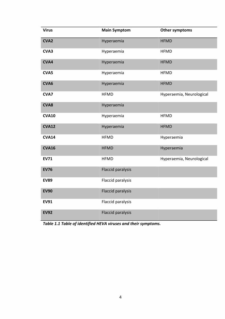
The virus species human enterovirus A (HEVA) is composed of several different
viruses that can cause a variety of diseases, including, hand, foot, and mouth
disease (HFMD), hyperaemia and flaccid paralysis. The details of what symptoms
each virus can cause can be found in table 1. Of the diseases caused by the HEVA
species, HFMD poses the largest public health problem, with sporadic outbreaks
occurring globally and regular seasonal outbreaks occurring in the Asian Pacific
region each year (reviewed in McMinn 2012). The disease usually manifests as self-
limiting infection of young children, causing small sores to appear on the hands,
feet, mouth and buttocks of the individual (Fig 1.1). However in more severe cases
it can cause brain stem encephalitis, cardiopulmonary disease and death (McMinn
et al. 2001, Zhang et al. 2011). The two most common viruses associated with
HFMD are enterovirus 71 (EV71) and Coxsackievirus A16 (CVA16) although CVA16 is

3
less commonly associated with severe symptoms (Mcminn 2003, McMinn et al.
2001, Zhang et al. 2011). In China alone, 7 million cases of HFMD were reported
over four years, and of those over 80,000 were classified as severe with EV71
confirmed to be the major causative agent (Xing et al. 2014).
Figure 1.1. Lesions typical of HFMD. Taken from WWW, nhs.uk 2012.Avaiable at:
http://www.nhs.uk/Conditions/Hand-foot-and-mouth-
disease/Pages/Introduction.aspx (Accessed: 09/03/2016)
4
Virus Main Symptom Other symptoms
CVA2 Hyperaemia HFMD
CVA3 Hyperaemia HFMD
CVA4 Hyperaemia HFMD
CVA5 Hyperaemia HFMD
CVA6 Hyperaemia HFMD
CVA7 HFMD Hyperaemia, Neurological
disorders CVA8 Hyperaemia
CVA10 Hyperaemia HFMD
CVA12 Hyperaemia HFMD
CVA14 HFMD Hyperaemia
CVA16 HFMD Hyperaemia
EV71 HFMD Hyperaemia, Neurological
disorders, respiratory
disorders
EV76 Flaccid paralysis
EV89 Flaccid paralysis
EV90 Flaccid paralysis
EV91 Flaccid paralysis
EV92 Flaccid paralysis
Table 1.1 Table of identified HEVA viruses and their symptoms.

5
1.2 The Picornavirus life cycle
1.2.1 Genome and capsid structure
The picornavirus family is composed of 29 different genera (WWW picornaviridae
2015. Available at; http://www.picornaviridae.com/ (Accessed: 09/03/2016)). All of
which possess a single stranded positive sense RNA genome that ranges between
7000 and 8500 nucleotides and encodes for a single polyprotein. The genome
arrangement between different genera is similar but not identical (Fig 1.2 reviewed
in Tuthill et al. 2010). The genome begins with the 5 prime untranslated region (5’
UTR) which contains a number of important features, such as the internal ribosome
entry site (IRES) and clover leaf that are involved in functions such as replication
and translation. It is also covalently linked to a small peptide (VpG) encoded by the
viral genome that is essential in replication (reviewed in Tuthill et al. 2010). The
genome ends with the 3 prime untranslated region which terminates with a poly-A
tract, the 3’UTR has functions in translation and replication (reviewed in Tuthill et
al. 2010). The rest of the genome encodes for a single polyprotein, which is co- and
post-translationally cleaved into its mature protein components by virally encoded
proteases. The first third of the polyprotein (P1) codes for structural proteins (VP0
(VP4/VP2), VP3, VP1) and the remaining two thirds (P2 and P3) consist of non-
structural proteins (2A, 2B, 2C and 3A 3B, 3C and 3D) that are involved in viral
replication and modification of the cellular environment. Certain picornaviruses
(e.g. aphthoviruses and cardioviruses) also encode for another non-structural
protein that precedes P1, called the leader protease (Lpro). This protein initiates host
cell shut off, which is a method of promoting viral gene expression by the

6
repressions of host gene translation. It performs this by cleaving eukaryotic
translation initiation factor 4GI (eIF4GI), which is essential for translation in
eukaryotic cells. Other picornaviruses (e.g. enteroviruses) do not possess a leader
protein and instead this function is carried out by 2A (e.g. PV). (Reviewed in Tuthill
et al. 2010)
Figure 1.2 Schematic of the enterovirus genome. The single ORF is indicated by a
rectangle flanked by 5’ and 3’ UTRs. A single polyprotein is translated from the
ORF which is subsequently cleaved into mature proteins by proteases encoded
within the polyprotein.
After translation the structural precursor protein P1, is co- translationally released
from the rest of the polyprotein by cis-cleavage carried out by 2Apro (Li et al. 2001).
In the majority of picornaviruses the N-terminus of P1 is modified by the covalent
addition of a myristic acid residue, which is thought to have important roles in
assembly and cell entry (Chow et al. 1987). However this does not occur in HAV and
viruses from the genera parechovirus and kobuvirus (Tesar et al. 1993; Johansson et
al. 2002). Post cleavage, the P1 region of enteroviruses and cardioviruses has been
shown to be dependent on heat shock protein (Hsp) 90 and possibly Hsp70, to
maintain a processing-competent conformation and as protection from
proteasomal degradation (Geller et al. 2007; Macejak & Sarnow 1992;
Mutsvunguma et al. 2011). These interactions allow P1 to be cleaved (although not
7
physically separated) into VP0, VP1 and VP3 by 3CDpro to form a protomer (Ansardi
et al. 1991; Ypma-Wong et al. 1988).
Cleavage allows protomers to then self-oligomerise into a pentameric subgroup and
12 of these associate to form a capsid (Guttman & Baltimore 1977; Hoey & Martin
1974; Lee et al. 1993). In most picornaviruses if viral RNA is present then
pentameric subunits will assemble round it and VP0 will undergo an autocatalytic
reaction to form VP2 and VP4 in the mature capsid. This VP0 cleavage does not
occur in parechoviruses and kobuviruses (Johansson et al. 2002). If no RNA is
present, capsid assembly will still occur, but it will form an empty capsid without
VP0 cleavage (Basavappa et al. 1994, Curry et al. 1997). Certain enteroviruses, such
as bovine enterovirus (BEV) and PV type 3, naturally produce high ratios of empty
particles to full particles, while others such as PV type 1 produce very few naturally
empty particles. The reason for production of empty capsids is not currently known,
one hypothesis is that they act as a reservoir of capsid proteins (Nugent and
Kirkegaard 1995). However work in BEV has shown this to be unlikely as it was
deemed energetically unfavourable to force the capsid to dissociate into its
components prior to reassembly (Guttman & Baltimore 1977; Marongiu et al. 1981;
Li et al. 2012). Another potential purpose of them is that they sequester antibodies
from the mature infectious virions to act as a form of immune evasion mechanism
(Shingler et al. 2015). Mature capsids will naturally become empty capsids after
uncoating, and this can be mimicked by heating. These particles are distinguishable
from naturally empty capsids as VP0 cleavage has occurred (Basavappa et al. 1994;
Ansardi & Morrow 1995).

8
It is possible to distinguish between mature and empty capsids by their
sedimentation on a sucrose gradient, with the denser mature capsids sedimenting
at 160S while empty capsids containing no RNA sediment at 80S. (Chow et al. 1987,
Fricks and Hogle 1990)
Picornavirus mature capsids are composed of VP1-3 externally and can be classified
as either a T=1 or a pseudo T=3, due to the fact that as mentioned earlier the
proteins VP1-3 never actually separate (Reviewed in Tuthill et al. 2010; Wang et al.
2012). VP4 is internal and forms an internal lattice, which is thought to stabilise the
capsid. Naturally empty particles do not possess VP4 because VP0 has not
undergone cleavage, due to RNA not being present at the time of encapsidation.
This causes them to be less structurally stable, readily converting from native (N)
type antigenicity to heated (H) type antigenicity (Rombaut and Jore 1997). In PV
this has been shown to occur at temperatures above 5 oC (Jim Hogle, personal
communication). The three major structural proteins (VP1-3) have a similar basic
structure, consisting of eight stranded beta barrels with connecting loops of various
lengths, and each has a wedge shape. The composition and shape of these three
proteins determine the antigenicity and receptor binding sites (Reviewed in Tuthill
et al. 2010). In aphthoviruses and HAV, capsids have a smooth surfaces with the
receptor binding site of aphthoviruses being a flexible loop protruding from the
surface, the receptor binding site of HAV is unknown (Acharya et al. 1989; Logan et
al. 1993; Wang et al. 2015). In cardioviruses the capsid is characterised by a series
of shallow depressions spanning the two fold axis, the depressions function as
receptor binding sites (Luo et al. 1987; Grant et al. 1994; Hertzler et al. 2000; Toth

9
et al. 1993). Enterovirus capsids are characterised by a series of much deeper
depressions, surrounding the 5-fold axis on VP1, these are referred to canyons
(Rossmann et al 1985). The canyon functions as the major receptor binding site,
although other areas of the capsid have also been implicated in receptor binding
(Olson et al. 1993; Chen et al. 2012; Colonno et al. 1988). Beneath the canyon is a
large hydrophobic space known as the VP1 pocket. In aphthoviruses and
cardioviruses this space is filled with large side chains (Acharya et al. 1989; Tuthill et
al. 2009). Generally the enterovirus VP1 pocket is filled with various hydrophobic
lipids known as pocket factors, this plays an important role in capsid stability and
uncoating (Wang et al. 2012; Plevka et al. 2012). Crystal structures have shown that
enterovirus pockets can dramatically vary in size, which influences the size of
pocket factor that they can accommodate. The predicted fatty acid sizes range from
between 10 carbons (e.g. enterovirus 68 (EVD68)) and 18 carbons (e.g. PV) (Liu et
al. 2015; Filman et al. 1989). One study using mass spectrometry to determine the
pocket factor of bovine enterovirus type 1 (BEV1) revealed that viruses will
naturally harbour several different pocket factors (Smyth et al. 2003). This explains
observations of crystal structures of different enteroviruses that showed electron
densities of the pocket factors can start to fade towards the end, as the structures
are an average the fading indicates that the pocket factors vary in size (Smyth et al.
1995). The presence of the pocket factor has been shown to be important for
enterovirus capsid stability and the expulsion of this molecule is necessary to
facilitate the structural rearrangements necessary for receptor binding and/or
uncoating of a number of different enteroviruses (Shepard et al. 1993; Dang et al.
2014).

10
Figure 1.3. Diagram of the icosahedral pseudo-T=3 capsid of EV71. Taken from
Solomon et al. 2010.
1.2.2 Receptors and Cell entry of picornaviruses
Binding to a receptor is very important for picornaviruses in terms of its life cycle as
it is essential for cell entry and sometimes uncoating. Most picornaviruses studied
have been shown to be able to utilise a variety of different receptors and co-
receptors. Which receptor it binds can affect what cell type it can enter, which
entry pathway it utilises, how it uncoats and pathogenesis (reviewed in Tuthill et al
2010).
The two best characterised entry pathways for the picornaviridae are the clathrin
and caveolae dependent pathways, although clathrin and caveolae independent
pathways have also been observed.
In the clathrin dependent pathway, the virus binds to a receptor, this initiates the
recruitment of clathrin to the surrounding plasma membrane by various adaptor
and accessory proteins (Schmid et al. 2006; reviewed in Doherty & McMahon
11
2009). After recruitment the clathrin polymerises, which forms a curved lattice and
induces a curvature of the plasma membrane, this is known as a clathrin coated pit.
This then causes deformation of the attached membrane creating a neck, so that it
forms a clathrin coated vesicle (reviewed in Doherty and McMahon 2009). After the
neck has formed, dynamin which is a large GTPase, forms a helical polymer around
the neck of the newly formed vesicle and upon GTP hydrolysis, induces the release
of the vesicle from the plasma membrane (Praefcke et al. 2004). The clathrin coated
vesicle becomes internalised, and clathrin is released from the vesicle by auxilin and
hsc70. The naked vesicle undergoes further trafficking to the early endosome
(reviewed in Doherty & McMahon 2009). By this pathway viruses usually enter
within a minute of attachment to the receptor. Aphthoviruses (FMDV and Equine
Rhinitis A Virus (ERAV)), enteroviruses (Human rhinoviruses (HRV), EV71, swine
vesicular disease virus (SVDV)), have all been shown to utilise the clathrin
dependent pathway of cell entry using a variety of different receptors between
them (reviewed in Tuthill et al. 2010).
Caveolin dependent endocytosis, like clathrin dependent endocytosis, also
functions via creating a curvature of the membrane. In this method of endocytosis,
receptor binding initiates the recruitment of caveolin 1 to the site of the receptor,
which is located in an area rich in sphingolipids and cholesterol (reviewed in
Doherty & McMahon 2009; Mulcahy et al. 2014). Oligomerisation of caveolins
facilitated by caveolin oligomerisation domains, mediates formation of caveolin rich
rafts in the plasma membrane. This causes a curvature in the plasma membrane
and forms a pit known as a caveola. The caveolae can be internalised, much like
12
clathrin coated pits and are released from the plasma membrane by dynamin
(reviewed in Doherty & McMahon 2009). Echovirus 1 (EV1) and CVB3 have been
shown to enter via the caveolar dependent pathway (Marjomaki et al. 2002,
Pelkmans et al 2005).
In addition to the clathrin and caveolin dependent entry pathways other
picornaviruses such as PV have been shown to be internalised via a non clathrin,
non caveolin pathway (Brandenburg et al. 2007)
1.2.3 Co-receptors
Sometimes viruses need multiple receptors to facilitate cell entry. One example is
Coxsackievirus B3 (CVB3) entry into polarized epithelial cells (reviewed in Tuthill et
al. 2010). CVB3 has two known receptors; decay accelerating factor (DAF) and
Coxsackie and adenovirus receptor (CAR) (Coyne et al 2007). It has been shown that
DAF is unable to facilitate cell entry on its own, yet in certain cell lines it is essential
for entry (Shafren et al. 1997). In polarized epithelial cells CVB3 requires the
presence of both DAF and CAR for cell entry. In these cells CAR is only expressed
within tight junctions, an area that is usually inaccessible to the virus. However
when the virus binds DAF, it triggers signalling-dependent transport of the
receptor-virus complex directly to the tight junctions. This allows the virus to access
CAR, and therefore facilitates cell-entry (Coyne & Bergelson 2006).

13
1.2.4 Viral uncoating
Once the virus has entered the cell, it needs to transport the genomic RNA out of
the capsid and into the cytoplasm to undergo translation and replication. This
process is known as uncoating. In certain picornaviruses, this has been shown to be
triggered by interactions with the receptor (PV), a drop in pH (aphthoviruses, HRV),
or both (EV71) (Tuthill et al 2009, Chen et al 2012).
Uncoating of picornaviruses takes place in the endosome. After cell entry the virus
is trafficked to the early endosome, which progresses to become a late endosome.
During the maturation process the endosomal pH is progressively reduced by the
action of an ATP dependent proton pump. Treatment of cells with compounds that
prevent either endosomal acidification (e.g. chloroquine) or maturation (e.g.
nocodazol) has proven that these functions are essential for infection by some
viruses (Baxt 1987; Berryman et al. 2005; Groppelli et al. 2010; Dimmock & Tyrrell
1963; Ashraf et al. 2013).
The following picornaviruses have been shown to be sensitive to disruptions in
endosome acidification; aphthoviruses (Groppelli et al. 2010), HRVs (Dimmock &
Tyrrell 1963; Ashraf et al. 2013) , EV71 (Lin et al. 2012; Lin et al. 2013), SVDV
(Martín-Acebes et al. 2009; Fry et al. 2003) and HAV (Superti et al. 1987; Feng et al.
2013). Aphthoviruses and HRVs are sensitive to acidic conditions, however this is
not the case in EV71, SVDV, HAV or PV. By tracking the conversion of mature
capsids into capsid subunits (aphthoviruses) or empty capsids (other
picornaviruses) it can be demonstrated that HRVs and aphthoviruses are sensitive
towards acidic conditions is because it induces viral uncoating, mimicking what

14
occurs in the cell during endosome acidification (Tuthill et al 2010). This conversion
can be demonstrated by sucrose gradient centrifugation analysis which can
separate mature particles and empty particles/capsid subunits which have
undergone uncoating and released their RNA. In EV71 conversion of mature to
empty capsids can only be triggered by incubation at pHs below six in the presence
of either scavenger receptor class B, member 2 (SCARB2, also known as LIMP2) or
cyclophilin A (Chen et al. 2012; Qing et al. 2014). While in PV incubation with its
receptor CD155 alone is sufficient to induce uncoating (Fricks & Hogle 1990).
Although it is fairly well established what triggers certain uncoating events, the
exact mechanisms and the structural rearrangements that occur during uncoating
are poorly understood in most non-enveloped viruses (reviewed in Suomalainen &
Greber 2013). Most of what is known about picornavirus uncoating has come from
comparisons of mature particles, uncoating intermediate particles and empty
particles in enteroviruses which are termed 160S, 135S and 80S particles
respectively based upon their sedimentation coefficients in sucrose gradients. The
RNA has been ejected from empty particles either following heating or receptor
binding and as they are derived from mature virions they have undergone VP0
cleavage unlike the natural empty particles discussed in section 1.2.1, where no
RNA encapsidation has occurred. From the comparison of crystal structures of PV
and EV71/CVA16, 160S, 135S and 80S particles, it is known that during the
conversion of 160S to 135S particles and 80S the particles expand, the pocket factor
leaves the VP1 pocket and the pocket becomes collapsed so that it can no longer
accommodate a pocket factor and openings form at the 5-fold axis, the 2 fold axis

15
and the base of the canyon (Basavappa et al. 1994; Xiangxi Wang et al. 2012; Ren et
al. 2013). During conversion N-myristoylated VP4 and the N-terminus of VP1
become externalised, most likely leaving from either the base of the canyon or the
2 fold axis (Bostina et al. 2011; Bubeck et al. 2005). This allows exit of VP4 and the
amphipathic helix on the N-terminus of VP1 (Ren et al. 2013). The VP1 N-terminus
is thought to tether the virus to the membrane while VP4 acts as a viroporin, to
form a hole in the endosomal membrane (Panjwani et al. 2014). Together they also
most likely form a 50 Å long tube between the virus and the membrane that allows
RNA egress from the capsid, through the endosomal membrane and into the
cytoplasm. This model has been supported by evidence derived by electron
tomography analyses (Danthi et al. 2003; Davis et al. 2008; Strauss et al. 2013). It is
known that RNA will not spontaneously exit a 135S particle and what triggers
release of the RNA and where it exits from the capsid is unknown (Xiangxi Wang et
al. 2012; Shingler et al. 2013; Chou et al. 2013). Once the RNA has left the
endosome into the cytoplasm it is able to be translated and then replicate.
1.2.5 The EV71 capsid and its involvement in cell entry and uncoating
EV71 has a genome structure and function typical of the picornavirus family Fig 1.2.
The capsid is a standard enterovirus capsid, described in section 1.2.1. In the
assembled structure VP2 and VP3 alternate around the threefold axis, which
resembles a three-bladed propeller-like structure, while VP1 components cluster
around the fivefold axis, which resembles a star shaped protrusion (Fig 1.4) and the
VP1 N-termini are situated in close proximity to the twofold axis (Wang et al 2012).

16
Figure 1.4. Crystal structure of EV71 capsid, radius coloured surface of mature
EV71 particle, taken from Wang et al 2012.(A) Cartoon of mature EV71 virion,
looking down an icosahedral two-fold axis, VP1, VP2, VP3 and VP4 are drawn in
blue, green, red and yellow, respectively. A single icosahedral promoter is
highlighted more brightly (taken from Wang et al 2012). (B) Radius coloured
surface representation of the EV71 mature particle, the surface is coloured
according to distance from the particle centre with blue to yellow (blue being the
closest). (C) Cartoon representation of a promoter drawn in PyMol. Individual
capsid proteins represented as cartoons using PyMol, VP1 with the hydrophobic

17
pocket represented as a grey mesh and the pocket factor modelled as a
sphingosine in hot pink (D), VP2 (E), VP3 (F) and VP4 (G).
VP1 of EV71 contains several different structures and features that are important
for cell entry and uncoating. There is a canyon situated below the fivefold axis and
the mesa located on the DE loop, which connects the βD and βE strands of the VP1
β barrel, which are the two receptor binding sites (Yamayoshi et al. 2009; Nishimura
et al. 2009). In addition there is a region on the VP1 HI loop, adjacent to the VP1 DE
loop, which is an interaction site for the uncoating initiator cyclophilin A. Finally
beneath the canyon is a large hydrophobic space known as the VP1 pocket.
Generally this hydrophobic pocket is filled with a lipid known as the pocket factor
and in the two crystal structures for EV71 it is predicted to be either sphingosine or
lauric acid (Wang et al. 2012; Plevka et al. 2012). The presence of the pocket factor
has been shown to be important for enterovirus capsid stability and the expulsion
of this molecule has been implicated in receptor binding and or uncoating of a
number of different enteroviruses including EV71 (described in more detail in
section 1.2.1) (Shepard et al. 1993; Dang et al. 2014).
In many enteroviruses the canyon is the binding site of the major uncoating
receptor, which for EV71 is SCARB2. In addition, the mesa on the DE loop is the
binding site and the predicted binding site of P-selectin glycoprotein ligand-1
(PSGL1 also known as SELPLG or CD162) and heparan sulphate glycosaminoglycan
(HS). There are other proposed receptors which are detailed in table 1.2, but their
binding sites are unknown or unconfirmed. SCARB2 and PSGL1 are the only two
receptors that have been shown to facilitate cell entry. SCARB2 is able to bind every
known isolate of EV71, while PSGL1 is predicted to bind ~20% of EV71 isolates
18
(Yamayoshi et al. 2012; Nishimura et al. 2013). PSGL1 binding isolates possess
residues in the mesa that cause it to have positively-charged amino acid side chains
exposed on the virus surface. This coincides with the fact the EV71 has been shown
to require a negatively charged sulphated tyrosine at the site of virus interaction
within the N-terminal region of PSGL-1 (Nishimura et al. 2013; Nishimura et al.
2010; Nishimura et al. 2009). Both SCARB2 and PSGL1 are able to induce
endocytosis using the clathrin and caveolin-mediated endocytosis receptively, and
both require endosome acidification to progress a cellular infection (Lin et al. 2012;
Lin et al. 2013). This is likely necessary to induce viral uncoating, as both known
uncoating initiators of EV71 (SCARB2 and cyclophilin A) require a low pH to induce
uncoating, as measured by conversion of S160 to S135 or S80 particles (Chen et al.
2012; Yamayoshi et al. 2013; Dang et al. 2014; Qing et al. 2014).
Cyclophilin A interacts with the HI loop on VP1, which is adjacent to the PSGL1
binding site (DE loop) but its full interaction with the capsid is not known, so it may
also interact with other areas (Qing et al. 2014). Interaction with cyclophilin induces
structural rearrangements that alter affinity towards certain receptors. Interaction
with cyclophilin A has been shown to reduce the capacity of the virus to bind
SCARB2, increases its ability to bind HS and does not affect its ability to bind PSGL1
(Qing et al. 2014). It has also been shown to require pH 6 to induce uncoating and is
completely ineffective at pHs 5.5 and 6.5 (Qing et al. 2014). However, the exact
mechanism of how uncoating is mediated via cyclophilin A is unknown.
The interaction between SCARB2 and the EV71 capsid is better understood and this
forms a plausible model for uncoating based on functional data and a docking

19
model of the two respective crystal structures (Dang et al. 2014). In this model
EV71 binds SCARB2, which induces clathrin dependent endocytosis. The endosome
undergoes acidification, leading to a reduction in pH which causes SCARB2 to
undergo a conformational change. This links the hydrophobic VP1 pocket to its own
hydrophobic channel, which is predicted to be a lipid transfer tunnel, thus creating
a hydrophobic channel between the two (Fig 1.5) (Dang et al. 2014). Using co-
Immunoprecipitation it was shown that at low pH SCARB2 was able to dislodge 3H
sphingosine from the virus capsid, sphingosine being the predicted pocket factor
(Dang et al. 2014; Wang et al. 2012). It was predicted that the pocket factor leaves
the pocket through the hydrophobic tunnel in SCARB2. Loss of the pocket factor
reduces capsid stability and allows the capsid to undergo the conformational
changes necessary to uncoat (Dang et al. 2014). SCARB2 induced uncoating has
been shown to occur at pHs between 4.8 and 5.6 but is most efficient at pH 5.6.
(Chen et al. 2012; Dang et al. 2014) (Fig 1.5).

20
Figure 1.5. Acid induced conformational change in SCARB2 creates a connection
between its own hydrophobic tunnel and the EV71 hydrophobic VP1 pocket.
PyMol cartoon representation of docking model SCARB2 in its neutral
conformation (A mint green) and acid induced conformation (B brick red) with an
EV71 protomer composed of VP1 (Blue), VP2 (Green), VP3 (Red), hydrophobic
spaces are represented by blue mesh, pocket factor is represented by cyan sticks
in the VP1 pocket. Produced in PyMol using the Protein Data Bank
(www.rcsb.org/pdb) from Dang et al. 2014.

21
Receptor Cell Line(s) Uncoating Interaction
sites
Reference
SCARB2/Lysosome
membrane
protein 2 (LIMP-2)
Vero, RD Yamayoshi et
al. 2009
P-selectin
glycoprotein
ligand-1 (PSGL1)/
SELPLG/CD162
Jurkat X Nishimura et
al. 2009
Heparan Sulphate RD, CHO X Tan et al. 2013
Annexin II RD X VP1 40-180 Yang et al.
2011
Sialic acid (SA) DLD-1
intestinal
cells
X Yang et al.
2009
DC-SIGN immature
DCs
X Lin et al. 2009
Vimentin RD, Hela,
Vero
X Du et al. 2014
Nucleolin RD, NIH 3T3 X Su et al. 2015
Table 1.2 Receptor table. Cell surface molecules that can bind to the capsid and
assist either cell entry or uncoating. confirms it is involved in that process, while
X denotes it is not. Sites of the virus capsid interacts are mentioned when known.

22
1.3 Capsid-binding inhibitors
As mentioned in section 1.2.1, enteroviruses have a structure known as the VP1
pocket, which for most enteroviruses contains a hydrophobic lipid known as the
“pocket factor”. This molecule stabilises the capsid and is expelled during receptor
binding. This destabilises the capsid and allows it to undergo structural
rearrangements that are associated with either receptor binding and/or uncoating
(Dang et al. 2014). Certain molecules are able to displace the pocket factor, due to a
higher binding affinity, and prevent infection by preventing the viruses from binding
to its receptor and/or uncoating (Pevear et al. 1999).
The original pocket binding compounds discovered were the WIN compounds;
these were effective against a small subset of rhinoviruses and had IC50 values of
~0.06 µM (Smith et al. 1986). Since the discovery of WIN compounds, many new
pocket binding compounds have been either discovered or developed, with
increased potency and different host ranges. Two such compounds that show
promise for clinical use are Pleconaril and Vapendavir (BTA798) which have both
undergone phase II clinical trials; these are to be used to treat asthmatic patients
with chronic rhinovirus infections (Rotbart et al. 1998; Feil et al. 2012).
These pocket binding compounds, along with many others have been tested against
a large range of enteroviruses. In the case of EV71 however it was shown that many
of these broad range acting pocket binding compounds had either no effect or
relatively high EC50/IC50 values. Vapendavir and another promising pocket binding
compound Pirodavir have been shown to have a relatively modest inhibitory
capacity when compared to other enteroviruses with EC50 values of 0.7 µM and 0.5

23
µM respectively in EV71, while in vitro studies for Pleconaril showed this to have no
anti-EV71 activity (Shia et al. 2002; Thibaut et al. 2012; Wildenbeest et al. 2012;
Tijsma et al. 2014). Due to the lack of EV71 capsid binding-inhibitors with
therapeutic level IC50/EC50 values, initiatives were taken to develop new compounds
with specific activity against EV71 (Shia et al. 2002; Ke & Lin 2006; De Colibus et al.
2014; Chern et al. 2004). In the initial studies, structure activity relationships were
employed using the skeletons of Pleconaril and other WIN compounds. This allowed
the design and creation of a novel class of imidazolidinones (3-(4-pyridyl)-2-
imidazolidinone) with a significant viral activity towards EV71 (Shia et al. 2002;
Chern et al. 2004). Following this, homology modelling and molecular dynamics
simulation techniques were used to predict the structure of VP1, so that the
compounds could be further refined using structure based design; this resulted in
an improved IC50 value, the most powerful of which was termed GPP3 (Ke & Lin
2006). When the crystal structure of EV71 became available (Xiangxi Wang et al.
2012; Plevka et al. 2012), it allowed further refinement, by studying the interactions
of a variety of compounds with varying anti-EV71 activity with the EV71 capsid (De
Colibus et al. 2014).
From this it was shown that there was an optimal size for the compounds, ones that
were too small will not be able to bind as deep in the pocket, thus reducing the
number of hydrogen bonds formed and reducing its affinity. If too large it is
possible that it is unable to fit in the pocket. For example a correctly positioned
aromatic moiety in the compound that forms links within a structure known as the
“hydrophobic trap” was shown to be important for binding and if the two do not
24
link, binding efficiency is reduced (Figure 1.6) (DeColibus et al. 2014). In addition,
the molecule also requires linkers to bind at the pore, near residues A112 and I113
and Pleconaril is unable to reach this region, which explains its inability to inhibit
EV71 (Tijsma et al. 2014; De Colibus et al. 2014; Plevka et al. 2013).
De Colibus et al 2014 developed two novel compound (termed NLD and ALD) using
the skeleton of GPP3, the previous most effective EV71 pocket-binding inhibitor
and modified it to improve its binding capacity. This was achieved characterising
the structure of EV71 in combination with four GPP compounds (GPP2, GPP3, GPP4
and GPP12). Then based on these interactions, In silico scanning was performed to
identify areas on the capsid which could act as additional binding sites for these
inhibitors, and the mouth of the VP1 pocket was identified as one such area. To
improve the binding to this area, an amide or an amine were added to the C-
terminus of GPP3 on the pyridine ring, thus creating NLD and ALD. Both these
molecules are predicted to have a higher binding efficiency than GPP3, by creating
extra binding site to the mouth of the VP1 pocket (DeColibus et al. 2014).

25
Figure 1.6 The enterovirus pocket. (A) Pymol diagram depicting the hydrophobic
trap; pocket binding inhibitor NLD in combination with EV71 VP1 pocket and
phenylalanine residues 155 and 135 trapping NLD in its place. Produced using
PyMol (B) The organization of the EV71 inhibitor-binding pocket, lying below the
canyon floor, shown occupied by a natural pocket factor (PF). An icosahedral five-
fold axis is marked. VP1 subunits are shown as a cyan surface. A segment around
the five-fold axis is cut away to reveal two pockets. (Taken from De Colibus et al.
2014).
A
B

26
1.3.1 Resistance against pocket binding compounds
A potential drawback in the development of pocket-binding compounds as useful
therapeutics is the rapid evolution of resistance. RNA viruses such as enteroviruses
characteristically have very high rates of mutation with between 1 and 7 mutations
occurring during every round of replication per genome (Rodriguez et al. 2001).
Also, the selection of resistance against pocket-binding compounds has been
demonstrated with several enteroviruses including EV71, HRV14, PV 1,2,3 and CVB3
(Shih et al. 2004; Heinz et al. 1989; Chang et al. 2012; Groarke & Pevear 1999a;
Salvati et al. 2004; Mosser et al. 1994). From studying resistant isolates, of namely
HRV14 and PV3, two different resistance mechanisms have been shown to evolve
towards these compounds. These are termed expulsion and compensation
mutations, both are associated with reduced capsid stability.
Expulsion mutations occur in the VP1 pocket and either shrink or fill the pocket to
prevent the compound from occupying the space (Mosser et al. 1994). These
mutations also likely prevent the natural pocket factor from binding, which may
explain their thermolability. Compensation mutations occur in other areas of the
virus capsid, and act to reduce the stability so that it is becomes more flexible. This
therefore allows the capsid to still undergo the conformational changes necessary
to either bind to the receptor or uncoat, even in the presence of these stabilising
inhibitors (Mosser et al. 1994). It is easy to distinguish between these two different
mutations as in a compensation mutation the compound is still able to protect the
virus from thermal inactivation, while with expulsion mutations the compound is
unable to prevent thermal inactivation.
27
Katpally et al. 2007 show that these mutations do not affect the fitness of the virus
in vitro, however reversion to WT genotype has been shown to occur within 10
passages in the absences of inhibitors (Liu et al. 2012). Also, in both PV and CVB3,
resistant mutants have been shown to be asymptomatic and associated with a
reduced viral load in in vivo models, similar results have been observed in
Pleconaril-resistant HRV isolated from patients (Kouiavskaia et al. 2011; Groarke &
Pevear 1999b; Pevear et al. 2005).
1.4 Vaccines
A variety of different vaccine strategies exist, the two most common being
attenuation and inactivation. In attenuated vaccines a live organism with reduced
virulence is used, while inactivated vaccines use a version of the pathogen that
cannot replicate due to chemical or physical treatment. Both strategies have
provided extremely successful vaccines. However, it is becoming evident that there
are issues with both these types of vaccines in terms of safety and cost, which have
generated a drive towards development of recombinant vaccines which are
pathogen free, as they are safer and have the potential to be cheaper.
1.4.1 Attenuated vaccines
Attenuated vaccines consist of replicating organisms with reduced virulence but are
still antigenically similar enough to induce an appropriate immune response. There
are a variety of different ways these can be created. Sometimes a naturally

28
avirulent strain with cross neutralising capacity towards the more virulent strain
can be used, an example of which is the smallpox vaccine. Here the closely-related
vaccinia virus was used as a vaccine. In most cases though, the pathogen is made to
be less virulent and there are two ways by which this can be achieved. One method
is to passage the virulent strain in an environment that is different from its natural
host such as in eggs or cell culture using animal cells and/or at lower temperatures.
The Oral Polio Vaccine (OPV) was developed in this way. After repeated passage,
the accumulation of mutations allows adaptations to the new environment, and the
mutations are often associated with reduced virulence for the original host.
Another more recent method of attenuating vaccines is the use of reverse genetics,
an example of which is the introduction of mutations into the 5’ UTR which has
been shown to be able to further attenuate PV (Macadam et al. 2006).
Efforts to design an EV71 attenuated vaccine have focused on the reverse genetics
approach. In these attempts the attenuating mutations seen in OPV were modelled
into EV71 using molecular genetics (Arita et al. 2005). The efficacy of this virus as a
vaccine was tested using rhesus macaque monkeys and immunisation with this
modified virus was shown to induce neutralising antibodies and to protect them
from a lethal challenge with wild type virus (Arita et al. 2008). However, the vaccine
itself produced mild neurological symptoms such as tremors when inoculated via
the intravenous route, showing that the vaccine has not been attenuated enough to
be used safely (Arita et al. 2008).
Complications of residual virulence are a major issue for attenuated vaccines along
with reversion to a virulent phenotype; however both occurrences are infrequent in
29
the field (reviewed in Kew et al. 2005). Problems with attenuated vaccines are more
prominent with small positive strand RNA viruses as they have small genomes and
are very prone to mutations. For example, the OPV is a largely successful and
effective vaccine (Paul 2009; John 2009), however it can mutate to a more virulent
strain and cause vaccine-derived paralytic poliomyelitis (VDPP). This occurs ~1 in
every 6.7 million doses of vaccine administered (reviewed in Guo et al. 2015). In
addition, if the vaccine is administered to patients with B cell immunodeficiency,
there is ~16% chance that the vaccine will not be cleared by the individual, and they
will become a life time excretor of VDPP. This occurs ~1 in 27 million doses of
vaccine administered and is especially dangerous in communities with low vaccine
coverage, as unvaccinated people in the community can be exposed to virulent
VDPP (Halsey et al. 2004, Reviewed in Kew et al. 2005). The existence of these life
time excretors means that VDPP is still present in the environment in countries that
are free of the disease. This has been demonstrated by testing the sewage of many
western countries (Esteves-Jaramillo et al 2014, Kuryk et al 2014, Centres for
Disease Control and Prevention (CDC 2008).
This poses major problems in terms of eradication, as if vaccine coverage dwindles
there is a real chance that PV cases will begin to emerge again, as has happened in
Syria (Gulland 2014).
1.4.2 Inactivated vaccine
Inactivated vaccines are produced by either physically or chemically treating
pathogens in a way that renders them non-infectious while maintaining their

30
antigenicity. This is usually achieved by treating with formaldehyde. Successful
inactivated vaccines have been produced for a number of human and non-human
diseases, such as influenza virus, HAV, PV and FMDV (Clarke et al. 2001, Clarke
2001, Martín-Acebes et al. 2011).
It is likely that a formaldehyde inactivated EV71 vaccine will be available soon.
There are currently three companies working on their own vaccine and they are all
in currently in phase III trials (Li et al 2014). The vaccine has been shown to induce
cross-neutralising antibodies to all genotypes in adults and children (Chou et al.
2013; Mao et al. 2013) and can produce long term immunity in rhesus macaque
models (Li et al. 2014).
Inactivated vaccines are much safer than attenuated vaccines as they cannot
replicate and therefore there is no chance of infection. However as large quantities
of live virus need to be grown there is always a chance of escape from the
production facility. An outbreak of FMDV in the UK in 2007 was linked to an
inactivated FMDV vaccine production plant (WWW,bbcnews 2007, Available;
http://news.bbc.co.uk/1/hi/uk/6930684.stm, Accessed; 09/03/2016). In addition to
this GSK also accidently released large amounts of live PV in Belgium from a vaccine
manufacturing plant (WWW,globalresearch 2015, Availiable;
http://www.globalresearch.ca/pharmaceutical-giant-glaxosmithkline-accidentally-
released-45-liters-of-concentrated-live-polio-virus-in-the-environment/5405801,
Accessed: 09/03/2016).

31
1.4.3 Pathogen free vaccines: subunit and virus-like particle (VLP) vaccines
Another type of vaccine is a “pathogen free” vaccine; generated by expression of
pathogen proteins in a recombinant system such as yeast, baculovirus or another
viral vector such as Adenovirus or Lentivirus. These can be just an epitope vaccine
that uses a single small antigenic feature, a whole virus protein, a combination of
pathogen proteins or the entire capsid region assembled into a virus like particle
(VLP). These have an advantage over traditional vaccines as they are much safer,
because there is no chance of infection or outbreak.
The most widely used virus-free vaccine is the hepatitis B (HBV) vaccine; this is a
subunit vaccine, in which one of the envelope proteins, hepatitis B surface antigen
(HBsAg), is expressed in Saccharomyces cerevisiae. This strategy has been very
successful for HBV, as HBsAg can self-assemble into VLPs. However most single
proteins do not self-assemble into VLPs, which means they do not have as rigid and
regular shape. Because of this they will form a less effective immune response,
compared to how effective they would be in the context of a VLP and are therefore
unsuitable to be used as a subunit vaccine.
With EV71 there have been several attempts to generate vaccines using entire
capsid proteins or just small epitopes. One strategy used E.coli to express a
recombinant VP1 protein, this proved successful at provoking an immune response
in mice, however only 30% of the mice survived a lethal challenge (Zhang et al.
2014). Other strategies have used the yeast Pichia pastoris to produce VP1 which
protected up to 90% of mice when used at its highest dose. However, the
neutralising titre was not tested (Wang et al. 2012).

32
As mentioned previously, proteins produced in this manner are not usually
successful as vaccines, as the conformation and therefore the antigenicity is often
not maintained as they do not form VLPs. If the protein is expressed in a VLP vector
then this can help maintain the structure, other attempts have focused on this
approach. In one such study a recombinant baculovirus VP1 EV71 vaccine that
expressed EV71 VP1 as a fusion protein with baculovirus surface protein (gp64) was
shown to express EV71 VP1 in insect cells. The particles maintained structural and
antigenic conformity of VP1 when analysed by immunofluorescence and western
blot (Meng et al. 2011). Furthermore the protein was able to protect neonatal mice
against a lethal challenge and elicit neutralising antibody titres of 1:64 compared to
1:128 in formaldehyde inactivated vaccines (Meng et al. 2011; Premanand et al.
2012). It also induced antibodies that could neutralise all sub-genotypes of the
virus. Studies with another VP1 baculovirus vaccine showed that it was able to
stimulate both humoral and cellular immunities (Kolpe et al. 2012).
Other EV71 subunit vaccines have focused on using small epitopes, using various
presentation vectors such as the HBVcore. The different epitopes expressed were
VP1 epitopes SP70 (amino acids 163–177) and SP55 (amino acids 208–222), the VP2
EF loop (amino acids 141-155) and VP4 N-terminus (amino acids 1-20), all were
expressed in HBVcore (Foo et al. 2007a; Foo et al. 2007b; Xu et al. 2014; Zhao et al.
2013). All four epitopes have been shown to produce neutralising antibodies and
are able to protect neonatal suckling mice from a lethal challenge. In the suckling
mouse model adult mice, that show no clinical symptoms of EV71 infection, are
immunised and their neo natal offspring, that are susceptible to EV71, are
33
challenged to assess if antibodies present in the mother’s milk are able to protect
the suckling mice (Ye et al. 2014; Xu et al. 2014; Zhao et al. 2013).
Although these types of vaccines can elicit protection in mice, they are often not as
efficient as an inactivated whole virus vaccine and they generate a lower titre of
neutralising antibodies (Chou et al. 2012). This is most likely because there are not
as many immunogenic epitopes to stimulate an immune response, as it lacks the
other proteins, the folds and the conformation of the protein is less well
maintained.
To overcome this problem the entire virus capsid must be used and, assembled into
a VLP.
Examples of commercial vaccines that use this strategy are the human
papillomavirus (HPV) vaccines Gardasil and Cervarix. These use S.cerevisiae and
insect cell expression systems respectively to produce the HPV major structural
protein L1 that is able to self-assemble into VLPs (Garland et al. 2007, Paavonen et
al. 2007, Lehtinen et al. 2012, Mulder et al. 2012). Other ongoing studies have
created a number of VLP vaccines, examples being influenza and severe acute
respiratory syndrome (SARS) (López-Macías et al. 2011, Lokugamage et al. 2008).
VLPs for influenza have proven effective at immunising ferrets for a number of
different strains and a VLP vaccine for H1N1 2009 Mexico is currently in phase II
trials (López-Macías et al. 2011).
There have also been attempts to create picornavirus VLPs, for FMDV, PV, EV71 and
CVB3 (Porta et al. 2013; Rombaut & Jore 1997; Ku et al. 2013; Liu et al. 2011; Zhang

34
et al. 2012). These essentially create the empty capsids described in section 1.2.2,
but produced in a recombinant system. In these systems the P1 structural protein
coding region and either 3Cpro or 3CDpro are expressed together. The protease
cleaves P1 into VP0, VP1 and VP3 which then self-assemble into an empty capsid
VLP as described in section 1.2.1.
Both baculovirus and S.cervisae have been tested as systems to produce EV71 VLPs.
EV71 VLPs produced by a baculovirus system were identical in size and shape to
naturally occurring empty particles and the antibodies produced through
immunisation have been shown to inhibit virus replication both pre and post
attachment (Ku et al. 2013). VLPs from both systems were able to induce an
immune response and offered protection to suckling neonatal mice with
efficiencies of 89% and 100% respectively, when the highest concentration of
vaccine was used (Hu et al. 2003; Chung et al. 2006; Chung et al. 2008; Chung et al.
2010; Li et al. 2013). Cryo-EM images of EV71 and CVA16 particles produced in the
baculovirus system highly resemble the natural empty particles and the majority of
the neutralising epitopes are well preserved (Gong et al. 2014).
Overall this looks like a more promising strategy than the subunit vaccine strategy
as full capsid VLPs are able to produce higher native neutralising titres than the
single recombinant proteins, the small immunogenic epitopes, heat inactivated
EV71 and heated VLPs. However they still produce lower levels compared to the
formaldehyde inactivated virus vaccines (Chung et al. 2008; Chou et al. 2012; Lin et
al. 2011; Zhao et al. 2015).
35
It is possible that the inactivated vaccine produces a higher level of neutralising
antibodies because of an adjuvant effect of RNA, which is lacking in the VLPs.
However it is more likely that this higher level of neutralising antibody is due to
greater antigenic stability of the inactivated vaccine. As both untreated VLPs and
formaldehyde inactivated vaccines can elicit higher titres of neutralising antibodies
than heated virus or heated VLPs it is likely that the non-heated (native)
conformation is important for producing high antibody levels. This is supported by
evidence in other picornaviruses such as PV where the heated conformation has
previously been shown to convert to an alternative form of antigenicity (Rombaut &
Jore 1997).
As PV empty capsids are less stable than mature virions and therefore more likely
to convert to an alternative antigenicity, it is likely that the reduced stability of the
empty capsids results in lower levels of neutralising antibodies (Porta et al. 2013,
Rombaut & Jore 1997). PV empty capsids have been shown to convert to an
alternative antigenicity at temperatures above 5 oC (Jim Hogle, personal
communication). This antigenic shift and reduction in neutralising titre would likely
be exacerbated further if used as a commercial vaccine, where it becomes more
difficult to maintain a cold chain.
Although this instability is inconvenient, there is work that shows that it is possible
to improve the stability of the empty particles. PV empty capsids grown in S.
cerevisiae expression systems were shown to be more stable and maintain native
antigenicity when they were grown and incubated in the presence of the pocket-
binding inhibitor Pirodavir (Rombaut & Jore 1997). Introduction of stabilising

36
mutations into FMDV and PV have been shown to improve the stability of empty
capsids (Porta et al 2013, unpublished data). Other work with EV71 has shown that
different buffers can prevent the expansion of empty particles, although it is not
clear if they are able to maintain N-antigenicity (Lin et al. 2014).
Incubation in stabilising compounds or introduction of stabilising mutations are two
approaches that could be used to improve the stability of EV71 VLPs.
1.5 Project objectives
Test efficacies of pocket binding inhibitors in vitro
Generate resistant isolates towards pocket binding compounds
Establish mechanism of action and resistance of pocket bind compound NLD
Assess heat stability of EV71
Determine methods to improve the heat stability
Explore mechanism of pH induced uncoating

37
Chapter 2
38
Chapter 2 Materials and methods
2.1 General Buffers, media and reagents
2.1.1 Luria-Bertoni (LB) Broth
10 g Nacl
10 g Tryptone
5 g Yeast extract
1 L dH2O
Add 15 g of Bacto agar to make LB agar
2.1.2 10xTBE (Tris-borate-EDTA) buffer pH 8.3
100 g Tris base
55 g Boric acid
9.3 g Na4EDTA
1 L dH20
Dilute 100 ml to 1 L in dH2O to make x1
50 mM EDTA (pH8.0)
2.1.3 6x DNA loading buffer
0.03 % bromophenol blue
0.03 % xylene cyanol FF
30 % glycerol

39
10 mM Tris-HCl (pH 7.5)
2.1.4 10x Sodium dodecyl sulphate - Polyacrylamide gel electrophoresis (SDS-
PAGE) running buffer
10 g SDS
30.3 g Tris-HCl
144.1 g glycine
1L dH2O
2.1.5 SDS-PAGE stacking gel
0.2 mM Tris-HCl (pH 6.8)
5 % 29:1 Acrylamide:bisacrylamide
10 % SDS
10 % Ammonium persulphate
2.1.6 15 % SDS-PAGE resolving gel
0.4 M Tris-HCl (pH8.8)
15 % 29:1 Acrylamide:bisarylamide
10 % SDS
10 % Ammonium persulphate
1 % NNNN-tetramethyl-ethane-1,2-diamine (TEMED)
1 % TEMED

40
Dilute 100ml to 1L in dH2O to make x1
2.1.7 Coomassie blue protein stain
2.5 g Coomassie Brilliant blue
450 ml methanol
100 ml acetic acid
400 ml dH2O
Mix and add dH2O to make 1L
2.1.8 Cell growth medium (Suspension/microcarrier)
Eagle’s Minimum Essentials Medium (EMEM)
10 % Foetal Bovine serum (FBS)
100 units/ ml penicillin
0.05 mg/ ml streptomycin
2.1.9 Coomassie de-stain solution
450 ml methanol
100 ml acetic acid
400 ml dH2O to make 1L
2.1.10 Tris EDTA (TE) buffer
10 mM Tris-HCl (pH7.5)
1 mM EDTA

41
2.1.11 Cell growth medium (Flask)
Dulbecco’s Modified Eagle’s Medium (DMEM)
10 % Foetal bovine serum (FBS)
100 units/ ml penicillin
0.05 mg/ ml streptomycin
1 % L-glutamine
2.1.12 Radio labelling medium
Roswell Park Memorial Institute medium (RPMI) 1640 media containing 1 µl per ml
of 35S labelled cysteine and methionine supplied by perkin elmer.
2.1.13 Radio-immuno precipitation assay (RIPA) buffer
50 mM Tris HCl (pH 8.0)
150 mM NaCl
1 % Nonidet P40
0.5 % Sodium deaxycholate
0.1 % SDS
2.1.14 Enhanced chemiluminescence (ECL)
Solution 1
2.5 mM luminal
400 µM p-coumaric acid

42
100 mM Tris-HCl (pH 8.5)
Solution 2
0.018 % H2O2
100 mM Tris-HCl (pH 8.5)
2.1.15 10 x Transfer buffer
30.3 g Tris-Base
144.1 g Glycine
5 g SDS
Make up to 1L dH20
To make 1 x buffer, dilute 100 ml in 200 ml methanol and 700 ml dH2O
2.1.16 Phosphate-buffered saline (PBS) TWEEN
10 PBS tablets
0.5 % TWEEN-20
1 L dH2O
Mix solution 1 and 2 in a 1:1 ratio prior to use.
2.1.17 RNA loading buffer
95 % formamide
0.025 % SDS
0.025 % bromophenol blue
43
0.0025 % xylene cyanol FF
0.0025 % ethidum bromide
0.05 mM EDTA
2.1.18 Crystal violet stain
40 ml 1% crystal violet
300 ml dH2O
80 ml 95% ethanol
2.1.19 Blocking buffer
1 % Skimmed milk powder
200 ml PBS TWEEN
2.1.20 Gel dry down buffer
250 ml isopropanol
100 ml acetic acid
50 ml glycerol
600 ml dd H2O
2.1.21 Virus re-suspension buffer
0.25 M NaCl
0.25 M HEPES
In dd H2O

44
2.1.22 pH 5.6 buffer
42 ml 0.1M citric acid
58 ml 0.2M Sodium phosphate dibasic
2.1.23 Carbonate Coating buffer
Na2CO3 1.590g
NaHCO3 2.930g
in 'ultrapure' water to 1 litre.
Check pH 9.6.
2.1.24 ELISA Dilution Buffer
Add 0.5g Bovine Serum Albumin to 100ml PBS w/o Ca Mg containing 0.1% TWEEN
20.
2.1.25 ELISA Wash Buffer
Salt PBS containing 2.0% dried milk and 0.5% Tween 20 - prepare on day of assay
and discard any unused buffer after use.
2.1.26 ELISA Assay diluent
PBS containing 2.0% dried milk
2.1.27 Antibodies
mAb 979 (Mouse monoclonal anti VP-2), AbCam
Goat anti-mouse IgG peroxidise conjugate, Sigma Aldrich
A9 mAb , The Chinese Academy of Science, Beijing

45
A6 mAb, The Chinese Academy of Science, Beijing
2.1.28 Lipids
Lauric acid (Sigma)
Myristic acid (Sigma)
Palmitic acid (Sigma)
Sphingosine (Cambridge Bioscience)
Arachidic acid (Sigma)
Stored at 10 mM in 100% ethanol, at temperature specified by the manufacturer.

46
2.2 Cell culture
2.2.1 Cell lines used
Cell line Isolated from Split Notes
Vero Cells Kidney epithelial
cells of an African
green monkey
1in10 Adherent cell line
that does not
secrete type 1
interferons when
infected by viruses
Rhabdomyosarcoma
(RD) Cells
Connective tissue
cancer cell line
1 in 4 Adherent cell line.
Is very sensitive to
cold temperatures.
Baby hamster kidney
cells (BHKs)
Kidney fibroblasts
of a hamster
1 in 10 Adherent cell line
Jurkat cells T lymphocyte cells 1 in 4 Suspension cell line
HeLa cells Cervical cancer
cells of a human
(Henrietta Lacks)
1 in 10 Adherent cell line
HeLa Suspension
cells
Cervical cancer
cells of a human
(Henrietta Lacks)
1 in 10 Suspension cell line
Table 2.1 Cell lines

47
2.2.2 Maintenance of cell culture Flasks
Maintenance of Vero, BHK and HeLa cells was managed in DMEM (Sigma Aldrich)
supplemented with 10 % foetal bovine serum (FBS), 100 units/ ml penicillin, 0.1mg/
ml streptomycin in T175 flasks. Cells were observed using a light microscope. Cell
passages were preformed every 2-4 days when they became 80-90% confluent. Old
medium was removed from the flask and cells were washed with 1 x PBS before
addition of 2 x trypsin. When cells detached from the flasks an equal volume of
medium containing FBS was added. Then 90% of the cell suspension was discarded
and 20ml medium was added to the remainder. Flasks were then placed in a
horizontal position in a humidified incubator (37oC; 5 % CO2). The same flask was
used for a maximum of 5 times.
2.2.3 Microcarrier cultures
Cultures were initiated by inoculating a 100 ml siliconised spinner flask with 1 x 10⁶
cells and 0.2g of microcarriers in 1/3 the final volume, this was then spun for three
hours with the cells being allowed to settle for one minute every half hour. After
three hours the culture was made to a final volume of 100 ml to make cell density
at 1 x 10⁵ cell ml-1 and microcarrier density of 2 g/l. Inoculating at 1/3 volume final
volume and allowing to settle every half hour was found to be very important to
achieve maximum cell attachment to microcarriers. Cell number was counted
everyday using 0.1 M citric acid crystal violet stain until they reached 1 x 10⁸ cells
ml-1. To perform cell counts, 1 ml of the culture was removed, microcarriers were
allowed to settle and 700 µl of medium was replaced with 0.1M citric acid crystal
violet stain. This was left for one hour before being used for cell counts. When cells
48
reached a density of 1 x 10⁸ cells ml-1, medium was removed and microcarriers
were washed with 2 X PBS. They were incubated with virus in a siliconised tube for
30 minutes on a rocking platform before being returned to a spinner flask and being
made to final volume of 100 ml with serum free medium.
2.2.4 Revival of frozen cells
A vial of cells from liquid nitrogen or -80oC was removed and thawed in a water
bath at 37oC. The content was then pipetted into a T70 flask and made to a final
volume of 15 ml with medium. This was then placed in a horizontal position in a
humidified incubator (37 oC; 5 % CO2). The next day the medium was changed and
cells were passaged into a T175 flask when confluent.
2.2.5 Preparation of preserved cell stocks
Cells were collected from flasks as mentioned previously. These were then
transferred into 50 ml falcon tubes. This was then centrifuged at 800 rpm for 10
minutes to pellet cells. Medium was removed and cell were re-suspended in 3 ml of
freezing mixture (10 % DMSO, 90 % FBS), which was aliquoted into three tubes.
Cells were then transferred to a -80 oC freezer.
2.2.6 Virus infection T175
Cells were grown to near confluency in T175 flasks before infection. Old medium
was removed from the flasks and cells were washed with 1 x PBS before addition of
trypsin. Virus was then made to an MOI of 1 TCID50’s/cell in 4 ml of serum free
medium. The cells were then incubated at room temperature on an oscillating
platform for 20 minutes before 10 ml of serum free medium was added and the

49
flasks placed in a horizontal position in a humidified incubator (37 oC; 5 % CO2), until
100 % cell death was observed.
2.2.7 Tissue culture infective dose 50 (TCID50) assay
96 cell well plates were seeded with Vero cells at 1 x 10⁴ cells per well in 100 µl of
complete medium. They were then left overnight. The next day serial dilutions of
samples were made ranging between 10-2 to 10-9 in serum free media. The first 10
wells of each row was incubated with 100 µl of the diluted samples and the last two
were incubated 100 µl of serum free media alone as controls. These were then
incubated for seven days. On the seventh day they were inactivated under UV for
30 minutes to kill all virus, then the media was removed into virkon and each well
was washed twice with PBS. Each well was then stained with 30µl of crystal violet.
Final titre was calculated using the Reed & Muench method, this works by
calculating the dilution at which 50% of cells are infected (Reed and Muench 1938).
2.2.8 Inhibition of pocket binding inhibitors IC50 curves
TCID50 assays were carried out, in the presence of either 50 µM of fatty acids lauric
acid, myristic acid and palmitic acid or increasing concentrations of either NLD,
GPP3 or ALD at 1% DMSO and GPP4 at 4% DMSO.
2.2.9 Drug inhibition curves knocking down host cell pathways
Vero cells were seeded into a 24-well plate at 105 cells suspended in 500 µl of 10 %
FBS-DMEM per well and incubated at 37 °C overnight. Media was removed from
the wells and the cells were pre-treated with different concentrations of drug (in
serum-free DMEM) (Table 2.1) at 37 °C. Cells were then infected on ice (MOI = 1
TCID50’s/cell) for 30 minutes. Unbound drug and virus were removed, and cells
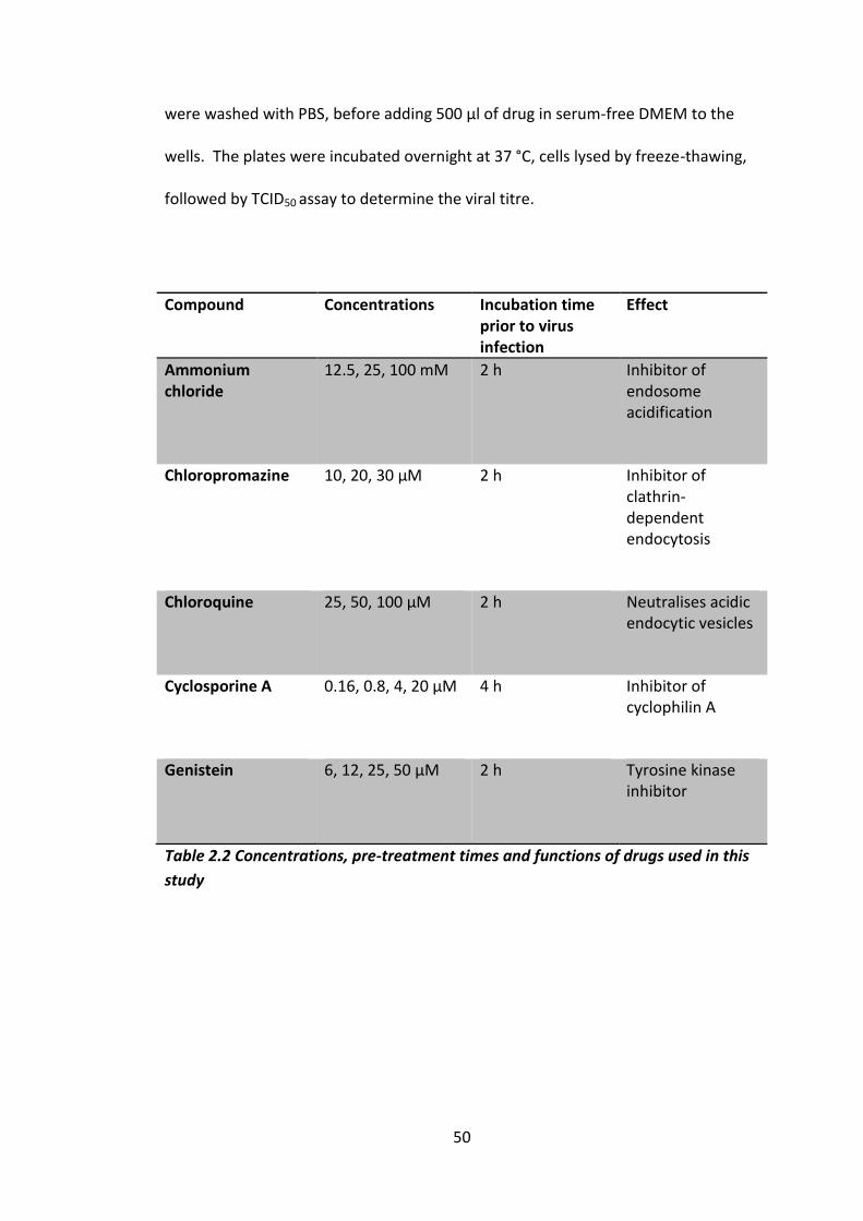
50
were washed with PBS, before adding 500 µl of drug in serum-free DMEM to the
wells. The plates were incubated overnight at 37 °C, cells lysed by freeze-thawing,
followed by TCID50 assay to determine the viral titre.
Compound Concentrations Incubation time prior to virus infection
Effect
Ammonium chloride
12.5, 25, 100 mM 2 h Inhibitor of endosome acidification
Chloropromazine 10, 20, 30 µM 2 h Inhibitor of
clathrin-dependent endocytosis
Chloroquine 25, 50, 100 µM 2 h Neutralises acidic
endocytic vesicles
Cyclosporine A 0.16, 0.8, 4, 20 µM 4 h Inhibitor of
cyclophilin A
Genistein 6, 12, 25, 50 µM 2 h Tyrosine kinase
inhibitor
Table 2.2 Concentrations, pre-treatment times and functions of drugs used in this
study
51
2.2.10 Time courses
Monolayers of Vero cells cells were infected with virus at an MOI of 1 TCID50’s/cell
and then cell were harvested at every time point by scraping the monolayer out
and re-suspending it in the lysate. Lysate was then freeze-thawed three times,
before titration by TCID50 assay.
2.2.11 Drug selection
1 ml of viral lysate was used to infect a T25 flask of confluent Vero cells before
being topped up to 5 ml with either 0.1 nM NLD, 0.9 nM GPP3 or 80 nM ALD. Cells
were left till 100% cell death had occurred and 1 ml of supernatant was taken and
used to infect another flask. Titrations were carried out by TCID50 assay between
each passage.
2.2.12 pH selection
1 ml of crude virus lysate was incubated in a pH 5.6 buffer for one hour at 4 oC. 5 ml
of solution was taken and incubated in a T175 flak of confluent Vero cells for 20
minutes on a rocking platform at room temperature, before it was topped up with
20 ml of refrigerated DMEM. The flask was then incubated at 37 oC until 100% cell
death was observed. Process was repeated until for five passages. Titrations were
carried out by TCID50 assay between each passage.
2.2.13 Heat selection
For the first passage enough virus to infect a T25 flask at an MOI of 10 TCID50’s/cell,
post heating was used, it was first concentrated by ultracentrifugation, then
resuspended in volume of 700 µl of serum free medium. A 10 µl sample was taken
for titration and the rest was heated to 50 oC for 30 minutes in a thermo cycler.

52
Another 10 µl of the heated virus was taken for titration and the rest was used to
infect a T25 flask of Vero cells. The flask was left until 100 % cell death was
observed, and then harvested. Cell debris was removed and freeze thawed three
times in RIPA buffer, before being added to the rest of the supernatant. This
supernatant was then concentrated by ultracentrifugation at 50,000 RPM for one
hour and the resulting pellet was re-suspended in 700 µl of serum-free media. The
re-suspended pellet was then heated to 50 oC and used to infect another T25 flask
of Vero cells. This process was repeated until passage eight, where there less than a
10% drop in logarithmic titre was observed before for four consecutive passages
2.2.14 Heat stability curves
Up to 70 µl of crude virus lysate added to PCR tube and different pocket factors or
PBS was added. Samples were then heated to different temperatures for 30
minutes using a thermo cycler, after 30 minutes samples were chilled to 4 oC and
then titrated by TCID50.
2.2.15 Radio labelling
Virus was used to infect a monolayer of cells and then left for either one hour (BEV)
or six hours (EV71), at which point medium was removed and cells were washed
with PBS. Media was replaces with 10 ml (T175) or 1 ml (6 well plate well) of RIMP
1640 media supplemented with 10 µCi per ml of 35S labelled cystine and methionine
(perkin elmer).

53
2.3 Gel Electrophoresis methods
2.3.1 Agarose gel electrophoresis
Gel electrophoresis were performed using 1 % agarose gels containing SYBR Safe at
0.3 µg/ml. Samples were loaded onto gels with 1x DNA loading buffer.
Electrophoresis for separation of DNA was carried out at 80V (constant) for 30-60
minutes in 1 X TBE buffer. Agarose gels were viewed and imaged using a Syngene
GeneDoc imaging system.
2.3.2 RNA gels
RNA samples were mixed with 2 x RNA loading dye and incubated at 95 oC for 10
minutes. RNA samples were then loaded along with RiboRulerTM low range RNA
ladder and electrophoresis was performed at 80 V (Constant) for 40 minutes. Gels
were viewed and imaged using a Syngene GeneDoc imaging system.
2.3.3 SDS-PAGE
Analysis of protein samples by SDS-PAGE. All analyses were performed on 15 % tris-
glycine resolving gels with 5% stacking gel. The Mini-PROTEAN r 3 electrophoresis
system (BioRad) was used for electrophoretic analysis. Samples were mixed in a
50/50 ratio of sample and 2x loading buffer and heated at 95 oC for 10 minutes,
they were then loaded into each well and run at 200 V (constant) for one hour. SDS-
PAGE gel analysis was carried out with precision plus protein molecular weight
ladder. Resolving gels were then submerged in Coomassie Brilliant Blue stain for + 2
hours on an oscillating platform (10 rpm) at room temperature. Gels were then
transferred to de-staining solution under the same conditions until blue bands were

54
evident on a clear back ground. The destain solution was replaced whenever it
turned a strong blue.
2.3.4 Western blotting
Polyvinylidene difluoride (PVDF) were cut according to the size of the SDS-page gel,
first they were put in 100% methanol for 10 seconds then they were soaked in
dH2O for 10 minutes and then in 1 x transfer buffer for 10 minutes. Sponges and
blotting paper were also soaked in 1 x transfer buffer for 10 minutes. The SDS-PAGE
gel and other components were assembled in a Bio Rad cassette and an ice pack
was put in the tanks. Transfer was then carried out either for 1 hour at 100 V or
overnight at 30 V. At this point the membranes could be dried and stored for a later
date, if membranes were to be used that day then they progressed directly to the
blocking stage. If membranes had been dried they were reconstituted in methanol
for 10 seconds and washed in PBS for 10 minutes. The membranes were then
washed in PBS for 10 minutes before transferring into blocking buffer for 1 hour.
They were then transferred into primarily antibody diluted in blocking buffer
1/5000 and incubated for 1 hour. Membranes were then washed 3 x 10 minutes in
PBS-Tween. They were then probed with an anti-mouse peroxidise conjugated
secondary antibody diluted in blocking buffer 1//2000 for 1 hour. Membranes were
then washed again in PBS-Tween 3 x 10 minutes. The excess buffer was drained off
and they were incubated in ECL for 1 minute at room temperature followed by
exposing to X-ray film (Hyperfilm ECL, Amersham Biosciences).
55
2.3.5 Gel dry down
After completion of electrophoresis of the SDS-page gel, gels were incubated in gel
dry down buffer for 30 minutes, then incubated for 15 minutes in Amplify GE heath
care (Amplify can be reused). Upon completion of incubation, the gel was placed
upon a 3 mm CHR Whatmann paper, and cling film was placed on top of the gel,
and an additional piece of 3 mm CHR Whatmann paper was placed on top of the
cling film. The gel was then placed in BioRad gel dryer, the plastic sheet of the gel
dryer was placed over the top and the vacuum was turned on, ensuring that the
plastic had formed a seal. Setting were then set as follows; slowest rate of heat
increase (Setting three), final heat 80 oC and for a total of 2 hours. Once the gel had
been dried down, it was exposed to film in a cassette at -80 oC, for 12 to 144 hours.
2.4 Cloning
2.4.1 Transformation of competent cells
Transformation of plasmid DNA into E.coli gold cell was performed using heat shock
method. 50 µl of competent cells were mixed with 20-40 ng of plasmid DNA and 2
µl of beta mercaptoethanol and incubated on ice for 30 minutes. Cells were heat
shocked at 42 oC for 45 seconds and then incubated on ice for 5 to 10 minutes. They
were then added to 3 ml of antibody free LB and incubated at 37 oC for ~ 1 hour. 50
µl and 100 µl samples were then plated out onto LB/amp plates and incubated
overnight; different amounts were plated to assess the effectiveness of the
transformation. The following day three colonies were picked from each plate and
grown on a shaking platform overnight at 37 oC in 5 ml of LB broth. The next day

56
glycerol stocks were prepared, by mixing equal volumes of 80 % glycerol and cell
suspension. These were then stored at -70 oC. These stocks were then used to
initiate cultures for plasmid extraction by mini or maxi prep (QIAGEN).
2.4.2 RNA Extraction
Viral RNA extractions were carried out from crude lysates of infected cells, using
QAIGEN universal RNA extraction kit following the manufactures instructions.
2.4.3 RNA Transcriptions
Transcription was carried out using a RiboMAX™ Large Scale RNA Production T7
System following the manufactures instructions.
2.4.4 Reverse transcriptions
Reveres transcriptions were carried out using Qiagen reverse transcription kit using
the manufactures instructions.
2.4.5 PCR
PCR was carried out using Qiangen fast cycle PCR, following the manufactures
instructions. Using primers in table 2.2.
2.4.6 TOPO cloning
TOPO cloning was carried out using the Life Technologies TOPO cloning kit,
following the manufactures instructions.
2.4.7 Sequencing
Samples for sequencing were sent to Beckman Coulter using their speciation’s using
the primers in table 2.3.
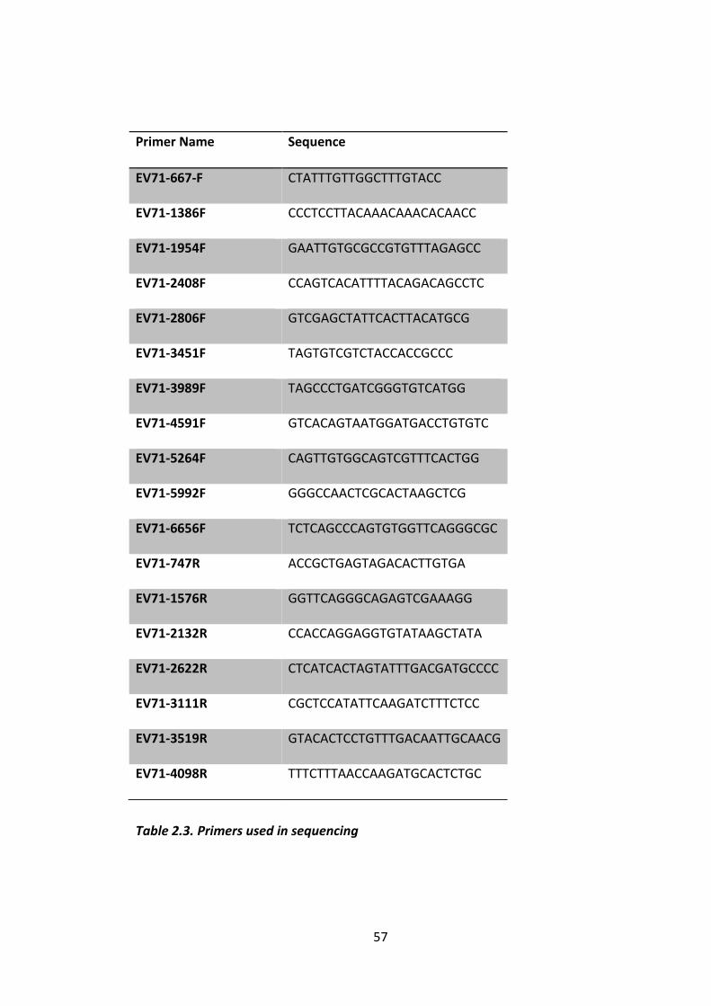
57
Table 2.3. Primers used in sequencing
Primer Name Sequence
EV71-667-F CTATTTGTTGGCTTTGTACC
EV71-1386F CCCTCCTTACAAACAAACACAACC
EV71-1954F GAATTGTGCGCCGTGTTTAGAGCC
EV71-2408F CCAGTCACATTTTACAGACAGCCTC
EV71-2806F GTCGAGCTATTCACTTACATGCG
EV71-3451F TAGTGTCGTCTACCACCGCCC
EV71-3989F TAGCCCTGATCGGGTGTCATGG
EV71-4591F GTCACAGTAATGGATGACCTGTGTC
EV71-5264F CAGTTGTGGCAGTCGTTTCACTGG
EV71-5992F GGGCCAACTCGCACTAAGCTCG
EV71-6656F TCTCAGCCCAGTGTGGTTCAGGGCGC
EV71-747R ACCGCTGAGTAGACACTTGTGA
EV71-1576R GGTTCAGGGCAGAGTCGAAAGG
EV71-2132R CCACCAGGAGGTGTATAAGCTATA
EV71-2622R CTCATCACTAGTATTTGACGATGCCCC
EV71-3111R CGCTCCATATTCAAGATCTTTCTCC
EV71-3519R GTACACTCCTGTTTGACAATTGCAACG
EV71-4098R TTTCTTTAACCAAGATGCACTCTGC

58
2.5 Purification
2.5.1 Purification of Virus through a sucrose gradient (A)
Once 100 % cell death was observed, the supernatant was removed and
centrifuged at 10,000 RPM for 30 minutes. The supernatant was then poured off
and stored at 4 oC if to be saved for future use. The pellet was re-suspended in an
appropriate amount of RIPA buffer and freeze-thawed 3 x before adding the
supernatant to the main supernatant. This would then be made up to 50 %
saturated ammonium sulphate or a 10 % polyethylene -glycol solution and stirred at
4 oC for 90 minutes. This was then centrifuged at 10,000 RPM for 1 hour; the
suspension supernatant would be poured off and saved for titration. The pellets
were re-suspended in an appropriate amount of PBS. These would then be
centrifuged at 29,000 RPM for 3 hours though a 30 % sucrose cushion after which
the supernatants were poured off and saved for titration. The pellets were re-
suspended overnight on a rocking platform at 4 oC. The re-suspended pellets were
made 1% SDS and loaded onto a 36 ml 15-45 % sucrose gradient in PBS and
centrifuged for 3 hours at 29,000 RPM/ 151,000 RCF at 20 oC. 2 ml fractions were
collected from the bottom of the centrifuge.
2.5.2 Purification of Virus through a sucrose gradient B
Virus was grown in culture flasks and labelled with 35S labelled cystine and
methionine, the supernatant was then clarified by centrifugation at 10,000 g for 30
minutes. The resultant pellet was then freeze-thawed three times in 1% NP40 to
release trapped virus particles, this was then clarified and added to the
supernatant. This was then subjected to ultracentrifugation at 32K rpm for one
59
hour through a 2 ml 30% sucrose cushion. The resulting pellet was then subjected
to ultracentrifugation through 15-45% sucrose gradient for 50 minutes at 296,000 x
g, this was then fractionated and the resulting fractions were added to scintillation
fluid and counted.
2.5.3 Nycodenz
Virus was grown in culture flasks and labelled with 35S labelled cystine and
methionine, the supernatant was then clarified by centrifugation at 10,000 g for 30
minutes. The resultant pellet was then freeze-thawed three times in RIPA or 1%
NP40 buffer to release trapped virus particles, this was then clarified and added to
the supernatant. This was then subjected to ultracentrifugation at 32K rpm for one
hour through a 2 ml 30% sucrose cushion. This was re-suspended overnight in virus
re-suspension buffer and then subjected to ultra-centrifugation either through a
40% self-forming Nycodenz gradient, or a 20%,30%,40%,50%,-60% layers step
gradient. This was then separated into 300 µl fractions.
2.5.4 3H Palmitic acid
Virus was grown in culture flasks and labelled with 35S labelled cystine and
methionine, the supernatant was then clarified by centrifugation at 10,000 g for 30
minutes. The resultant pellet was then freeze-thawed three times in RIPA or 1%
NP40 buffer to release trapped virus particles, this was then clarified and added to
the supernatant. This was then subjected to ultracentrifugation at 32K rpm for one
hour through a 2 ml 30% sucrose cushion. It was then incubated in 3H palmitic acid
overnight and then split into two equal volumes, 2 nM NLD was added to one tube
and the other was left untreated. After two minutes of NLD treatment the two

60
fractions were subjected to ultracentrifugation through a self-forming nycodenz
gradient.
2.5.5 Scintillation counting
10 to 200 µl of sample was added to 2 ml of perkin elmer scintillation fluid in a
scintillation fluid tube, samples were then counted using a scintillation counter.
Counting for one minute per tube.
2.6 Immunoprecipitation
50 µl of crude virus lysates were incubated with 10 µl of sepharose G beads (GE
Healthcare) and 5 µl of either D6 or A9 antibody and then topped up to 500 µl with
PBS. This was left spinning on a rotating platform overnight at 4 oC. The following
day the beads were subjected to centrifugation at 10,000 RPM to pellet the beads,
the supernatant was removed and the beads were re-suspended in PBS. The
process was repeated three times, then the beads were re-suspended in 50 µl of LB
buffer, and were then heated to 100 oC for 10 minutes. The samples pelted for 5
minutes at 13,000 RPM, and the sample was then analysed by SDS-PAGE.
2.7 ELISA
Purified SCARB2 or PSGL1 were diluted to 0.1 µg in Carbonate Coating buffer and
50 µl was incubated individual wells on a 96 well plate. The plate was incubated
overnight at 2-8 oC in a box with a humidified atmosphere. The next day each well
was washed 4 x with wash buffer, leaving the plate containing the last wash at

61
room temperature for at least 30 minutes. 50 µl of sample was added to each well.
The plate was then sealed and incubated for two hours at 37 oC and then washed 3
x with ELISA wash buffer. Then 50 µl of either mAb D6 or A9 diluted 1 in 100 in
ELISA assay diluent and added to each well. The plate was then sealed and
incubated for 1 hour and then washed with ELISA wash buffer three times. 50 µl of
peroxidase conjugated anti-mouse diluted in ELISA assay diluent at 1/400 was
added to each well. After 30 minutes 1M H2SO4 was added to each well and the
optical density at 492 was read as soon as possible.
2.8 PyMol
Structural figure were prepared using PyMol

62
Chapter 3

63
Chapter 3 EV71 Compounds – In vitro characterisation and
selection of resistant isolates
There are currently no compounds available for clinical use against EV71, although in
vitro studies have shown that several different approaches could potentially lead to
effective EV71 therapeutics (Reviewed in Kok, 2015; Kuo & Shih, 2013; Shang, Xu, &
Yin, 2013). One such strategy is the use of small inhibitory compounds that bind the
enterovirus VP1 pocket (discussed in section 1.3). In an untreated enterovirus
population the VP1 pocket is filled with a hydrophobic lipid known as the pocket
factor. The pocket factor is expelled from the pocket upon receptor binding; this
initiates viral uncoating and genome release and is associated with capsid instability
(Ren et al. 2013; Dang et al. 2014). The presence of compounds that bind to the VP1
pocket with a higher affinity are therefore able to prevent receptor binding/uncoating
and infection of the cell (Rotbart 2002). In the case of BEV1 the use of certain fatty
acids, shown to be natural pocket factors by mass spectrometry have also been shown
to have an inhibitory effect (Ismail-Cassim et al. 1990; Smyth et al. 2003).
Since the discovery of the first of these pocket binding compounds (refer to
Introduction 1.3), other pocket binding compounds have been found for a wide
variety of enterovirus including; HRV, CVB3 and PV (Mckinlay et al. 1992; Groarke &
Pevear 1999a; Liu et al. 2012). With certain compounds, such as the HRV pocket
binding compound Pleconaril in phase III trials (Feil et al. 2012). Although they are
shown to be effective inhibitors, resistance has been shown to evolve rapidly towards
them (Groake and Pevear 1999; Liu et al. 2012).

64
In addition to the many other pocket binding compounds, 3-(4-pyridyl)-2-
imidazolidinone derivatives have been shown to be the most effective pocket binding
compounds against EV71 which were termed the GPP compounds, the most inhibitory
of which was GPP3 (Chern et al. 2004; Ke & Lin 2006). Since the generation of these
compounds the crystal structure of EV71 has been solved (Wang et al. 2012; Plevka et
al. 2012), and the interactions between four GPP compounds (GPP2, GPP3, GPP4 and
GPP12) and the EV71 capsid were modelled (DeColibus et al. 2014) ( Figure 3.1).
Figure 3.1 Structures of different3-(4-pyridyl)-2-imidazolidinone derivatives with
anti-EV71 activity. Images modified from DeColibus et al. 2014.
In addition to this, in silico docking was used to predict the energy of interaction
between the compounds and VP1. This was performed by computing the docking
poses of the compounds in the VP1 pocket using quantum mechanics-polarised ligand
docking (QMPLD). Compounds that were predicted to have a higher energy of
interaction had previously been shown to have lower IC50 value (DeColibus et al.
2014). Based on these observations, in silico scanning was performed to identify areas

65
on the capsid which could act as additional binding sites for these inhibitors, and the
mouth of the VP1 pocket was identified as one such area. To improve the binding to
this area, an amide or an amine were added to the C-terminus of GPP3 on the pyridine
ring, thus creating NLD and ALD. Both these molecules are predicted to have a higher
binding efficiency by QMPLD than GPP3, by creating an extra binding site to the
mouth of the VP1 pocket (DeColibus et al. 2014).
In this chapter, the effect of NLD and ALD on EV71 grown in Vero cells is characterised
and compared to two compounds previously identified (GPP3 and GPP4) and potential
pocket factors (lauric acid, myristic acid and palmitic acid). Isolates of virus that were
found to be resistant to these compounds are also identified and characterised.

66
3.1 Characterisation of viral inhibition and cellular cytotoxicity
The effectiveness of NLD and ALD as inhibitors towards EV71 and other enteroviruses
was assessed and compared to previously characterised inhibitors GPP3 and GPP4, as
well as to fatty acids/natural pocket factor candidates, lauric acid, myristic acid and
palmitic acid. This was carried out by titrating EV71 via TCID50 assays (Refer to section
2.2.7, 2.2.8), in a range of different concentrations of each inhibitor for seven days.
Prior to this the minimum amount of DMSO able to dissolve 1000 nM of each
compound was assessed. This was carried out by adding a range of DMSO
concentrations from 0.5 v/v % to 5 v/v % DMSO to each compound and observing at
what concentration it dissolved each compound. This was 1 v/v % for NLD, ALD and
GPP3 and 5 v/v % for GPP4. These DMSO concentrations were kept constant in each
TCID50 assay and DMSO only control wells were included in each assay. All fatty acids
were dissolved in ethanol to make a 10 mM stock and remained soluble at
concentrations of 50 µM when ethanol was diluted down to 0.05 v/v % ethanol.
The TCID50 values were represented as a sigmoidal curve, modelled using a nonlinear
regression (curve fit) to fit the data (Figure 3.2). This showed that NLD was the most
effective inhibitor, with an IC50 value of 0.025 nM and the ability to reduce titres
below 5% at 0.05 nM. GPP3 was the next most effective inhibitor with an IC50 of 0.39
nM, then ALD which had an IC50 value of 8.54 nM although a more complex relation
was seen in the curve and it did not fit the curve perfectly, which will reduce the
precision of the IC50 values. Control wells were exposed to the equivalent amount of
DMSO (1%) to ensure that this was not causing any cytopathic effect or an effect on
viral titre. Also GPP4 was shown to have no inhibitory effect up to concentrations of 1
µM (Figure 3.2) (De Colibus et al. 2014).
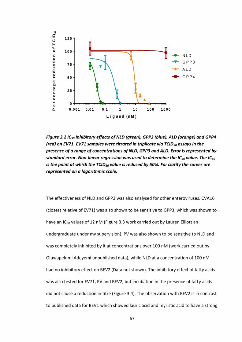
67
L i g a n d (n M )
Pe
rc
en
ta
ge
re
du
ctio
n o
f T
CID
50
0 .0 0 1 0 .0 1 0 .1 1 1 0 1 0 0 1 0 0 0
0
2 5
5 0
7 5
1 0 0
1 2 5
N LD
A L D
G P P 3
G P P 4
Figure 3.2 IC50 Inhibitory effects of NLD (green), GPP3 (blue), ALD (orange) and GPP4
(red) on EV71. EV71 samples were titrated in triplicate via TCID50 assays in the
presence of a range of concentrations of NLD, GPP3 and ALD. Error is represented by
standard error. Non-linear regression was used to determine the IC50 value. The IC50
is the point at which the TCID50 value is reduced by 50%. For clarity the curves are
represented on a logarithmic scale.
The effectiveness of NLD and GPP3 was also analysed for other enteroviruses. CVA16
(closest relative of EV71) was also shown to be sensitive to GPP3, which was shown to
have an IC50 values of 12 nM (Figure 3.3 work carried out by Lauren Elliott an
undergraduate under my supervision). PV was also shown to be sensitive to NLD and
was completely inhibited by it at concentrations over 100 nM (work carried out by
Oluwapelumi Adeyemi unpublished data), while NLD at a concentration of 100 nM
had no inhibitory effect on BEV2 (Data not shown). The inhibitory effect of fatty acids
was also tested for EV71, PV and BEV2, but incubation in the presence of fatty acids
did not cause a reduction in titre (Figure 3.4). The observation with BEV2 is in contrast
to published data for BEV1 which showed lauric acid and myristic acid to have a strong

68
inhibitory effect and palmitic acid to have a weak inhibitory effect (Ismail-Cassim et al.
1990). This could be due to the fact that the two BEV serotypes have different sized
pockets. Higher concentrations were not used due to the cytotoxic effect.
1 1 0 1 0 0
2 0
4 0
6 0
8 0
1 0 0
L ig a n d (n M )
Pe
rc
en
tag
e r
ed
uc
tio
n o
f T
CID
50
Figure 3.3 Inhibition of CVA16 by GPP3. CVA16 samples were titrated via TCID50
assay in the presence of a range of concentrations of GPP3. Non-linear regression
was used to determine the IC50 value. The IC50 is 14 nM the point at which the TCID50
value is reduced by 50%. For clarity the curves are represented on a logarithmic
scale. N=1 (Work was conducted by Lauren Elliott an undergraduate student under
my supervision).
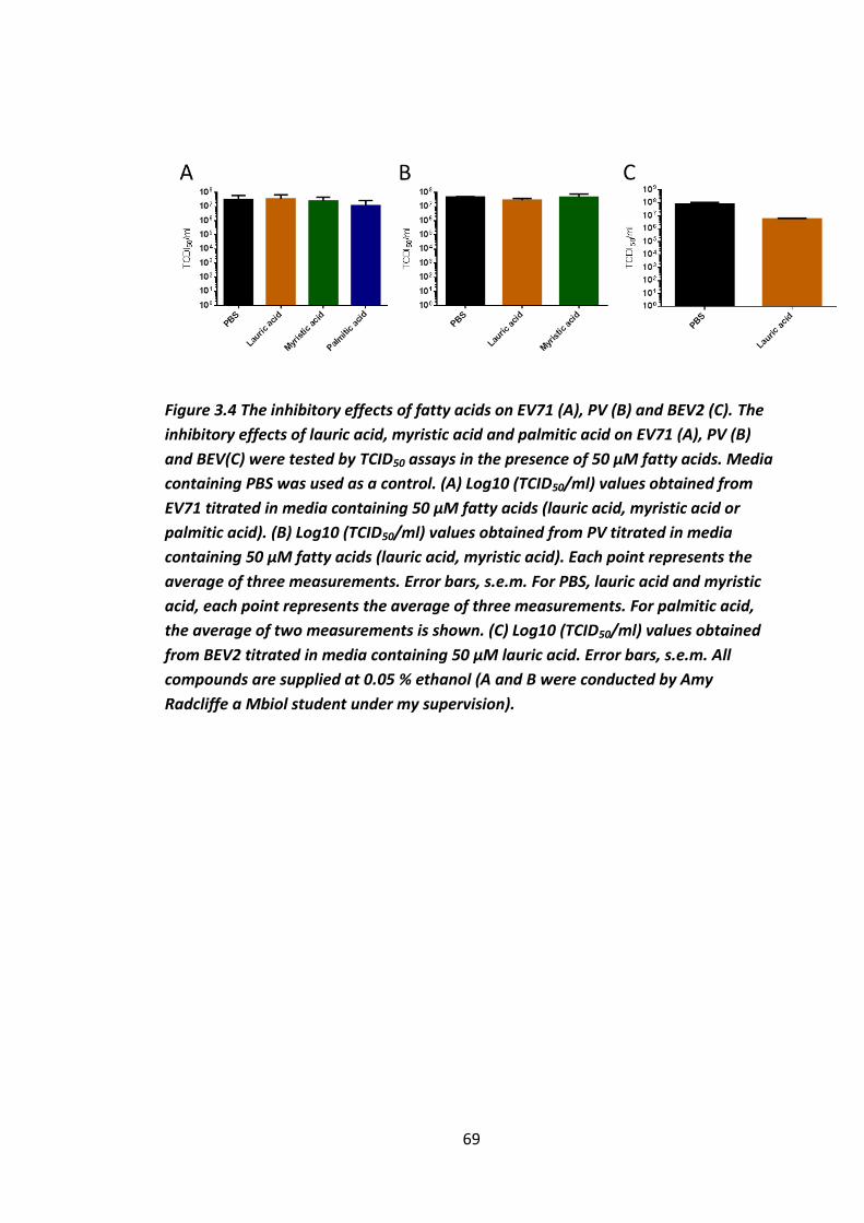
69
Figure 3.4 The inhibitory effects of fatty acids on EV71 (A), PV (B) and BEV2 (C). The
inhibitory effects of lauric acid, myristic acid and palmitic acid on EV71 (A), PV (B)
and BEV(C) were tested by TCID50 assays in the presence of 50 µM fatty acids. Media
containing PBS was used as a control. (A) Log10 (TCID50/ml) values obtained from
EV71 titrated in media containing 50 µM fatty acids (lauric acid, myristic acid or
palmitic acid). (B) Log10 (TCID50/ml) values obtained from PV titrated in media
containing 50 µM fatty acids (lauric acid, myristic acid). Each point represents the
average of three measurements. Error bars, s.e.m. For PBS, lauric acid and myristic
acid, each point represents the average of three measurements. For palmitic acid,
the average of two measurements is shown. (C) Log10 (TCID50/ml) values obtained
from BEV2 titrated in media containing 50 µM lauric acid. Error bars, s.e.m. All
compounds are supplied at 0.05 % ethanol (A and B were conducted by Amy
Radcliffe a Mbiol student under my supervision).

70
These results show that low concentrations of NLD, GPP3 and ALD could potentially be
used to treat EV71 infections. However, the cytotoxicity of these compounds needed
to be assessed so that the therapeutic window could be calculated, i.e. the amount of
inhibitor that can be used before it becomes toxic to cells still needed to be assessed.
Vero cells were incubated in the presence of media containing a range of
concentrations of NLD, GPP3, ALD or DMSO for seven days, after which the cellular
cytotoxicity was evaluated by MTT assay (Figure 3.5). From these results we can see
that the CC50 values for the three inhibitors respectively are 270 nM, 250 nM and 1000
nM. The CC50 value is the value at which the compounds cause a 50% reduction in the
OD reading judged by MTT assay. DMSO did not have a cytotoxic effect at the
concentration of 1% that was used for NLD, GPP3 or ALD. This gives a therapeutic
window of 1 x 10⁴ for NLD, 6.8 x 10² for GPP3 and > 1.3 x 10³ for ALD (Table 3.1). Both
NLD and ALD therefore have a larger therapeutic window than the previous best EV71
inhibitor GPP3.

71
C o n c e n tra t io n (M )
Pe
rc
en
ta
ge
re
du
ctio
n i
n O
D
0 .0 1 0 .1 1 1 0 1 0 0
0
1 0
2 0
3 0
4 0
5 0
6 0
7 0
8 0
9 0
1 0 0
A L D
G P P 3
N LD
1 0 1 0 0
-2 0
-1 0
0
1 0
2 0
3 0
4 0
5 0
6 0
7 0
8 0
9 0
1 0 0
C o n c e n tra t io n o f D M S O %
Pe
rc
en
tag
e r
ed
uc
tio
n i
n O
D
Figure 3.5 Toxicity of NLD, GPP3 and ALD to Vero cells. Vero cells were incubated
with a range of concentrations of NLD (green filled triangle), GPP3 (blue filled
square), ALD (orange filled circle). (A) or DMSO (B) for seven days. MTT assays were
then performed and the CC50 was derived from the OD (692 nm) by non-linear
regression. Each point represents an average of three measurements and error was
measured using standard error.
A
B

72
NLD GPP3 ALD
CC50
(Cell toxicity)
250 nM 270 nM >1000 nM
IC50
(EV71 inhibition)
0.025 nM 0.39 nM 8.7 nM
Therapeutic window
(CC50 : IC50)
1 x 10⁴ 6.8 x 10² > 1.3 x 10³
Table 3.1 Therapeutic window of NLD, GPP3 and ALD against EV71. This table
displays the CC50 (from Figure 3.5) and IC50 (from Figure 3.2). The CC50 value has been
divided by the IC50 value to give the therapeutic window for each compound.

73
3.2 Selection of resistant mutants
As stated previously (section 1.3), enteroviruses are known to evolve resistance
rapidly towards pocket binding inhibitors, therefore it is likely that resistance can arise
to NLD, GPP3 and ALD.
To investigate this, EV71 or CVA16 were grown in the presence of these pocket
binding compounds at a concentration able to reduce the TCID50 value by over 99.9%
(Figure 3.7). Concentrations of 0.1 nM NLD, 0.9 nM GPP3, 70 nM ALD or a
combination of 0.1 nM NLD/ 0.9 nM GPP3 were used as the selection pressure for
EV71 and concentrations of 20 nM GPP3 was used for CVA16. Virus was grown under
of one of these selection pressures until 100% cell death was achieved, samples were
then titrated and passaged in the presence of the same concentration of inhibitor,
until 100% cell death was achieved again. This was repeated for eight passages (Figure
3.7). Titres were seen to rise after just one or two passages and continued to rise until
either passage six or eight at which point the titres were equivalent to WT virus in the
absence of any compounds (EV71 ~ 1 x 10⁷ TCID50/ml and CVA16 ~ 1 x 10⁵ TCID50/ml)
(Figure 3.7).
Each isolate was then sequenced to determine the mutations which may have arisen
to generate resistance. Isolates were sequenced from purified PCR products that had
been sub-cloned into a vector (See methods sections 2.4.5, 2.4.6), this was to ensure
that the mutations seen in combination were present in individual isolates. 18 of the
sequenced sub-clones possessed the same two VP1 amino acid changes, I113M and
L123V. These were observed in the GPP3 resistant isolates (4/4), the ALD resistant
isolate (1/1), one of the NLD resistant isolates (1/13), and all the isolates taken from
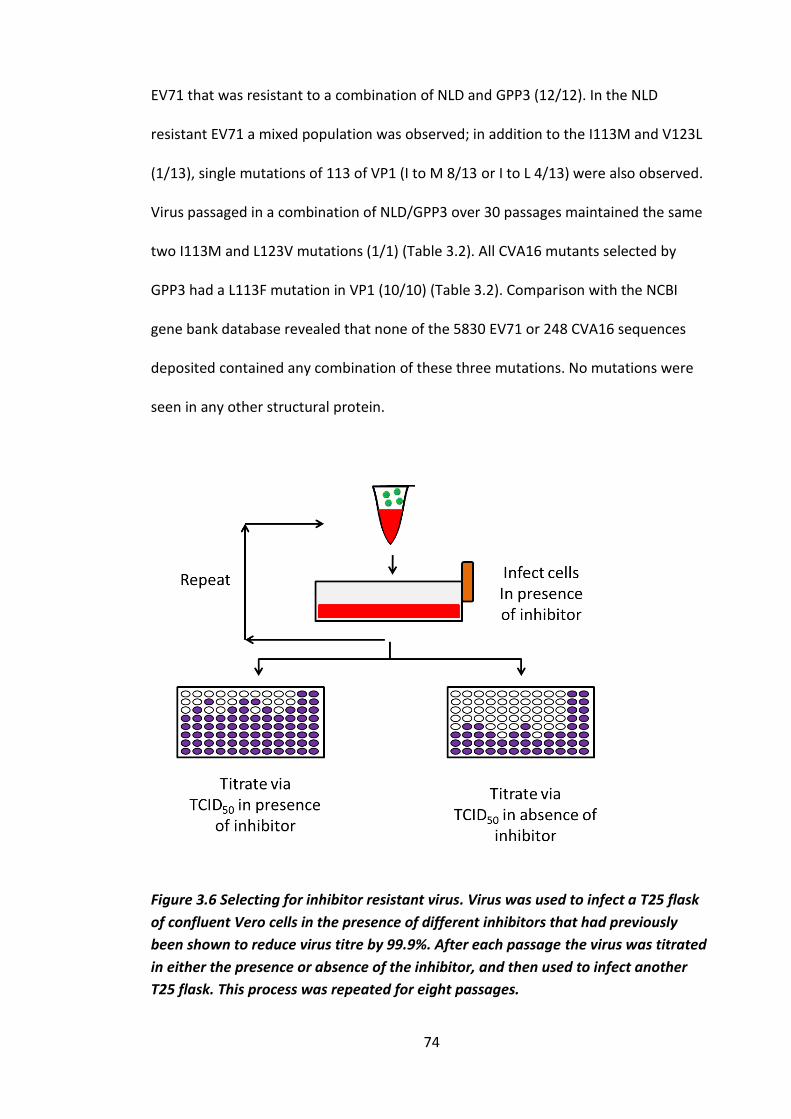
74
EV71 that was resistant to a combination of NLD and GPP3 (12/12). In the NLD
resistant EV71 a mixed population was observed; in addition to the I113M and V123L
(1/13), single mutations of 113 of VP1 (I to M 8/13 or I to L 4/13) were also observed.
Virus passaged in a combination of NLD/GPP3 over 30 passages maintained the same
two I113M and L123V mutations (1/1) (Table 3.2). All CVA16 mutants selected by
GPP3 had a L113F mutation in VP1 (10/10) (Table 3.2). Comparison with the NCBI
gene bank database revealed that none of the 5830 EV71 or 248 CVA16 sequences
deposited contained any combination of these three mutations. No mutations were
seen in any other structural protein.
Figure 3.6 Selecting for inhibitor resistant virus. Virus was used to infect a T25 flask
of confluent Vero cells in the presence of different inhibitors that had previously
been shown to reduce virus titre by 99.9%. After each passage the virus was titrated
in either the presence or absence of the inhibitor, and then used to infect another
T25 flask. This process was repeated for eight passages.

75
Figure 3.7 Generation of resistant isolates. WT EV71 was passaged in the presence of either (A) 0.1 nM NLD, (B) 0.9 nM GPP3, (C) 80
nM ALD or, (D) a combination of 0.1 nM NLD and 0.9 nM GPP3, (E) WT CVA16 was passaged in the presence of 20 nM GPP3. Each
isolate was passaged a total of 8 times and after each passage a sample was titrated in the presence of the selecting concentration
of compound.
N L D re s is ta n t- is o la te
P a s s a g e n u m b e r
Lo
g T
CID
50/m
l
0 2 4 6 8
4
5
6
7
A L D re s is ta n t- is o la te
P a s s a g e n u m b e r
Lo
g T
CID
50/m
l
0 2 4 6 8
3
4
5
6
7
G P P 3 re s is ta n t- is o la te
P a s s a g e n u m b e r
Lo
g T
CID
50/m
l
0 2 4 6 8
2
3
4
5
6
7
G P P 3 /N L D re s ita n t- is o la te
P a s s a g e n u m b e r
TC
ID5
0/m
l
0 2 4 6 8
2
3
4
5
6
7
C V A 1 6 G P P 3 r e s is ta n t- is o la te
P a s s a g e n u m b e r
Lo
g T
CID
50/m
l
0 2 4 6 8
3
4
5
6E
D
C B A

76
Virus/Compound Mutation(s) Number sequenced
EV71/NLD I113M (62%), I113L
(31%), I113M/V123I
(7%)
n=13
EV71/GPP3 I113M/V123I n=4
EV71/ALD I113M/V123I n=1
EV71/NLD + GPP3 I113M/V123I n=12
CVA16/GPP3 L113F n=10
Table 3.2 Resistance mutations. Mutations identified in EV71/CVA16 after
passage in the presence of NLD, GPP3, ALD or a combination of NLD/GPP3. CVA16
isolates resistant to NLD or ALD were not selected.
To better understand the effects these mutations are having on the structure,
computational modelling using Rosetta was carried out by Luigi De Colibus (Oxford
University). Figure 3.8 illustrates the crystal structure of EV71 in complex with NLD
and shows the interactions between NLD and the VP1 pocket. This demonstrates
that NLD is able to bind the capsid in several places, including residue I113 where
resistance mutations occurred. Predictive modelling of mutation I113M suggested
that the methionine side chain comes out to fill the pocket, possibly causing a steric
clash with NLD. While predictions of the effect of mutation V123L suggested that
the leucine residue causes the pocket to shrink, leaving less space for the
compound to occupy. In CVA16 the L113F mutation was predicted to have the same
effect as I113M in EV71, with phenylalanine causing a steric clash with GPP3. To

77
further assess how the mutations could be affecting stability, the change in free
energy of folding (ΔΔG-folding) between WT and the mutant viruses was assessed
and compared. This works on the principle that proteins with the lowest free-
energy states are more stable. This was carried out by Luigi De Colibus at Oxford
University using Rosetta. This software computes atom-atom interactions, and is
able to predict the hydrophobic effects, electrostatic desolvation cost associated
with burial of polar atoms and an explicit hydrogen-bonding potential to predict a
ΔΔG-folding value. Here structures of WT EV71 in complex with NLD and WT CVA16
in complex with GPP3 (De Colibus. et al 2014, De Colibus. et al 2015) were used.
Differences in ΔΔG-folding were calculated by Rosetta by relaxing the VP1 subunit
structure with coordinate constraints, and applying monomer ΔΔG-folding
calculation without sampling alternative backbone conformations (Fowler et al.
2010, Kellogg et al. 2011, Tyka et al. 2011).
This predicted that differences between ΔΔG-folding between the WT and EV71
I113M mutant was +0.73 kcal/mol, WT and V123I -0.98 kcal/mol whereas the
combination of both mutations gave ΔΔG-folding of ~ +1.1 kcal/mol, this suggests
that the mutant virus capsid may be less stable than WT (Fowler et al. 2010; Kellogg
et al. 2011; Tyka et al. 2011 carried out by Luigi DeColibus Oxford University).
However, the ΔΔG-folding for CVA16 mutant was -1.7 Kcal/mol, suggesting that this
mutant may be more stable than WT.
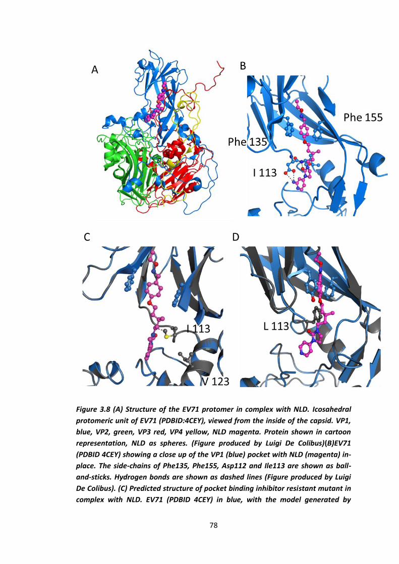
78
Figure 3.8 (A) Structure of the EV71 protomer in complex with NLD. Icosahedral
protomeric unit of EV71 (PDBID:4CEY), viewed from the inside of the capsid. VP1,
blue, VP2, green, VP3 red, VP4 yellow, NLD magenta. Protein shown in cartoon
representation, NLD as spheres. (Figure produced by Luigi De Colibus)(B)EV71
(PDBID 4CEY) showing a close up of the VP1 (blue) pocket with NLD (magenta) in-
place. The side-chains of Phe135, Phe155, Asp112 and Ile113 are shown as ball-
and-sticks. Hydrogen bonds are shown as dashed lines (Figure produced by Luigi
De Colibus). (C) Predicted structure of pocket binding inhibitor resistant mutant in
complex with NLD. EV71 (PDBID 4CEY) in blue, with the model generated by

79
Rossetta shown in grey. The two mutated side chains I113M and V123L are shown
as grey balls-and-sticks (Figure produced by Luigi De Colibus). (D) Predicted
structure of CVA16 GPP3 resistant mutant in complex with GPP3. CVA16 in
complex with GPP3 (unpublished). I113F is shown (blue and grey balls-and-sticks).
(Figure produced by Luigi De Colibus).
3.3 Fitness cost of inhibitor resistant mutations
As mutations that deviate from WT often incur a fitness cost, the genetic stability of
the inhibitor-resistant EV71 isolate selected using both NLD and GPP3 (I113M,
V123L) was tested by repeated passage in the absence of inhibitors (Figure 3.9).
After each passage, virus was titrated in the presence or absence of inhibitor. This
showed that while the titre remained unaffected in the absence of inhibitor, in the
presence of the inhibitor the titre dropped dramatically after just one passage. This
continued for the next two passages, after which the titre became equivalent to the
WT when in the presence of the inhibitors (Figure 3.9). Sequencing confirmed that
the virus had reverted to a WT genotype (n=1), indicating a strong selection
pressure for reversion. However, as the experiment was performed with a “pool” of
virus it is not possible to determine if this is back mutation or if it is amplification of
residual WT virus.
80
Figure 3.9 Inhibitor resistant isolates are genetically unstable. An inhibitor-
resistant isolate that was selected with a combination of 0.1 nM NLD and 0.9 nM
GPP3 was passaged four times in the absence of any inhibitor. It was then titrated
in the presence (red circles) and absence of inhibitor (black squares) using TCID50
assay. n = 1.
To determine the reason for the reduced fitness of the inhibitor-resistant virus, the
growth kinetics of the both WT EV71 and the inhibitor-resistant isolate were
compared. To do this a one-step growth curve was performed; samples from both
isolates were taken every three hours for 18 hours and then titrated via TCID50. This
revealed no difference between the growth kinetics of the two variants (Figure
3.10).

81
Figure 3.10 One step growth curves of inhibitor-resistant and WT EV71. WT (black
filled circle) and inhibitor-resistant EV71 selected in the presence of 0.1 nM NLD and
0.9 nM GPP3 (grey open square) were used to infect six wells each of a 96 well plate
at an MOI of 10 TCID50’s/cell. Every three hours the supernatant and cells of a well
from each isolate were removed, and freeze-thawed to lyse the cells. These samples
were then titrated by TCID50 assay. n = 1.
As the growth kinetics did not appear to be the fitness cost, the thermostability was
then assessed. The thermal stabilities of WT EV71 and inhibitor-resistant mutant
virus selected in the presence of NLD/GPP3 were evaluated in the absence and
presence of 0.1 nM NLD / 0.9 nM GPP3 or 2 nM NLD. To do this all isolates were
exposed to a range of temperatures using a thermocycler for 30 minute periods and
each sample was then titrated (Figure 3.11A). This showed that WT heated in the
presence of NLD was able to tolerate temperatures 6 oC higher before inactivation
began to occur and 3.5 oC higher before total inactivation occurred compared to
the inhibitor-resistant virus. In contrast to WT virus, inhibitor-resistant virus could
not be rescued from heat inactivation by the presence of 2 nM NLD or by a
combination of 0.1 nM NLD and 0.9 nM GPP3. The inhibitor-resistant virus was also

82
shown to be more thermolabile than the WT, with initial inactivation occurring at
~3 oC lower and the complete inactivation temperature occurring 4 oC lower. This
suggests that the I113M/V123L mutation prevented the compounds from binding
within the VP1 pocket and results in increased thermolability. Also each stability
curve appeared to have a step, indicating that there is population of viruses with
different heat stabilities. Similar results were obtained with inhibitor-resistant
CVA16 where heat inactivation was shown to occur at 54 oC in the WT and at 51 oC
in the GPP3 resistant L113F mutant, although there was a less obvious step here
(Figure 3.11B). These results are consistent with observations in PV and HRVs in
which pocket-binding compounds were able to increase thermostability while
inhibitor resistant isolates with mutations located in their pockets were shown to
be more thermolabile (Katpally et al. 2007; Mosser et al. 1994; Shepard et al. 1993).
In addition to a reduced stability at higher temperatures, the resistant isolates were
also noted to be less stable during long term storage at ~4 oC in the fridge and after
storage at -20 oC and -80 oC, when no cryoprotectant was used. Inhibitor-resistant
isolates start to become non-viable after 3 weeks storage at ~4 oC or after one
freeze thaw after being stored at -20 Co or -80 Co. In contrast, WT virus titre remains
constant after months of storage at 4 oC and after multiple freeze thaw cycles.

83
Figure 3.11 Inhibitor-resistant mutants are more thermolabile than WT virus. (a)
Thermolability curves of EV71 WT (black filled circle) and inhibitor-resistant EV71
(grey open square), WT EV71 in the presence of 2 nM NLD (hot pink open circle),
inhibitor-resistant EV71 in the presence of 0.9 nM GPP3 and 0.1 nM NLD (blue
open triangle) and 2 nM NLD (red filled triangle) (b) Thermolability curves of WT
CVA16 (black filled circle) and inhibitor-resistant CVA16 (purple open square). All
samples were heated at a range of temperatures for 30 minutes using a
thermocycler, prior to titration by TCID50 assay. Samples containing NLD were first
diluted to a level at which the inhibitor has no effect before titration. Titrations
were carried out in triplicate and error was measured using standard error.
A

84
3.4 Chapter summary
In this chapter it has been shown that two novel EV71 inhibitors, NLD and ALD, are
active against EV71, while short chain fatty acids that bind to the same site as these
compounds are ineffective at inhibiting infections in cell culture. NLD was shown to
be more than 10 fold more potent against EV71 than the previous best inhibitor,
GPP3 (De Colibus, et al. 2014). It was also shown that when EV71 or CVA16 are
grown in the presence of these compounds, they rapidly acquired resistance,
however this appears to come with a fitness cost as the viruses reverted back to a
WT phenotype and genotype after very few passages in the absence of selection.
Similar mutations associated with resistance were seen in EV71 for each of the
compounds used. For GPP3, ALD or a combination of NLD and GPP3 two mutations
in VP1, I113M and V123L, were seen in every isolate sequenced. However, EV71
passaged in the presence of NLD alone resulted in a population containing three
different mutations; two single mutations in VP1, I113M or I113L, and the same
double mutation described above (I113M, V123L). CVA16 passaged in the presence
of GPP3 gained a single L113F mutation, which is analogous to the I113M mutation
seen in EV71. The mutations selected are likely to prevent the compounds from
binding, as the thermostability of the resistant isolates was not increased in the
presence of NLD. Similar results have been reported for WIN51711-resistant PV3
and WIN52035-2-resistant HRV14, with mutations identified in the VP1 pocket
(Mosser et al. 1994; Shepard et al. 1993).
Using the crystal structures of EV71 in complex with NLD (De Colibus et al. 2014)
and CVA16 in complex with GPP3, (unpublished data) modelling was used to predict
85
the effect of resistance mutations. From this analysis, the bulkier side chains of the
I113M and L113F mutations were predicted to point directly into the pocket,
reducing the space available for inhibitor binding and likely causing a steric clash
with the compounds, while the V123L mutation was predicted to reduce the space
further. It is important to note that I113 is one of the residues involved in
compound binding, as shown in Figure 3.7 B (De Colibus, et al. 2014). Also a change
to M or F could be achieved by a single point mutation for both EV71 and CVA16
and it is interesting that the same selection pressure has resulted in different
mutations in the two viruses. Furthermore, Gfolding calculations predicted that
there is little difference in the stability of the viruses carrying the resistance
mutations (in the absence of pocket factor) which suggests that the thermolability
of the mutant virus potentially arises instead from the reduced affinity of the
pocket factor. Studies with other enteroviruses have documented many different
mutations associated with resistance to a variety of pocket-binding compounds
(Appendix table 1); for example all PV types are susceptible to the antiviral capsid
inhibitor V-073 and growing PV in the presence of V-073 results in the selection of
variants with reduced susceptibility to the drug (Liu et al., 2012). The majority of
the mutant viruses exhibited a VP1 I194F/M substitution (equivalent position 192 in
type 3). Generally, drug-resistant viruses have acquired mutations which interfered
with the correct placement of the inhibitors in the binding pocket. However, this
also has a deleterious effect on the binding of the natural pocket factor, which is
known to be a virion stabiliser, and results in greater thermolability of the drug
resistant viruses compared to the WT, however there have been no observable

86
difference in growth kinetics (Heinz, et al. 1989; Groarke & Pevear 1999b; Mosser
et al. 1994; Salvati et al. 2004; F. Liu et al. 2012; Lacroix et al. 2014).
Infections with pocket inhibitor-resistant CVB3 and PV were asymptomatic or
associated with reduced virulence in murine models (Groarke & Pevear 1999b;
Kouiavskaia et al. 2011). Similarly, Pleconaril-resistant HRV B isolated from patients
was shown to be asymptomatic and be associated with a greatly reduced viral load
(Pevear et al. 2005b). In addition, the PV Sabin strains, which are avirulent, are also
thermolabile and other thermolabile EV71 mutants have previously been shown to
have reduced pathogenesis in Cynomolgus monkeys (Arita et al. 2005). Further
work will be necessary in order to evaluate if inhibitor-resistant EV71 and CVA16
mutants behave in a similar fashion.
To summarise, the effect of different inhibitory compounds in EV71 and CVA16 has
been accessed and characterised as well as resistance towards these compounds.
For further work on the subject it would be interesting to look at virulence of the
mutant viruses in mice and also to see if the same mutations arise. The effect of
fatty acids on virus stability is further explored in section 5.1.4

87
Chapter 4
88
Chapter 4 Growth, cell entry and uncoating of EV71
In this chapter the growth and life cycle of EV71 is described, to allow a better
understanding of the life cycle as a whole and to improve yield of virus for
purification.
In addition to this, purification of virus is studied in an attempt to analyse different
types of particles that occur at different points in the virus life cycle; these being
mature (160S), uncoating intermediate (135S) and empty particles (80S) (discussed
in section 1.2.4). The mature particles are the infectious particle that would
typically infect a cell, the uncoating intermediates are particles that have
undergone structural rearrangements to allow uncoating, and empty particles are
virus capsids containing no RNA. This can occur naturally, or they can be generated
by forcing mature particles to uncoat by heating or treatment with receptor
(discussed in more detail in section 1.2.4).
The possibility of EV71 producing enveloped virus particles is also explored, as there
is currently evidence that two other picornaviruses (HAV and PV) are able to
produce enveloped virus particles, as well as the standard non-enveloped virus
particles (Feng et al. 2013; Chen et al. 2015).
Further to the role of different particles involved in the EV71 life cycle, specific
aspects of EV71 cell entry and uncoating are also studied here. EV71 has two known
receptors that are able to facilitate cell entry, SCARB2 and PSGL1 (discussed in more
detail in section 1.2.5). The function of these entry receptors has been
demonstrated by loss of function studies. Knock down of SCARB2 in RD cells and

89
PSGL1 in Jurkat cells, renders the cells non-susceptible to EV71 infection
(Yamayoshi et al. 2009; Nishimura et al. 2009). In addition, expression of SCARB2 or
PSGL1 in cells that are generally non-susceptible to EV71 renders them susceptible
to infection (Yamayoshi et al. 2009; Nishimura et al. 2009). Both receptors have
been shown to facilitate endocytosis, although they use different pathways, with
SCARB2 utilising the clathrin-dependant pathway and PSGL1 utilising the caveolar
pathway. This has been demonstrated by blocking these pathways with inhibitors
or by knocking down various components of each pathway with siRNA (Yamayoshi
et al. 2009; Nishimura et al. 2009). In addition to inducing endocytosis, SCARB2 has
also been implicated in viral uncoating, along with the proline isomerase cyclophilin
A. Each of these host factors induces uncoating in a pH-dependant manner (Figure
4.1). SCARB2 functions most efficiently at pH 5.6 and docking models of the EV71
crystal structure and the SCARB2 crystal structure in its low pH and neutral pH
conformations show that at low pH SCARB2 undergoes a conformation shift that
could allow additional interactions with the EV71 capsid (Chen et al. 2012; Dang et
al. 2014). Cyclophilin A was shown to function best at pH 6, and is completely
inactive at pH 5.5 and 6.5. There is no structural data of EV71 and cyclophilin A in
combination available so exactly where it binds is unknown, however residues
S243, P246 on VP1 have been implicated in cyclophilin A interaction (Figure 4.1)
(Qing et al. 2014) (described in more detail in the 1.2.5). In addition, endosome
acidification has been shown to be essential for infection through both SCARB2 and
PSGL1 mediated cell entry (Lin et al. 2012; Lin et al. 2013).

90
To study the mechanisms of uncoating, an evolution experiment was conducted to
analyse the effect of low pH on virus fitness. The resultant mutant’s susceptibility
towards different inhibitors was compared to the WT and the mutation was
mapped onto the crystal structure. In addition, the timings of key events in relation
to the life cycle, such as cell entry, uncoating, protein production and host cell shut
off were also analysed.

91
Figure 4.1. Acid induced conformational change in SCARB2 creates a connection
between its own hydrophobic tunnel and the EV71 hydrophobic VP1 pocket.
PyMol representation of docking model SCARB2 in its neutral conformation (A
mint green) and acid induced conformation (B brick red) with an EV71 protomer
composed of VP1 (Blue), VP2 (Green), VP3 (Red), hydrophobic spaces are
represented by blue mesh, pocket factor is represented by cyan sticks in the VP1
pocket. Produced in PyMol using the Protein Data Bank (www.rcsb.org/pdb) from
Dang et al. 2014. C VP1 is represented by in blue, residues that have previously
been reported by Qing et al 2014 to be involved in cyclophilin A (S243, P246)
interactions are represented in hot pink. Produced in PyMol using the PDB from
Dang et al. 2014

92
4.1 Cells and purification
4.1.1 Optimisation of viral growth
The literature details a number of different methods of growing EV71 (Lin et al.
2012; Plevka 2012; Nishimura et al. 2009). To ensure that the maximum yield of
virus could be achieved a number of different virus strains, cell lines and growth
conditions were explored.
The virus strains tested were ED1, a clinical isolate (Supplied by Prof Peter
Simmonds), a genotype B2 isolate MS742387 (Supplied by SingVac), and BrCr a
genotype A isolate and prototype EV71 strain (Supplied by Prof Frank van
Kupperveld). This initial work was carried out by infecting Vero cells grown in a
T175 flask. It was determined that MS742387 gave the highest yields and all further
work was conducted with this strain (Figure 4.2).
Next, the yield of EV71 grown in Vero cells, Rhabdomyosarcoma (RD) cells, HeLa
cells, suspension HeLa cells, and Jurkat cells was evaluated; Vero, RD, HeLa and
Jurkat cells have previously been shown to support growth of EV71 (Lin et al. 2012;
Plevka, Perera & Rossmann 2012; Nishimura et al. 2009). Vero and RD cells have
been shown to allow entry through the receptor SCARB2, although SCARB2 was
shown to be non-essential for entry in Vero cells (Lin et al. 2012). Jurkat cells allow
entry via the PSGL1 receptor and the entry mechanism of EV71 in HeLa cells has not
been studied. To assess which cell line can produce the highest yield, monolayers of
cells were grown in T175 flasks, except for the Jurkat cells and suspension HeLa
cells, which are non-adherent cell lines. These were grown in suspension. After

93
100% cell death had been achieved, samples were titrated via TCID50 assay in Vero
cells (Figure 4.3).
Vero and RD cells produced the highest yields of virus, with titres of between 1 x
107 to 1 x 10⁸ TCID50 per/ml (Figure 4.3). However, it had previously been reported
that HeLa cells were also able to yield titres close to this, but here only a titre of 3.5
x 105 TCID50 per/ml was achieved (Figure 4.3) (Plevka et al. 2012). An attempt was
made to adapt the virus to grow to higher yields in HeLa cells. However, after 5
passages the titre did not increase (data not shown). As Vero cells produced the
highest yields and grew faster than RD cells, work was subsequently continued in
Vero cells.
TC
ID5
0/m
l
MS
742387
BrC
r
ED
1
1 0 0
1 0 1
1 0 2
1 0 3
1 0 4
1 0 5
1 0 6
1 0 7
1 0 8
1 0 9
V iru s s tra in s
Figure 4.2 Comparative yields of different strains of EV71.EV71 strains MS74387,
BrCr and ED1 were used to infect Vero cell monolayers at MOI 1 TCID50’s/cell
TCID50’s/cell. They were incubated at 37 oC until 100% cell death had been
achieved and harvests were titrated by TCID50 assay. Each bar represents an
average of three measurements and error was measured using s.e.m.

94
TC
ID5
0/m
l
Vero
R
D
HeL
a
HeL
a s
usp
en
sio
n
Ju
rkat
1 0 0
1 0 1
1 0 2
1 0 3
1 0 4
1 0 5
1 0 6
1 0 7
1 0 8
1 0 9
Figure 4.3 Comparative yields of EV71 in different cell lines. Monolayers of Vero
cells, RD cells and HeLa cells and suspension cultures of HeLa cells and Jurkat cells,
were infected with EV71 strain MS74387 at an MOI 10 TCID50’s/cell. When 100%
cell death had been achieved, samples were titrated by TCID50 assay. Each bar
represents an average of three measurements and error was measured using
s.e.m.
A number of different methods of cell culture were employed in attempts to
improve virus yields. These were; flasks, roller bottles and microcarrier beads. All
have been shown to be successful for growth of EV71 (Chou et al. 2012; Liu et al.
2007). In standard flasks the adherent cells stick to the bottom and are incubated
with media. With roller bottles the entire bottle is coated with cells and media is
slowly rolled over the inner surface, which reduces the volume of media required.
With microcarrier beads adherent cells are attached to the beads so that the cells
can be grown in suspension, which enables a greater number of cells per ml.
Greater number of cells could help obtain higher virus yields and reduce the
resources needed for bulk virus growth. The latter two methods are generally used

95
for large scale preparation, with microcarriers being used on an industrial scale.
Figure 4.4 summarises the results from these experiments. It was evident that there
was relatively little difference between the titre per ml that could be achieved
between each technique. However, flasks were the least labour-intensive so this
was the method chosen for bulk growing virus.
TC
ID5
0/m
l
Fla
sk
Ro
ller
bo
ttle
Mic
rocarr
ier
1 0 0
1 0 1
1 0 2
1 0 3
1 0 4
1 0 5
1 0 6
1 0 7
1 0 8
1 0 9
Figure 4.4. Optimising growth techniques for EV71. Vero cells were grown in
either flasks, roller bottles or on microcarrier beads in suspension. Once cells were
confluent, they were infected at MOI 10 TCID50’s/cell and left until 100% cell death
was achieved and then titrated by TCID50 assay. Each bar represents an average of
three measurements and error was measured using s.e.m.
4.1.2 Replication kinetics of WT EV71 in Vero cells
Next the viral replication kinetics were studied. This is useful in understanding the
virus life cycle, as it can identify when certain events occur, such as cell entry,
uncoating and viral replication. In PV it is known that a full replication cycle from
96
receptor binding to cell lysis takes around nine hours, and cell entry, uncoating and
replication occurs within the first 30 minutes of infection (Brandenburg et al. 2007).
Initially EV71 growth kinetics were studied to establish the time required to
undergo a full life cycle. This was done by performing a one-step growth curve
experiment in which Vero cell monolayers were infected with virus at an MOI of 1
TCID50’s/cell and a sample for titration was taken each hour. These were taken by
scraping cells from the wells and freeze-thawing three times to release intracellular
virus, total virus was then titrated via TCID50 assay. This revealed that there is a drop
in titre at three to four hours post infection. At seven hours post-infection titres
began to increase, and continued to until they peaked at 20 hours (Figure 4.5 A).
The drop in titre at 2-3 hours post infection indicates that uncoating has occurred
as uncoated virions are not infectious and the increase in titre at seven hours post
infection is indicative that mature infectious virions have been produced (Lu et al
2011). This indicates that virus uncoating continues until three hours after infection
and infectious virus is produced between seven and 20 hour post-infection, at
which point replication ends due to cell lysis. This is consistent with the findings of
Lu et al 2011.
Further analysis to determine the time of uncoating were carried out using time of
addition studies with the uncoating inhibitor NLD previously discussed in Chapter 3.
NLD is able to bind the virus capsid and stabilise it to prevent conformational
changes necessary for uncoating, and infection of cells (De Colibus et al. 2014; Dang
et al. 2014, discussed in more detail in section 1.3 and chapter 3). In this
experiment monolayers of Vero cells were infected with EV71 and from zero to

97
seven hours post-infection NLD was added to a new well and left for 24 hours
before cells were harvested. Virus was then titrated via TCID50 assay and the
percentage loss in titre was calculated for each time point by comparing it to an
infected monolayer that had not been treated with NLD. This allows the time of
uncoating to be estimated, as a reduction in titre will only occur when NLD has
been added prior to virus uncoating. This experiment revealed that NLD had
complete activity against EV71 until five hours post infection (Figure 4.5B). Six hours
post infection the inhibitory effect of NLD was greatly reduced. Results in PV have
shown that NLD only has maximum activity up to one hour post infection
(unpublished data Oluwapelumi Adeyemi). This indicates that uncoating does not
complete until after five hours post infection (Figure 4.5B). Taken together with the
infectivity data (Figure 4.5B), it indicates that uncoating occurs for a prolonged
period of time, between three and five hours post-infection.
To attain a better understanding of when post entry events such as translation and
replication occurred, protein production was analysed using 35S labelling. Cells were
infected with virus at an MOI of 1 TCID50’s/cell using RPMI cysteine and methionine
free media and 35S labelled cysteine and methionine was added to a different well
every hour for 24 hours. Cells from all wells were harvested after 24 hours, meaning
that each subsequent well had been incubated in the presence of 35S labelled
cysteine and methionine for an hour less than the previous well. As the 35S labelled
cysteine and methionine, will be incorporated into all proteins that are synthesised
after its addition, it is possible to detect when the proteins were synthesised. The
cell samples were taken and analysed by SDS PAGE (Figure 4.5C). From these

98
analyses it is evident that viral protein production continues until very late in the
infection, with viral banding patterns still visible up until 22 hours after infection,
just prior to lysis. To better understand the portion of the labelled protein that
forms mature virions, samples were subjected to immunoprecipitation using an
antibody that has previously been shown to recognise mature EV71 virus particles
(Wang et al. 2012). Samples were then analysed using an SDS PAGE gel (Figure
4.5C). This shows that the majority of mature particles are produced between three
and nine hours post-infection, and by 12 hours post-infection significantly fewer
mature particles are formed. The labelling data and the infectivity data are
contradictive and are n=1 experiments, so both would need to be repeated further
before any conclusions could be made.

Figure 4.5 Growth of EV71. (A) Vero cells were grown in six well plates and infected with EV71 at an MOI 10 TCID50’s/cell. Every hour whole
cell samples were taken. The viral titre of each sample was measured by TCID50 assay. n = 1. (B) Cells were infected with EV71 at MOI 1
TCID50’s/cell and NLD was then added to a different well of cells every hour. After 24 hours, samples were titrated by TCID50 assay. n = 1
(Performed by Amy Radcliffe).(C) Vero cells were grown in six well plates and every hour 35S labelled cysteine and methionine was added to
a new well and left for
99

100
the duration of the infection. After 24 hours the samples were harvested and
analysed by SDS PAGE gel. n = 1. (D) Vero cells were grown in 6 well plates and
every hour 35S labelled cysteine and methionine was added to a new well, after 24
hours the samples were harvested, samples were then immunoprecipitated with
mature capsid recognising antibody mAb A6 and analysed by SDS PAGE gel. n = 1.
The sizes are accessed by using a BioRad precession plus protein marker.
4.1.3 Viral purification and empty capsid production
To better understand the EV71 life cycle, the different particles that occur at
different stages in the life cycle were analysed. These particles can be distinguished
by density and buoyant density. Initially particles were separated by sucrose density
gradient centrifugation, as is commonly performed for other enteroviruses. To
ensure that the purification techniques employed were working, 35S labelled EV71
and BEV2 (as a control) were grown side by side and purified at the same time using
the purification technique in the Materials and Methods section 2.5.1. Particles
were then separated on sucrose gradients and fractions analysed by either
scintillation counting or SDS PAGE.
When the BEV2 gradient was analysed by scintillation counting there were two
peaks, one towards the bottom of the gradient, which is where full particles are
expected to sediment, and one around the middle of the gradient, where empty
particles were expected to sediment. A large amount of labelled material was also
observed towards the top of the gradient (Figure 4.6A). This profile is consistent
with sucrose gradients profiles of BEV and other enteroviruses (Ismail-Cassim et al.
1990). However, for EV71 there was only detectable material towards the top of
the gradient, which was speculated to be viral subunits, cellular debris or both

101
(Figure 4.6B). It was suspected that no EV71 particles were detectable because the
level of virus was too low for detection. Consequently the scale of EV71 production
was increased so that there was 10 times more virus than in the BEV experiment;
however, there were still no detectable peaks (Figure 4.6C). The total virus present
after each purification step was then measured using TCID50 assay to assess if virus
was lost at any stage. This showed that over 1 log (1 x 10⁹ TCID50/ml) of infectious
virus was lost after the precipitation stage and then a further 2 logs (1 x 10⁸
TCID50/ml) was lost after the sucrose cushion stage while less than one log of BEV
was lost in total (Table 4.1).

102
F ra c tio n
35
S C
ou
nts
pe
r m
inu
te
0 5 1 0 1 5 2 0
0
1 0 0 0
2 0 0 0
3 0 0 0
4 0 0 0
5 0 0 0
T o p B o tto m
F ra c tio n
35
S C
ou
nts
pe
r m
inu
te
0 5 1 0 1 5 2 0
0
2 0 0 0
4 0 0 0
6 0 0 0
8 0 0 0
1 0 0 0 0
T o p B o tto m
F ra c tio n
35
S C
ou
nts
pe
r m
inu
te
0 5 1 0 1 5 2 0
0
2 0 0 0 0
4 0 0 0 0
6 0 0 0 0
8 0 0 0 0
1 0 0 0 0 0
T o p B o tto m
Figure 4.6 Sucrose gradient profiles of 35S labelled BEV and EV71 BEV grown in a
well of a six well plate (A) EV71 grown in one T175 flask (B) or 20 T175 flasks (C).
Mature particles (circled blue 14) and empty particles (circled red fraction 9) were
separated by centrifugation through a continuous 15-45 (w/v) % sucrose gradient.
300 µl fractions were taken and analysed by scintillation counting.
A
B
C
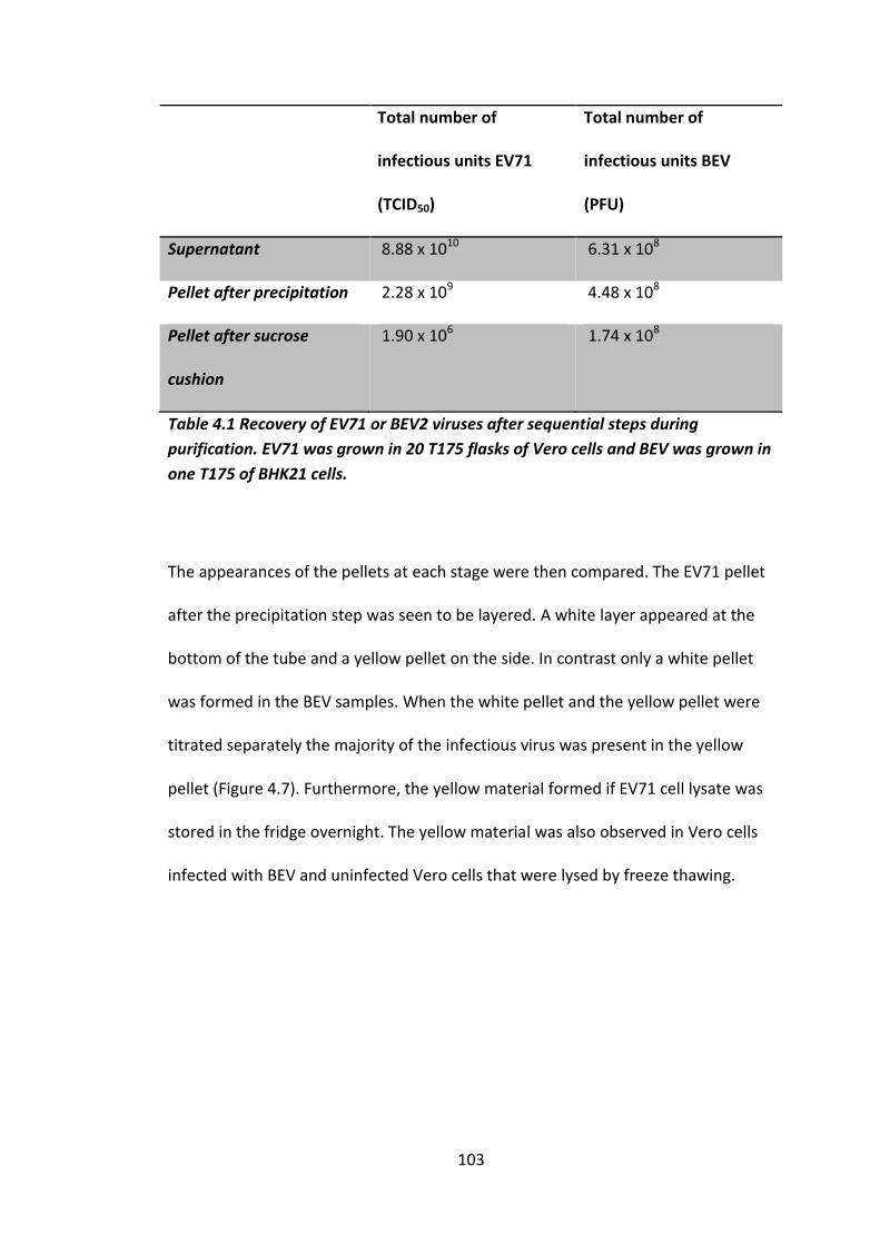
103
Total number of
infectious units EV71
(TCID50)
Total number of
infectious units BEV
(PFU)
Supernatant 8.88 x 10¹⁰ 6.31 x 10⁸
Pellet after precipitation 2.28 x 10⁹ 4.48 x 10⁸
Pellet after sucrose
cushion
1.90 x 10⁶ 1.74 x 10⁸
Table 4.1 Recovery of EV71 or BEV2 viruses after sequential steps during
purification. EV71 was grown in 20 T175 flasks of Vero cells and BEV was grown in
one T175 of BHK21 cells.
The appearances of the pellets at each stage were then compared. The EV71 pellet
after the precipitation step was seen to be layered. A white layer appeared at the
bottom of the tube and a yellow pellet on the side. In contrast only a white pellet
was formed in the BEV samples. When the white pellet and the yellow pellet were
titrated separately the majority of the infectious virus was present in the yellow
pellet (Figure 4.7). Furthermore, the yellow material formed if EV71 cell lysate was
stored in the fridge overnight. The yellow material was also observed in Vero cells
infected with BEV and uninfected Vero cells that were lysed by freeze thawing.

104
TC
ID5
0/m
l
Wh
ite p
ellet
Yello
w p
ellet
1 0 0
1 0 1
1 0 2
1 0 3
1 0 4
1 0 5
1 0 6
1 0 7
1 0 8
1 0 9
1 0 1 0
Figure 4.7 Virus titres of different EV71 pellets. 10 flasks of Vero cells were
infected with EV71 at MOI 10 TCID50’s/cell. The virus harvest was clarified and the
cell debris was freeze-thawed three times in a small volume and the supernatant
added to the first. The pooled supernatant was then precipitated in 50% saturated
ammonium sulphate overnight at 4 oC. This was then centrifuged at 10,000 RPM
for one hour. The supernatant was then decanted, leaving two separate pellets, a
white pellet at the bottom of the tube and a yellow pellet up the side of the tube.
Both pellets were resuspended in 1 ml of PBS and titrated by TCID50 assay.
It was then attempted to separate the virus from the yellow material with
detergents and then analysing the virus after centrifugation through a 15-45 w/v %
sucrose gradient. However, this was not successful as it did not separate the virus
from this material, as virus was still present at the top of the gradient. It was then
decided to omit the precipitation stage and progress directly to the sucrose cushion
stage. However, a yellow insoluble pellet was formed after ultracentrifugation that
could not be solubilised, even with 10 w/v % SDS, in which the suspension was very
cloudy. When this suspension was separated on a sucrose gradient the yellow
insoluble matter pelleted but a white cloudy substance remained at the top of the
gradient (Figure 4.8 A). The highest virus titres were found in the high count
fractions towards the top of the gradient (Figure 4.8 B). This implies that the
majority of virus associates with the cloudy substance.

105
F ra c tio n
35
S C
ou
nts
pe
r m
inu
te
0 5 1 0 1 5 2 0
0
2 0 0 0 0
4 0 0 0 0
6 0 0 0 0
8 0 0 0 0
1 0 0 0 0 0
T o p B o tto m
TC
ID5
0/m
l
Fra
ct i
on
1-3
Fra
ct i
on
9-1
1
fract i
on
16-1
7
1 0 0
1 0 1
1 0 2
1 0 3
1 0 4
1 0 5
1 0 6
1 0 7
1 0 8
1 0 9
Figure 4.8 Sucrose gradient profiles and titrations of 35S labelled EV71 after
treatment with SDS. (A) Virus was separated by centrifugation through a 15-45
w/v % sucrose cushion. 300 µl fractions were taken and analysed by scintillation
counting. (B) Titration of an EV71 sucrose gradient. Different fractions were
pooled together and then titrated by TCID50 assay.
A
B

106
It was hypothesised that this yellow/cloudy substance was specific to Vero cells and
so RD cells were used to grow the virus. Virus grew in RD cells to a similar level to
Vero cells (Figure 4.2), and the pellet formed was less yellow, but still insoluble
after purification through a sucrose cushion. When analysed by sucrose density
gradient centrifugation after purification through a sucrose cushion, the majority of
the counts were still seen at the top of the gradient, but a small peak was
consistently seen just below the material at the top (Methods section 2.5.2) (Figure
4.9 A). Next, virus supernatant was treated with 50 µM DNase (AlphiChem) or 1%
w/v SDS for 3.5 hours prior to the sucrose cushion as performed in Kattur
Venkatachalam et al. 2014. Both treatments resulted in a post-sucrose cushion
pellet that could be solubilised. After sucrose density gradient centrifugation the
DNase treated sample produced a slightly larger peak compared to the small one
seen in Figure 4.9 A, although the majority of counts were still seen at the top of
the gradient. However, after SDS treatment the number of counts at the top of the
tube was reduced, but there were still no faster sedimenting peaks (Figure 4.9 A).
The small peak with DNase treatment was reproducible (Figure 4.9 B) and when
examined by SDS PAGE gel a viral capsid band profile was visible (Figure 4.9 C). The
large amount of material seen at the top of the EV71 gradient also had bands at
positions compatible with viral capsid proteins. This indicates that the virus is
associated with the cloudy material, indicating that the particles could potentially
be envelope

107
Figure 4.9 Sucrose gradient profiles of 35S labelled EV71 grown in RD cells. (A) Samples
untreated, treated with SDS or DNase prior the sucrose gradient were run alongside each
other. (B) Repeat of EV71 gradient treated with DNase. All particle were separated by
centrifugation through a 15-45 w/v % sucrose gradient. 300 µl fractions were taken and
analysed by scintillation counting. (C) SDS PAGE gel Sample 1, 2 and 3 correspond to A, 1 is
the peak from the DNAse treated sample (fraction 10) and 2 is top of the gradient (fraction
2), sample 3 is from the top of the gradient that was treated with SDS (Fraction 2).
Expected band sizes; VP1 36 kDa, VP2 25 kDa, VP3 23 kDa

108
To further analyse the virus another purification method was employed using
Nycodenz gradients (Methods 2.5.3). This separates particles based upon buoyant
density rather than rate of sedimentation. Both 20-60 w/v % step gradients and 40
w/v % self-forming gradients were used. Bands visible by eye were formed in both
gradients, and these correlated with the 35S peaks observed by scintillation
counting (Figure 4.10). In the step gradients, peaks were seen towards the centre of
the gradient, but for the self-forming gradients there was one peak near the top of
the gradient and another towards the bottom of the gradient. However if the cell
pellet was freeze-thawed with RIPA buffer, instead of 1 v/v % NP40 then the higher
(less dense) peak was greatly diminished (Figure 4.10).

109
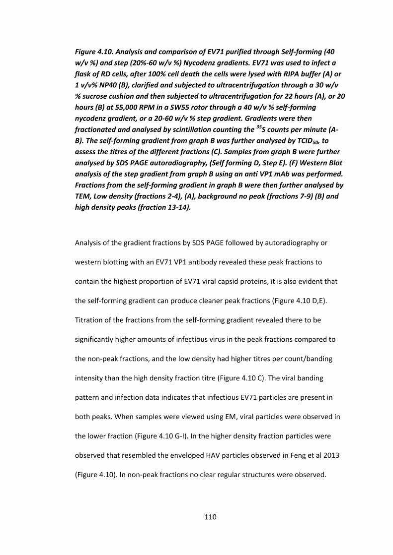
110
Figure 4.10. Analysis and comparison of EV71 purified through Self-forming (40
w/v %) and step (20%-60 w/v %) Nycodenz gradients. EV71 was used to infect a
flask of RD cells, after 100% cell death the cells were lysed with RIPA buffer (A) or
1 v/v% NP40 (B), clarified and subjected to ultracentrifugation through a 30 w/v
% sucrose cushion and then subjected to ultracentrifugation for 22 hours (A), or 20
hours (B) at 55,000 RPM in a SW55 rotor through a 40 w/v % self-forming
nycodenz gradient, or a 20-60 w/v % step gradient. Gradients were then
fractionated and analysed by scintillation counting the 35S counts per minute (A-
B). The self-forming gradient from graph B was further analysed by TCID50, to
assess the titres of the different fractions (C). Samples from graph B were further
analysed by SDS PAGE autoradiography, (Self forming D, Step E). (F) Western Blot
analysis of the step gradient from graph B using an anti VP1 mAb was performed.
Fractions from the self-forming gradient in graph B were then further analysed by
TEM, Low density (fractions 2-4), (A), background no peak (fractions 7-9) (B) and
high density peaks (fraction 13-14).
Analysis of the gradient fractions by SDS PAGE followed by autoradiography or
western blotting with an EV71 VP1 antibody revealed these peak fractions to
contain the highest proportion of EV71 viral capsid proteins, it is also evident that
the self-forming gradient can produce cleaner peak fractions (Figure 4.10 D,E).
Titration of the fractions from the self-forming gradient revealed there to be
significantly higher amounts of infectious virus in the peak fractions compared to
the non-peak fractions, and the low density had higher titres per count/banding
intensity than the high density fraction titre (Figure 4.10 C). The viral banding
pattern and infection data indicates that infectious EV71 particles are present in
both peaks. When samples were viewed using EM, viral particles were observed in
the lower fraction (Figure 4.10 G-I). In the higher density fraction particles were
observed that resembled the enveloped HAV particles observed in Feng et al 2013
(Figure 4.10). In non-peak fractions no clear regular structures were observed.
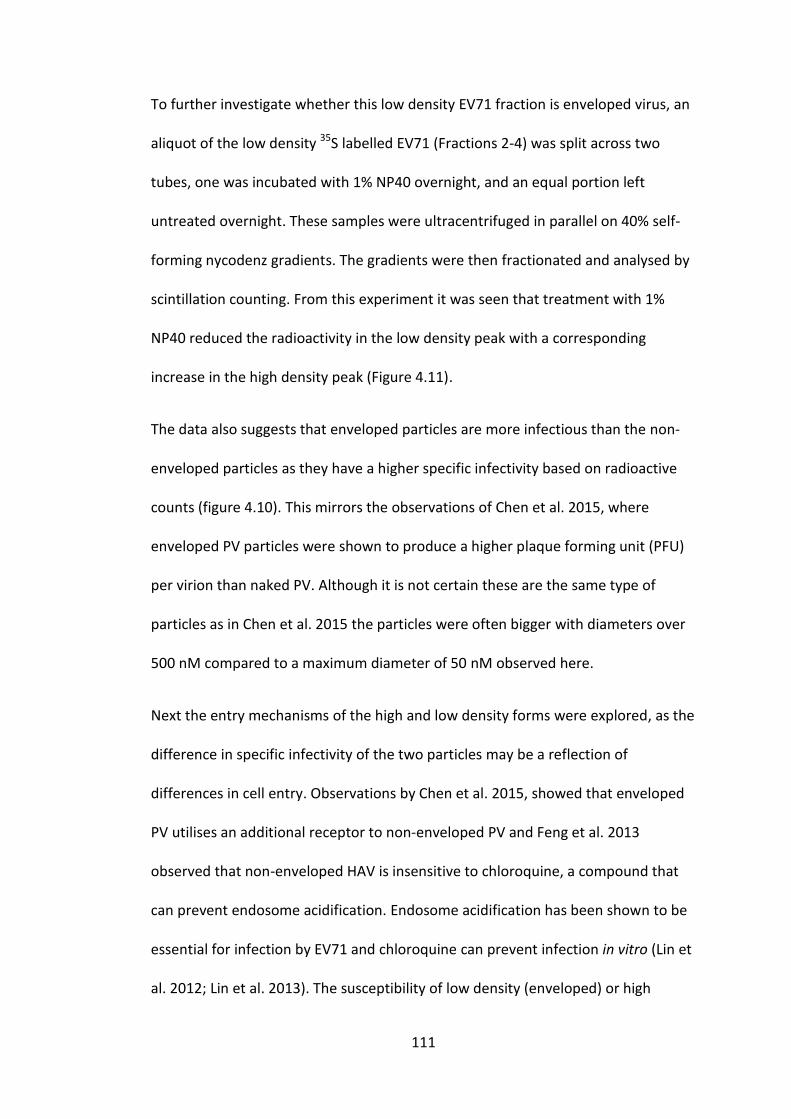
111
To further investigate whether this low density EV71 fraction is enveloped virus, an
aliquot of the low density 35S labelled EV71 (Fractions 2-4) was split across two
tubes, one was incubated with 1% NP40 overnight, and an equal portion left
untreated overnight. These samples were ultracentrifuged in parallel on 40% self-
forming nycodenz gradients. The gradients were then fractionated and analysed by
scintillation counting. From this experiment it was seen that treatment with 1%
NP40 reduced the radioactivity in the low density peak with a corresponding
increase in the high density peak (Figure 4.11).
The data also suggests that enveloped particles are more infectious than the non-
enveloped particles as they have a higher specific infectivity based on radioactive
counts (figure 4.10). This mirrors the observations of Chen et al. 2015, where
enveloped PV particles were shown to produce a higher plaque forming unit (PFU)
per virion than naked PV. Although it is not certain these are the same type of
particles as in Chen et al. 2015 the particles were often bigger with diameters over
500 nM compared to a maximum diameter of 50 nM observed here.
Next the entry mechanisms of the high and low density forms were explored, as the
difference in specific infectivity of the two particles may be a reflection of
differences in cell entry. Observations by Chen et al. 2015, showed that enveloped
PV utilises an additional receptor to non-enveloped PV and Feng et al. 2013
observed that non-enveloped HAV is insensitive to chloroquine, a compound that
can prevent endosome acidification. Endosome acidification has been shown to be
essential for infection by EV71 and chloroquine can prevent infection in vitro (Lin et
al. 2012; Lin et al. 2013). The susceptibility of low density (enveloped) or high

112
density (non-enveloped) virus to chloroquine was investigated. Here cells were pre-
treated with chloroquine, to prevent endosome acidification and after two hours
were infected with either low density (enveloped) or high density (non-enveloped)
EV71. Cells were then incubated until 100% cell death had occurred, when samples
were titrated by TCID50 assay (Figure 4.12, performed by a PhD student Özlem
Cesur). This showed that low density (enveloped) EV71 is susceptible to chloroquine
while high density (non-enveloped) virus was not. This suggests a different
entry/uncoating mechanism between the two particles, although more repeats are
necessary as it currently n = 1.
F ra c tio n
35
S C
ou
nts
pe
r m
inu
te
0 5 1 0 1 5 2 0
0
1 0 0 0
2 0 0 0
3 0 0 0
1 % N P 4 0
U n tre a ted
T o p B o tto m
Figure 4.11 High buoyancy EV71 is sensitive to NP40 treatment. 35S labelled low
density EV71 that had previously been purified through a self-forming nycodenz
gradient was either treated with 1% NP40 over night or left untreated. They were
then ultracentrifuged through further self-forming nycodenz gradients in parallel.
Gradients were then fractionated and analysed by scintillation counting.
113
C h lo ro q u in e C o n c e n tra t io n (M )
TC
DI 5
0/m
l
0 1 0 2 0 3 0 4 0 5 0
0
1 .01 0 7
2 .01 0 7
3 .01 0 7
H ig h d e n s ity
(n o n -e n v e lo p e d )
L o w d e n s ity
(e n v e lo p e d )
Figure 4.12 Effect of chloroquine on high and low buoyancy EV71. High density
and low density particles were used to infect cells pre-treated with increasing
concentrations of chloroquine and then titrated by TCID50 after 100% cell death
had occurred. (Performed by Özlem Cesur). n = 1.

114
As enveloped and non-enveloped particles appear to separate on nycodenz
gradients they were also used to distinguish between empty and mature virions.
Empty particles can occur naturally (‘natural empties’ as discussed in section 1.2.4)
but they can also be produced by heating virions. These types of particles sediment
similarly on sucrose gradients, but they have different protein profiles as natural
empty particles have not undergone VP0 cleavage (discussed in section 1.2.4). Virus
was heated at 60 oC to convert to ‘artificial’ empty particles and separated by
nycodenz density gradient centrifugation. After centrifugation the sample was
fractionated and analysed by scintillation counting this revealed that a different
sedimentation pattern to the untreated virus (Figure 4.13). This indicates that it is
possible to distinguish between the two particles via Nycodenz gradients.
F ra c tio n
35
S C
ou
nts
pe
r m
inu
te
5 1 0 1 5 2 0
0
1 0 0 0
2 0 0 0
3 0 0 0
T o p B o tto m
H ea ted
U n tre a ted
Figure 4.13 The effect of heating on the nycodenz gradient profile of EV71. Heated
(60 oC for 30 minutes) or unheated EV71 was separated on self-forming Nycodenz
gradients.

115
4.2 A mutant with an altered uncoating phenotype
To further study the effect of pH on EV71 uncoating the effect of low pH on cell
entry and uncoating of the virus was assessed. Virus was incubated in pH 5.6 buffer
for one hour at 4 oC. 5 ml of the solution was added directly to Vero cells and
incubated on them for 30 minutes at room temperature, before being topped up
with 20 ml of serum free DMEM. At this point cells were incubated at 37 oC until
100% cell death had occurred. This treatment reduced the virus titre by 1 x 10³
TCID50 per/ml (i.e. three orders of magnitude) (Figure 4.14). However, when media
was neutralised with 1 M Tris before infection or if the incubation was at pH 7.2
there was no reduction in virus titre compared to incubating in pH 5.6 buffer(Figure
4.14B). When the virus was passaged under low pH conditions resistance developed
after one passage and the resulting virus has been termed the acid-resistant virus
(Figure 4.14C).

116
Figure 4.14. pH treatment of virus on cells has a fitness cost. Virus was incubated
in either PBS pH 7.2 (green) or a pH 5.6 buffer (red) for one hour, then used to
infect Vero cells at an MOI of 1 TCID50’s/cell. Virus incubated in pH 5.6 buffer was
also neutralised with a 1 M Tris prior to infection of cells (yellow) (A). (B) After
100% cell death, lysates were then titrated via TCID50 assay. Each bar represents
an average of three measurements and error was measured using standard error.
(C) Evolution of an acid-resistant virus. EV71 was incubated in a pH 5.6 buffer for
one hour at 4 oC and then 5 ml of this solution was used to infect cells for 20
minutes at room temperature before adding 20 ml of DMEM. Cells were then
incubated at 37 oC until 100% cell death. The process was repeated, for 5
passages. Virus was titrated via TCID50 assay after each passage.
117
Sequencing of the capsid coding region of the acid-resistant virus, revealed a single
point mutation between every isolate observed (VP1 N104S n=6/6) and a
duplication of three codons in VP2 (n = 1/5). Sequence alignments show that N104
is conserved among all EV71 genotypes and is close to N102 which is a highly
conserved residue among all EV71/CVA16 genotypes and among all enterovirus A
species that utilize SCARB2 as their major receptor, but it is not conserved among
viruses that have not been shown to utilize SCARB2 (Yamayoshi et al. 2012). Similar
observations were made in Dang et al. 2014 (Figure 4.15). This indicates that it
could be an important residue for SCARB2 mediated cell entry.
Mapping N104 in a docking model of the crystal structures of EV71 and SCARB2
reveals that it is located in loop of VP1, which is predicted to be a SCARB2 binding
site (Dang et al. 2014) (Figure 4.16). Comparing the docking models of SCARB2 in its
neutral and acid conformations predicts that an alpha helix of SCARB2 moves
towards VP1 residue N104 in its acid induced form (Figure 4.16). This indicates that
N104 may play a role in binding to the acid-induced form, which is the
conformation that is able to induce uncoating. These results, taken together with
experiments performed by Dang et al 2014 that showed co-immunoprecipitation
with SCARB2 and an EV71 peptide sequence containing residue N104, suggest that
this residue is involved in SCARB2 binding. Given that the mutation is to a serine,
which is smaller, it is possible that this serine is no longer able to bind SCARB2.

118
EV71A NASDESMIETRCVLNSHSTAETTLDSFFSRAGLVGEIDLPLKGTTNPNGYANWDIDITGY 116
EV71C4 ............................................................ 116
EV71C1 .........................................E.................. 116
EV71B2 .T.......................................E.................. 116
EV71B3 .T.......................................E.................. 116
CVA16A ..QENL.........H.......IGN........SI.TM.TT..Q.TD..V.....LM.. 116
CVA16B ....KNL........H...Q..AIGN........SI.TM.TT..Q.TD..V.....LM.. 116
CVA7 LV..KNM......I.K..VE..SITN.Y.......VVNM.VQ..S.TE.F.K.E...M.F 115
CVA14 LV...........VNR...EE..VGH.........V.....Q..A.TG.F.S....VM.. 115
EVA120 ..T.KNM......V.K........TH.Y..SGL..V....TQS.K-.V.F.T.T..VM.. 114
EVA76 .TT..N.....A.I.K...E.T.IEH........SM.S.LVSEKT-TQA.......VM.. 115
EVA90 .T...N.L.....V....VAE.SVSH........GML.L.TSDE.-.I.FTT.....M.F 114
EVA89 .T...N.....A.V.K..V...SVEH.....A...MVE.LTSD..-NI..T.....VM.. 115
EVA91 TT...N.......V.K..V....VSH.YGK..LV.W.Q.LTSTKN-NH.FK...V.VM.F 115
CVA92 VT...........H..HSV...S.EN..GRA....MAT.LTSEDS-A..FT..P...M.. 116
SiEV46 TT...........V.R..V...SITN.YARA....YLK.LTSKD.-...FTNW....MAF 111
BaEV T....N.......V.K.GV..TSIQH..A.SA..GMVT.LTSE..-A..FT......M.. 101
SiEV19 .T...G.......V.K..V...SIEH.YG..A...LVSILTSE.S-A..FT...V..M.F 107
SiEV43 .............V.K..V...SVEH.....A...Y.SILTST..-A..FT......M.. 108
CVA2 ..T..NL......V.KN.VE..SINH.....A...KVE.NDT..S-AT.FT......M.Y 116
CVA4 T.T.GNL......V...G.R..HIEH....S....VMEVDDT..A-GK.FS......MAF 116
CVA6 .....NL......M.RNGVN.ASVEH.Y.......VVEVKDS..S-LD..TV.P..VM.F 111
CVA3 .TT..........V.RNGVV..SV.H....S....VMNILDG..--SK.FKT.E..VM.F 113
CVA8 .....N.........RNGVV..S..H.........VINVQD...--QK.FEV....VM.F 113
CVA10 ..T..N.......I.KNGVL...INH....S....VVN.TDG..D-TT...T....IM.F 115
CVA12 ..T..........V.K.GVS..SVEY....S..A.VVVVEDATA.-NK...T.E..VM.F 114
CVA5 ..T..........VNR.GVME.SVEH....S..AGILIIEDSGTS-TK...T.E..VM.F 115
EV71A TVGSSKSEYSLVIRIYMRMKHVRAWIPRPMRNQNYLFKSNPNYAGDSIKPTGTSRTAITT 296
EV71C4 ...T...K.P..V.........................A......N......A....... 296
EV71C1 ...T...K.P............................A......N.............. 296
EV71B2 .......K.P..V.........................A......N.........N.... 296
EV71B3 .......K.P..V.........................A......N.............. 296
CVA16A ...TE..P...TL.V...I..........L...P....T-...K.ND..C.S...DK... 295
CVA16B ...TE..PH.ITL.V...I..........L...P....T....K.ND..C.S...DK... 296
CVA7 MI.EG.PTQ.VT......L..I...V...L.S...TMRNY...N.GA..C.AK..DK... 295
CVA14 I..EGAPSVHF.V..........V.......S.P.VAKNY...K.SE..CASS...S... 295
EV120 II..GTPPT.LT......L..I.G.......S.D.TARNY...N.SN..C.AV..QS... 294
EV76 V...GQLTEAV....F..I......V...I.S.K.IVKNY..FDPAV-..HS...AS... 294
EV90 VL..E.LTEA.RVRIY..I..........L.S.R..L.NY..FD.AN.T..SA..AQ... 294
EV89 V...EQITDKITV..F..L...KT.V...L.S.K..L.NY..FD.ANLTMSSA..AD.KN 295
EV91 V...GQITDQ.DV.....I...K..L...L.S.P.ML.NY..FD.TKL.C.SK...S.K. 295
CVA92 V...ENITTK.TV.....I............S.P.IL.NY..FN.ND.RHMAKD..S... 296
SiEV46 V...GSITSKIKV..F..I..........L.S.Q.MIRNY..FPT-TV.CLAA..NN... 290
BaEV V...GSIKD.IIV.....L..I...V...I.S.PYMLKNY..FD.NN..HVTNR-NS.K. 280
SiEV19 A...GNIKSNIR..V......I...V...F.S.P..L.NY..MD.TNL.CAST..AK... 287
SiEV43 A...GSITANIR..V......I.......L...P.VL.NY..FD.TN..CSS...QS... 288
CVA2 F..EEITNERIT......L......V...L.SEP.VL.NF...TAV-T-HVTAN..T..N 294
CVA4 V..KQITNQKFQ....L.L.R....V...L.S.P.IYRNY.T.GTT-.QHLAKD.RK..E 295
CVA6 T..ESTTGKNVHV.V...I...K..V...L.S.A.MV..Y.T.SQT-.TNTA.D.AS... 290
CVA3 I.SKTPTERD.RV....KL......V...I.S.PYVLKNY...D.AN.V.CAKD.DD.KN 293
CVA8 F.SKTPATRD.RV.V...L......V...I.S.P.IL.NY...D.TK.TS.SKD.QS.K. 293
CVA10 V.SREA.QLK.QT.V..KL..........I.S.P..L.NF...DSSK.TNSARD.SS.KQ 295
CVA12 T.SGEA.GKNITV..F..L......V...I.S.L..L.NY..FDNTK.LNASHN..S..A 294
CVA5 M..GVSTGKDVTV..F.KL......V...I.S.P..L.NY..FDKAN.IDASSN..S... 295
Figure 4.15 Sequence alignments of VP1 of Enterovirus A species. Alignments of SCARB2 dependent (EV71(genotypes/sub-genotypes; A, B2, B3, C1 and C4), CVA7, CVA14 and CVA16 (Genotypes; A and B)) and SCARB2 independent (CVA2, CVA3, CVA4, CVA5, CVA6, CVA8, CVA10 and CVA12) enterovirus A viruses as identified by (Yamayoshi et al 2012) and unknown receptor binding (EV76, EV89, EV90, EV91, EV120, CVA92, Simian enterovirus 19, Simian enterovirus 43, Simian enterovirus 46 and Baboon enterovirus). Conserved N104 residue is highlighted in yellow, conserved residues predicted to be involved in Cyclophilin A binding are highlighted in pink, Conserved N103 residue is highlighted in green. Cyclophilin A binding area of the HI loop found by Qing et al 2014 is highlighted in a green box. Only displaying partial alignments, full alignment can be found in the appendix.

119
Figure 4.16 Acid induced conformational change in SCARB2 causes an alpha helix
to move towards VP1 N104. Cartoon PyMol representation of docking model
SCARB2 in it neutral conformation (A. green) and acid induced conformation (B.
red) with EV71 VP1 (blue), N104 is highlighted in yellow.

120
Given the role of SCARB2 in cell entry and uncoating and the structural and co-
immunoprecipitation data from Dang et al. 2014 indicating that N104 may interact
with SCARB2, the cell entry and uncoating phenotypes of both WT and the acid-
resistant mutant were compared.
To examine differences between cell entry, both isolates were used to infect cells
that had been pre-treated with either chlorpromazine or gestin, which block the
clathrin and caveolae-dependent endocytosis pathways, respectively at
concentrations that have previously been shown to be effective against WT EV71 in
RD cells (Lin et al. 2012; Lin et al. 2013) (work conducted by Amy Radcliffe). This
revealed that both isolates were sensitive to chlorpromazine although the acid-
resistant mutant was shown to be significantly more sensitive at the lowest
concentration (10 µM). This indicates that clathrin-dependent endocytosis is
essential for both WT and the acid-resistant mutant, but the acid-resistant mutant
is more sensitive to disruption of the clathrin-mediated endocytosis pathway
(Figure 4.20). Both samples were also susceptible to the caveolar inhibitor gestin,
although it was not possible to completely prevent infection. This indicates that the
caveolar endocytosis is utilised by EV71 in Vero cells but is not essential for either
WT or the acid-resistant mutant. It has previously been shown that gestin has no
effect on cell lines expressing only SCARB2 as a functional receptor; this indicates
another receptor on Vero cells may function in a caveolin-dependent manner
(Figure 4.17).

121
Figure 4.17 Analysis of the effect of inhibitors of cell entry. Vero cells were
incubated in the presence of increasing concentrations of either chlorpromazine
(A) or gestin (B) for two hours and then infected with either WT EV71 (Green) or
the acid-resistant mutant (Red). After 24 hours samples were taken for titration
via TCID50 assay. Each bar represents an average of three measurements and error
was measured using standard error * = p ≥ 0.01,. Experiments were conducted by
Amy Radcliffe.
Now that it has been established that this mutant has an effect on cell entry, the
effect on uncoating was further analysed. As a drop in pH is known to be essential
for WT EV71 uncoating, the effect of endosome acidification inhibitors was
analysed. Cells were incubated with a range of concentrations of endosome
acidification inhibitors, ammonium chloride (NH4Cl) or chloroquine for two hours
prior to infection, with WT or acid-resistant virus isolates and were then titrated
after 24 hours (work conducted by Amy Radcliffe). Both of these inhibitors have
previously been shown to prevent infection by WT EV71 (Lin et al. 2012; Lin et al.
2013). Both the WT and the acid-resistant mutant were susceptible to inhibitors of
endosome acidification, with the trend showing that the acid-resistant mutant
appears to be less susceptible, although only one data point was shown to be
significantly different (Figure 4.18). This suggests that the acid resistant mutant is
less sensitive to inhibition of endosome acidification.
A B

122
Figure 4.18 Endosome acidification plays an important role in WT EV71 and acid-
resistant EV71. Vero cells were incubated in the presence of increasing
concentrations of either Chloroquine (A) or NH4Cl (B) for two hours and then
infected with either WT EV71 (Green) or the acid resistant mutant (Red). After 24
hours samples were taken for titration via TCID50. Experiments were performed in
triplicate and error is measured by s.e.m, * = p ≥ 0.01. Experiments were
conducted by Amy Radcliffe.
Next the effect of the proline isomerase cyclophilin A was analysed (work
conducted by Amy Radcliffe), which is the only known EV71 uncoating initiator
other than SCARB2 (Qing et al. 2014). To analyse the effect of cyclophilin A both
isolates were grown in Vero cells that had been treated with the cyclophilin A
inhibitor, cyclosporine A. Cyclosporine A functions by binding to cyclophilin A
making it less readily available in the cell for the virus to use. The Cyclosporine A
analogue (HL051001P2) has previously been shown to inhibit EV71 infections (Qing
et al. 2014). Here Vero cells were incubated in the presence of cyclosporine A for
two hours prior to infection and incubated at 37 oC for 24 hours before samples
were taken for titration (Figure 4.19). Titrations revealed that the acid-resistant
mutant was significantly less susceptible to cyclosporine A.
A B

123
C y c lo s p o r in e A c o n c e n tra t io n M
Pe
rc
en
tag
e r
ed
uc
tio
n o
f T
CID
50
0.0
0
0.1
6
0.8
0
4.0
0
20.0
0
0
2 0
4 0
6 0
8 0
1 0 0
W T
A c id -re s is ta n t m u ta n t
* * *
*
* * *
*
Figure 4.19 Cyclosporine A can prevent EV71 infection but is significantly less
effective against the acid-resistant mutant. Vero cells were incubated in the
presence of increasing concentrations cyclosporine A for two hours and then
infected with either WT EV71 (Green) or the acid-resistant mutant (Red). After 24
hours samples were taken for titration via TCID50 assay. Experiments were
performed in triplicate and error is measured by s.e.m, * = p ≥ 0.01, ** = ≥ 0.001,
*** = p ≥ 0.0001 . Experiment was conducted by Amy Radcliffe.
To gain a better understanding of the effect of mutation N104S has on the
interaction with cyclophilin A, its location on the crystal structure was analysed in
comparison to two previously identified cyclophilin A interaction sites (VP1; S243,
P246) (Qing et al. 2014). This revealed that the site of the acid-resistance mutation
N104S, is adjacent to the HI loop which has been shown to be a cyclophilin A
interaction site (S243, P246) (Qing et al. 2014), meaning that N104 is present in an
area that is involved in cyclophilin A interactions (Figure 4.20).
To determine whether the mutation may affect binding preferences for cyclophilin
A this sequence was compared to known cyclophilin A target sequences. However,
124
no preference for either asparagine (N) or serine (S) was revealed and so it is
unlikely that this residue directly effects cyclophilin A binding. However
examination of residues neighbouring N104 revealed that residue 103 is a proline,
which has been shown to be conserved across all EV71 genotypes (Figure 4.15). This
is not found in other members of Enterovirus A species which also do not contain
P246, a residue that has been shown to be important for cyclophilin A binding in
EV71 (Figure 4.20) (Qing et al. 2014). So it is possible that VP1 residue 104 affects
the availability of P103, and effects its interaction with cyclophilin A. Mutation of
residue 243 to a proline has previously been shown to increase cyclophilin A
binding while mutation of P246 has been shown to decrease cyclophilin A affinity
(Qing et al. 2014). This demonstrates that prolines in this region are important for
cyclophilin A interactions, and can increase capsid affinity towards cyclophilin A and
reduce the effectiveness of cyclophilin A inhibitors. Given that N104S generates
resistance to cyclosporine A and it is located next to a proline, it could be effecting
the interaction between cyclophilin A and EV71 by affecting the availability of P103.
If N104 blocks access to P103 then mutation to a smaller residue such as serine may
release this blocking. Alternatively, if this mutation does indeed prevent interaction
with SCARB2, then it is possible that SCARB2 usually blocks cyclophilin A
interactions, and when it is not able to bind here, then it is not able to block
cyclophilin A interactions.

Figure 4.20 Potential role of cyclophilin A using crystal structure. Cartoon representation of VP1 (blue), acid-resistant mutation site
is highlighted in yellow N104, residues that have previously been reported by Qing et al 2014 to be involved in cyclophilin A (S243,
P246) interactions are represented in hot pink, and proline residue (P103) that we hypothesise could be involved in cyclophilin A
interactions is highlighted in peach.
124

126
4.3 Discussion and future work
From, the work described in this Chapter it has been demonstrated that EV71 can
grow in Vero or RD cells to reach titres of up to 1 x 10⁸ TCID50/ml and that growth in
flasks was the most effective method of growing virus. Roller bottles and micro-
carriers give no significant increase in virus yield and are more labour intensive to
use.
Cell entry and uncoating has been studied in detail, showing that incubation of virus
with cells in a low pH buffer can reduce the titre of the virus by 1 x10³ TCID50/ml
(three orders of magnitude). As EV71 uncoating is only induced when it binds to its
receptor at a low pH it is likely that the reduction in virus titre is the result of the
virus binding to the receptor and then uncoating outside the cell before
endocytosis occurs. When EV71 is repeatedly exposed to these conditions, it
evolves resistance and gains a mutation in VP1 (N104S). Immunoprecipitation and
docking models have predicted N104 to be involved in binding SCARB2 the primary
receptor and an uncoating initiator of EV71 (Dang et al. 2014). Further analysis of
the docking model revealed that an alpha helix of SCARB2 is predicted to move
towards N104 in its acid-induced conformation. The mutation N104S likely effects
this interaction, which is potentially why the acid-resistant mutant is more sensitive
to chlorpromazine, an inhibitor of the clathrin-dependent endocytosis pathway,
which is the entry pathway SCARB2 utilizes. N104 was also shown to be in close
proximity to residues previously shown by mutagenesis and immunoprecipitation
to be involved in cyclophilin A interaction, which is a proline isomerase (Qing et al.
2014). This N104S mutant is less susceptible to cyclosporine A an inhibitor of

127
cyclophilin A. It is likely that this mutation reduces the binding capacity of EV71 to
SCARB2 therefore leaving SCARB2 unable to induce uncoating even in the presence
of low pH or unable to bind SCARB2 at all. A potential reason for increased binding
capacity of cyclophilin A to the acid-resistant mutant could be that SCARB2 blocks
access of cyclophilin A to P103 and if N104S cannot bind SCARB2, P103 is free to
bind cyclophilin A, making it less susceptible to cyclosporine A. To further test this,
uncoating assays, to detect if SCARB2 and cyclophilin A can induce conversion of
mature particles to empty particles need to be performed, using the following
isolates; EV71 WT, N104S and two additional mutants P103A and N104S/P103A.
The susceptibility of the new mutants to cyclophilin A will also need to be assessed.
Work to assess the toxicity of the inhibitors will also need to be done to confirm
that the drop in titre observed is due to inhibition of virus entry and not due to a
toxic effect on the cells.
In addition to greater knowledge of how EV71 uncoats/what residues it uses,
greater insight into the timings of uncoating has also been gained. Figure 4.4-4.7
gives an insight into EV71 uncoating, from the viral growth curves there is a visible
drop in titre after three hours which indicates that an uncoating event is occurring
and from seven hours onwards titre begins to increase, this coincides with data
from use with NLD inhibition which coincides with data previously seen in Lu et al
2011. NLD is known to prevent viral uncoating and from an inhibition time course it
is evident that it is fully active until five hours post-infection. This indicates that
between hours three and six EV71 is undergoing a prolonged uncoating event that
causes the virion to become less infectious at three hours post infection, but six

128
hours post infection the virus become less susceptible to the uncoating inhibitor
NLD, indicating that it the virus has undergone uncoating. From seven hours
onwards the virus titre increases indicating that replication has occurred. Labelling
experiments using an MOI of 10 for infection show that all proteins that become
incorporated into mature virions are formed within nine to 12 hours and no virus
capsid was detected to be produced after this point. However after this point virus
titres continue to rise and make a sudden sharp increase at twenty hours post
infection suggesting that virus replication is a multistep process infection.
This process differs from PV which undergoes a complete round of replication after
nine hours and is known to complete uncoating and begin replication within one
hour post-infection (Brandenburg et al. 2007). The long uncoating phase partially
explains the reason for long life cycle of EV71 compared to PV.
In addition it was shown that standard sucrose gradients were not sufficient to
purify EV71; this appeared to be in part due to a large population of enveloped
particles. Use of Nycodenz gradients, which separates particles based on buoyant
density, revealed two populations of particles; a high density population and a low
density population. EM analysis revealed the high density particles were standard
virus particles, but in the low density fractions there were what appeared to be
black blobs. When the low density particles were treated with 1 v/v % NP40, and
subjected to ultracentrifugation they sedimented at the same density as the high
density particles. This suggests that treatment with detergent can disrupt the
membranes to release naked particles. Western blot and SDS PAGE gel analysis
revealed that both populations have standard EV71 protein profiles and were

129
detectable by a VP1 antibody. In HAV the enveloped particles were shown to have
the VP1 precursor protein, VPX (Feng et al. 2013). Titrations revealed that the
enveloped particles are more infectious than the non-enveloped particles. This is
similar to what was observed for PV but differs from HAV where both particles were
shown to be equally infectious (Chen et al 2015, Feng et al 2013). It was also
demonstrated that EV71 enveloped particles may have a slightly different entry
mechanism to non-enveloped viruses, as high density particles did not appear to be
susceptible to chloroquine while low density particles were. The same observations
were made in HAV (Feng et al. 2013). Further characterisation of these particles
would be necessary as well as characterising the entry pathway and their
susceptibility to neutralising antibodies.

130
Chapter 5

131
Chapter 5 Heat stability of EV71 and ways to increase it.
A major problem with some viral vaccines is their thermolability. This can be an
issue for reasons such as a break in the cold chain, which can result in the vaccines
undergoing structural re-arrangement that cause an antigenic shift. As a
consequence they will no longer produce as powerful an appropriate immune
response. This problem is especially exacerbated in picornavirus VLP vaccine
candidates, which have been shown to be thermally unstable and lose native
antigenicity at temperatures above 5 oC (Rombaut & Jore, 1997, personal
communication with James Hogle, Harvard Medical School, described in more detail
in sections 1.2.4, 1.4.3). These proposed vaccine candidates are produced in
recombinant systems that express the viral structural protein P1 and viral protease
3C (described in more detail in section 1.4.3). When co-expressed, 3C cleaves P1
into its constituent structural proteins VP0, VP1 and VP3, which self-assemble into
an empty capsid containing no viral RNA. In the absence of viral RNA VP0 does not
undergo the cleavage into VP2 and VP4, which allows VP4 to rearrange and form an
internal lattice that can improve virus stability (described in more detail in sections
1.2.4, 1.4.3). The thermal/antigenic instability of these vaccine candidates is an
impediment to their development as practical products. Therefore to produce a
viable EV71 VLP vaccine candidate, methods to improve its thermostability and
assessment of its antigenicity need to be developed.
In this section different strategies to improve the EV71 stability and detect the
antigenic shift are explored.

132
Several methods have previously been shown to improve capsid stability of
picornaviruses. One is by mutagenesis, by selecting for mutations that stabilise the
virus or by site-directed mutagenesis. Previously it has been shown that mutations
identified by structural observation of the FMDV empty capsid crystal structure are
able to improve the thermostability of FMDV VLPs (Porta et al. 2013).
Another strategy to improve VLP stability that is specific for enteroviruses, is to
incubate in the presence of pocket-binding compounds that are able to enhance
virus stability. Enteroviruses contain a structure in the VP1 structural protein known
as the VP1 pocket. This naturally harbours a hydrophobic lipid known as the pocket
factor, which is a virion stabiliser (Smith et al. 1986; Wang et al. 2012). The pocket
factor binds here and it must be released before uncoating can occur (discussed in
section 1.2.1, 1.2.4, Chapter 3). Certain compounds have been shown to displace
the natural pocket factor due to higher binding affinities, and increase capsid
stability so that it is unable to undergo structural rearrangements necessary for
uncoating (discussed in section 1.3, chapter 3). These compounds have been shown
to inhibit conformational changes and so improve the thermostability of a number
of different enteroviruses, including EV71 (Katpally et al. 2007; Mosser et al. 1994;
Shepard et al. 1993). They have also been shown to successfully increase the
antigenic stability of PV VLPs produced in a recombinant system (Rombaut & Jore
1997). In Chapter 3, it was shown that the pocket-binding inhibitor NLD was able to
increase the temperature at which mature EV71 particles were heat inactivated by
~3.5 oC (Figure 3.7), and an EV71 mutant resistant to NLD was shown to be resistant
to NLD-mediated stabilisation. The mutations identified were predicted to prevent

133
NLD binding and as a result the mutant was more thermolabile than WT (Figure
3.7).
In addition to inhibitory compounds, natural pocket factors have also been shown
to have a similar effect. In BEV1 it has been shown that a number of different fatty
acids are able to function as natural pocket factors as identified by mass-
spectroscopy (Ismail-Cassim et al. 1990; Smyth et al. 2003). Incubation with one
natural pocket factor, lauric acid, was shown to improve the thermostability of
BEV1 (discussed in section 1.2.1, Ismail-Cassim et al. 1990; Smyth et al. 2003). The
natural pocket factor for other enteroviruses is unknown and all other data to date
has been based upon predictions from crystal structures from the electron density
filling the VP1 pocket. This data has often indicated that there could be a
population of different sized fatty acids functioning as the pocket factor (discussed
in section 1.2.1, Smyth et al. 1995). For EV71, the pocket factor has been predicted
to be either sphingosine or lauric acid from two different crystal structures (Wang
et al. 2012; Plevka et al. 2012).
Mutagenesis and/or pocket-binding compounds could be useful strategies to
improve the capsid stability of EV71. However a method to detect EV71 native
antigenicity also needs to be developed, so that the antigenic stability can be
assessed.
Previous studies with PV have shown it is possible to develop antibodies that can
distinguish between the antigenicity of native and heated forms (Rombaut et al.
1990; Rombaut & Jore 1997). Antibodies have been developed (mAbs D6 and A9)
that have been shown to exclusively recognise EV71 full virions but not the

134
expanded empty capsids, indicating that they should only be able to recognise EV71
in its native antigenic form (Wang et al. 2012). In addition, it has been shown that
certain receptors (e.g. SCARB2) are only able to bind the EV71 capsids in their
native form (Qing et al. 2014).
In this section mutagenesis by artificial selection and the use of different pocket-
binding compounds are assessed as methods to improve the thermostability of
EV71. Also the available antibodies and receptors are used in IPs and ELISAs to
analyse their capacity to detect native EV71 antigenicity.
5.1 Capsid stability
5.1.1 Long term stability of EV71/CVA16
The long term stability of EV71 and CVA16 after storage at temperatures of 37 oC, 4
oC or -20 oC was assessed for 120-240 hours followed by titration via TCID50 assay
(Figure 5.1) (Work performed by Joseph Ward, an undergraduate under my
supervision).
Stability for both viruses at 37 oC started to decrease after 23 hours. For CVA16 this
continued until between 72 and 96 hours by which time the titre had dropped to 0,
while that of EV71 had dropped by over 2 logs, although it was still viable. EV71
titres continued to drop by a further three logs over the next 150 hours, at which
point the experiment was discontinued. Storage at 4 or -20 oC showed that CVA16
was very stable for over 196 hours and EV71 for over 120 hours, at which point the
experiments were discontinued.

135
E V 7 1 S ta b ility
H o u r
TC
ID5
0/m
l (lo
g)
0 1 0 0 2 0 0 3 0 0
1 0 3
1 0 4
1 0 5
1 0 6
1 0 7
1 0 8
1 0 9
3 7o
C
-2 0o
C
4o
C
C V A 1 6 S ta b ility
H o u r
TC
ID5
0/m
l (lo
g)
0 5 0 1 0 0 1 5 0 2 0 0
1 0 0
1 0 1
1 0 2
1 0 3
1 0 4
1 0 5
1 0 6
1 0 7
3 7o
C
4o
C
-2 0o
C
Figure 5.1 Long term stability of EV71 (A) and CVA16 (B). EV71 and CVA16 were
stored at either 37 oC, 4 oC or -20 oC, and titrated every 24 hours. n = 1 (Performed
by Joseph Ward)
A
B

136
5.1.2 Evolution of an EV71 isolate with increased thermal stability
One strategy to stabilise protein structures is the introduction of stabilising
mutations. In an attempt to identify mutations that stabilise the EV71 capsid, an
evolution experiment was carried out to select for stabilising mutations. The virus
was repeatedly passaged at 37 oC after being heated between each passage. The
selection temperature used was 50 oC as it was previously shown in chapter 3 that
exposure to this temperature reduced the TCID50/ml titre by over 99.9% (Figure
3.10 A, Figure 5.2). For the first passage, enough virus to infect a confluent T25 flask
at an MOI of 10 TCID50’s/cell post-heating, was used. The virus was first
concentrated from cell culture medium by ultracentrifugation, and then re-
suspended in a volume of 700 µl of serum-free medium (Figure 5.3). A 10 µl sample
was taken for titration and the rest was heated to 50 oC for 30 minutes in a
thermocycler. Another 10 µl of the heated virus was taken for titration and the rest
was used to infect a confluent T25 flask of Vero cells. The flask was incubated at 37
oC until 100% cell death had occurred, and then harvested. Cell debris was removed
and the cells were freeze-thawed three times in RIPA buffer, before being added to
the tissue culture supernatant. This supernatant was then concentrated by
ultracentrifugation and the resulting pellet was resuspended in 700 µl of serum-free
media. The resuspended pellet was then heated to 50 oC and used to infect another
T25 flask of Vero cells, this process was repeated until passage eight, at which less
than a 10% drop in titre was observed for four consecutive passages (Figure 5.4).
From analysing the effect of heating on each passage, it was apparent that over the
first three passages there was initially a large drop in the post-heating titre,
unusually at this point the virus actually appeared to be become less heat stable. At

137
passage four there was a slight increase in titre indicating that the virus was
becoming more heat stable. Then at passage five there was a large increase in titre
and subsequently there was only a small difference between the post-heating and
pre-heating titres, indicating that the virus was more heat stable than before.
Once it was established that the virus was able to tolerate a temperature of 50 oC,
the thermostability of this isolate (now referred to as thermostable EV71) was
assessed by creating a thermostability curve in the same manner as the WT heat
stability curve generated in chapter 3. Figure 5.2 shows that WT virus could tolerate
temperatures up to 45 oC before a loss in titre was observed and temperatures of
51 oC before complete inactivation occurred. The thermostable mutant could
tolerate temperatures up to 51 oC before there was an observed loss in titre, and
56.5 oC before complete inactivation occurred. The growth kinetics of WT and
thermostable EV71 were compared, revealing that thermostable EV71 did not
achieve a titre equivalent to WT until 18 hour post infection, suggesting that
thermostable EV71 has slower growth kinetics (Figure 5.5).

138
T e m p e ra tu re /o
C
Lo
g T
CID
50
/ml
3 7 4 0 4 3 4 6 4 9 5 2 5 5 5 8
0
1
2
3
4
5
6
7
8
W T
T h e rm o s ta b le E V 7 1
Figure 5.2 Heat stability curves of WT EV71 and thermostable EV71. WT (black
circles) and thermostable EV71 (gray squares), were heated to a variety of
temperatures for 30 minutes using a thermocycler and then titrated by TCID50
assay. WT curve is previously shown in figure 3.7 and is included for comparison.

139
Figure 5.3 Selection of a thermostable EV71 isolate. A T25 was infected with EV71,
after 100% cell death. The virus was then concentrated from cell culture medium
by ultracentrifugation, and then re-suspended in a volume of 700 µl of serum-free
medium. A 10 µl sample was taken for titration and the rest was heated to 50 oC
for 30 minutes in a thermocycler. Another 10 µl of the heated virus was taken for
titration and the rest was used to infect a confluent T25 flask of Vero cells. The
flask was incubated at 37 oC until 100% cell death had occurred, and then
harvested. Cell debris was removed and the cells freeze-thawed three times in
RIPA buffer, before being added to the tissue culture supernatant. This
supernatant was then concentrated by ultracentrifugation and the resulting pellet
was resuspended in 700 µl of serum-free media. The resuspended pellet was then
heated to 50 oC and used to infect another T25 flask of Vero cells, this process was
repeated until passage eight

140
P a s s a g e n u m b e r
TC
ID5
0/m
l
0 2 4 6 8
1 0 3
1 0 4
1 0 5
1 0 6
1 0 7
1 0 8
1 0 9
1 0 1 0
V ira l t itre a fte r h e a tin g
V ira l t itre b e fo re h e a tin g
Figure 5.4 Difference in titres between EV71 thermostable mutants before and
after heating. EV71 was repeatedly passaged in cell culture after being heated to
50 oC for 30 minutes each time using a thermocycler. Viral titres were determined
before and after heating by TCID50 assay (n=1).
h p o s t in fe c tio n
Lo
g T
CID
50/m
l
0 3 6 9 1 2 1 5 1 8
0
1
2
3
4
5
6
7
8
T h e rm o s ta b le E V 7 1
W T
Figure 5.5 Grow kinetics of WT EV71 and thermostable EV71. WT and
thermostable virus were used to infect wells in a six well plate at MOI 1
TCID50’s/cell and whole cell samples were taken every three hours for 18 hours.
Samples were then titrated via TCID50 assay. WT curve is previously shown in
figure 3.8 and is included for direct comparison.

141
5.1.3 Determination of heat stability mutations
In order to determine the mechanism of increased thermostability, the entire
capsid coding region of thermostable EV71 was sequenced using the methods
described in the materials and methods (section 2.4.5 - 2.4.7). This revealed two
point mutations located on VP1 (V179A and L183V). Mapping the mutations onto
the crystal structure of VP1 using PyMol showed that they were located towards
the end of the inside of the VP1 pocket (Figure 5.6). The VP1 pocket is the site of
binding of the pocket factor, a fatty acid known to act as an enterovirus capsid
stabiliser (discussed in sections 1.2.1, 1.2.4). Predictive modelling of the mutations
into the virus capsid, not taking into account the effect of pocket factor stability,
were performed by Luigi De Colibus (Oxford University). These models did not
predict any significant increase in the inherent stability of the virus capsid (personal
communication with Luigi De Colibus). This suggests that the mutations could
improve stability by affecting the virus’ interaction with the pocket factor (Figure
5.6). Both mutations, V179A and L183V are to smaller amino acid s, which indicate
that they could cause an increase in the size of the VP1 pocket. Using PyMol to
substitute the amino acids gives further indication that these mutations could affect
the size of the pocket. In WT EV71 V179 is at the upper edge of the hydrophobic
space that represents the pocket. However when mutated to an alanine residue, it
no longer touches this hydrophobic area, which could result in an increase in the
size of the pocket (Figure 5.6). Most of the other molecules lining the space around
the VP1 pocket appear to point directly out of the space, indicating that they do not
fill the pocket. The other heat-stabilising mutation L183V, is much higher up and at
the edge of the space that pocket could potentially occupy, therefore a mutation to

142
a smaller amino acid could allow for an increase in pocket size (Figure 5.6). This
potentially indicates that these mutations allow for a larger pocket factor to bind,
or that the mutations allow the pocket factor to bind deeper into the pocket.
Figure 5.6 Location of thermostabilising mutations. Crystal structure of WT EV71,
VP1 is blue, pocket factor is represented in hot pink, pocket is represented by blue
mesh and mutations (L183V, V179A) highlighted as yellow spheres. The lower
panels show a close up view of top of VP1 pocket, all residues lining the pocket
are presented as sticks, with mutations highlighted in yellow. In the right hand
side panel, mutated residues have been inserted using PyMol mutagenesis tool for
illustration purposes.

143
5.1.4 Effect of different pocket factors on the thermostability of WT EV71 and
isolates with VP1 pocket mutations
Previous work performed in chapter 3 showed that the EV71 inhibitor NLD is able to
bind the VP1 pocket to displace the natural pocket factor and increase virus
thermostability (Figure 3.8, 5.6). Mutations that allow resistance to NLD reduce the
thermostability of EV71 (Figure 3.8, 5.6). These mutations are predicted to prevent
NLD and the natural pocket factor from binding and, accordingly, NLD was unable
improve the thermostability of this mutant. In addition to this, previous work in
BEV1 shows that one of its pocket factors (lauric acid) can function as an inhibitor in
the same manner as NLD and improve the thermostability of BEV1 (Ismail-Cassim et
al. 1990; Smyth et al. 2003). Mutations that were observed in thermostable EV71
are within the VP1 pocket and could affect the way the virus interacts with the
pocket factor. Together, this indicates that which pocket factor binds and how it
interacts with the pocket is important for capsid stability.
To get a better understanding of the role the pocket factor plays, the effect of
different potential pocket factors on virus stability was studied by comparing WT
EV71 to thermolabile (NLD resistant, I113M, V123L) and thermostable (V179A,
L183V) EV71, which are predicted to have altered interactions with the pocket
factor. To perform this, heat-stability curves of the three different isolates in the
presence of different pocket factors were carried out (Figure 5.6). The pocket
factors used were the fatty acids, acetic acid, lauric acid, palmitic acid, stearic acid,
sphingosine and arachidic acid, along with NLD as a pocket-binding inhibitor (De
Colibus et al. 2014, Chapter 3) (table 5.1). The main difference between the fatty

144
acids is that they are different sizes with carbon backbones ranging from C2 to C20.
NLD is a pocket-binding inhibitor (described in more detail in chapter 3) that was
shown to improve the heat stability of WT EV71 while having no effect on the heat
stability of the thermolabile/inhibitor-resistant EV71 (Figure 3.7, 5.6). This data
provided evidence that incubation of WT EV71 in the presence of the mentioned
fatty acids caused a reduction in thermostability, except for sphingosine which had
no effect, while incubation in the presence of NLD (as previously shown in chapter
3, figure 3.7) caused an increase (Figure 5.7, 5.8A). Incubation of thermostable EV71
in the presence of these fatty acids, including sphingosine, caused a dramatic
decrease in thermostability (Figure 5.7). Incubation with NLD still caused an
increase in heat stability but not as dramatic as in WT. Incubation of
thermolabile/inhibitor-resistant EV71 in the presence of any of the fatty acids or
NLD did not affect thermostability (Figure 5.8). This was expected as structural
modelling in chapter 3 predicted that the mutations present prevented both NLD
and (likely) the natural pocket factor from binding.
A possible reason for the reduction in heat stability observed when incubated in the
presence of different fatty acids at a high concentration is that this caused the
natural pocket factor to be displaced, and these now bound fatty acids function less
effectively at improving heat stability.
Interestingly, these alternative pocket factors are only able to decrease the
temperature at which the virus begins to inactivate but does not affect the final
inactivation temperature. Incubation in the presence of NLD, however, increases
both the temperature at which the virus begins to lose infectivity and the

145
temperature at which complete inactivation occurs. The differences in responses to
the different pocket factors between the three isolates show that mutations in the
virus pocket do affect the way EV71 interacts with pocket factors. It should be
noted that the concentration of compound the virus was incubated at was very at a
very high concentration (1000 nM).
Name Structure
Acetic acid
C:2
Lauric acid
C:12
Myristic acid
C:14
Palmitic acid
C:16
Stearic acid
C:18
Sphingosine
C:18X
Arachidic acid
C:20
NLD
Table 5.1 Pocket factor table. This table lists the different pocket factors used in
stability experiments and shows their chemical structures.

3 8 4 0 4 2 4 4 4 6 4 8 5 0 5 2 5 4 5 6
1 0 0
1 0 1
1 0 2
1 0 3
1 0 4
1 0 5
1 0 6
1 0 7
1 0 8
C 2
W T U n tre a te d
A c e tic a c id C 2 1 m M
3 8 4 0 4 2 4 4 4 6 4 8 5 0 5 2 5 4 5 6
1 0 0
1 0 1
1 0 2
1 0 3
1 0 4
1 0 5
1 0 6
1 0 7
1 0 8
C 6
W T U n tre a te d
C a p ro ic a c id C 6 1 m M
3 8 4 0 4 2 4 4 4 6 4 8 5 0 5 2 5 4 5 6
1 0 0
1 0 1
1 0 2
1 0 3
1 0 4
1 0 5
1 0 6
1 0 7
1 0 8
C 1 0
W T U n tre a te d
C a p r ic a c id C 1 0 1 m M
3 8 4 0 4 2 4 4 4 6 4 8 5 0 5 2 5 4 5 6
1 0 0
1 0 1
1 0 2
1 0 3
1 0 4
1 0 5
1 0 6
1 0 7
1 0 8
C 1 2
W T U n tre a te d
L a u ric a c id C 1 2 1 m M
H S U n tre a te d
H S L a u r ic a c id C 1 2 1 m M
H L U n tre a te d
H L L a u r ic a c id C 1 2 1 m M
3 8 4 0 4 2 4 4 4 6 4 8 5 0 5 2 5 4 5 6
1 0 0
1 0 1
1 0 2
1 0 3
1 0 4
1 0 5
1 0 6
1 0 7
1 0 8
C 1 4
W T U n tre a te d
M y r is t ic a c id C 1 4 1 m M
3 8 4 0 4 2 4 4 4 6 4 8 5 0 5 2 5 4 5 6
1 0 0
1 0 1
1 0 2
1 0 3
1 0 4
1 0 5
1 0 6
1 0 7
1 0 8
C 1 6
U n tre a ted
P a lm itic a c id C 1 6 1 m M
H S U n tre a te d
H S P a lm a tic a c id C 1 6 1 m M
H L U n tre a te d
H L P a lm a tic a c id C 1 6 1 m M
3 8 4 0 4 2 4 4 4 6 4 8 5 0 5 2 5 4 5 6
1 0 0
1 0 1
1 0 2
1 0 3
1 0 4
1 0 5
1 0 6
1 0 7
1 0 8
C 1 8
WT
ND
W T U n tre a te d
S te a r ic a c id C 1 8 1 m M
3 8 4 0 4 2 4 4 4 6 4 8 5 0 5 2 5 4 5 6
1 0 0
1 0 1
1 0 2
1 0 3
1 0 4
1 0 5
1 0 6
1 0 7
1 0 8
C 1 8 .1
W T U n tre a te d
W T S p h in g o s in e 1 m M
H S U n tre a te d
H S S p h in g o s in e C 1 8 .1 1 m M
3 8 4 0 4 2 4 4 4 6 4 8 5 0 5 2 5 4 5 6
1 0 0
1 0 1
1 0 2
1 0 3
1 0 4
1 0 5
1 0 6
1 0 7
1 0 8
C 2 0
W T U n tre a te d
W T A ra c h id ic a c id 1 m M
H S A ra c h id ic a c id C 2 0 1 m M
H S U n tre a te d
Figure 5.7 Effect of fatty acids on heat stability of EV71 isolates with different pocket mutations. Heat stability curves of WT EV71,
Thermostable (VP1; V179A, L183V), (HS) EV71 and Thermolabile/Inhibitor-resistant (VP1; I113M, V123L) (HL) EV71 after incubation
in the presence of either PBS (untreated) after incubation in the presence of 1 mM of different fatty acids. Samples in PBS and fatty
145

acids were incubated at 37 oC for one hour, prior to heating while samples incubated in NLD were heated within 10 minutes of
addition. After heating, samples were titrated by TCID50 assay and values plotted. The data for samples of WT and thermolabile,
untreated and NLD-treated were previously depicted in figure 3.7 of chapter 3 and the data for thermostable untreated was
previously depicted in figure 5.2, these are included here for comparison.
3 8 4 0 4 2 4 4 4 6 4 8 5 0 5 2 5 4 5 6 5 8 6 0
1 0 0
1 0 1
1 0 2
1 0 3
1 0 4
1 0 5
1 0 6
1 0 7
1 0 8
N L D
U n tre a ted
N L D 2 n M
H S N L D
HL
H L N L D
Figure 5.8 Effect of a pocket-binding inhibitor on heat stability of EV71 isolates with different pocket mutations. Heat stability
curves of WT EV71, Thermostable (VP1; V179A, L183V), (HS) EV71 and Thermolabile/Inhibitor-resistant (VP1; I113M, V123L) (HL)
EV71 after incubation in the presence of either PBS (untreated) in the presence of 2 nM NLD.
146

148
5.1.5 Binding of pocket factors to virus
It is predicted that thermolabile/inhibitor-resistant EV71 is unaffected by incubation
in the presence of pocket factors and is thermolabile due to mutations that prevent
pocket factors from binding.
To test this, the ability of WT and thermolabile/inhibitor-resistant EV71 to associate
with NLD or palmitic acid was evaluated. Palmitic acid is a candidate pocket factor
and is available as a labelled molecule. It was shown to reduce WT EV71
thermostability but not affect thermolabile/inhibitor-resistant EV71 stability (shown
in section 5.1.4, Figure 5.7). Virus was purified and concentrated through a sucrose
cushion and then incubated in the presence of 3H labelled palmitic acid. This was
then subjected to ultracentrifugation through a nycodenz gradient. The gradients
were then fractionated and each fraction was analysed by scintillation counting to
assess where 3H palmitic acid was sedimenting in relation to where EV71 was
shown to sediment in section 4.1.3. Because of the concentration of the material
supplied, it was not possible to incubate the virus in the presence of 1000 µM 3H
palmitic, as was done in the thermostability curve experiments, and instead the
isolates were incubated in 20 µM 3H palmitic acid. When co-incubated for 1 hour at
37 oC no association between palmitic acid with either WT or
thermolabile/inhibitor-resistant EV71 was observed (Data not shown), however
when incubated overnight at 4 oC palmitic acid was shown to associate with both
isolates (Figure 5.9A and B, black lines). The ability of NLD to displace 3H palmitic
acid was assessed by incubating in the presence of NLD for two minutes prior to
nycodenz gradient ultracentrifugation. This revealed that NLD consistently reduced
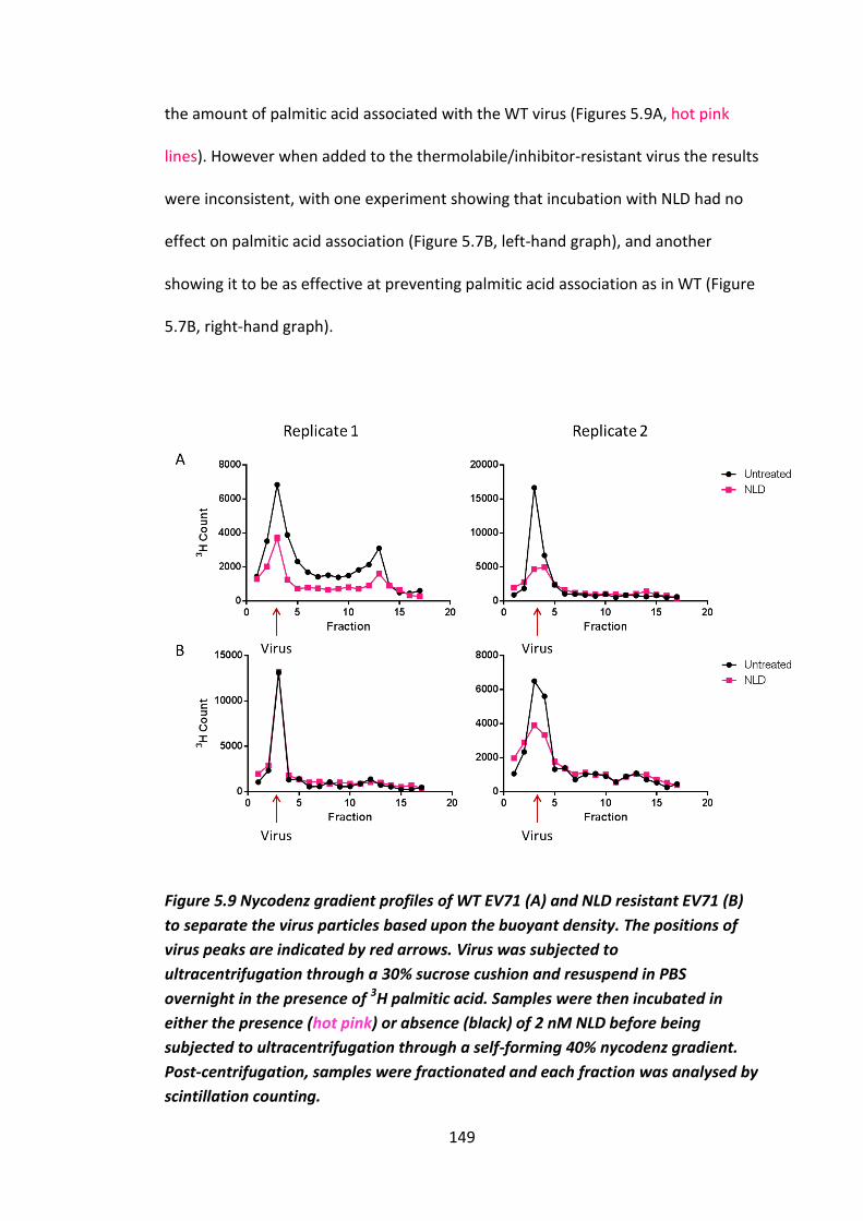
149
the amount of palmitic acid associated with the WT virus (Figures 5.9A, hot pink
lines). However when added to the thermolabile/inhibitor-resistant virus the results
were inconsistent, with one experiment showing that incubation with NLD had no
effect on palmitic acid association (Figure 5.7B, left-hand graph), and another
showing it to be as effective at preventing palmitic acid association as in WT (Figure
5.7B, right-hand graph).
Figure 5.9 Nycodenz gradient profiles of WT EV71 (A) and NLD resistant EV71 (B)
to separate the virus particles based upon the buoyant density. The positions of
virus peaks are indicated by red arrows. Virus was subjected to
ultracentrifugation through a 30% sucrose cushion and resuspend in PBS
overnight in the presence of 3H palmitic acid. Samples were then incubated in
either the presence (hot pink) or absence (black) of 2 nM NLD before being
subjected to ultracentrifugation through a self-forming 40% nycodenz gradient.
Post-centrifugation, samples were fractionated and each fraction was analysed by
scintillation counting.
150
5.1.6 Effect of pocket factors in other enteroviruses
In the previous section it was demonstrated that incubation of EV71 in the presence
of different fatty acids was able to reduce its thermostability (Figure 5.8). This is in
contrast to previous data for BEV1 where incubation in the presence of lauric acid,
one of its predicted pocket factors, was shown to improve thermostability (Ismail-
Cassim et al. 1990; Smyth et al. 2003).
As it has been shown that BEV1 can be stabilised by lauric acid, experiments were
conducted to test the effect of lauric acid and other fatty acids on the stability of
BEV2. A comparison of BEV1 and BEV2 VP1 pockets reveals that BEV2 is predicted
to harbour a larger pocket factor due to having a larger pocket, therefore it would
be expected that they behave differently in the presence of different fatty acids
(Figure 5.10, produced by Jingshan Ren at Oxford University).

151
Figure 5.10 Crystal structures reveal BEV1 and BEV2 have different sized VP1
pockets. Crystal structure overlay of BEV1 (green) and BEV2 (yellow) pockets and
predicted pocket factors lauric acid and palmitic acid produced by Jingshan Ren at
Oxford University.
Initially BEV2 was incubated either in PBS or the different fatty acids which were
shown to be able to reduce the stability of EV71 (Figure 5.8 and Table 5.1). Samples
were then titrated by TCID50 assay before and after heating to 50 oC (Figure 5.11A).
This revealed that the titre of BEV2 was dramatically reduced when heated in the
presence of any of the fatty acids tested, compared to heating in the presence of
PBS. This demonstrated that incubation in the presence of different fatty acids,
including lauric acid, was able to decrease the thermostability of BEV2. The
destabilising effect was also more dramatic than that observed for WT EV71. In
EV71 the reduction in titre of the WT virus was generally between 0.5 and 2 orders
of magnitude, however for BEV2 it was between 4 and 5 orders of magnitude
(Figure 5.11A).

152
Following confirmation that incubation of BEV2 in the presence of fatty acids
reduced the thermostability when measured via TCID50, the effect on conversion of
160S particles to 80S particles was analysed, as this was the mode of assessment
used to show lauric acid could improve the heat stability of BEV1 (Ismail-Cassim et
al. 1990). This was to ensure it was not affecting titre by any other mode of action.
In this experiment 35S-labelled BEV2 from crude cell lysate was purified through a
sucrose gradient and resuspended in PBS. This was split into three separate
samples, one was left untreated, the other was heated to 50 oC and the final sample
was incubated in the presence of lauric acid for one hour and subsequently heated
to 50 oC. All three samples were then subjected to centrifugation through a sucrose
gradient which separates particles based upon their density. Each gradient was
then fractionated and each fraction was analysed by scintillation counting to detect
the amount of 35S-labelled BEV2. When the untreated virus was subjected to
centrifugation through a gradient, a peak was visible at fractions 10-12 (Figure
5.11B), which has previously been identified as the point at which mature BEV 160S
particles sediment in 15-45% sucrose gradient (shown in section 4.1.3). A peak at
the same position was also identified in the sample heated to 50 oC, but there was
no observable peak in the sample heated in the presence of lauric acid and there
does appear to be a shift towards a peak at the 80S position (Figure 5.11B). This
indicated that heating BEV2 to 50 oC was not sufficient to destabilise the capsid, but
heating to 50 oC after incubation in the presence of lauric acid disrupted the virus.
This complements what was observed in the titration experiments where heating
BEV2 in the presence of PBS only caused a small reduction in titre of less than one
153
order of magnitude, but incubation in the presence of pocket factors was able to
reduce the titre by over four orders of magnitude (Figure 5.11 A). This indicates that
incubation in the presence of different pocket factors can reduce virus stability as
well as improve it. Work should, however, be repeated with BEV1 alongside to
confirm the observation made previously (Ismail-Cassim et al. 1990).

154
Figure 5.11 Effect of heating on BEV2 after incubation in the presence of short
chain fatty acids. (A) BEV2 was incubated in the presence of either PBS, lauric acid
(green), myristic acid (red), palmitic acid (blue), sphingosine(purple) for one hour
at 37 oC prior to heating to 50 oC for 30 minutes in a thermocycler. Samples were
then titrated before and after heating by TCID50 assay (B) Sample of 35S-labelled
BEV that was either untreated, heated to 50 oC for 30 minutes or incubated in the
presence of lauric acid for one hour prior to heating to 50 oC for 30 minutes and
were subjected to ultracentrifugation through a 15-45% sucrose gradient. The
gradient was fractionated and each sample was analysed by scintillation counting
to detect the sedimentation position of each sample. n = 1
F ra c tio n
Co
un
ts
0 5 1 0 1 5 2 0
0
2 0 0 0
4 0 0 0
6 0 0 0
8 0 0 0
U n tre a ted
5 0o
C
L a u r ic a c id 5 0o
C
B o tto mT o p
A
B
155
5.2 The potential use of antibodies and/or purified receptor to
distinguish between heated and native empty particles
Now that methods to stabilise the virus capsid had been established, methods to
distinguish between native and non-native antigenicity were required. Previous
work had shown that an antibody known as D6 was discriminatory between empty
and full virus particles (Wang et al. 2012). Purified receptors PSGL1 or SCARB2 were
also tested as reagents to detect conformational changes as they had previously
been shown to be specific to non-expanded virus particles (Qing et al. 2014).
5.2.1 Immunoprecipitation of EV71 using capsid antibodies D6 and A9
Initially antibodies that had been shown to bind natively antigenic EV71 were
tested using immunoprecipitation.
The antibodies used were monoclonal antibodies D6 and A9 that had been shown
to only be able to interact with natively antigenic EV71 (Wang et al. 2012). Initially
both antibodies were used in immunoprecipitation assays to establish if they could
distinguish between virus in heated and non-heated (native) crude lysates (Figure
5.12). Immunoprecipitations were performed with 35S-methionine/cysteine
radiolabeled EV71 crude lysate and analysed by SDS-PAGE and autoradiography. It
was evident from this that both antibodies recognised native EV71 but not the
heated EV71 (“heated antigenicity”). The autoradiograph shows VP0, VP1, VP2 and
VP3 bands, with a lower intensity observed for VP2 than VP0. This lower intensity
could be due to differences in methionine and cysteine residues in the VPs, as VP1
has 13, VP3 has 12 and VP0 has 10, whereas VP2 has 9. However it is more likely
that this sample contains as high proportion of empty capsids compared to mature

156
virions. As empty capsids contain uncleaved VP0 and in mature virions VP0 is
cleaved into VP2 and VP4. Both antibodies were also tested by Western blot,
however nothing was detected, suggesting that they recognise conformational
epitopes only.
Figure 5.12 Antibodies recognise native virus particles but not heated virus
particles. Immunoprecipitations (See materials and methods section 2.6) were
performed with 35S labelled EV71 crude lysate that had either been heated to 50 oC for 30 minutes or was untreated. The resulting immunoprecipitation was then
analysed by SDS PAGE and autoradiography. D6 and A9 are only able to detect
native antigenicity (Wang et al. 2012).

157
5.2.2 Use of ELISA in combination with EV71 receptors and antibodies to detect
native EV71 antigenicity
As it was established that antibodies D6 and A9 could distinguish between native
and heated antigenicity, it was attempted to use the antibodies in an ELISA using
purified receptor as a capture molecule.
ELISA plates were coated with one of two EV71 purified receptors (PSGL1 or
SCARB2, supplied by Prof Ian Jones, University of Reading) overnight at 4 oC. The
following day the plates were incubated with either EV71 crude lysate, heated EV71
crude lysate (50 oC), EV71 VLPs produced in a baculovirus system (provide by Ian
Jones, University of Reading) or control non-infected Vero cell crude lysate. After
washing the plates, samples were then incubated with either A9 or D6, followed by
an HRP-conjugated anti-mouse secondary antibody and developed with OPD
substrate. The OD 492 nm values showed the untreated EV71 crude lysate sample
to have a significantly higher reading than VLPs and Vero cell lysate, indicating that
WT antigenicity was detected (Figure 5.13). Although a higher reading was
generally observed in the untreated EV71 compared to heated, a significant
difference was only seen in one of the receptor antibody combinations against
heated EV71 (SCARB2/A9), the error bars were very large in the other samples. All
readings were quite low therefore this appears to be an ineffective method of
detecting EV71 antigenicity.

158
Figure 5.13 Graphs to illustrate ELISA results of EV71 captured with purified
receptor (SCARB2 or PSGL1) and detected using antibodies raised against the
whole capsid (D6 or A9). * = p ≥ 0.01, ** = ≥ 0.001.

159
5.3 Discussion
In this section methods for increasing thermostability and detecting antigenic
stability of EV71 were explored. This work has highlighted the role of the VP1
pocket in enterovirus stability, and that compounds which bind residues within this
structure were shown to have dramatic effects on the thermostability of both EV71
and BEV.
Initially, attempts to improve the thermostability of EV71 used natural selection by
heating the virus between each passage to 50 oC, a temperature which was shown
to reduce the titre by over 99.9% (Figure 5.3, 5.4). This resulted in identification of a
mutant of EV71 with increased thermostability (Figure 5.2), which possessed two
mutations in the VP1 pocket (V179A, L183V). Observations of the WT crystal
structure indicated that these mutations could increase the pocket size (Figure 5.6,
5.10). This could have two possible effects; it could allow the pocket factor to bind
deeper into the pocket, it could allow it to bind to a larger pocket factor, or both. It
was also shown that inhibitors that bind the VP1 pocket can improve enterovirus
stability, and mutations that give resistance to these inhibitors reduce
thermostability (Figure 5.14). Work described in Chapter 3 showed that the pocket-
binding inhibitor NLD was able to improve the thermostability of WT EV71, but had
no effect on thermolabile/inhibitor-resistant EV71. The same effect is seen in other
enteroviruses, with pocket-binding inhibitors improving thermostability, while
thermolabile/inhibitor-resistant mutants with bulky residues filling the pocket were
unaffected by the inhibitor (Groarke & Pevear 1999; Heinz et al. 1989; Lacroix et al.
2014; Liu et al. 2012; Mosser et al. 1994; Salvati et al. 2004). In addition, it has also

160
previously been demonstrated that natural pocket factors improve thermostability
and function as inhibitors to prevent infection in BEV1 (Ismail-Cassim et al. 1990;
Smyth et al. 2003).
Figure 5.14 Comparison of location of mutations in (A) inhibitor-
resistant/thermolabile EV71 and (B) thermostable. Crystal structure of WT EV71
with mutations of thermolabile/inhibitor resistant EV71 and thermostable EV71
highlighted by yellow spheres, VP1 is in blue and the pocket factor is represented
as hot pink spheres. Modelled using PyMol.
Together, this work shows that pocket factor binding is important for enterovirus
stability. To test how the different mutations in the pocket affected pocket factor
interactions, the effect of different compounds that are able to bind and occupy the
pocket have on the thermostability of the different EV71 mutants was compared
(Figure 5.6).
A B

161
Here, virus was incubated in the presence of fatty acids at a very high concentration
(1000 nM) for a prolonged period of time to displace the natural pocket factor. NLD
has a high binding affinity so can displace the natural pocket factor at low
concentrations (Discussed in section 1.3, chapter 3). Incubation in the presence of
NLD was able to improve the thermostability of both WT and thermostable EV71.
NLD-binding increased the temperature at which WT began to lose titre from ~46 oC
to ~51 oC and increased the temperature at which thermostable EV71 began to lose
titre from ~52 oC to ~54 oC (Figure 5.8). Incubation of WT EV71 in the presence of
different fatty acids reduced thermostability to varying degrees, with the exception
of sphingosine, which has a more bulky head that potentially allows it to bind better
because it can make more contacts (Figure 5.7). In thermostable EV71 all of the
fatty acids tested, including sphingosine, were shown to decrease thermostability
to a more dramatic degree than for WT EV71 (Figure 5.7). The reason for the
increase in stability when the two isolates are incubated in NLD is postulated to be
that the compound displaced the natural pocket factor due to a higher binding
affinity. This stabilised the virus by making it more difficult to release this unnatural
pocket factor due to a higher binding affinity and therefore was unable to undergo
conformational changes associated with loss of native antigenicity. However, in the
case of the fatty acids reducing virus stability, it is likely that because the virus had
been incubated in such high concentrations of these pocket factors the natural
pocket factor had been displaced by mass action. These compounds do not bind
with a higher affinity and are therefore more easily displaced, causing the
thermostability to be reduced. The potential reasons for sphingosine having no
effect on the stability of WT virus capsid, are either because it is the natural pocket

162
factor or it binds with the same affinity as the natural pocket factor, therefore
incubation in its presence has no visible effect. This suggests that the mode of
action of these mutations is to change the affinity for pocket factors. As in WT,
sphingosine had no effect on thermostability, but reduced the thermostability of
the thermostable isolate. This indicates that thermostable EV71 no longer uses
sphingosine as its natural pocket factor. It is not possible to tell whether the
mutations do affect the size of the pocket factor, as a pocket factor has not yet
been identified which has no effect on the thermostability of the thermostable
mutant. More fatty acids could be to be tested to determine this.
Pocket factor preference, however, cannot be the only effect on thermostability
due to the effect observed for NLD. Thermostable EV71 was more stable than WT in
the presence of NLD and NLD was still able to further increase the thermostability
of thermostable EV71. If pocket factor preference was the only reason for an
increase in thermostability then both isolates would have the same thermostability
in the presence of NLD as it is not available in the cell. Therefore, it appears that
thermostable EV71 interacts with NLD with a higher affinity. If the mutations were
merely affecting which pocket factor binds, and what pocket factor binds was the
sole determinant of capsid stability then the thermostability of both isolates would
be the same when incubated in same pocket factor.
This work so far has shown that the pocket factor is important for enterovirus
stability and that binding of certain pocket factors is able to reduce virus stability.
The role of the pocket factor and its ability to destabilise enteroviruses is further
demonstrated using BEV. Lauric acid was previously shown to improve the

163
thermostability of BEV1 (Ismail-Cassim et al. 1990; Smyth et al. 2003), while in this
work it was shown to reduce thermostability in BEV2, along with several other fatty
acids of different sizes (Figure 5.11). It would be useful to repeat this experiment
with BEV1 to observe the effects of other fatty acids on heat stability and to
confirm the effects of lauric acid. It would also be useful to test the effect of a larger
range of fatty acids to see if it was possible to find one that was able to improve the
heat stability of EV71. It would also be useful to test a wide range of
concentrations.
To further demonstrate the importance of pocket factor binding on enterovirus
stability, the thermolabile/inhibitor-resistant EV71 isolate was incubated in the
presence of different pocket factors and its thermostability was then assessed
revealing that the presence of different pocket factors and NLD had no effect on its
thermostability (Figure 5.7 C). It is predicted that the mutations fill the entrance of
the pocket with bulky side chains so that NLD and the natural pocket factor cannot
bind the pocket, which causes a reduction in stability. To test if these mutations do
prevent the pocket factor from binding WT virus and the thermolabile/inhibitor-
resistant virus, these samples were incubated in the presence of 3H palmitic acid
overnight. Palmitic acid is a pocket factor that was shown to reduce WT stability
and have no effect on the stability of the thermolabile/inhibitor-resistant EV71
(Figure 5.7). When the association of 3H palmitic acid and the two virus isolates was
assessed by analysis on nycodenz gradients, it was revealed that palmitic acid was
able to associate with both isolates (Figure 5.9). This is unexpected as it was
predicted that palmitic acid would be unable to bind the thermolabile/inhibitor-

164
resistant isolate, it should be noted that the incubation time of this experiment was
overnight, in contrast to experiments detailed in section 5.1.4 where incubations
were one hour. The experiments here therefore need to be repeated over the same
time range. However, these results do appear to establish that palmitic acid can still
bind thermolabile/inhibitor-resistant EV71. The effects of NLD remain unclear.
When this incubation experiment was repeated in the presence of NLD for two
minutes, it was shown that NLD was able to prevent palmitic acid from associating
with WT, but for the thermolabile/inhibitor-resistant mutant, NLD prevented
palmitic acid binding in one experiment but had no effect in another (Figure 5.9).
These observations require further investigation.
Finally, strategies to detect an antigenic shift away from native antigenicity have
been developed. It has been demonstrated that two different antibodies (D6 and
A9) are only able to detect EV71 in its native non-heated form using IPs (Figure
5.12). ELISAs using these antibodies and purified receptors as capture molecules
were also shown to be able to detect the difference between native and heated
antigenicity, although it was not very sensitive (Figure 5.13). It would be useful to
further optimise this method to make it more sensitive. It would be useful to use
these antibodies to produce an EV71 antigenicity curve to see if it was possible to
detect the actual point of a shift away from native antigenicity and to correlate with
infectivity.
In the future this work would need to be carried out with EV71 VLPs to see if the
strategies used here were able to improve the antigenic stability of VLPs

165
Chapter 6

166
Chapter 6 Concluding remarks and future perspective
The virus capsid plays many important roles in the virus life cycle such as; host cell
recognition, cell entry, uncoating and protection of viral RNA from degradation. In this
thesis artificial selection, inhibitory compounds and structural analysis have been combined
to further understand aspects of these multiple roles.
One aspect studied here was cell entry of EV71 in Vero cells. It was revealed that incubation
of EV71 in a low pH buffer on Vero cells causes a reduction in titre (discussed in section
4.2). Given that incubation of the virus in the presence of its receptor SCARB2 is known to
induce uncoating (Chen et al. 2012; Dang et al. 2014) it was hypothesised that the
prolonged treatment at a low pH on the cell surface in the presence of SCARB2 induced the
virus to uncoat outside of the cell, which caused the observed reduction in titre. Repeated
exposure to these conditions resulted in a mutant, termed the acid-resistant mutant (VP1
N104S) (discussed in section 4.2). Structural data and comparisons of susceptibility of WT
and the acid-resistant mutant phenotypes to different inhibitors, indicated that the acid-
resistant mutant (VP1 N104S) may not undergo SCARB2-mediated uncoating and is instead
reliant on another EV71 uncoating initiator, the proline isomerase cyclophilin A. As the acid-
resistant mutant is less susceptible to the cyclophilin A inhibitor cyclosporine A, it was
predicted that the acid-resistant mutant has a higher binding affinity for cyclophilin A, as
has been demonstrated for other EV71 isolates resistant to cyclosporine A analogues (Qing
et al. 2014). It was speculated that the mechanism by which N104S causes an increased
affinity for cyclophilin A is by making residue P103 more accessible/available than in the
WT. We predicted that N104 blocks cyclophilin A interaction with P103, either directly or
indirectly by allowing interactions with SCARB2. To confirm this proposed mode of action
several things must be established such as the role of binding to SCARB2/cyclophilin A, the
167
ability of SCARB2/cyclophilin A to induce uncoating at neutral pH and low pH and how the
acid-resistant mutant (N104S) interacts with SCARB2 and cyclophilin A in cells. Initially this
work would require the creation of the following mutants to act as controls for the inhibitor
experiments; VP1 P103A, VP1 P103A/N104S. This work would be needed to confirm that
P103 is involved in cyclophilin A interactions. To assess the roles that N104 and P103 play in
binding to SCARB2 and cyclophilin A, co-immunoprecipitation of SCARB2/cyclophilin A with
the following EV71 isolates WT, VP1 N104S, VP1 P103A, VP1 N104S/P103A could be
performed and compared. Mature virions or small peptide sequences (Dang et al. 2014)
could be used in these experiments. The same approach could be used to assess how
binding of SCARB2 and cyclophilin A mutually influence each other by pre-treating with one
uncoating initiator and observing the binding of the other. Such experiments would show
whether the mutations are be able to either increase or decrease binding. The structures of
each mutant in combination with both SCARB2 and cyclophilin A would need to solved
using either crystallography and/or cryo-EM to determine how each residue interacts with
the uncoating initiators.
This would help to determine how the mutations affect binding, but further work would
need to be done to determine their functionality. To determine if N104S effects SCARB2-
mediated cell entry, infectivity experiments could be carried out comparing WT and the
acid-resistant mutant using a cell line that is artificially expressing SCARB2 but no other
EV71 receptors (Yamayoshi et al. 2009). This would confirm that virus could only have
entered via SCARB2 and no other unknown receptors which are present in Vero cells.
To elucidate the role of these residues in uncoating, uncoating assays of each mutant in the
presence and absence of acidic conditions with both purified SCARB2 and cyclophilin A
would need to be performed. This could be done by tracking conversion of mature 160S

168
particles into uncoating intermediate 135S or empty 80S particles by use of a sucrose
gradient (described in sections 1.2.1, 1.2.4, Chen et al 2012, Dang et al 2014).
Finally toxicity experiments (such as MTT assays) of the compounds used would need to be
performed to ensure that the observed effects were not due to the cell death (Mosmann
1983).
Further knowledge of this could aid strategies to prevent the interactions between
SCARB2/cyclophilin A and could potentially lead to structure-based design of inhibitors that
are able to bind the virus capsid and block these interactions.
Further to the role of certain residues in cell entry and uncoating, this thesis has
demonstrated that EV71 takes an unusually long time to uncoat in comparison to other
picornaviruses (Section 4.1.2). In most picornaviruses, cell entry and uncoating occurs
within 30 minutes of infection (Brandenburg et al. 2007). Here using an infectivity time
course, where EV71 samples were taken for titration every hour post-infection it was shown
that the virus titre dropped in the first three hours post-infection. This reduction in
infectivity suggests that the virus has undergone uncoating, as reported by Lu et al 2011.
However, another time course experiment examining the time at which EV71 lost
susceptibility to the uncoating inhibitor NLD, revealed that this did not occur until five hours
post-infection (Section 4.1.2). Therefore the virus cannot have completed uncoating until
after five hours post-infection. Together, these data indicate that EV71 begins to uncoat
three hour post-infection, but does not complete it until six hours post-infection. It is
possible that initial structural rearrangements occur by three hours post-infection, which
reduces its infectivity, but the virus then takes a further two hours to releases its RNA. To
confirm what is occurring between three and five hours post-infection, a further time
course should be carried out to assess what uncoating stage the virus particle is at each
time point, e.g. mature 160S, uncoating intermediate 135S or empty 80S (described in
169
1.2.1, 1.2.4). This could be achieved by analysing samples from each time point on a
sucrose gradient, which is able to separate these three particle types based upon their
density. If it is confirmed that viral particles undergo structural rearmaments, to uncoating
intermediate particles (135S) before they uncoat at five hours post-infection, it would need
to be tested if these particles are less infectious and still susceptible to NLD. This would
reveal what is happening to the EV71 capsid during its unusually long uncoating time, but it
would not reveal why it takes so long. To get a better understanding of what is happening to
the virus at each stage and what induces uncoating, a time course of compounds that affect
cell entry, uncoating, virus trafficking, endosome acidification and endosome maturation
could be carried out. This would aid in annotating exactly when certain events occur and
give an idea of when the virus interacts with different uncoating factors. Live cell and
confocal imaging, staining for EV71 and different cellular markers could be used to
determine where it is in the cell and what it is associating with at each time point. This
would be very useful in terms of further understanding the virus life cycle, which could have
implications in therapeutic strategies.
This work discussed so far has focused on how uncoating effects the virus in relation to the
cell. Another major focus of the work in this thesis has been on the effect compounds that
bind the VP1 pocket have on enterovirus capsid stability (Chapters 3 & 5). The VP1 pocket is
a hydrophobic cavity that harbours a lipid that must be ejected before uncoating can occur
(Discussed in section 1.2.4). Previous work has shown that compounds that bind this pocket
with high affinity and displace the natural pocket factor can increase the capsid stability.
This stabilising effect can make the virus unable to uncoat and these compounds can
therefore act as inhibitors. Both novel synthetic compounds and natural pocket factors are
able to do this (Discussed in section 1.3). Here it was established that two novel compounds
NLD and ALD were able to inhibit EV71 infection in vitro and NLD was shown to improve the
virus thermostability (sections 3.1, 3.3). NLD was shown to be more than an order of

170
magnitude more potent than the previous most powerful EV71 pocket-binding inhibitor
(GPP3) and was not toxic to cells at inhibitory concentrations. In addition, the effects of
EV71 resistance towards these three compounds (NLD, GPP3 and ALD) were studied. WT
virus was repeatedly passaged in their presence, until the titre of virus grown in the
presence of the compounds was equivalent to WT grown without inhibitors. These resistant
isolates were shown to be genetically and thermally unstable and thermostability could not
be increased by incubation with NLD. Sequencing revealed that they all possessed the same
two mutations in the VP1 pocket (I113M and V123L). Modelling predicted that these
mutations fill the pocket and prevent binding of NLD and the natural pocket factor.
Experiments conducted to test if the mutations did prevent NLD and pocket factor binding
were inconclusive and need to be repeated. Natural fatty acids were not shown to be able
to inhibit uncoating.
This work has demonstrated the potential effectiveness of NLD as an EV71 therapeutic.
However further work needs to be carried out before it could be used in humans, such as
testing its efficacy against other enteroviruses, the efficiency of the compounds at
preventing disease in animal models, virulence of the resistant virus and further
characterisation of the action of NLD and resistance.
It would be useful to test the efficiency of NLD at inhibiting other enteroviruses, given that
HFMD is caused by a broad range of viruses and it is impossible to tell if EV71 is the
causative agent without either serum testing or sequencing. NLD would be a better
therapeutic if it was able to treat a wider variety of viral infections. Work so far has shown
NLD is able to inhibit CVA16 (De Colibus et al. 2014) and PV (Adeyemi, University of Leeds,
unpublished, Neyts, KU Leuven, unpublished), but not CVB4 (Neyts, KU Leuven,
unpublished). Viruses that should be tested with high priority are CVA6 and CVA10, which

171
are the next largest causative agents of HFMD after EV71 and CVA16, and also CVA7 which,
like EV71, can be neurotropic (Table 1.1).
While establishing if NLD has a broad range effect, it will be essential to test if it is effective
and non-toxic in vivo as well as in vitro. Further to the testing of the in vivo efficiency of the
compound, the virulence and rate at which resistance evolves must be further evaluated. It
would be useful to establish if resistance would evolve in vivo. Work with PV has shown that
it takes much more time for resistance to evolve in vivo than in vitro (Tanner et al. 2014).
This could be investigated by PCR analysis of mouse faeces or blood after infection with
EV71 and treatment with NLD.
Finally it would be good to confirm the exact mechanism action of NLD. Work by Dang et al
2014 confirmed that it prevents uncoating as measured by preventing conversion from
160S particles to 135S or 80S particles (section 4.1.2). However, some pocket-binding
inhibitors in other enteroviruses can also prevent receptor binding (Mckinlay et al. 1992).
This experiment could be conducted using co-immunoprecipitation with EV71 to investigate
if receptors bind EV71 in the presence of NLD. Further to this, live cell imagining could be
used to assess if NLD can prevent endocytosis, by measuring the distance travelled into the
cell as performed in Brandenburg et al. 2007.
The work described above has highlighted the role of the pocket factor in capsid stability
and the use of compounds such NLD as enterovirus inhibitors. To further study capsid
thermostability a selection experiment was carried out by repeatedly exposing the virus to
50 oC, a temperature which reduced the WT titre by over 99.9% (Figure 3.7, Section 5.1.2).
This resulted in selection of an isolate that tolerated a temperature 5.5 oC higher than WT.
Sequencing revealed two mutations in the VP1 pocket (V179A, L183V) which appear to
increase the size of the pocket. In an attempt to better understand the effect of these heat
stabilising mutations and the role of the pocket factor, the effect of different fatty acids on
172
heat stability was compared between WT, thermostable and inhibitor-
resistant/thermolabile EV71. This work showed that all fatty acids tested (other than
sphingosine) lowered the thermostability of WT, indicating that sphingosine could be the
natural pocket factor. All fatty acids tested reduced the stability of thermostable EV71 and
to a higher degree than the WT, indicating that none of these are the natural pocket factor.
Therefore the mutations change the pocket factor preference. In addition to this the
mutations also appear to make the capsid more sensitive to changes in pocket factor. This
work has given further insight into how the “wrong” pocket factor effects heat stability,
however work in BEV1 has shown that this virus naturally associates with multiple different
pocket factors and that some natural pocket factors can have a stabilising effect. To better
determine the role of the pocket factor, the exact pocket factor population must be
examined and the way in which virus interacts with each pocket factor must be determined.
To determine the natural pocket factor population, purified virus could be analysed by
mass-spectrometry as was previously done for BEV1 (Smyth et al. 2003) or by NMR. It
would be useful to investigate virus produced from several different cell lines as well, to see
if this made an impact on the pocket factor population. Recently, structural analysis has
indicated that different cell lines can affect which pocket factor binds (Ren et al. 2015).
Further screening of different fatty acids could be employed; a suggested list is in the
appendix 2. A pocket factor that was able to stabilise EV71 could potentially be used as an
inhibitor or aid inhibitor design.
To understand the interactions occurring with the different pocket factors, predictive
modelling or solving the structure of the viruses in the presence of different pocket factors
would need to be carried out. This would give information about exactly what interactions
form between the pocket and the different pocket factors.

173
Another way to study the interactions of different pocket factors would be to perform a
series of artificial selection experiments in the presence of different pocket factors at
elevated temperatures. This could select for a change in pocket factor. The mutations
identified could give further information about how pocket factor preference is formed.
The work described here has provided information about how the VP1 pocket is involved in
structural stability, if it was desired to select for further heat stabilising mutations that are
not involved in pocket factor binding, a heat stability selection experiment could be set up
using the inhibitor resistant mutant, which it is hypothesised not to harbour a pocket factor.
Now that heat stabilising strategies have been established, the question remains regarding
whether incubation with NLD and/or the mutations V179A and L183V are able to also
improve the stability of VLPs. First the antigenic stability of untreated WT virus and VLPs
produced in a recombinant system would need to be evaluated and then compared to the
various different stabilising strategies. This could be done by creating heat stability curves in
the same manner as previous experiments but instead of assessing titre, the native
antigenicity could initially be assessed using antibodies D6 and A9. Section 5.1.2 showed
that D6 and A9 were able to distinguish between native and heated antigenicity. This could
be done via IP or ELISA, but ELISA would require further optimisation. Although these
antibodies will be useful in assessing the virus antigenicity, further antibodies that could
bind other sites on the capsid would need to be developed. This would enable further
assessment of the antigenicity of a variety of epitopes on the capsid and therefore give a
fuller view of the capsids antigenicity. Once a method to maintain VLP antigenic stability is
established it would be required to test their efficacy as a vaccine in an animal model. There
are several different animal models available for EV71 such as the suckling mouse model
described in section 1.4.3, murine-adapted EV71 virus model (Johan Neyts, KU Leuven,
personal communication), transgenic line of mice that have been engineered to express

174
human SCARB2 (Y. W. Lin et al. 2013). It would also be useful to test if the same techniques
could be used to improve efficacy of other enterovirus VLPs. A further potential method of
improving VLP stability would be to over-express a natural pocket factor that has a
stabilising effect in the recombinant system.
A final observation made during this thesis it that two distinct types of viable virus particles
exist in EV71, non-enveloped and enveloped particles, which can be distinguished by their
buoyant densities and the enveloped particles are sensitive to detergent treatment (section
4.1.3). As picornaviruses have traditionally been considered to be non-enveloped viruses
this is interesting but not completely unexpected as similar observations have been made
for HAV and PV (Feng et al. 2013; Chen et al. 2015). Studies with these viruses have
indicated that these two particles have different entry and/or uncoating strategies. Similar
observations were made here, although only in an n = 1 experiment and so it would be
necessary to undertake a more comprehensive analysis. The presence of two particles with
different entry pathways would have implications on our understanding on EV71 cell entry.
The work presented here and that reported by others to annotate different cell entry
pathways by EV71 has been performed on a population of the two different particles
present in cell lysates, which appears to be predominantly enveloped virus. It would be
useful to understand the pathways of both particles and look at receptor binding in
different cells to assess if there is a difference. Different pathways could have implications
in pathogenesis and potentially affect disease treatment. In HAV it has been demonstrated
that enveloped particles could be used as a form of immune evasion, therefore testing the
neutralising capacity of a panel of different antibodies, such as D6 and A9, against both the
enveloped and non-enveloped form would be useful. Determining the structure of the
enveloped virus would be useful as it could give a greater insight into how enveloped virus
enters a cell and if key areas of the capsid are covered by the envelope, and potentially give
an idea of how the envelope is attached to the capsid. In addition to this it would be useful

175
to use the techniques (buoyancy, EM and detergent treatment) used to determine that
EV71 was enveloped, to screen the rest of the picornavirus family to assess if there are
other members of the picornavirus family able to produce enveloped particles.
In conclusion, this thesis has produced a body of work that has contributed to a greater
understanding of EV71 uncoating and could have implications in the design of future
vaccines and antiviral

176
Chapter 7
Bibliography
177
Chapter 7 Bibliography
Acharya, R. et al., 1989. The three-dimensional structure of foot-and-mouth disease virus at 2.9 Å resolution. Nature, 337(6209), pp.709–716.
Ansardi, D.C. & Morrow, C.D., 1995. Amino acid substitutions in the poliovirus maturation cleavage site affect assembly and result in accumulation of provirions. Journal of virology, 69(3), pp.1540–1547.
Ansardi, D.C., Porter, D.C. & Morrow, C.D., 1991. Coinfection with recombinant vaccinia viruses expressing poliovirus P1 and P3 proteins results in polyprotein processing and formation of empty capsid structures. Journal of virology, 65(4), pp.2088–2092.
Arita, M. et al., 2008. Cooperative effect of the attenuation determinants derived from poliovirus sabin 1 strain is essential for attenuation of enterovirus 71 in the NOD/SCID mouse infection model. Journal of virology, 82(4), pp.1787–1797.
Arita, M. et al., 2005. Temperature-sensitive mutants of enterovirus 71 show attenuation in cynomolgus monkeys. The Journal of general virology, 86, pp.1391–401.
Ashraf, S. et al., 2013. Biological characteristics and propagation of human rhinovirus-C in differentiated sinus epithelial cells. Virology, 436, pp.143–146.
Basavappa, R. et al., 1994. Role and mechanism of the maturation cleavage of VP0 in poliovirus assembly: structure of the empty capsid assembly intermediate at 2.9 A resolution. Protein science : a publication of the Protein Society, 3(10), pp.1651–1669.
Baxt, B., 1987. Effect of lysosomotropic compounds on early events in foot-and-mouth disease virus replication. Virus research, 7(3), pp.257–271.
Berryman, S. et al., 2005. Early Events in Integrin alpha v beta 6-Mediated Cell Entry of Foot-and-Mouth Disease Virus. Journal of virology, 79(13), pp.8519–8534.
Bostina, M. et al., 2011. Poliovirus RNA is released from the capsid near a twofold symmetry axis. Journal of virology, 85(2), pp.776–783.
Le Bouvier, G.L., 1955. The modification of poliovirus antigens by heat and ultraviolet light. Lancet, 269(6898), pp.1013–1016.
Brandenburg, B. et al., 2007. Imaging Poliovirus Entry in Live Cells. PLoS Biology, 5(7), p.e183.
178
Bubeck, D., Filman, D.J. & Hogle, J.M., 2005. Cryo-electron microscopy reconstruction of a poliovirus-receptor-membrane complex. Nature structural & molecular biology, 12(7), pp.615–618.
Chang, J.-Y. et al., 2012. Selection and characterization of vaccine strain for Enterovirus 71 vaccine development. Vaccine, 30(4), pp.703–711.
Chen, P. et al., 2012. Molecular determinants of enterovirus 71 viral entry: a cleft around Q172 on VP1 interacts with a variable region on scavenge receptor B2. The Journal of biological chemistry, 71, pp.6406–6420.
Chen, Y.-H. et al., 2015. Phosphatidylserine Vesicles Enable Efficient En Bloc Transmission of Enteroviruses. Cell, 160(4), pp.619–630.
Chern, J.-H. et al., 2004. Design, synthesis, and structure-activity relationships of pyrazolo[3,4-d]pyrimidines: a novel class of potent enterovirus inhibitors. Bioorganic & medicinal chemistry letters, 14(10), pp.2519–2525.
Chou, A.H. et al., 2013. Formalin-inactivated EV71 vaccine candidate induced cross-neutralizing antibody against subgenotypes B1, B4, B5 and C4A in adult volunteers. PLoS ONE, 8(11), e79783.
Chou, A.H. et al., 2012. Immunological Evaluation and Comparison of Different EV71 Vaccine Candidates. Clinical and Developmental Immunology, 2012, p.831282.
Chou, A.-H. et al., 2012. Pilot scale production of highly efficacious and stable enterovirus 71 vaccine candidates. PloS one, 7(4), p.e34834.
Chow, M. et al., 1987. Myristylation of picornavirus capsid protein VP4 and its structural significance. Nature, 327(6122), pp.482–486.
Chung, C. et al., 2010. Enterovirus 71 virus-like particle vaccine : Improved production conditions for enhanced yield. Vaccine, 28(43), pp.6951–6957.
Chung, Y.-C. et al., 2006. Expression, purification and characterization of enterovirus-71 virus-like particles. World journal of gastroenterology, 12(6), pp.921–927.
Chung, Y.-C. et al., 2008. Immunization with virus-like particles of enterovirus 71 elicits potent immune responses and protects mice against lethal challenge. Vaccine, 26(15), pp.1855–1862.
Clarke, P., Kitchin, N. & Souverbie, F., 2001. A randomised comparison of two inactivated hepatitis A vaccines, AvaximTM and VaqtaTM, given as a booster to subjects primed with AvaximTM. Vaccine, 19(31), pp.4429–4433.
Clarke, T., 2001. Polio’s last stand. Nature, 409(6818), pp.278–280.

179
De Colibus, L., Wang, X., Spyrou, J.A.B., et al., 2014. More-powerful virus inhibitors from structure-based analysis of HEV71 capsid-binding molecules. Nature structural & molecular biology, 21(3), pp.282–288.
De Colibus, L., Wang, X., Spyrou, J. a B., et al., 2014. More-powerful virus inhibitors from structure-based analysis of HEV71 capsid-binding molecules. Nature structural & molecular biology, 21(3), pp.282–288.
Colonno, R.J. et al., 1988. Evidence for the direct involvement of the rhinovirus canyon in receptor binding. Proceedings of the National Academy of Sciences of the United States of America, 85(15), pp.5449–5453.
Coyne, C.B. & Bergelson, J.M., 2006. Virus-induced Abl and Fyn kinase signals permit coxsackievirus entry through epithelial tight junctions. Cell, 124(1), pp.119–131.
Coyne, C.B. et al., 2007. Coxsackievirus entry across epithelial tight junctions requires occludin and the small GTPases Rab34 and Rab5. Cell Host Microbe,2, pp. 181-192
Curry, S. et al., 1997. Dissecting the roles of VP0 cleavage and RNA packaging in picornavirus capsid stabilization: the structure of empty capsids of foot-and-mouth disease virus. Journal of virology, 71(12), pp.9743–9752.
Dang, M. et al., 2014. Molecular mechanism of SCARB2-mediated attachment and uncoating of EV71. Protein Cell, 5(9), pp.692–703.
Danthi, P. et al., 2003. Genome delivery and ion channel properties are altered in VP4 mutants of poliovirus. Journal of virology, 77(9), pp.5266–5274.
Davis, M.P. et al., 2008. Recombinant VP4 of human rhinovirus induces permeability in model membranes. Journal of virology, 82(8), pp.4169–4174.
Dimmock, N.J. & Tyrrell, D.A.J., 1963. Some physico-chemical properties of rhinoviruses. Br J Exp Pathol, 45, pp.271–280.
Doherty, G.J. & McMahon, H.T., 2009. Mechanisms of endocytosis. Annual review of biochemistry, 78, pp.857–902.
Du, N. et al., 2014. Cell Surface Vimentin Is an Attachment Receptor for Enterovirus 71. Journal of virology, 88(10), 5816-5833.
Feil, S.C. et al., 2012. An Orally Available 3-Ethoxybenzisoxazole Capsid Binder with Clinical Activity against Human Rhinovirus. ACS Med Chem Lett, 3(4), pp.303–307.
Feng, Z. et al., 2013. A pathogenic picornavirus acquires an envelope by hijacking cellular membranes. Nature, 496(7445), pp.367–371.

180
Filman, D.J. et al., 1989. Structural factors that control conformational transitions and serotype specificity in type 3 poliovirus. The EMBO journal, 8(5), pp.1567–1579.
Foo, D.G.W., Alonso, S., Phoon, M.C., et al., 2007. Identification of neutralizing linear epitopes from the VP1 capsid protein of Enterovirus 71 using synthetic peptides. Virus Research, 125(1), pp.61–68.
Foo, D.G.W., Alonso, S., Chow, V.T.K., et al., 2007. Passive protection against lethal enterovirus 71 infection in newborn mice by neutralizing antibodies elicited by a synthetic peptide. Microbes and Infection, 9(11), pp.1299–1306.
Fowler, D.M. et al., 2010. High-resolution mapping of protein sequence- function relationships. Nature methods, 7(9), pp.741–746.
Fricks, C.E. & Hogle, J.M., 1990. Cell-induced conformational change in poliovirus: externalization of the amino terminus of VP1 is responsible for liposome binding. Journal of virology, 64(5), pp.1934–1945.
Fry, E.E. et al., 2003. Crystal structure of Swine vesicular disease virus and implications for host adaptation. Journal of virology, 77(9), pp.5475–5486.
Geller, R. et al., 2007. antiviral approach refractory to development of drug resistance Evolutionary constraints on chaperone-mediated folding provide an antiviral approach refractory to development of drug resistance. Genes & Development, 21, pp.195–205.
Gong, M. et al., 2014. Cryo-electron microscopy study of insect cell-expressed enterovirus 71 and coxsackievirus a16 virus-like particles provides a structural basis for vaccine development. Journal of virology, 88(11), pp.6444–6452.
Grant, R. a et al., 1994. Structures of poliovirus complexes with anti-viral drugs: implications for viral stability and drug design. Current biology : CB, 4(9), pp.784–797.
Groarke, J.M. & Pevear, D.C., 1999a. Attenuated Virulence of Pleconaril-Resistant Coxsackievirus B3 Variants. Journal of Infectious Diseases, 179, pp.1538–1541.
Groarke, J.M. & Pevear, D.C., 1999b. Attenuated Virulence of Pleconaril-Resistant Coxsackievirus B3 Variants. JID, 179, pp.1538–1541.
Groppelli, E., Tuthill, T.J. & Rowlands, D.J., 2010. Cell entry of the aphthovirus equine rhinitis A virus is dependent on endosome acidification. Journal of virology, 84(12), pp.6235–6240.
Gulland, A., 2014. World has been slow to act on polio outbreak in Syria, charity warns. BMJ, 348, p.1947.

181
Guo, J. et al., 2015. Immunodeficiency-related vaccine-derived poliovirus (iVDPV) cases: A systematic review and implications for polio eradication. Vaccine, 33(10), pp.1235–1242.
Guttman, N. & Baltimore, D., 1977. Morphogenesis of poliovirus. IV. existence of particles sedimenting at 150S and having the properties of provirion. Journal of virology, 23(2), pp.363–367.
Halsey, N.A. et al., 2004. Search for poliovirus carriers among people with primary immune deficiency diseases in the United States, Mexico, Brazil, and the United Kingdom. Bulletin of the World Health Organization, 82(1), pp.3–8.
Heinz, B.A., Rueckert, R.R., Shepard, D.A., Dutko, F.J., Mckinlay, M.A., Fancher, M., et al., 1989. Genetic and molecular analyses of that are resistant to an antiviral compound . Genetic and Molecular Analyses of Spontaneous Mutants of Human Rhinovirus 14 That Are Resistant to an Antiviral Compound. Journal of virology, 63(6), pp.2476-2485.
Hertzler, S., Luo, M. & Lipton, H.L., 2000. Mutation of Predicted Virion Pit Residues Alters Binding of Theiler’s Murine Encephalomyelitis Virus to BHK-21 Cells. Journal of virology, 74(4), pp.1994–2004.
Hoey, E.M. & Martin, S.J., 1974. A possible precursor containing RNA of a bovine enterovirus: the provirion 11. Journal of General Virology, 24(3), pp.515–524.
Hu, Y.-C. et al., 2003. Formation of enterovirus-like particle aggregates by recombinant baculoviruses co-expressing P1 and 3CD in insect cells. Biotechnology Letters, 25(12), pp.919–925.
Huang, S.-W. et al., 2012. Mutations in VP2 and VP1 capsid proteins increase infectivity and mouse lethality of enterovirus 71 by virus binding and RNA accumulation enhancement. Virology, 422(1), pp.132–143.
Ismail-Cassim, N., Chezzi, C. & E, N.J.F., 1990. Inhibition of the uncoating of bovine enterovirus by short chain fatty acids. Journal of General Virology, 71, pp.2283–2289.
Johansson, S. et al., 2002. Molecular analysis of three Ljungan virus isolates reveals a new, close-to-root lineage of the Picornaviridae with a cluster of two unrelated 2A proteins. Journal of virology, 76(17), pp.8920–8930.
John, J., 2009. Role of injectable and oral polio vaccines in polio eradication. Expert review of vaccines, 8(1), pp.5–8.
Katpally, U., Smith, T.J. & Rhinovirus, H., 2007. Pocket Factors Are Unlikely To Play a Major Role in the Life Cycle of Human Rhinovirus Pocket Factors Are Unlikely To Play a Major Role in the Life Cycle of. Journal of virology, 81(12), pp.6307-6315.
182
Kattur Venkatachalam, A.R. et al., 2014. Concentration and purification of enterovirus 71 using a weak anion-exchange monolithic column. Virology journal, 11(1),p p.99-108.
Ke, Y. & Lin, T., 2006. Modeling the Ligand - Receptor Interaction for a Series of Inhibitors of the Capsid Protein of Enterovirus 71 Using Several Three-Dimensional Quantitative Structure - Activity Relationship Techniques. J. Med. Chem, 49, pp.4517–4525.
Kellogg, E.H., Leaver-fay, A. & Baker, D., 2011. Role of conformational sampling in computing mutation-induced changes in protein structure and stability. Proteins, 79(3), pp.830–838.
Kew, O.M. et al., 2005. Vaccine-derived polioviruses and the endgame strategy for global polio eradication. Annual review of microbiology, 59, pp.587–635.
Kok, C.C., 2015. Therapeutic and prevention strategies against human enterovirus 71 infection. World Journal of Virology, 4(2), pp.78-95.
Kolpe, A.B. et al., 2012. Display of enterovirus 71 VP1 on baculovirus as a type II transmembrane protein elicits protective B and T cell responses in immunized mice. Virus Research, 168(1-2), pp.64–72.
Kouiavskaia, D. V et al., 2011. Original article Immunological and pathogenic properties of poliovirus variants selected for resistance to antiviral drug V-073. Antiviral Therapy, 16(7), pp.999–1004.
Ku, Z. et al., 2013. Neutralizing Antibodies Induced by Recombinant Virus-Like Particles of Enterovirus 71 Genotype C4 Inhibit Infection at Pre- and Post-attachment Steps. PLoS ONE, 8(2), pp.1–8.
Kuo, R.-L. & Shih, S.-R., 2013. Strategies to develop antivirals against enterovirus 71. Virology journal, 10(1), pp.28-36.
Lacroix, C. et al., 2014. A novel benzonitrile analogue inhibits rhinovirus replication. Journal of Antimicrobial Chemotherapy, 69(June), pp.2723–2732.
Lee, W.M., Monroe, S.S. & Rueckert, R.R., 1993. Role of maturation cleavage in infectivity of picornaviruses: activation of an infectosome. Journal of virology, 67(4), pp.2110–2122.
Lehtinen, M. et al., 2012. Overall efficacy of HPV-16/18 AS04-adjuvanted vaccine against grade 3 or greater cervical intraepithelial neoplasia: 4-year end-of-study analysis of the randomised, double-blind PATRICIA trial. The lancet oncology, 13(1), pp.89–99.
Li, C. et al., 2012. In Vitro Assembly of an Empty Picornavirus Capsid follows a Dodecahedral Path. Journal of Virology, 86(23), pp.13062–13069.

183
Li, H. et al., 2013. Virus-like particles for enterovirus 71 produced from Saccharomyces cerevisiae potently elicits protective immune responses in mice. Vaccine, 31(32), pp.3281–3287.
Li, X. et al., 2001. The C-terminal residues of poliovirus proteinase 2Apro are critical for viral RNA replication but not for cis- or trans-proteolytic cleavage. Journal of General Virology, 82(2), pp.397–408.
Lin, H.-Y. et al., 2013. Caveolar Endocytosis Is Required for Human PSGL-1-Mediated Enterovirus 71 Infection. Journal of virology, 87(16), pp.9064–9076.
Lin, S.-Y. et al., 2014. Evaluation of the stability of enterovirus 71 virus-like particle. Journal of bioscience and bioengineering, 117(3), pp.366–371.
Lin, Y.-L. et al., 2011. Enterovirus type 71 neutralizing antibodies in the serum of macaque monkeys immunized with EV71 virus-like particles. Vaccine, 30, pp 1305-1312
Lin, Y.W. et al., 2013. Human SCARB2 Transgenic Mice as an Infectious Animal Model for Enterovirus 71. PLoS ONE, 8(2), p.e57591.
Lin, Y.-W. et al., 2009. Enterovirus 71 infection of human dendritic cells. Experimental biology and medicine (Maywood, N.J.), 234(10), pp.1166–1173.
Lin, Y.-W. et al., 2012. Human SCARB2-Mediated Entry and Endocytosis of EV71. PloS one, 7(1), p.e30507.
Liu, C.-C. et al., 2007. High immunogenic enterovirus 71 strain and its production using serum-free microcarrier Vero cell culture. Vaccine, 25(1), pp.19–24.
Liu, F. et al., 2012. Research in Veterinary Science Virus-like particles : potential veterinary vaccine immunogens. Research in Veterinary Science, 93(2), pp.553–559.
Liu, H. et al., 2012. Characterization of Poliovirus Variants Selected for Resistance to the Antiviral Compound V-073. Antimicrobial agents and chemotherapy, 56(11), pp.5568–5574.
Liu, J. et al., 2011. Lycorine reduces mortality of human enterovirus 71-infected mice by inhibiting virus replication. Virology Journal, 8(1), pp.483-500.
Liu, Y. et al., 2015. Structure and inhibition of EV-D68, a virus that causes respiratory illness in children. Science, 347(6217), pp.71–74.
Logan, D. et al., 1993. Structure of a major immunogenic site on foot-and-mouth disease virus. Nature, 362(6420), pp.566–568.

184
Lokugamage, K.G. et al., 2008. Chimeric coronavirus-like particles carrying severe acute respiratory syndrome coronavirus (SCoV) S protein protect mice against challenge with SCoV. Vaccine, 26(6), pp.797–808.
López-Macías, C. et al., 2011. Safety and immunogenicity of a virus-like particle pandemic influenza A (H1N1) 2009 vaccine in a blinded, randomized, placebo-controlled trial of adults in Mexico. Vaccine, 29(44), pp.7826–7834.
Luo, M. et al., 1987. The atomic structure of Mengo virus at 3.0 A resolution. Science, 235(4785), pp.182–191.
Macadam, A.J. et al., 2006. Rational design of genetically stable, live-attenuated poliovirus vaccines of all three serotypes: relevance to poliomyelitis eradication. Journal of virology, 80(17), pp.8653–8663.
Macejak, D.G. & Sarnow, P., 1992. Association of heat shock protein 70 with enterovirus capsid precursor P1 in infected human cells. Journal of virology, 66(3), pp.1520–1527.
Mao, Q. et al., 2013. The cross-neutralizing activity of enterovirus 71 subgenotype C4 vaccines in healthy chinese infants and children. PLoS ONE, 8(11), pp.16–20.
Marongiu, M.E. et al., 1981. Poliovirus morphogenesis. I. Identification of 80S dissociable particles and evidence for the artifactual production of procapsids. Journal of virology, 39(2), pp.341–347.
Martín-Acebes, M. a et al., 2009. Internalization of swine vesicular disease virus into cultured cells: a comparative study with foot-and-mouth disease virus. Journal of virology, 83(9), pp.4216–4226.
Martín-Acebes, M.A. et al., 2011. Foot-and-mouth disease virus particles inactivated with binary ethylenimine are efficiently internalized into cultured cells. Vaccine, 29(52), pp.9655–9662.
Mckinlay, M.A. et al., 1992. Treatment of the picornavirus common cold by inhibitors or viral uncoating and attachment. Annual review of microbiology, 46, pp.635–654.
McMinn, P. et al., 2001. Neurological manifestations of enterovirus 71 infection in children during an outbreak of hand, foot, and mouth disease in Western Australia. Clinical infectious diseases, 32(2), pp.236–242.
Mcminn, P.C., 2003. Enterovirus 71 in the Asia-Pacific region : An emerging cause of acute neurological disease in young children. Neurol J Southeast Asia, 8, pp.57–63.
McMinn, P.C., 2012. Recent advances in the molecular epidemiology and control of human enterovirus 71 infection. Current opinion in virology, 2(2), pp.199–205.

185
Meng, T. et al., 2011. Display of VP1 on the surface of baculovirus and its immunogenicity against heterologous human enterovirus 71 strains in mice. PloS one, 6(7), p.e21757.
Mosmann, T., 1983. Rapid colorimetric assay for cellular growth and survival: application to proliferation and cytotoxicity assays. Journal of immunological methods, 65(1-2), pp.55–63.
Mosser, A.G., Sgro, J. & Rueckert, R.R., 1994. Distribution of drug resistance mutations in type 3 poliovirus identifies three regions involved in uncoating functions . Distribution of Drug Resistance Mutations in Type 3 Poliovirus Identifies Three Regions Involved in Uncoating Functions. Journal of virology, 68(12), pp.8193-8201.
Mulcahy, L.A., Pink, R.C. & Carter, D.R.F., 2014. Routes and mechanisms of extracellular vesicle uptake. Journal of extracellular vesicles, 3, pp.1–14.
Mulder, A.M. et al., 2012. Toolbox for Non-Intrusive Structural and Functional Analysis of Recombinant VLP Based Vaccines: A Case Study with Hepatitis B Vaccine. PloS one, 7(4), p.e33235.
Mutsvunguma, L.Z. et al., 2011. Theiler’s murine encephalomyelitis virus infection induces a redistribution of heat shock proteins 70 and 90 in BHK-21 cells, and is inhibited by novobiocin and geldanamycin. Cell Stress and Chaperones, 16(5), pp.505–515.
Nishimura, Y. et al., 2013. Enterovirus 71 Binding to PSGL-1 on Leukocytes: VP1-145 Acts as a Molecular Switch to Control Receptor Interaction. PLoS Pathogens, 9(7), p.e1003511.
Nishimura, Y. et al., 2009. Human P-selectin glycoprotein ligand-1 is a functional receptor for enterovirus 71. Nature medicine, 15(7), pp.794–797.
Nishimura, Y., Wakita, T. & Shimizu, H., 2010. Tyrosine sulfation of the amino terminus of PSGL-1 is critical for enterovirus 71 infection. PLoS pathogens, 6(11), p.e1001174.
Olson, N.H. et al., 1993. Structure of a human rhinovirus complexed with its receptor molecule. Proceedings of the National Academy of Sciences of the United States of America, 90(2), pp.507–511.
Panjwani, A. et al., 2014. Capsid Protein VP4 of Human Rhinovirus Induces Membrane Permeability by the Formation of a Size-Selective Multimeric Pore. PLoS pathogens, 10(8), p.e1004294.
Paul, Y., 2009. Oral polio vaccines and their role in polio eradication in India. Expert review of vaccines, 8(1), pp.35–41.
186
Pevear, D.C. et al., 1999. Activity of Pleconaril against Enteroviruses Activity of Pleconaril against Enteroviruses. Antimicrobial agents and chemotherapy, 43(9), pp.2109-2115.
Pevear, D.C. et al., 2005a. Relationship of Pleconaril Susceptibility and Clinical Outcomes in Treatment of Common Colds Caused by Rhinoviruses. Antimicrobial agents and chemotherapy, 49(11), pp.4492–4499.
Pevear, D.C. et al., 2005b. Relationship of Pleconaril Susceptibility and Clinical Outcomes in Treatment of Common Colds Caused by Rhinoviruses Relationship of Pleconaril Susceptibility and Clinical Outcomes in Treatment of Common Colds Caused by Rhinoviruses. Antimicrobial agents and chemotherapy, 49(11), pp.4492–4499.
Plevka, P., Perera, R., Cardosa, J., et al., 2012. Crystal Structure of Human Enterovirus 71. Science, 336(6086), p.1274.
Plevka, P. et al., 2013. Structure of human enterovirus 71 in complex with a capsid-binding inhibitor. Proceedings of the National Academy of Sciences, 110, pp.3–7.
Plevka, P., Perera, R. & Rossmann, M.G., 2012. Structure determination of enterovirus 71 research papers. Biological Crystallography, 68, pp.1217–1222.
Porta, C. et al., 2013. Rational Engineering of Recombinant Picornavirus Capsids to Produce Safe, Protective Vaccine Antigen. PLoS Pathogens, 9(3), p.e1003255.
Praefcke, G.J.K. et al., 2004. Identification of residues in the human guanylate-binding protein 1 critical for nucleotide binding and cooperative GTP hydrolysis. Journal of Molecular Biology, 344(1), pp.257–269.
Premanand, B. et al., 2012. Induction of protective immune responses against EV71 in mice by baculovirus encoding a novel expression cassette for capsid protein VP1. Antiviral research, 95, pp. 311-315.
Qing, J. et al., 2014. Cyclophilin A Associates with Enterovirus-71 Virus Capsid and Plays an Essential Role in Viral Infection as an Uncoating Regulator. PLoS Pathogens, 10(10), p.e1004422.
Ren, J. et al., 2013. Picornavirus uncoating intermediate captured in atomic detail. Nature Communications, 4, pp.1927–1929.
Ren, J. et al., 2015. Structures of coxsackievirus A16 capsids with native antigenicity, implications for particle expansion, receptor binding and immunogenicity. Journal of Virology, 4343, pp 1102–1115.
Rombaut, B. et al., 1990. Creation of an antigenic site in poliovirus type 1 by assembly of 14 S subunits. Virology, 174(1), pp.305–307.

187
Rombaut, B. & Jore, J.P., 1997. Immunogenic, non-infectious polio subviral particles synthesized in Saccharomyces cerevisiae. The Journal of general virology, 78 ( Pt 8), pp.1829–1832.
Rossmann, M.G.; et al., 1985. Structure of a human common cold virus and functional relationship to other picornaviruses. Nature, 317, pp. 145–153
Rotbart, H.A., 2002. Treatment of picornavirus infections. Antiviral Research, 53, pp.83–98.
Rotbart, H.A., Connell, J.F.O. & Mckinlay, M.A., 1998. Treatment of human enterovirus infections. Antiviral research, 38, pp.1–14.
Salvati, A.L. et al., 2004. Mechanism of Action at the Molecular Level of the Antiviral Drug 3 ( 2H ) -Isoflavene against Type 2 Poliovirus Mechanism of Action at the Molecular Level of the Antiviral Drug 3 ( 2H ) -Isoflavene against Type 2 Poliovirus. Antimicrobial agents and chemotherapy, 48(6), p.2233-2243.
Schmid, E.M. et al., 2006. Role of the AP2 β-Appendage Hub in Recruiting Partners for Clathrin-Coated Vesicle Assembly. PLoS Biology, 4(9), pp.1532–1548.
Shafren, D.R., Williams, D.T. & Barry, R.D., 1997. A decay-accelerating factor-binding strain of coxsackievirus B3 requires the coxsackievirus-adenovirus receptor protein to mediate lytic infection of rhabdomyosarcoma cells. Journal of virology, 71(12), pp.9844–9848.
Shang, L., Xu, M. & Yin, Z., 2013. Antiviral drug discovery for the treatment of enterovirus 71 infections. Antiviral Research, 97(2), pp.183–194.
Shepard, D. a, Heinz, B. a & Rueckert, R.R., 1993. WIN 52035-2 inhibits both attachment and eclipse of human rhinovirus 14. Journal of virology, 67(4), pp.2245–2254.
Shia, K. et al., 2002. Design, Synthesis, and Structure- Activity Relationship of Pyridyl Imidazolidinones: A Novel Class of Potent and Selective Human Enterovirus 71. J. Med. Chem, 45, pp.1644–1655.
Shih, S. et al., 2004. Mutation in Enterovirus 71 Capsid Protein VP1 Confers Resistance to the Inhibitory Effects of Pyridyl Imidazolidinone Mutation in Enterovirus 71 Capsid Protein VP1 Confers Resistance to the Inhibitory Effects of Pyridyl Imidazolidinone. Antimicrobial agents and chemotherapy, 48(9), pp.3523–3529.
Shingler, K.L. et al., 2013. The Enterovirus 71 A-particle Forms a Gateway to Allow Genome Release: A CryoEM Study of Picornavirus Uncoating. PLoS Pathogens, 9(3), p.e1003240.
188
Shingler, K.L. et al., 2015. The Enterovirus 71 Procapsid Binds Neutralizing Antibodies and Rescues Virus Infection In Vitro. Journal of Virology, 89(3), pp.1900–1908.
Smith, T.J. et al., 1986. The site of attachment in human rhinovirus 14 for antiviral agents that inhibit uncoating. Science, 233(4770), pp.1286–1293.
Smyth, M. et al., 2003. Identification of the pocket factors in a picornavirus. Archives of Virology, 148, pp.1225–1233.
Smyth, M. et al., 2003. Identification of the pocket factors in a picornavirus. Archives of Virology, 148(6), pp.1225–1233.
Smyth, M. et al., 1995. Implications for viral uncoating from the structure of bovine enterovirus. Structural biology, 2, pp.224–231.
Solomon, T. et al., 2010. Virology, epidemiology, pathogenesis, and control of enterovirus 71. The Lancet infectious diseases, 10(11), pp.778–790.
Strauss, M. et al., 2013. RNA transfer from poliovirus 135S particles across membranes is mediated by long umbilical connectors. Journal of virology, 87(7), pp.3903–3914.
Su, P.-Y. et al., 2015. Cell Surface Nucleolin Facilitates Enterovirus 71 Binding and Infection. Journal of Virology, 89, pp.4527–4538.
Suomalainen, M. & Greber, U.F., 2013. Uncoating of non-enveloped viruses. Current Opinion in Virology, 3(1), pp.27–33.
Superti, F. et al., 1987. The effect of lipophilic amines on the growth of hepatitis A virus in Frp/3 cells. Archives of Virology, 96(3-4), pp.289–296.
Tan, C.W. et al., 2013. Enterovirus 71 Uses Cell Surface Heparan Sulfate Glycosaminoglycan as an Attachment Receptor. Journal of virology, 7(7), p e41381.
Tanner, E.J. et al., 2014. Dominant drug targets suppress the emergence of antiviral resistance. eLife, 3, pp.1–16.
Tesar, M. et al., 1993. Analysis of a potential myristoylation site in hepatitis A virus capsid protein VP4.pdf. Virology, 194, pp.616–626.
Thibaut, H.J., De Palma, A.M. & Neyts, J., 2012. Combating enterovirus replication: State-of-the-art on antiviral research. Biochemical Pharmacology, 83(2), pp.185–192.

189
Tijsma, a. et al., 2014. The Capsid Binder Vapendavir and the Novel Protease Inhibitor SG85 Inhibit Enterovirus 71 Replication. Antimicrobial Agents and Chemotherapy, 58(11), pp.6990–6992.
Toth, K.S. et al., 1993. Crystallization and preliminary X-ray diffraction studies of Theiler’s virus (GDVII strain). Journal of Molecular Biology, 231, pp.1126–1129.
Tuthill, T.J. et al., 2009. Equine Rhinitis A Virus and Its Low pH Empty Particle : Clues Towards an Aphthovirus Entry Mechanism ? PLoS pathogens, 5(10), p.e1000620.
Tuthill, T.J. et al., 2010. Picornaviruses. Current topics in microbiology and immunology, 343, pp.43–89.
Tyka, M.D. et al., 2011. Landscape Mapping. J Mol Biol, 405(2), pp.607–618.
Wang, X. et al., 2012. A sensor-adaptor mechanism for enterovirus uncoating from structures of EV71. Nature structural & molecular biology, 19(4), pp.424–429.
Wang, X. et al., 2012. Characterization of full-length enterovirus 71 strains from severe and mild disease patients in northeastern China. PloS one, 7(3), p.e32405.
Wang, X. et al., 2015. Hepatitis A virus and the origins of picornaviruses. Nature, 517(7532), pp.85–88.
Wildenbeest, J.G. et al., 2012. Pleconaril revisited: Clinical course of chronic enteroviral meningoencephalitis after treatment correlates with in vitro susceptibility. Antiviral Therapy, 17(3), pp.459–466.
Xing, W. et al., 2014. Hand , foot , and mouth disease in China , 2008 – 12 : an epidemiological study. Lancet Infect Dis, 14(April), pp.308–318.
Xu, L. et al., 2014. Protection against Lethal Enterovirus 71 Challenge in Mice by a Recombinant Vaccine Candidate Containing a Broadly Cross-Neutralizing Epitope within the VP2 EF Loop. Theranostics, 4(5), pp.498–513.
Yamayoshi, S. et al., 2013. Functional comparison of SCARB2 and PSGL1 as receptors for enterovirus 71. Journal of virology, 87(6), pp.3335–3347.
Yamayoshi, S., Iizuka, S., et al., 2012. Human SCARB2-Dependent Infection by Coxsackievirus A7, A14, and A16 and Enterovirus 71. Journal of virology, 86(10), pp.5686–5696.
Yamayoshi, S. et al., 2009. Scavenger receptor B2 is a cellular receptor for enterovirus 71. Nature medicine, 15(7), pp.798–801.

190
Yamayoshi, S., Fujii, K. & Koike, S., 2012. Scavenger receptor b2 as a receptor for hand, foot, and mouth disease and severe neurological diseases. Frontiers in microbiology, 3, pp.32-40.
Yang, B., Chuang, H. & Yang, K.D., 2009. Sialylated glycans as receptor and inhibitor of enterovirus 71 infection to DLD-1 intestinal cells. Virology journal, 6, p0.141-146.
Yang, S.-L. et al., 2011. Annexin II binds to capsid protein VP1 of enterovirus 71 and enhances viral infectivity. Journal of virology, 85(22), pp.11809–118020.
Ye, X. et al., 2014. Chimeric Virus-Like Particle Vaccines Displaying Conserved Enterovirus 71 Epitopes Elicit Protective Neutralizing Antibodies in Mice through Divergent Mechanisms. Journal of Virology, 88(1), pp.72–81.
Ypma-Wong, M.R. et al., 1988. Protein 3CD is the major Poliovirus proteinase responsibe for cleavage of the P1 capsid precursor. Virology, 270(1988), pp.265–270.
Zhang, F. et al., 2014. Oral immunization with recombinant enterovirus 71 VP1 formulated with chitosan protects mice against lethal challenge. Virology Journal, 11(1), p.80-85.
Zhang, L. et al., 2012. Vaccination with coxsackievirus B3 virus-like particles elicits humoral immune response and protects mice against myocarditis. Vaccine, 30(13), pp.2301–2308.
Zhang, Y. et al., 2011. Pathogenesis study of enterovirus 71 infection in rhesus monkeys. Laboratory investigation; a journal of technical methods and pathology, 91(9), pp.1337–1350.
Zhao, D. et al., 2015. Enterovirus71 virus-like particles produced from insect cells and purified by multistep chromatography elicit strong humoral immune responses in mice. Journal of Applied Microbiology, 6, pp.12922-12930.
Zhao, M. et al., 2013. Immunization of N terminus of enterovirus 71 VP4 elicits cross-protective antibody responses. BMC microbiology, 13(289), pp.1–9.

191
Website
WWW, nhs.uk 2012.Avaiable at: http://www.nhs.uk/Conditions/Hand-foot-and-
mouth-disease/Pages/Introduction.aspx Accessed: 09/03/2016
WWW picornaviridae 2015. Available at; http://www.picornaviridae.com/
Accessed: 09/03/2016
WWW,bbcnews 2007, Available; http://news.bbc.co.uk/1/hi/uk/6930684.stm,
Accessed; 09/03/2016
WWW,globalresearch 2015, Availiable;
http://www.globalresearch.ca/pharmaceutical-giant-glaxosmithkline-accidentally-
released-45-liters-of-concentrated-live-polio-virus-in-the-environment/5405801,
Accessed: 09/03/2016

192
Appendix
Virus Mutation Inhibitor Ref
EV71 I1113M NLD I1113L NLD I1113M/L1123V NLD,GPP3,ALD V1192M BPR0Z-194 Shih et al. 2004 CVA16 L1113F GPP3 Polio 1 A1088T/A3024V V-073 Liu et al 2012 P1161S V-073 Liu et al 2012 I1183T V-073 Liu et al 2012 I1194M V-073 Liu et al 2012 I1194F V-073 Liu et al 2012 A3024V V-073 Liu et al 2012 Polio 2 D1131V 3(2H)-Isoflavene Salvati et al 2004 I1194M 3(2H)-Isoflavene/V073 Salvati et al 2004
/Liu et al 2012 I1194F V073 Liu et al 2012 N1053S 3(2H)-Isoflavene Salvati et al 2004 K4058G 3(2H)-Isoflavene Salvati et al 2004 Polio 3b M1105T WIN51711 Mosser et al. 1994 F1237L V-073 Mosser et al.
1994/Liu et al 2012
D1129V WIN51711 Mosser et al. 1994 I1192M V-073 Mosser et al.
1994/Liu et al 2012
I1192F WIN51711/V-073 Mosser et al. 1994/Liu et al 2012
V1194L WIN51711 Mosser et al. 1994 M1260L WIN51711 Mosser et al. 1994 A3024V V-073 Liu et al 2012 S4046L WIN51711 Mosser et al. 1994 P1052S WIN51711 Mosser et al. 1994 N1051S WIN51711 Mosser et al. 1994 A1049V WIN51711 Mosser et al. 1994 A2205V WIN51711 Mosser et al. 1994 V2208I WIN51711 Mosser et al. 1994 I3049M WIN51711 Mosser et al. 1994 T4053A WIN51711 Mosser et al. 1994

193
HRV14 N1100S WIN 52035-2 Shepard et al 1993 N1105S WIN 52035-2 Shepard et al 1993 A1150T LPCRW_0005 Lacroix et al. 2014 A1150V LPCRW_0005 Lacroix et al. 2014 Y1152F Pleconaril Ledford et al.
2005 V1153 WIN 52035-2 Shepard et al 1993 V1191L Pleconaril Ledford et al.
2005 C1199Y WIN 52084 Heinz et al 1989 C1199A WIN 52084 Heinz et al 1989 C1199W WIN 52084 Heinz et al 1989 C1199F WIN 52084 Heinz et al 1989 C1119TC WIN 52035-2. Shepard et al 1993 V1188M WIN 52084/ WIN
52035-2 Heinz et al 1989/ Shepard et al 1993
V1188L/L1112F WIN 52084 Heinz et al 1989 V1188L WIN 52084 Heinz et al 1989
Shepard et al 1993 S1223G WIN 52084, WIN 52035-
2 Heinz et al 1989
N1105S/V1176A WIN 52084 Heinz et al 1989 N1219S WIN 52084 Heinz et al 1989 V1188M/C1119W CVB3 I1092M Pleconaril Groarke and
Pevear 1999 I1092L Pleconaril Groarke and
Pevear 1999 I1092L/L1207V Pleconaril Groarke and
Pevear 1999 Echovirus 11
V117I Pleconaril Benschop et al 2015
V119M Pleconaril Benschop et al 2015
I183M Pleconaril Benschop et al 2015
I188L Pleconaril Benschop et al 2015
Appendix 1 Table of mutations that give enteroviruses resistance towards pocket
binding inhibitors.

194
Name Strructure
Hexanoic acid C:6
CAPRYLIC ACID
C:8
C:14
Palmitoleic acid
C16.1
Elaidic acid
C18:1(9trans)
Petroselinic Acid
6c-18:1
Vaccenic acid 18:1
cis-11 -11
Oleic acid
C18:1(9cis)
Phytosphingosine
t18:0
Dihydroceramide
C2-C20

195
Sphingomyelin
Linoleic Acid 18:2
cis,cis-9,12
alpha-Linolenic
acid C18:3
Gama- Linolenic
acid 18:3 ω−6
Stearidonic acid
18:4 W3 C18H28O2
Behenic acid C:22
Melissic acid C:30
Appendix 2.Other fatty acids that could be tested