the endoplasmic reticulum and the golgi structures in maize root
TRANSCRIPT
The Endoplasmic Reticulum and the Golgi Structures in Maize Root Cells
By W. GORDON WHALEY, Ph.D., HILTON H. MOLLENHAUER, and JOYCE E. KEPHART
(From The Plant Research Institute, The University of Texas, Austin)
PLATES 216 TO 218
(Received for publication, January 4, 1959)
ABSTRACT
Maize root tips were fixed in potassium permanganate, embedded in epoxy resin, sectioned to show silver interference color, and studied with the electron microscope. All the cells were seen to contain an endoplasmic reticulum and ap- parently independent Golgi structures.
The endoplasmic reticulum is demonstrated as a membrane-bounded, vesicular structure comparable in many aspects to that of several types of animal cells. With the treatment used here the membranes appear smooth surfaced. The endoplasmic reticulum is continuous with the nuclear envelope and, by contact at least, with structures passing through the cell wall. The nuclear envelope is characterized by discontinuities, as previously reported for animal cells. The reticula of adjacent cells seem to be in contact at or through the plasmodesmata. Because of these contacts the endoplasmic reticulum of a given cell appears to be part of an in- tercellular system.
The Golgi structures appear as stacks of platelet-vesicles which apparently may, under certain conditions, produce small vesicles around their edges. Their form changes markedly with development of the cell.
The existence of a major cytoplasmic com- ponent consisting of a membrane-bounded, vesic- ular structure or structures has been clearly established in many types of animal cells by electron microscope studies (originally by Por- ter, Claude, and Fullam, 1954; key references have been summarized by Palade, 1956). Ele- ments of this structure have been observed also under phase contrast optics (Fawcett and Ito, 1958). The presence of a similar cytoplasmic component in plant cells has been suggested in studies by Wyckoff (1954), Hodge et al. (1955), Mercer et al. (1955), Mtihlethaler (1955); Set- terfield and Bayley (1958), and others, but most electron microscope studies of plant cells have failed to reveal such a structure. Failure to demonstrate it generally has led to the sugges- tion, by Chayen and Jackson (1957), that this vesicular system found in animal cells may be homologous to the major vacuole system of plant cells. The implication is that such a vesicu- lar system as has been repeatedly demonstrated
in many types of animal cells may not exist in the more commonly vacuolate plant cells.
This "endoplasmic reticulum" is remarkably pleiomorphic. Its appearance differs among cell types and with the status of cell development, as well as with the method of handling and prep- aration of the material. Much confusion exists over questions of the character, extent, and possible importance of the system. Sj/3strand (1956) has laid the emphasis on the membranous components of the system and applied a ter- minology classifying these among several types of cytomembranes. Palade (1956) has laid the emphasis on the vesicular nature of the system. He has made a case for continuing, for the time being, to describe the system as an "endoplasmic reticulum." This case is made in full recognition of the limitations of this term and its frequent inapplicability. There is also confusion as to whether certain vesicular components of the cytoplasm are part of this system. The term "ergastoplasm" has been applied to the system
501
J. BloPl~vsIc. AND BIOCttE~. CYTOL,, 1959, Vol. 5, No. 3

502 ENDOPLASMIC RETICULUM
by some investigators, and basophilic substances or structures not clearly constituting a reticulum have sometimes been considered as a part of it. The questions regarding the indusion of various vesicular components, including the Golgi com- plex, as elements of the endoplasmic reticulum are obviously related to questions concerning the origin of, functioning of, changes in, and breakdown of the reticulum.
The structures that are the subject of the first part of this paper would be variously dassified by investigators in the different schools of thought. With no more knowledge than we have at present concerning the character of these structures, their relation to other structures in the cell, the variations in their appearance among different types of ceils and by different methods of preparation, and regarding their functions, nothing is to be gained by argument over the terminology to be used. Accordingly, we shall adopt the term "endoplasmic reticulum" in the sense that Palade (1956) has summarized the use of this term, recognizing that the system could be described quite as aptly within SjiSs- trand's classification of cytomembranes (Sj6s- trand, 1956). The system we shall deal with here is definitely vesicular in character, and it con- stitutes a reticulum. It is independent of the long recognized vacuole system of the cells.
It has been suggested (Palade, 1955 b) that an interrelationship exists between the membranous structures of the Golgi apparatus of animal cells and the endoplasmic reticulum. There have been numerous suggestions (Bowen, 1928; Scott, 1929, 1955; Guillermond, 1941) that "osmiophilic plate- lets" in certain types of plant cells may be homo- logues of the Golgi apparatus of animal cells. However, "Golgi bodies" and "osmiophilic plate- lets" generally proved elusive in traditional plant cytology. In the cytoplasm of ceils of the maize root in which the endoplasmic reticulum has been studied, there are discrete components that are apparently comparable to the Golgi apparatus of animal cells as revealed by electron microscopy (Dalton and Felix, 1957).
In an electron microscope study of the growing and differentiating portion of the maize root, an endoplasmic reticulum and Golgi structures both have been found in the cytoplasm of all cells.
Definite changes in the character of both struc- tures are associated with growth and develop- ment. These changes will be dealt with in sub-
AND GOLGI STRUCTURES
sequent papers. In this paper consideration will be limited to certain features of the endoplasmic reticulum and the Golgi structures in the cells of the rootcap. Comparable systems exist in the cells of the root proper, but as this paper is writ- ten, more complete information concerning both is available for the cap ceils.
Materials and Methods
The tips of primary roots of 6-day-old seedlings of maize grown on filter paper in the dark at 24°C. were prepared for electron microscope study by fixation with a 5 per cent KMnO4 solution (Luft, 1956) for 2 minutes at 24°C. (The fixing and/or "staining" action of the permanganate solution on this material will be dis- cussed in a later paper. The simple term "fixation" is used here for convenience.) The roots were dehy- drated in ethanol and embedded in araldite epoxy resin (Glauert el al., 1956; Glauert and Glauert, 1958). Sections showing silver interference color were cut on a Porter-Blum ultra-microtome with a diamond knife (Fernandez-Moran, 1956). The sections were examined with an RCA-EML-eleetron microscope.
OBSERVATIONS
Endoplasmic Reticulum :
The particular usefulness of permanganate treatment in membrane studies and especially in studies of the endoplasmic retlculum has been noted by Porter (1957) and Lu[t (1956). Ele- ments of an endoplasmic reticulum have been observed in osmium-fixed cells of the maize root. In any study of the endoplasmic reticulum or Golgi structures, detailed observation of osmiam- fixed material embedded in methacrylate is de- sirable because most tissues in which these structures are studied are so fixed and embedded. Unfortunately, osmium tetroxide is generally a much less satisfactory fixative for plant cells than for animal cells. In addition, our use of methacrylate, even with many modifications of procedure, did not result in successful embedding of these maize root tips. When cells of the root- cap are permanganate-fixed and araldite-em- bedded as in the present instance, an extensive endoplasmic reticulum is revealed (Fig. 1). The basic structure is composed o[ two membranes, actually the apposed sides of a flattened vesicle, each about 40 A thick. The distance between the two membranes varies. The extent to which swelling or shrinkage changes this distance is not known. Either the system is branched or it is composed of anastomosing elements. Fre-

W. G. WHALEY, H. H. MOLLENHAUER, AND J. E. KEPHART 503
quently the edges appear to be associated with vesicles (Fig. 1). Whether such vesicles can be seen or not, the system usually appears to be somewhat expanded along the edges. Many por- tions of the reticulum have considerable depth and extend through a large mass of cytoplasm (Figs. 2 a to d). Such portions are comparable to the parts or forms of the reticulum designated by Palade (1956), Palade and Porter (1954), Fawcett and Ito (1958), and others as cisternae. Other portions are more limited in extent.
The profiles of this reticulum appear to be smooth. Whether there are "Palade" particles on the outer surfaces of the reticulum membranes has not been determined in these permanganate preparations. The apparent absence of these RNA particles in permanganate-fixed materials also has been noted by Luft (1956). In micro- graphs of roots treated with permanganate for short periods the contents of the vesicles are not distinguished from the ground substance of the cytoplasm.
The thickness of the individual membranes of the reticulum is the same as that of the two membranes of the nuclear envelope. The outer membrane of the nuclear envelope is frequently seen to be continuous with the membranes of the reticulum (Figs. 3 and 5). The reticulum extends, sometimes apparently uninterruptedly, from the nucleus to the margin of the cell.
Several features of the organization of the reticulum at or near the margin of the cell are conspicuous. Frequently, close to the margin of the cell, portions of the reticulum, which may consist of several layers, are more or less paral- lel to the cell surface (Figs. 2, 3, and 6). In these portions, the membranes are often nearly planar.
In some instances, profiles of the reticulum run to the cell margin. In most micrographs these appear to end as expanded edges just short of the plasma membrane, frequently, as already noted, after coursing along parallel to it. How- ever, it is occasionally apparent that portions of the reticulum contact the plasma membrane or cell wall (Figs. 3, 5, and 8). Cisternal elements of two adjacent cells frequently are associated with the same plasmodesma and occasionally, as in Fig. 4, what appear to be portions of reticu- lum membranes are found in the intercellular bridges.
In many instances, as in Fig. 5, the nuclear envelope is seen to be part of a system that ex-
tends, at least by contact, to the reticulum of another cell. The arrangement provides cell-to- cell contacts involving the reticulum, of which the "tri-dimensional moat" surrounding the nu- cleus (Palade, 1956) is a part.
The discontinuities in the nuclear envelope reported by many investigators (see Watson, 1955) are clearly visible in nearly all the nuclei of this material. The inner membrane of the nuclear envelope is continuous with the outer membrane at the edges of the openings. Hence the inner nuclear membrane as well as the outer has continuity with the membrane of the retic- ulum.
In several cells an elaborate development of the reticulum has been found localized in one region of the cell (Fig. 6). In such instances, the system appears as a tortuous one of branched and/or anastomosing components, usually with suggestions of changes in plane. The elements of the reticulum may appear grouped around a central structure or mass (Fig. 6). Such complex structures are not found consistently and they might possibly represent some type of functional center, or they could represent forms of the reticulum resulting from preparation procedures or "unfavorable conditions" as described by Fawcett and Ito (1958).
Golgi Structures:
There is another system of membranous struc- tures, apparently independent of the endoplasmic reticulum, which corresponds in several features to some of the descriptions of the Golgi apparatus in animal ceils (Figs. 2, 6 to 9). These structures appear to be comparable to double membrane structures reported by Hodge et el. (1956) in the shoot apices of Nitella, by Heitz (1958) in the cells of a number of cryptogams, and by Perner (1957) in cells of several higher plants including those of the maize root. The compo- nents of this system will be referred to here as Golgi structures with recognition that there may be questions about the applicability of the term. They are considered here primarily because of their character as membrane-bounded vesicles, their association with small vesicles in the cytoplasm, and the suggestions in the literature (see Palade, 1955 b) that such structures may be related to the endoplasmic reticulum.
This system, too, changes markedly with de- velopment of the cells. In each of the inner cells

504 ENDOPI,ASMIC RETICULUM AND GOLGI STRUCTURES
of the rootcap there are many structures com- posed of several elements that are either plate- like or definitely vesicular. At least in certain stages of their development, the components of this system can aptly be termed cisternae. Inas- much as they seem independent of the endo- plasmic reticulum, to segments of which this term is frequently applied, they will here be referred to as Golgi-cisternae. The most common number of Golgi-cisternae is six, but there is considerable variation. The structure resembles somewhat the dictyosome of certain animal cells (Dalton and Felix, 1956). Near their centers the cisternae often appear contiguous. Each Golgi- cisterna is apparently disc-shaped, or nearly so, but it may be bent so that in section the profile of it appears as an arc. Sometimes the margins of the disc come in contact and the structure ap- pears in section as a full circle. Small, discrete vesicles (Figs. 7 and 8) often appear in close proximity to the edges of the Golgi-cisternae. The impression gained from studying these Golgi- like structures in many cells is one of the evolu- tion of the vesicles from the edges of the individua] cisternae (Figs. l and 7) as suggested by Buvat (1958) from his study of Elodea cells. Hodge et al. (1956) have postulated that the apparently comparable structures found in shoot apex cells of Nitella are formed by the coalescence of vesi- cles. These various possibilities will be considered in a later paper devoted to these structures. Sections are sometimes cut only through the vesicles around the edges. In some instances the entire diameter of a platelet-cisterna appears as a vesicular structure of "beaded" aspect (Fig. 8).
With development of the rootcap cell, the Golgi-cisternae may come to resemble somewhat the small vacuoles of the cell except that some precipitated material is always apparent in the latter. The Golgi-cisternae apparently never fuse with the vacuoles, even though they may be appressed to the surface of the latter (Fig. 9). With longer periods of permanganate treatment the Golgi-cisternae are seen to have contents quite different from those of the vacuoles.
DISCUSSION
When considered in terms of its appearance in individual sections and in serial sections, the system of membranous profiles revealed in these rootcap cells is seen to be made up of flattened, tortuously reticulate, vesicular elements extending
from the margins of the cell to the nucleus, the envelope of which is a component of the system. This reticulum provides the cytoplasm with a tremendous amount of internal surface, as Palade (1956) has noted. The bounding membranes are distinct. In reference to the observation that the substance between the membranes appears the same as the ground substance of the cyto- plasm, Palade (1955b) has emphasized that failure to demonstrate structural distinctions here does not mean that such do not exist, and that physiological and/or biochemical differences may not be apparent as morphological differ- ences.
There could be true vesicles along the edges of the reticulum as a result of breakdown or other modifications of the system or the evolution of some material along the edges. Such vesicles might represent substances moving toward fu- sion with the edge of the reticulum. A similar appearance might result if the system had a "lace-like" character along its edges and the sections were cut through the "lacy" zone, or if along the edges, at least, the system were com- posed of tubular elements.
There is no basis for making a choice among these alternative explanations for the frequent, apparent association of discrete vesicles with the edges of the reticulum. The formation of discrete vesicles at the edges of the cisternae of the Golgi- like structures seems probable, and there may be similar activity along the edges of the reticulum (Palade, 1955 a). Alternatively, formation of all components of such lamellar systems by the coalescence of vesicles with the edges must Ire considered (Hodge el al., 1956). Except for the possibility of the association of "Palade" gran- ules with the membranes of the reticulum, there is no morphological suggestion of activity over the surfaces of the system, but it seems unlikely that such a system could exist in living cytoplasm without surface activity being important.
The structure of the cellulose walls in this material has been well established. Passages through these walls, represented by the plasmo- desmata, are likely to be, at least to some extent, fixed structures. The association of the reticulum with these passages makes it, in effect, part of an intercellular system. Although elements of the reticulum are found within the wall, the ques- tion of whether the rcticulum itself actually ex- tends through the cell wall has not been
W. G. WHALEY, H. H. MOLLENHAUER, AND J. E. KEPHART 505
answered. There are clearly contacts, if not con- tinuities, between the reticulum of one cell and those of adjacent cells. In the younger cells of the rootcap, at least, some of the elements of the system are continuous from the nuclear en- velope to the cell margin and, by contact at least, into adjacent cells.
The movement seen in living cells in darkfield microscopy makes it logical to suppose that the cytoplasm may flow over the surfaces of the reticulum and/or that membrane flow through the cytoplasm may take place. The activity of lipoprotein membranes of the character assumed for those of the endoplasmic reticulum has been considered by several investigators (see Fawcett and Ito, 1958). Assuming such activity, the reticulum presumably has a great importance in the biochemical activities of the cell.
The significance of such a system for questions of transport within the cell has been emphasized by Palade (1956), who noted particularly the extension of the endoplasmic reticulum from the cell membrane to the nucleus. The demonstra- tion of intercellular contacts connecting the reticula of adjoining cells further extends the possible significance of the reticulum in transport. The demonstration of these connections seems to call for a re-evaluation of many of the supposi- tions concerning the movement of the substances within and between plant cells. In the younger portions of the rootcap (and, as will be shown in later papers, in the root proper), the possi- bility of "systemic" transport appears to exist.
The association of the endoplasmic reticulum with the nuclear envelope on one hand, and with intercellular contacts through the cell walls on the other, suggests that in this material the endoplasmic reticulum may not be as labile as has been assumed for some animal cells.
Without more evidence, discussion of the manner of formation of the reticulum in this material can be only speculation. I t has been demonstrated in cells in different stages of de- velopment and it seems reasonable to expect that its ontogeny can be followed. It is possible that the reticulum originates with infolding of the plasma membrane. If so, it would be com- parable to Sj6strand's (1956) 3-cytomembranes as described for tubule cells of the kidney. Minor infoldings of the plasma membrane are sometimes seen in the micrographs, and the contacts of the reticulum at the cell surface (Fig. 3 a) might
further suggest this as a possible mode of forma- tion, but study of many micrographs has failed to present any evidence for such an origin.
The structure could originate by the coalescing of vesicles or tubules in the cytoplasm, from the nuclear envelope, or by the activity of discrete centers in the cytoplasm. The choice among the possible alternatives must await further study.
Quite frequently the plasma membrane is seen to course around small "bays" of wall material or around particles which seem to be distinct from the wall material (Fig. 5). Often the plasma membrane appears quite irregular. Again, dis- turbances during preparation may be responsible for some or all of this appearance. I t seems pos- sible, however, that some of the activities at the cell surface in this material may be detectable by a detailed study concentrated on the plasma membrane.
In a later paper the structures described here as Golgi structures will be considered in detail with particular reference to their appearance with modifications of the fixation procedures and their changes through the developmental history of the cells. As already noted, they are included in this discussion because of the suggested inter- relationship of the vesicles of this system to those of the endoplasmic reticulum. The validity of describing them as Golgi structures depends not on any tested homology with the structures described by Golgi in the nerve cells of vertebrates, or on the application of "Golgi techniques" to their demonstration (see Baker, 1957), but on certain morphological characteris- tics and a histochemical feature (see Dalton and Felix, 1956, 1957). They are constituted of mem- brane-bounded, flattened vesicles which may at certain stages assume the character of vacuoles (apparently not true ones), and the structures are capable of reducing osmic acid. They appear regularly in preparations that pass the usual tests for artifacts. The choice is between referring to them as Golgi structures or coining a new term in a field already confused by many terms, The former seems more desirable, particularly since these structures appear to be directly com- parable to the structures in cryptogam cells deemed by Heitz (1958) to be identical with the Golgi apparatus and to the structures reported as Golgi apparatus in maize root cells by Perner (1957).
In osmium-fixed preparations (Buvat, 1958)

506 ENDOPLASMIC RETICULUM
these structures appear in the same positions in the cytoplasm and have the same plaquette character as that described here. However, in the maize root their definition is much clearer in permanganate-treated material. The form of the cisternae is such as to suggest that they well might constitute the "osmiophilic platelets" re- ported by several plant cytologists.
REFERENCES
Baker, J. R., Symp. Soc. Exp. Biol., 1957, 10, 1. Bowen, R. H., Z. Zellforsch. u. mikr. Anat., 1928, 6,
689. Buvat, R., Ann. sc. nat., bol., 1958, Series 11, 19,
121. Chayen, J., and Jackson, S. F., Syrup. Soc. Exp. Biol.,
1957, 10, 134. Dalton, A. J., and Felix, M. D., J. Biophysic. and Bio-
chem. Cytol., 1956, 9., No. 4, suppl., 79. Dalton, A. J., and Felix, M. D., Syrup. Soc. Exp. Biol.,
1957, 10, 148. Fawcett, D. W., and Ito, S., J. Biophysic. and Bio-
chem. Cytol., 1958, 4, 135. Fernfmdez-Morfm, R., J. Biophysic. and Biochem.
Cytol., 1956, 9., No. 4, suppl., 29. Glauert, A. M., Rogers, G. E., and Glauert, R. H.,
Nature, 1956, 178, 803. Glauert, A. M., and Glauert, R. H., J. Biophysie. and
Biochem. Cytol., 1958, 4, 191. Guillermond, A., The Cytoplasm of The Plant Cell,
Waltham, Massachusetts, Chronica Botanica Co., 1941.
AND GOLGI STRUCTURES
Heitz, E., Z. Naturforsch., 1958, 136, 663. Hodge, A. J., McLean, D., and Mercer, F. U., J. Bio-
physic, and Biochem. Cytol., 1955, 1, 605. Hodge, A. J., McLean, D., and Mercer, F. U., J. Bio-
physic, and Biochem. Cytol., 1956, 2, 597. Luft, J. H., J. Biophysic. and Biochem. Cytol., 1956,
9., 799. Mercer, F. U., Hodge, A. J., Hope, A. B., and McLean,
J. D., Aust. J. Biol. Sc., 1955, 8, I. Miihlethaler, K., Protoplasma, 1955, 43, 264. Palade, G. E., and Porter, K. R., J. Exp. Med., 1954,
I00, 641. Palade, G. E., J. Biophysic. and Biochem. Cytol.,
1955 a, 1, 59. Palade, G. E., J. Biophysic. and Biochem. Cytol.,
1955 b, 1, 567. Palade, G. E., J. Biophysic. and Biochem. Cytol.
1956, 9., 85. Perner, E. S., Naturwissenschaflen, 1957, 44, 336. Porter, K. R., Claude, A., and Fullam, E. F., J. Exp.
Med., 1954, 81, 233. Porter, K. R., Harvey Lectures, 1955-56, 1957, 51, 175. Scott, F. M., Am. J. Bot., 1929, 16, 598. Scott, F. M., Am. J. Bot., 1955, 49., 475. Setterfield, G., and Bayley, S. T., Plant Physiol., 1958,
33, suppl., 46. Sj6strand, F., Internat. Rev. Cytol., 1956, 5, 455. Watson, M. L., J. Biophysic. and Biochem. Cytol.,
1955, 1, 257. Wyckoff, R. W. G., Fine Structure of Cells, Interna-
tional Union of Biological Sciences, New York, Interscience Publishers, Inc. 1954, Series B, 21, 201.
EXPLANATION OF PLATES
Key to Labelling
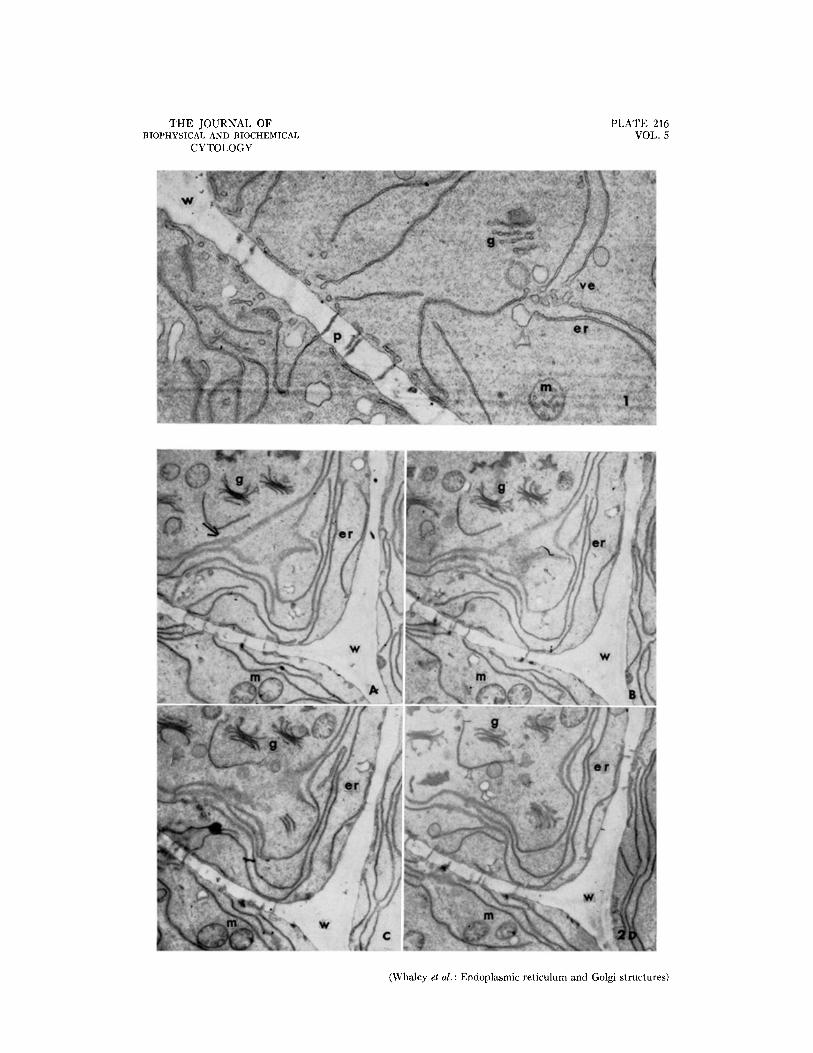
er, Endoplasmic reticulum, n, Nucleus. g, Golgi-like structure, v, Vacuole. gv, Golgi-vesicles. ve, Vesicle. p, Plasmodesma (intercellular connection), w, Cell wall. m, Mitochondrion.
PLATE 216
Fro. 1. Portions of two cells showing elements of the endoplasmic reticulum with discrete vesicles along their free edges, intercellular connections through the wall, a mitochondrion, and a Golgi-like component sectioned near its edge. Approximately )< 19,000.
FIGS. 2 a to d. Serial sections of portions of three cells showing the extensive endoplasmic reticulum, Golgi- like structures, and mitochondria. The sections are approximately ~/~0 # in thickness. The arrow indicates a por- tion of the endoplasmic reticulum sectioned parallel to its membranes. Approximately )< 8,500.
THE JOURNAL OF BIOPHYSICAL AND BIOCHEMICAL
CYTOLOGY
PLATE 216 VOL. 5
(Whaley et al.: Endoplasmic reticulum and Golgi structures)
PLATE 217
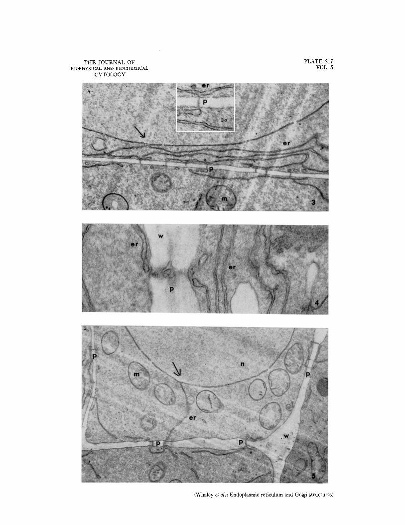
FIo. 3. Portions of two cells showing the relationship of the nuclear envelope to the endoplasmic reticulum and to intercellular connections through the wall. At the arrow the nuclear envelope and the endoplasmic re- ticulum are continuous. X 12,000.
The inset (Fig. 3 a) is an enlargement of the area of the labelled intercellular connection to show portions of the endoplasmic reticulum parallel to the wall, and a contact with an intercellular connection. Approximately X 30,000.
FIG. 4. Portions of two cells and the intervening wail showing part of an intercellular connection. The connec- tion appears to incorporate a membranous component comparable to that of the endoplasmic reticulum. Ap- proximately X 38,000.
FIG. 5. Portions of four cells showing the endoplasmic reticulum and intercellular connections. The endoplasmic reticulum of the upper cell, which is continuous with the nuclear envelope at the arrow, extends directly to an intercellular connection. Discontinuities are clearly visible in the nuclear envelope. Approximately X 12,000.
THE JOURNAL OF BIOPHYSICAL AND BIOCHEMICAL
CYTOLOGY
PLATE 217 VOL. 5
(Whaley et al.: Endoplasmic reticulum and Golgi structures)

PLATE 218
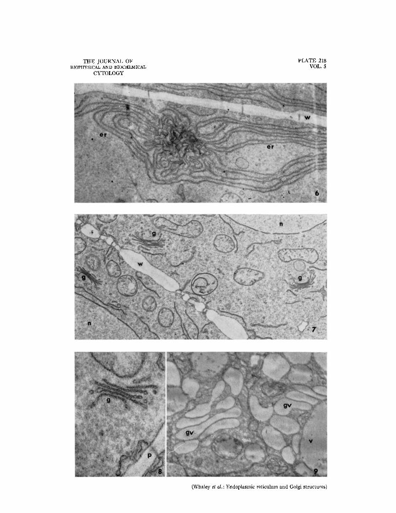
FIo. 6. Portion of a cell showing an extensive, localized development or differentiation of the endoplasmic reticulum. Approximately X 14,000.
FIG. 7. Portions of two cells showing Golgi-like structures, nuclei, and mitochondria. Approximately X 15,000. FIG. 8. A Golgi-]ike structure showing the relation of the platelet-cisternae to the vesicles at their edges. Ap-
proximately X 50,000. FIG. 9. Golgi vesicles developed from the platelet-cisternae of the Golgi-like structures in older cells. These
vesicles remain distinct from the vacuoles. Approximately X 26,000. All figures are electron mlcrographs of thin sections of rootcap cells of Zea mays fixed in permanganate and
embedded in araldite epoxy resin.
THE JOURNAL OF BIOPHYSICAL AND BIOCHEMICAL
CYTOLOGY
PLATE 218 VOL. 5
(Whaley et al.: Endoplasmic reticulum and Golgi structures)