rna processing bodies, peroxisomes, golgi bodies ...rna processing bodies, peroxisomes, golgi...
TRANSCRIPT

RNA Processing Bodies, Peroxisomes, Golgi Bodies,Mitochondria, and Endoplasmic Reticulum TubuleJunctions Frequently Pause at Cortical MicrotubulesTakahiro Hamada1,2,6,*, Motoki Tominaga3, Takashi Fukaya4, Masayoshi Nakamura2, Akihiko Nakano3,5,Yuichiro Watanabe4, Takashi Hashimoto2 and Tobias I. Baskin1
1Biology Department, University of Massachusetts Amherst, Amherst, MA 01003, USA2Graduate School of Biological Sciences, Nara Institute of Science and Technology, Ikoma, 630-0101 Japan3Molecular Membrane Biology Laboratory, RIKEN Advanced Science Institute, Wako, Saitama, 351-0198 Japan4Department of Life Sciences, Graduate School of Arts and Sciences, The University of Tokyo, Komaba, Tokyo, 153-8902 Japan5Department of Biological Sciences, Graduate School of Science, The University of Tokyo, Bunkyo-ku, Tokyo, 113-0033 Japan6Present address: Department of Botany, Graduate School of Science, Kyoto University, Sakyo-ku, Kyoto, 606-8502, Japan.*Corresponding author: E-mail, [email protected]; Fax, +81-75-753- 4141(Received October 18, 2011; Accepted February 24, 2012)
Organelle motility, essential for cellular function, is drivenby the cytoskeleton. In plants, actin filaments sustain thelong-distance transport of many types of organelles, andmicrotubules typically fine-tune the motile behavior.In shoot epidermal cells of Arabidopsis thaliana seedlings,we show here that a type of RNA granule, the RNA process-ing body (P-body), is transported by actin filaments andpauses at cortical microtubules. Interestingly, removal ofmicrotubules does not change the frequency of P-body paus-ing. Similarly, we show that Golgi bodies, peroxisomes, andmitochondria all pause at microtubules, and again the fre-quency of pauses is not appreciably changed after microtu-bules are depolymerized. To understand the basis forpausing, we examined the endoplasmic reticulum (ER),whose overall architecture depends on actin filaments. Bythe dual observation of ER and microtubules, we find thatstable junctions of tubular ER occur mainly at microtubules.Removal of microtubules reduces the number of stable ERtubule junctions, but those remaining are maintained with-out microtubules. The results indicate that pausing onmicrotubules is a common attribute of motile organellesbut that microtubules are not required for pausing. We sug-gest that pausing on microtubules facilitates interactionsbetween the ER and otherwise translocating organelles inthe cell cortex.
Keywords: ER tubule junction � Golgi body � Microtubule �
Mitochondrion � Peroxisome � Processing body.
Abbreviations: ER, endoplasmic reticulum; GFP, green fluor-escent protein; mRFP, monomeric red fluorescent protein;P-body, RNA processing body; PTS1, peroxisomal targetingsignal 1; SP, signal peptide.
Introduction
A fundamental attribute of life is polarity. Cells are polarized, andcellular polarity forms the base supporting the polarity of theorgan and organism. Among the many features that contributeto polarizing cells, the cytoskeleton is paramount. Both microtu-bules and actin filaments are themselves polar, being polymer-ized from asymmetric subunits so that the polymer retainsasymmetry, and hence is polar (Li and Gundersen 2008). Thecytoskeleton anchors and also transports organelles (Hirokawaet al. 2009). By virtue of cytoskeletal filaments being polarized,the cell controls this flow of material and consequently ofinformation. Therefore, cytoskeletal-driven motility, more thanovercoming the inefficiency of diffusion over tens of microm-eters, reinforces and even establishes the polarity of cells.
In animal cells, microtubules are involved principally inorganelle transport, whereas actin filaments are concernedwith cell shape and cell contacts, being enriched at the cellperiphery (Hirokawa et al. 2009). In plant cells, these rolesare reversed, with actin supporting most organelle transport(Shimmen and Yokota 2004) and microtubules being involvedin expansion and enriched at the cell periphery (Sedbrook andKaloriti 2008).
The pivotal role of cortical microtubules in controlling plantexpansion was suggested in the first publication to image amicrotubule (Ledbetter and Porter 1963) and since then hasbeen amply confirmed with both chemical inhibitors and gen-etics (Sedbrook and Kaloriti 2008). The role of actin filaments insupporting organelle motility in plants was first observed withrespect to cytoplasmic streaming (Kamitsubo 1966, Nagai andRebhun 1966). Indeed, powering cytoplasmic streaming inplants was among the first roles documented for actin andmyosin in any non-muscle cell type (Shimmen 2007).Subsequently, actin filaments in plants were demonstrated to
Plant Cell Physiol. 53(4): 699–708 (2012) doi:10.1093/pcp/pcs025, available online at www.pcp.oxfordjournals.org! The Author 2012. Published by Oxford University Press on behalf of Japanese Society of Plant Physiologists.All rights reserved. For permissions, please email: [email protected]
699Plant Cell Physiol. 53(4): 699–708 (2012) doi:10.1093/pcp/pcs025 ! The Author 2012.
Regu
larP
aper
at Univ. of M
assachusetts/Am
herst Library on A
pril 13, 2012http://pcp.oxfordjournals.org/
Dow
nloaded from
support the transport of various organelles, including theGolgi apparatus, peroxisomes, mitochondria, plastids, andendoplasmic reticulum (ER) (Boevink et al. 1998, Nebenfuhret al. 1999, Mathur et al. 2002, Van Gestel et al. 2002, Uedaet al. 2010). Interestingly, although actin filaments sustainlong-distance transport of plant organelles, it has recentlybeen observed that microtubules influence short-distance be-havior, causing pauses for both peroxisomes (Chuong et al.2005) and the Golgi (Crowell et al. 2009, Gutierrez et al. 2009).
In addition to deploying membrane-bound organelles, thecytoskeleton deploys other kinds, including those related to RNAmetabolism. The most familiar of these is the ribosome, but cellscontain several types of large RNA–protein complex, collectivelytermed RNA granules. In animal cells, RNA granules—includingthe RNA-processing body (P-body), stress granule, neuronal gran-ule, and germ cell granule—are transported on microtubules(Hirokawa et al. 2009). In plants, RNA granules homologous tothe stress granule and the P-body have been described(Bailey-Serres et al. 2009), but the involvement of the cytoskeletonin their transport remains for the most part uncharacterized.
One exception where cytoskeletal involvement has beencharacterized is the movement of viral RNA. Many plant virionsspread systemically by moving from cell to cell through plasmo-desmata. To reach plasmodesmata, the viral RNA is often trans-ported on actin filaments, although microtubules might berequired for efficient systemic spread (Niehl and Heinlein2011). In another example, pertaining to rice endosperm, actinfilaments support the motility of an ER-associated, prolaminemRNA particle (Hamada et al. 2003, Wang et al. 2008).
Here, we studied the movement of the P-body. This organ-elle is a cytoplasmic aggregation of protein and RNA, without amembrane. The P-body functions to degrade mRNA and toproduce small RNAs. In animal cells, P-bodies undergo directedmotility based on microtubules and pause around actinfilaments (Aizer et al 2008). In plants, although P-bodieshave been reported (Xu et al. 2006, Iwasaki et al. 2007), theirmovements have been little studied.
We report that P-bodies undergo long-distance transport onactin but pause at cortical microtubules. Interestingly, thepattern of pauses is apparently unaffected by the loss of micro-tubules. Likewise, we find that pauses in the transport ofmitochondria, peroxisomes, and Golgi, as well as the mainten-ance of stable branches of tubular ER, occur near microtubulesbut do not require the microtubules. We suggest that micro-tubules are associated with sites in the cortex that promoteinteractions among the various organelles as they are trans-ported through the large plant cell.
Results
P-bodies are transported by actin filaments andpause at cortical microtubules
To image the P-body, we used plants in which one of the majorP-body components, the mRNA de-capping enzyme, DCP2, was
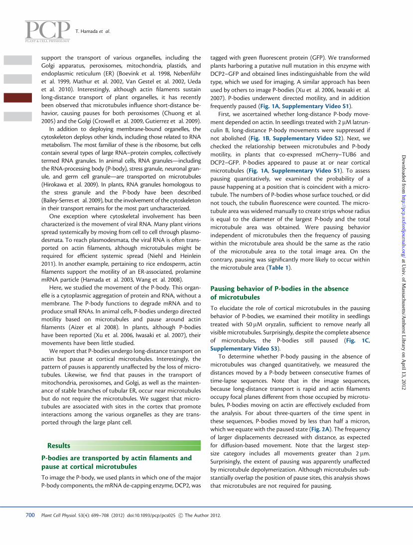
tagged with green fluorescent protein (GFP). We transformedplants harboring a putative null mutation in this enzyme withDCP2–GFP and obtained lines indistinguishable from the wildtype, which we used for imaging. A similar approach has beenused by others to image P-bodies (Xu et al. 2006, Iwasaki et al.2007). P-bodies underwent directed motility, and in additionfrequently paused (Fig. 1A, Supplementary Video S1).
First, we ascertained whether long-distance P-body move-ment depended on actin. In seedlings treated with 2 mM latrun-culin B, long-distance P-body movements were suppressed ifnot abolished (Fig. 1B, Supplementary Video S2). Next, wechecked the relationship between microtubules and P-bodymotility, in plants that co-expressed mCherry–TUB6 andDCP2–GFP. P-bodies appeared to pause at or near corticalmicrotubules (Fig. 1A, Supplementary Video S1). To assesspausing quantitatively, we examined the probability of apause happening at a position that is coincident with a micro-tubule. The numbers of P-bodies whose surface touched, or didnot touch, the tubulin fluorescence were counted. The micro-tubule area was widened manually to create strips whose radiusis equal to the diameter of the largest P-body and the totalmicrotubule area was obtained. Were pausing behaviorindependent of microtubules then the frequency of pausingwithin the microtubule area should be the same as the ratioof the microtubule area to the total image area. On thecontrary, pausing was significantly more likely to occur withinthe microtubule area (Table 1).
Pausing behavior of P-bodies in the absenceof microtubules
To elucidate the role of cortical microtubules in the pausingbehavior of P-bodies, we examined their motility in seedlingstreated with 50mM oryzalin, sufficient to remove nearly allvisible microtubules. Surprisingly, despite the complete absenceof microtubules, the P-bodies still paused (Fig. 1C,Supplementary Video S3).
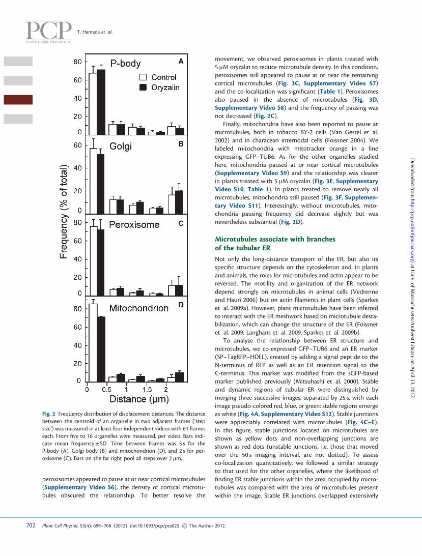
To determine whether P-body pausing in the absence ofmicrotubules was changed quantitatively, we measured thedistances moved by a P-body between consecutive frames oftime-lapse sequences. Note that in the image sequences,because long-distance transport is rapid and actin filamentsoccupy focal planes different from those occupied by microtu-bules, P-bodies moving on actin are effectively excluded fromthe analysis. For about three-quarters of the time spent inthese sequences, P-bodies moved by less than half a micron,which we equate with the paused state (Fig. 2A). The frequencyof larger displacements decreased with distance, as expectedfor diffusion-based movement. Note that the largest step-size category includes all movements greater than 2 mm.Surprisingly, the extent of pausing was apparently unaffectedby microtubule depolymerization. Although microtubules sub-stantially overlap the position of pause sites, this analysis showsthat microtubules are not required for pausing.
700 Plant Cell Physiol. 53(4): 699–708 (2012) doi:10.1093/pcp/pcs025 ! The Author 2012.
T. Hamada et al.
at Univ. of M
assachusetts/Am
herst Library on A
pril 13, 2012http://pcp.oxfordjournals.org/
Dow
nloaded from

Golgi, peroxisomes, and mitochondria also pausein the absence of microtubules
Because it is surprising that complete removal of cortical micro-tubules does not affect the frequency of P-body pauses, wechecked whether removal of microtubules affected the pausingbehavior of other organelles. Previously, Golgi have been
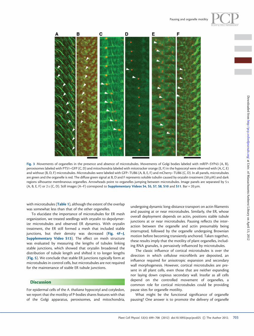
reported to pause at cortical microtubules (Crowell et al.2009, Gutierrez et al. 2009). To label Golgi, we used a trans-Golgi network marker, monomeric red fluorescent protein(mRFP)–SYP43 (Ebine et al. 2008). Consistent with previousreports, we observed that Golgi bodies frequently stopped ator near a microtubule, sometimes jumping to a neighboringmicrotubule (Fig 3A, Supplementary Video S4), and theco-localization of paused Golgi with microtubules was signifi-cant (Table 1). Without microtubules, Golgi bodies also pausedfrequently (Fig. 3B, Supplementary Video S5), and similarly tothe P-body, the removal of microtubules had little if any effecton the frequency of pausing (Fig. 2B).
Likewise, in onion epidermal cells, peroxisomes have alsobeen reported to pause on microtubules (Chuong et al.2005). That report labeled peroxisomes with GFP-taggedmultifunctional protein, which also labeled microtubulesnon-specifically; while the double labeling was convenient forimaging, the pausing of peroxisomes might reflect a non-physiological interaction. Therefore, we examined peroxisomeslabeled with a peroxisomal signal peptide construct, peroxi-somal targeting signal 1 (PTS1)–GFP (Mano et al. 2002), in aline also expressing mCherry–TUB6. Although in control plants,
Fig. 1 Movements of P-bodies. Confocal time series of a hypocotyl expressing DCP2–GFP, which labels P-bodies (red), and mCherry–TUB6(green). Image panels are separated by 5 s. (A) Control. One P-body jumps between microtubules and stops around a microtubule.(B) Latrunculin B treatment (2 mM) to depolymerize actin filaments. Long-distance movements of P-bodies are not observed. (C) Oryzalintreatment (50 mM) to depolymerize microtubules. Both pausing and moving P-bodies are seen (arrowheads show moving P-bodies). Still imagescorrespond to Supplementary Videos S1–S3. Bar = 20 mm.
Table 1 Correlation of paused organelles with cortical microtubules
Organelle Microtubulearea, % ofimage total
Overlappingorganelles,% of total
Replication
P-bodies 50 ± 6.2 94 ± 0.7 n = 1,137
Golgi bodies 60 ± 5.0 90 ± 1.7 n = 1,420
Peroxisomes 26 ± 5.3 95 ± 2.5 n = 1,215
Mitochondria 36 ± 2.3 95 ± 4.4 n = 2,105
ER stable junctions 50 ± 15.1 84 ± 6.5 n = 1,296
Data are the mean ± SD, with n representing the number of scored organelles.The microtubule area for peroxisomes and mitochondria is less than for Golgibodies and P-bodies because plants were treated with subsaturating oryzalin(5 mM) to decrease microtubule density. Note that the stable ER junctions arefar less motile than the other tabulated organelles.
701Plant Cell Physiol. 53(4): 699–708 (2012) doi:10.1093/pcp/pcs025 ! The Author 2012.
Pausing and organelle motility
at Univ. of M
assachusetts/Am
herst Library on A
pril 13, 2012http://pcp.oxfordjournals.org/
Dow
nloaded from
peroxisomes appeared to pause at or near cortical microtubules(Supplementary Video S6), the density of cortical microtu-bules obscured the relationship. To better resolve the
movement, we observed peroxisomes in plants treated with5 mM oryzalin to reduce microtubule density. In this condition,peroxisomes still appeared to pause at or near the remainingcortical microtubules (Fig. 3C, Supplementary Video S7)and the co-localization was significant (Table 1). Peroxisomesalso paused in the absence of microtubules (Fig. 3D,Supplementary Video S8) and the frequency of pausing wasnot decreased (Fig. 2C).
Finally, mitochondria have also been reported to pause atmicrotubules, both in tobacco BY-2 cells (Van Gestel et al.2002) and in characean internodal cells (Foissner 2004). Welabeled mitochondria with mitotracker orange in a lineexpressing GFP–TUB6. As for the other organelles studiedhere, mitochondria paused at or near cortical microtubules(Supplementary Video S9) and the relationship was clearerin plants treated with 5 mM oryzalin (Fig. 3E, SupplementaryVideo S10, Table 1). In plants treated to remove nearly allmicrotubules, mitochondria still paused (Fig. 3F, Supplemen-tary Video S11). Interestingly, without microtubules, mito-chondria pausing frequency did decrease slightly but wasnevertheless substantial (Fig. 2D).
Microtubules associate with branchesof the tubular ER
Not only the long-distance transport of the ER, but also itsspecific structure depends on the cytoskeleton and, in plantsand animals, the roles for microtubules and actin appear to bereversed. The motility and organization of the ER networkdepend strongly on microtubules in animal cells (Vedrenneand Hauri 2006) but on actin filaments in plant cells (Sparkeset al. 2009a). However, plant microtubules have been inferredto interact with the ER meshwork based on microtubule desta-bilization, which can change the structure of the ER (Foissneret al. 2009, Langhans et al. 2009, Sparkes et al. 2009b).
To analyze the relationship between ER structure andmicrotubules, we co-expressed GFP–TUB6 and an ER marker(SP–TagRFP–HDEL), created by adding a signal peptide to theN-terminus of RFP as well as an ER retention signal to theC-terminus. This marker was modified from the sGFP-basedmarker published previously (Mitsuhashi et al. 2000). Stableand dynamic regions of tubular ER were distinguished bymerging three successive images, separated by 25 s, with eachimage pseudo-colored red, blue, or green: stable regions emergeas white (Fig. 4A, Supplementary Video S12). Stable junctionswere appreciably correlated with microtubules (Fig. 4C–E).In this figure, stable junctions located on microtubules areshown as yellow dots and non-overlapping junctions areshown as red dots (unstable junctions, i.e. those that movedover the 50 s imaging interval, are not dotted). To assessco-localization quantitatively, we followed a similar strategyto that used for the other organelles, where the likelihood offinding ER stable junctions within the area occupied by micro-tubules was compared with the area of microtubules presentwithin the image. Stable ER junctions overlapped extensively
Fig. 2 Frequency distribution of displacement distances. The distancebetween the centroid of an organelle in two adjacent frames (‘stepsize’) was measured in at least four independent videos with 61 frameseach. From five to 16 organelles were measured, per video. Bars indi-cate mean frequency ± SD. Time between frames was 5 s for theP-body (A), Golgi body (B) and mitochondrion (D), and 2 s for per-oxisome (C). Bars on the far right pool all steps over 2 mm.
702 Plant Cell Physiol. 53(4): 699–708 (2012) doi:10.1093/pcp/pcs025 ! The Author 2012.
T. Hamada et al.
at Univ. of M
assachusetts/Am
herst Library on A
pril 13, 2012http://pcp.oxfordjournals.org/
Dow
nloaded from
with microtubules (Table 1), although the extent of the overlapwas somewhat less than that of the other organelles.
To elucidate the importance of microtubules for ER meshorganization, we treated seedlings with oryzalin to depolymer-ize microtubules and observed ER dynamics. With oryzalintreatment, the ER still formed a mesh that included stablejunctions, but their density was decreased (Fig. 4F–J,Supplementary Video S13). The effect on mesh structurewas evaluated by measuring the lengths of tubules linkingstable junctions, which showed that oryzalin broadened thedistribution of tubule length and shifted it to longer lengths(Fig. 5). We conclude that stable ER junctions typically form atmicrotubules in control cells, but microtubules are not requiredfor the maintenance of stable ER tubule junctions.
Discussion
For epidermal cells of the A. thaliana hypocotyl and cotyledon,we report that the motility of P-bodies shares features with thatof the Golgi apparatus, peroxisomes, and mitochondria,
undergoing dynamic long-distance transport on actin filamentsand pausing at or near microtubules. Similarly, the ER, whoseoverall deployment depends on actin, positions stable tubulejunctions at or near microtubules. Pausing reflects the inter-action between the organelle and actin presumably beinginterrupted, followed by the organelle undergoing Brownianmotion before becoming transiently anchored. Taken together,these results imply that the motility of plant organelles, includ-ing RNA granules, is pervasively influenced by microtubules.
The classic influence of cortical microtubules is over thedirection in which cellulose microfibrils are deposited, aninfluence required for anisotropic expansion and secondarywall morphogenesis. However, cortical microtubules are pre-sent in all plant cells, even those that are neither expandingnor laying down copious secondary wall. Insofar as all cellsdepend on the controlled movement of organelles, acommon role for cortical microtubules could be providingpause sites for organelle motility.
What might be the functional significance of organellepausing? One answer is to promote the delivery of organelle
Fig. 3 Movements of organelles in the presence and absence of microtubules. Movements of Golgi bodies labeled with mRFP–SYP43 (A, B),peroxisomes labeled with PTS1–GFP (C, D) and mitochondria labeled with mitotracker orange (E, F) in the hypocotyl were observed with (A, C, E)and without (B, D, F) microtubules. Microtubules were labeled with GFP–TUB6 (A, B, E, F) and mCherry–TUB6 (C, D). In all panels, microtubulesare green and the organelle is red. The diffuse green signal at B, D and F represents soluble tubulin caused by oryzalin treatment (50 mM) and darkregions silhouette membranous organelles. Arrowheads point to organelles jumping between microtubules. Image panels are separated by 5 s(A, B, E, F) or 2 s (C, D). Still images (A–F) correspond to Supplementary Videos S4, S5, S7, S8, S10 and S11. Bar = 20mm.
703Plant Cell Physiol. 53(4): 699–708 (2012) doi:10.1093/pcp/pcs025 ! The Author 2012.
Pausing and organelle motility
at Univ. of M
assachusetts/Am
herst Library on A
pril 13, 2012http://pcp.oxfordjournals.org/
Dow
nloaded from
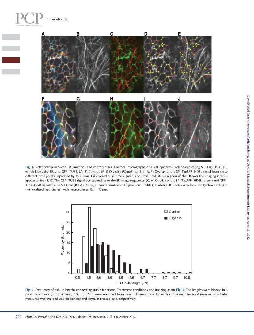
Fig. 4 Relationship between ER junctions and microtubules. Confocal micrographs of a leaf epidermal cell co-expressing SP–TagRFP–HDEL,which labels the ER, and GFP–TUB6. (A–E) Control. (F–J) Oryzalin (50 mM) for 1 h. (A, F) Overlay of the SP–TagRFP–HDEL signal from threedifferent time points, separated by 25 s. Time 1 is colored blue, time 2 green, and time 3 red; stable regions of the ER over the imaging intervalappear white. (B, G) The GFP–TUB6 signal corresponding to the ER image sequences. (C, H) Overlay of the SP–TagRFP–HDEL (green) and GFP–TUB6 (red) signals from (A, F) and (B, G). (D, E, I, J) Characterization of ER junctions. Stable (i.e. white) ER junctions co-localized (yellow circles) ornot localized (red circles) with microtubules. Bar = 10 mm.
Freq
uenc
y (%
of t
otal
)
30
25
20
15
10
5
0 0.5 1.5 2.6 3.6 4.6 5.6 6.7 7.7 8.7 9.7 10.8
ER tubule length (mm)
Control
Oryzalin
Fig. 5 Frequency of tubule lengths connecting stable junctions. Treatment conditions and imaging as for Fig. 4. The lengths were binned in 3pixel increments (approximately 0.5 mm). Data were obtained from seven different cells for each condition. The total number of tubulesmeasured was 396 and 284 for control and oryzalin-treated cells, respectively.
704 Plant Cell Physiol. 53(4): 699–708 (2012) doi:10.1093/pcp/pcs025 ! The Author 2012.
T. Hamada et al.
at Univ. of M
assachusetts/Am
herst Library on A
pril 13, 2012http://pcp.oxfordjournals.org/
Dow
nloaded from
contents to the surrounding region. This idea has beensupported for the pausing of Golgi bodies, which apparentlydeliver cellulose synthase complexes to the plasma membraneat the sites of cortical microtubules (Crowell et al. 2009,Gutierrez et al. 2009). Notwithstanding that the cargo in thiscase (cellulose synthase) continues to interact with the micro-tubule long after delivery, the ability of various organelles todeliver cargo might be enhanced by frequent and predictablestopping points. What is more, organelles often need toexchange components. The efficiency of such exchangewould be increased by organelles pulling off the actin highwayand parking side by side at a rest stop.
We hypothesize that microtubules establish and maintainsites within the cortical cytoplasm, sites that potentiate deliveryof organelle cargo as well as interactions among organelles.Fixed, cortical sites, associated with the cytoskeleton and ERhave been proposed previously (Reuzeau et al. 1997). For thesake of further discussion, we will call these sites cortical ‘land-marks’. The proposed landmark sites in the cortex would allowcells to supplement chance encounters and diffusion withdirected contacts, an enhancement that might be particularlyvaluable in large cells such as those of the hypocotyl and leavesstudied here.
This hypothesis of landmarks is supported by the sharedmotility pattern observed here, as well as by finding manyannotated organelle and RNA-processing proteins amongmicrotubule-associated proteins (T. Hamada and T. Hashimoto,unpublished data). Furthermore, microtubules mediateinteractions between peroxisomes and plastids that arerequired for chloroplast development (Albrecht et al. 2010).Nonetheless, organelles and P-bodies pause (and ER junctionspersist) in the absence of microtubules. These apparentlycontradictory observations can be reconciled by positing thatthe organelles can pause at a landmark site either throughassociating with microtubules or through binding one ormore components of the site itself.
In principle, pausing of motile organelles could be caused bydirect interactions between organelle and microtubule or byindirect interactions between the organelle and some thirdcomponent, which also interacts with the microtubule. Thatdirect interactions occur is indicated by microtubule-associatedproteins known to be organelle components. For example, twokinesins (microtubule-based motor proteins) are localized toGolgi bodies as well as to secretory vesicles (Lee et al. 2001, Luet al. 2005) and a different kinesin is localized to mitochondria(Ni et al. 2005). Experiments in vitro have found that micro-tubules are bound by enoyl-CoA hydratase, which is a peroxi-some protein (Chuong et al. 2002), and also by dynamin, whichis localized to peroxisomes and mitochondria (Hamada et al.2006). Although proteins mediating an interaction betweenmicrotubules and P-bodies have yet to identified, microtubulebinding in vitro has been reported for several proteins involvedwith RNA metabolism, including elongation factor 1-a(Durso and Cyr 1994), MPB2C (Kragler et al. 2003), THO2(Hamada et al. 2009), and Rae1 (Lee et al. 2009).
Direct organelle–microtubule interactions are supported bycertain of our observations. For mitochondria, the frequency ofpausing is modestly but significantly reduced in the absence ofmicrotubules. For ER, direct interactions seem even clearer:stable tubule junctions are localized preferentially at corticalmicrotubules and their removal reduces the number of suchjunctions (Fig. 4). This result suggests that ER tubule junctionsare stabilized at microtubules. Nevertheless, that indirectinteractions also contribute to pausing is indicated by thefact that removal of microtubules scarcely affects pausingfrequency (Fig. 2).
We compared P-body motility not only with three kinds ofrelatively small and motile organelles but also with the ER.The latter comprises a complex network of tubules, sheets,and junctions that pervades the cell. The network is dynamic,with tubules extending and retracting, forming and dissolvingjunctions and branches. While the overall network structuredepends on the actin cytoskeleton, there has been debate onwhether ER motility or network structure involves microtu-bules (Sparkes et al. 2009a). When microtubules are depoly-merized chemically, some studies have reported little if anychange in ER motility (Quader et al. 1989, Knebel et al. 1990,Lichtscheidl and Hepler 1996), whereas others did observechanges (Foissner et al. 2009, Langhans et al. 2009, Sparkeset al. 2009c). Also, the reported effect of microtubule depoly-merization on ER structure differs: on one hand, oryzalin treat-ment produced aggregations of ER termed ‘nodules’ in A.thaliana roots, tobacco BY-2 cells, and tobacco leaf protoplasts(Langhans et al. 2009); however, we were unable to observeoryzalin-induced ER nodules either in tobacco BY-2 cells or inroot, hypocotyl, and leaf epidermis of A. thaliana (data notshown). On the other hand, in elongating internodes ofNitella translucens, removing microtubules increased themesh size within the ER network (Foissner et al. 2009), whichis consistent with our results.
It is still unclear how ER tubules extend and branch. Theextension of tubular ER is correlated with movements of Golgibodies, which sometimes physically interact with the ERmembrane (Sparkes et al. 2009b); however, the extension ofER tubules continues more or less unchanged when the Golgimembranes are decimated by brefeldin (Sparkes et al. 2009c).Here, we show that ER tubule junctions are partially associatedwith microtubules. That ER tubule junctions are related to thepresence of microtubules, but do not require them, is reminis-cent of the pausing movements we observed for organelles andP-bodies, which occur at or near microtubules but continuewhen microtubules are depolymerized. This leads us tohypothesize that the positioning of ER tubule junctionsshares motility characteristics with those of organelles andP-bodies, depending on actin for sustained localization but sub-ject to local modification in the cell cortex based ultimately onthe microtubule system.
Pausing, widespread among plant organelles, even whilethey otherwise undergo sustained long-distance transport, illus-trates the fact that anchoring and stillness are as much the
705Plant Cell Physiol. 53(4): 699–708 (2012) doi:10.1093/pcp/pcs025 ! The Author 2012.
Pausing and organelle motility
at Univ. of M
assachusetts/Am
herst Library on A
pril 13, 2012http://pcp.oxfordjournals.org/
Dow
nloaded from

province of the cytoskeleton as are locomotion and dynamics.Understanding how these opposite requirements are inte-grated now stands as a challenge for the future.
Materials and Methods
Plant material and growth conditions
All material was A. thaliana L. (Heynh), Columbia background.Seeds were sterilized in 5% sodium hypochlorite and 1% TritonX-100 for 5 min. After sterilization, seeds were rinsed five timeswith sterile water and plated on agar containing 2% sucrose,1.5% agar, and half-strength A. thaliana nutrient solutiondescribed in Haughn and Somerville (1986). Plates were set ata near vertical position at 22�C with a 16 h light/8 h dark photo-period. Five-day-old plants were used for analyses of Golgibodies, peroxisomes, mitochondria, and P-bodies; 7-day-oldplants were used for analyses of the ER.
Construction of plasmids and transformation
For P-body labeling, to construct the DCP2–GFP (At5g13570)plasmid, a genomic DNA fragment containing a 2,555 bp 50
upstream sequence from the start codon of DCP2 and imme-diately 30 of the DCP2 open reading frame (ORF) was amplifiedby PCR with 50-CACCCTGTCCAAAAGCAGCCAAAG-30 as theleft border primer and 50-AGCTGAATTACCAGATTCCAACGC-30 as the right border primer. The PCR product was cloned intopENTR/D-TOPO vector (Invitrogen) and moved intopGWB550, which provides a C-terminal G3GFP fusion protein(Nakagawa et al. 2007, Nakagawa et al. 2008). The dcp2-1(SALK_000519, Xu et al. 2006, Iwasaki et al. 2007) line wastransformed by the floral dip method (Clough and Bent1998) using Agrobacterium tumefaciens GV3101 strain withthe pGWB550 vector containing the insert.
For ER labeling, a plasmid containing SP–TagRFP–HDELwas constructed according to the sequence of SP–sGFP–HDEL (Mitsuhashi et al. 2000) using pGWB502 (Nakagawaet al. 2007). The left border primer (50-CACCATGGCCAGACTCACAAGCATCATTGCCCTCTTCGCAGTGGCTCTGCTGGTTGCAGATGCGTACGCCTACCGCATGGTGTCTAAGGGCGAAGAG-30) and the right border primer (50-TCAAAGCTCATCGTGGTGGTGGTGGTGGTGCCCCCCCCCATTAAGTTTGTGCCCCAGTTTGCTAGGGAG-30) were used for PCR. Theplasmid was introduced into A. tumefaciens GV3101 strainand used to transform GFP–TUB6 plants by the floral dipmethod.
Microscopy and quantitative methods
Observations were performed on a fluorescence microscope(BX51, Olympus) equipped with a confocal spinning disk unit(CSU22, Yokogawa), with the Dualview (Optical Insight) andEMCCD (Hamamatsu Photonics) system. Images were takenwith MetaMorph software (Molecular Devices) and analyzedwith ImageJ (NIH, http://rsbweb.nih.gov/ij/). For drug
treatments, plants were soaked in small tubes and incubatedfor the appropriate time at 22�C in the light. At least 10 differ-ent plants were observed in each condition. For the analyses ofcorrelation of paused organelles with microtubules, the micro-tubule area was widened manually to create strips whose radiusis equal to the largest diameter of each organelle of interest, andthe total microtubule area was obtained. The numbers oforganelles whose surface touched, or did not touch, the micro-tubule fluorescence lines were counted. For analyzing thedisplacement distances of organelles, ImageJ was used toextract centroid coordinates from image sequences, and thePythagorean distance between the positions of a given organ-elle in successive frames tabulated for Fig. 2.
Supplementary data
Supplementary data are available at PCP Online.
Funding
This work was supported by the Nara Institute of Scienceand Technology [Global COE Program (Frontier Biosciences:strategies for survival and adaptation in a changing globalenvironment)]; TOYOBO BIOFOUNDATION [Long-termResearch Fellowship to T. Hamada]; the Division of ChemicalSciences, Geosciences, and Biosciences, Office of BasicEnergy Sciences of the US Department of Energy [research onmorphogenesis in the Baskin laboratory through grantDE-FG-03ER15421].
Acknowledgments
We thank Makoto Hayashi and Mikio Nishimura (NIBB, Japan)for the gift of PTS1–GFP, Takashi Ueda (University of Tokyo) forthe gift of mRFP–SYP43, and Tsuyoshi Nakagawa (ShimaneUniversity) for the gift of the pGWB series of plasmids. Wealso thank Noriko Inada (NAIST) for technical advice and help-ful discussion regarding imaging.
References
Aizer, A., Brody, Y., Ler, L.W., Sonenberg, N., Singer, R.H. and Shav-Tal, Y. (2008) The dynamics of mammalian P body transport,assembly, and disassembly in vivo. Mol. Biol. Cell 19: 4154–4166.
Albrecht, V., Simkova, K., Carrie, C., Delannoy, E., Giraud, E., Whelan, J.et al. (2010) The cytoskeleton and the peroxisomal-targetedSNOWY COTYLEDON3 protein are required for chloroplast devel-opment in arabidopsis. Plant Cell 22: 3423–3438.
Bailey-Serres, J., Sorenson, R. and Juntawong, P. (2009) Getting themessage across: cytoplasmic ribonucleoprotein complexes. TrendsPlant Sci. 14: 443–453.
Boevink, P., Oparka, K., Cruz, S.S., Martin, B., Betteridge, A. andHawes, C. (1998) Stacks on tracks: the plant Golgi apparatus trafficson an actin/ER network. Plant J. 15: 441–447.
706 Plant Cell Physiol. 53(4): 699–708 (2012) doi:10.1093/pcp/pcs025 ! The Author 2012.
T. Hamada et al.
at Univ. of M
assachusetts/Am
herst Library on A
pril 13, 2012http://pcp.oxfordjournals.org/
Dow
nloaded from
Chuong, S.D.X., Mullen, R.T. and Muench, D.G. (2002) Identification ofa rice RNA- and microtubule-binding protein as the multifunctionalprotein, a peroxisomal enzyme involved in the beta-oxidation offatty acids. J. Biol. Chem. 277: 2419–2429.
Chuong, S.D.X., Park, N.I., Freeman, M.C., Mullen, R.T. andMuench, D.G. (2005) The peroxisomal multifunctional proteininteracts with cortical microtubules in plant cells. BMC Cell Biol.6: 40.
Clough, S.J. and Bent, A.F. (1998) Floral dip: a simplified method forAgrobacterium-mediated transformation of Arabidopsis thaliana.Plant J. 16: 735–743.
Crowell, E.F., Bischoff, V., Desprez, T., Rolland, A., Stierhof, Y.D.,Schumacher, K. et al. (2009) Pausing of Golgi bodies on microtu-bules regulates secretion of cellulose synthase complexes inarabidopsis. Plant Cell 21: 1141–1154.
Durso, N.A. and Cyr, R.J. (1994) A calmodulin-sensitive interactionbetween microtubules and a higher plant homolog of elongationfactor-1 alpha. Plant Cell 6: 893–905.
Ebine, K., Okatani, Y., Uemura, T., Goh, T., Shoda, K., Niihama, M. et al.(2008) A SNARE complex unique to seed plants is required forprotein storage vacuole biogenesis and seed development ofArabidopsis thaliana. Plant Cell 20: 3006–3021.
Foissner, I. (2004) Microfilaments and microtubules control the shape,motility, and subcellular distribution of cortical mitochondria incharacean internodal cells. Protoplasma 224: 145–157.
Foissner, I., Menzel, D. and Wasteneys, G.O. (2009)Microtubule-dependent motility and orientation of the corticalendoplasmic reticulum in elongating characean internodal cells.Cell Motil. Cytoskeleton 66: 142–155.
Gutierrez, R., Lindeboom, J.J., Paredez, A.R., Emons, A.M. andEhrhardt, D.W. (2009) Arabidopsis cortical microtubules positioncellulose synthase delivery to the plasma membrane and interactwith cellulose synthase trafficking compartments. Nat. Cell Biol. 11:797–806.
Hamada, S., Ishiyama, K., Choi, S.B., Wang, C., Singh, S., Kawai, N. et al.(2003) The transport of prolamine RNAs to prolamine proteinbodies in living rice endosperm cells. Plant Cell 15: 2253–2264.
Hamada, T., Igarashi, H., Taguchi, R., Fujiwara, M., Fukao, Y.,Shimmen, T. et al. (2009) The putative RNA-processing protein,THO2, is a microtubule-associated protein in tobacco. Plant CellPhysiol. 50: 801–811.
Hamada, T., Igarashi, H., Yao, M., Hashimoto, T., Shimmen, T. andSonobe, S. (2006) Purification and characterization of plantdynamin from tobacco BY-2 cells. Plant Cell Physiol. 47: 1175–1181.
Haughn, G.W. and Somerville, C. (1986) Sulfonylurea-resistantmutants of Arabidopsis thaliana. Mol. Gen. Genet. 204: 430–434.
Hirokawa, N., Noda, Y., Tanaka, Y. and Niwa, S. (2009) Kinesin super-family motor proteins and intracellular transport. Nat. Rev. Mol. CellBiol. 10: 682–696.
Iwasaki, S., Takeda, A., Motose, H. and Watanabe, Y. (2007)Characterization of Arabidopsis decapping proteins AtDCP1 andAtDCP2, which are essential for post-embryonic development.FEBS Lett. 581: 2455–2459.
Kamitsubo, E. (1966) Motile protoplasmic fibrils in cells of Characeae.II. Linear fibrillar structure and its bearing on protoplasmic stream-ing. Proc. Jpn. Acad. 42: 640–643.
Knebel, W., Quader, H. and Schnepf, E. (1990) Mobile and immobileendoplasmic reticulum in onion bulb epidermis cells: short- andlong-term observations with a confocal laser scanning microscope.Eur. J. Cell Biol. 52: 328–340.
Kragler, F., Curin, M., Trutnyeva, K., Gansch, A. and Waigmann, E.(2003) MPB2C, a microtubule-associated plant protein binds toand interferes with cell-to-cell transport of tobacco mosaic virusmovement protein. Plant Physiol. 132: 1870–1883.
Langhans, M., Niemes, S., Pimpl, P. and Robinson, D.G. (2009) Oryzalinbodies: in addition to its anti-microtubule properties, the dinitroa-niline herbicide oryzalin causes nodulation of the endoplasmicreticulum. Protoplasma 236: 73–84.
Ledbetter, M.C. and Porter, K.R. (1963) A ‘microtubule’ in plant cellfine structure. J. Cell Biol. 19: 239–250.
Lee, J.Y., Lee, H.S., Wi, S.J., Park, K.Y., Schmit, A.C. and Pai, H.S. (2009)Dual functions of Nicotiana benthamiana Rae1 in interphase andmitosis. Plant J. 59: 278–291.
Lee, Y.R., Giang, H.M. and Liu, B. (2001) A novel plant kinesin-relatedprotein specifically associates with the phragmoplast organelles.Plant Cell 13: 2427–2439.
Li, R. and Gundersen, G.G. (2008) Beyond polymer polarity: how thecytoskeleton builds a polarized cell. Nat. Rev. Mol. Cell Biol. 9:860–873.
Lichtscheidl, I.K. and Hepler, P.K. (1996) Endoplasmic reticulum in thecortex of plant cells. In Membranes: Specialised Functions in Plants.Edited by Smallwood, M., Knox, P. and Bowles, D. pp. 383–402. BIOSScientific Publishers, Wallingford, UK.
Lu, L., Lee, Y.R., Pan, R., Maloof, J.N. and Liu, B. (2005) An internalmotor kinesin is associated with the Golgi apparatus and plays arole in trichome morphogenesis in Arabidopsis. Mol. Biol. Cell 16:811–823.
Mano, S., Nakamori, C., Hayashi, M., Kato, A., Kondo, M. andNishimura, M. (2002) Distribution and characterization ofperoxisomes in Arabidopsis by visualization with GFP: dynamicmorphology and actin-dependent movement. Plant Cell Physiol.43: 331–341.
Mathur, J., Mathur, N. and Hulskamp, M. (2002) Simultaneous visual-ization of peroxisomes and cytoskeletal elements reveals actin andnot microtubule-based peroxisome motility in plants. Plant Physiol.128: 1031–1045.
Mitsuhashi, N., Shimada, T., Mano, S., Nishimura, M. and Hara-Nishimura, I. (2000) Characterization of organelles in thevacuolar-sorting pathway by visualization with GFP in tobaccoBY-2 cells. Plant Cell Physiol. 41: 993–1001.
Nagai, R. and Rebhun, L.I. (1966) Cytoplasmic microfilaments instreaming Nitella cells. J. Ultrastruct. Res. 14: 571–589.
Nakagawa, T., Kurose, T., Hino, T., Tanaka, K., Kawamukai, M., Niwa, Y.et al. (2007) Development of series of gateway binary vectors,pGWBs, for realizing efficient construction of fusion genes forplant transformation. J. Biosci. Bioeng. 104: 34–41.
Nakagawa, T., Nakamura, S., Tanaka, K., Kawamukai, M.,Suzuki, T., Nakamura, K. et al. (2008) Development of R4gateway binary vectors (R4pGWB) enabling high-throughputpromoter swapping for plant research. Biosci. Biotechnol.Biochem. 72: 624–629.
Nebenfuhr, A., Gallagher, L.A., Dunahay, T.G., Frohlick, J.A.,Mazurkiewicz, A.M., Meehl, J.B. et al. (1999) Stop-and-go move-ments of plant Golgi stacks are mediated by the acto-myosinsystem. Plant Physiol. 121: 1127–1141.
Ni, C.Z., Wang, H.Q., Xu, T., Qu, Z. and Liu, G.Q. (2005) AtKP1, akinesin-like protein, mainly localizes to mitochondria inArabidopsis thaliana. Cell Res. 15: 725–733.
Niehl, A. and Heinlein, M. (2011) Cellular pathways for viral transportthrough plasmodesmata. Protoplasma 248: 75–99.
707Plant Cell Physiol. 53(4): 699–708 (2012) doi:10.1093/pcp/pcs025 ! The Author 2012.
Pausing and organelle motility
at Univ. of M
assachusetts/Am
herst Library on A
pril 13, 2012http://pcp.oxfordjournals.org/
Dow
nloaded from
Quader, H., Hofmann, A. and Schnepf, E. (1989) Reorganization of theendoplasmic reticulum in epidermis cells of onion bulb scales aftercold stress: involvement of cytoskeletal elements. Planta 177:273–280.
Reuzeau, C., McNally, J.G. and Pickard, B.G. (1997) The endomembranesheath: a key structure for understanding the plant cell?.Protoplasma 200: 1–9.
Sedbrook, J.C. and Kaloriti, D. (2008) Microtubules, MAPs and plantdirectional cell expansion. Trends Plant Sci. 13: 303–310.
Shimmen, T. (2007) The sliding theory of cytoplasmic streaming: fiftyyears of progress. J. Plant Res. 120: 31–43.
Shimmen, T. and Yokota, E. (2004) Cytoplasmic streaming in plants.Curr. Opin. Cell Biol. 16: 68–72.
Sparkes, I.A., Frigerio, L., Tolley, N. and Hawes, C. (2009a) The plantendoplasmic reticulum: a cell-wide web. Biochem. J. 423: 145–155.
Sparkes, I.A., Ketelaar, T., de Ruijter, N.C. and Hawes, C. (2009b) Grab aGolgi: laser trapping of Golgi bodies reveals in vivo interactionswith the endoplasmic reticulum. Traffic 10: 567–571.
Sparkes, I., Runions, J., Hawes, C. and Griffing, L. (2009c) Movementand remodeling of the endoplasmic reticulum in nondividing cellsof tobacco leaves. Plant Cell 21: 3937–3949.
Ueda, H., Yokota, E., Kutsuna, N., Shimada, T., Tamura, K., Shimmen, T.et al. (2010) Myosin-dependent endoplasmic reticulum motilityand F-actin organization in plant cells. Proc. Natl Acad. Sci. USA107: 6894–6899.
Van Gestel, K., Kohler, R.H. and Verbelen, J.P. (2002) Plant mitochon-dria move on F-actin, but their positioning in the corticalcytoplasm depends on both F-actin and microtubules. J. Exp.Bot. 53: 659–667.
Vedrenne, C. and Hauri, H.P. (2006) Morphogenesis of the endoplas-mic reticulum: beyond active membrane expansion. Traffic 7:639–646.
Wang, C., Washida, H., Crofts, A.J., Hamada, S., Katsube-Tanaka, T.,Kim, D. et al. (2008) The cytoplasmic-localized, cytoskeletal-associated RNA binding protein OsTudor-SN: evidence for anessential role in storage protein RNA transport and localization.Plant J. 55: 443–454.
Xu, J., Yang, J.Y., Niu, Q.W. and Chua, N.H. (2006) Arabidopsis DCP2,DCP1, and VARICOSE form a decapping complex required forpostembryonic development. Plant Cell 18: 3386–3398.
708 Plant Cell Physiol. 53(4): 699–708 (2012) doi:10.1093/pcp/pcs025 ! The Author 2012.
T. Hamada et al.
at Univ. of M
assachusetts/Am
herst Library on A
pril 13, 2012http://pcp.oxfordjournals.org/
Dow
nloaded from